Introduction
Polycystic ovary syndrome (PCOS) is the most common
reproductive endocrine and metabolic disease with a prevalence of
4-10% in reproductive-age women globally (1). The main manifestations of the disease
are oligomenorrhea, chronic anovulation, polycystic ovarian
ultrasonography changes and clinical or biochemical
hyperandrogenism. Reportedly, 50-60% of patients with PCOS have
different degrees of hyperandrogenemia (HA) (2), and 44-70% of patients have insulin
resistance (IR) and hyperinsulinemia (3-5).
In addition, 44-70% of patients with PCOS are obese (6). Although identification of the
mechanism of PCOS remains elusive, the interaction of genetic,
endocrine and environmental factors likely leads to PCOS (7).
In recent years, an increasing number of studies
have confirmed that androgen status, IR and obesity play notable
roles in the pathogenesis of PCOS (8-10).
Previous studies have suggested that hyperandrogen status (11,12),
IR (13-15)
and obesity (16-19)
increase the rate of miscarriage and decrease the pregnancy rate
among patients with PCOS. Chang et al (13) indicated that IR affects endometrial
function and the implantation process. Patel and Carr (20) revealed that hyperandrogen status
and IR are associated with adverse fertility outcomes in patients
with PCOS. Cohort studies of >9,500 cycles (21-23),
indicate that obesity may induce embryo implantation failure by
impairing endometrial receptivity.
Endometrial receptivity is an important factor for
successful embryo implantation, which is accompanied by the
fluctuations of steroid hormones and endometrial
receptivity-related factors, the influx of uterine natural killer
cells (uNK) cells and the inhibition of inflammatory factors.
Endometrial receptivity is regulated by multiple factors, such as
receptors of hormones (estrogen and progesterone), pro-inflammatory
cytokines [IL-6, IL-8, IL-15 and monocyte chemoattractant protein-1
(MCP-1)] and endometrial decidualization-related factors [integrin
β3 (avβ3) and insulin-like growth factor binding protein-1
(IGFBP-1)] (24).
In addition, adiponectin is also a biomarker of
endometrial receptivity (25,26).
Adiponectin, an insulin sensitizer secreted by adipocytes, is
closely associated with the insulin signal transduction pathway
(25,26). Adiponectin exerts its effects by
binding to adapter protein containing PH domain, PTB domain and
leucine zipper motif 1 (APPL1), together with AdipoR1 and AdipoR2,
to activate different signaling pathways, such as MAPK, p38,
ERK1/2, Akt and AMP-activated protein kinase (AMPK) (27). Adiponectin contains three main
forms: Trimer, hexamer and high molecular weight (HMW). Some
studies hypothesize that HMW adiponectin is the form most closely
associated with insulin sensitivity (28). The binding of the APPL1 to
adiponectin receptors is regulated by the APPL2; AdipoR1- and
R2-dependent signaling is mediated through APPL 1 and APPL2. In the
absence of adiponectin signal, APPL2 can bind to the adiponectin
receptors or it can form an APPL1/APPL2 heterodimer which prevents
the binding of APPL1/adiponectin receptors (29). The adiponectin signaling pathways
play notable roles in anti-atherosclerosis, improving the insulin
resistant state, reducing blood glucose and reducing
anti-inflammatory effects.
Metabolic disorders (high androgen status, IR and
obesity) may be the reason for embryo implantation failure, as
androgen status and IR are two interacting factors. However,
previous studies (13,21-23,30)
did not assess different androgen status combining with different
IR levels, nor consider the BMI cutoff value for Asian PCOS women.
In addition, the majority of previous studies were performed in
spontaneous ovulation. Therefore, based on previous findings
(24,25) the present study investigated the
endometrial receptivity in patients with PCOS with different
metabolic abnormalities by assessing well-characterized endometrial
receptivity markers, including adiponectin and its signaling
protein, pro-inflammatory cytokines (MCP-1, IL-6 and IL-8) mediated
through adiponectin, estrogen receptor (ERα), ERβ, progesterone
receptor (PR), IL-15, avβ3 and IGFBP-1 in the endometrium during
the window of implantation.
Materials and methods
Ethical approval
The present study was approved by the Clinical
Scientific Research and Experimental Animal Ethics Committee of the
First Affiliated Hospital of Sun Yat-sen University [approval no.
Ethics (2020) no. 422-1]. Written informed consent was obtained
from all participants.
Participants
Infertile patients with PCOS attending the
Department of Reproductive Endocrine of the First Affiliated
Hospital of Sun Yat-sen University (Guangzhou, China) from November
2020 to January 2021 were recruited. The patients' PCOS diagnoses
were based on the 2003 Rotterdam criteria (31). Each participant met at least two of
the following criteria: i) Oligomenorrhea and/or chronic
anovulation; ii) clinical and/or biochemical hyperandrogenism; and
iii) polycystic ovarian ultrasonography changes. The exclusion
criteria were patients with Cushing's syndrome, congenital adrenal
hyperplasia, androgen-secreting tumor, tubal effusion, intrauterine
adhesions, multiple endometrial polyps, chromosomal diseases,
recurrent fertilization failures, organic diseases of the uterus or
ovaries (such as endometriosis or adenomyosis), history of surgery,
pelvic radiotherapy and chemotherapy in ovaries, thyroid diseases,
hyperprolactinemia, adrenal gland tumors, cardiovascular and
cerebrovascular diseases and mental illness and associated
disorders. Patients who took hormones or drugs in the past 3 months
(that would affect insulin) were also excluded. The diagnosis was
confirmed by a pathologist, and the pathologist was independent
from the study.
Levels of testosterone (T), sex hormone-binding
globulin (SHBG), oral glucose tolerance test (OGTT) and fasting
insulin were measured on the 2-5 days of menstruation. High
androgen status was determined when the free androgen index (FAI)
(%) [T (nmol/l)/SHBG (nmol/l)] was >4.5% (32). IR was considered when the
homeostasis model assessment IR (HOMA-IR) [fasting insulin (µ
international unit (IU)/ml) x fasting plasma glucose (FPG)
(mmol/l)/22.5] was >2.4(33).
According to the OGTT test, the present study defined FPG as ≥5.6
mmol/l and/or 2-h postprandial blood glucose (2hPBG) ≥7.7 mmol/l as
impaired glucose tolerance (IGT); furthermore, FPG ≥7.0 mmol/l
and/or 2hPBG ≥11.1 mmol/l was defined as diabetes mellitus
(34).
The patients were classified into five different
groups depending on their FAI, HOMA-IR, and OGTT results: i)
Hyperandrogenemia combined with impaired glucose tolerance (HA +
IGT) group (n=8), FAI ≥4.5% and HOMA-IR >2.4, 5.6 mmol/l ≤FPG
≤6.9 mmol/l, and/or 7.7 mmol/l ≤2 h PBG ≤11 mmol/l; ii)
hyperandrogenemia combined with diabetes mellitus (HA + DM) group
(n=8), FAI ≥4.5%, HOMA-IR >2.4, FPG ≥7.0 and/or 2 h PBG ≥11.1;
iii) hyperandrogenemia combined with non-IR (HA + NIR) group
(n=10), FAI ≥4.5%, HOMA-IR ≤2.4; iv) non-hyperandrogenemia androgen
combined with IR (NHA + IR) group (n=8), FAI <4.5%, HOMA-IR
>2.4; or v) non-hyperandrogenemia combined with non-IR (NHA +
NIR) group (n=9), FAI <4.5%, HOMA ≤2.4. In addition, according
to the BMI value, the subjects were divided into a lean/normal
group (n=27; BMI<24 kg/m2), an overweight group (n=8;
24≤ BMI <28 kg/m2), and an obese group (n=8; BMI ≥28
kg/m2) (35).
Sample collection and treatments
The number of antral follicles was measured by
vaginal B-ultrasound on the 2-5 days of menstruation for all
participants, and ultrasound examinations were conducted every few
days to monitor the number and size of the follicles as well as the
endometrial thickness. In addition, the basic information of each
patient was collected on the 2-5 days of menstruation, including
age, BMI, years of infertility, and blood samples. In this sample,
other possible interference in the diagnosis of PCOS [such as
follicle-stimulating hormone (FSH), luteinizing hormone (LH),
estrogen, progesterone, testosterone, prolactin (PRL),
anti-Mullerian hormone (AMH), thyroid-stimulating hormone (TSH),
high-density lipoprotein (HDL), low-density lipoprotein (LDL),
triglyceride (TG) and cholesterol (CHOL)] were included on Table I.
 | Table IChemical and endocrine
characteristics of the studied groups. |
Table I
Chemical and endocrine
characteristics of the studied groups.
| Parameters | HA + IGT | HA + DM | HA + NIR | NHA + IR | NHA + NIR | P-value |
|---|
| Number | 8.00 | 8.00 | 10.00 | 8.00 | 9.00 | |
| Age (years) | 25.63±3.02 | 26.43±3.87 | 28.20±3.36 | 26.60±2.51 | 28.22±2.39 | 0.33 |
| WHR | 0.89±0.03 | 0.92±0.03 | 0.89±0.04 | 0.86±0.03 | 0.90±0.02 | 0.16 |
| BMI
(kg/m2) | 25.05±3.2 | 27.45±5.05 | 21.25±1.93 | 23.37±3.69 |
19.11±2.37a | <0.001 |
| FSH (mIU/ml) | 5.35±1.60 | 4.94±0.91 | 5.29±1.29 | 5.40±0.64 | 5.29
(4.68,5.97) | 0.81 |
| LH (mIU/ml) | 7.26±4.39 | 7.53±2.51 | 8.68±4.83 | 40.94±11.03 | 7.58±3.66 | 0.76 |
| E2 (pg/ml) | 33.75±9.15 | 35.3±10.41 | 40.81±17.88 | 40.94±11.03 | 39.49±15.92 | 0.86 |
| PRL (ng/ml) | 13.01
(9.29,23.37) | 17.67±8.06 | 15.22
(10.25,27.47) | 12.41±5.29 | 14.39
(12.81,20.96) | 0.78 |
| AMH | 11.44±5.17 | 7.57±4.56 | 11.05±5.57 | 10.46
(6.62,11.15) | 9.04
(6.61,12.97) | 0.38 |
| TSH | 1.80±1.11 | 1.55
(1.25,2.14) | 2.16±1.09 | 1.52±0.63 | 1.97±0.58 | 0.62 |
| CHOL | 5.15±0.85 | 4.9(4.6,5.3) | 5.11±0.99 | 4.6(4.6,5.95) | 4.6±0.89 | 0.48 |
| TG | 1.01
(0.89,2.03) | 1.3±0.65 | 1.05±0.39 | 1.11±0.43 | 0.85±0.13 | 0.42 |
| HDL | 1.28±0.36 | 1.24
(1.14,1.31) | 1.54±0.30 | 1.35±0.20 | 1.40±0.25 | 0.34 |
| LDL | 3.29±0.62 | 3.22±0.22 | 1.54±0.30 | 1.35±0.20 | 1.40±0.25 | 0.22 |
In order to guarantee the transformation of the
endometrium to the secretory phase, hormone replacement therapy
(HRT) was performed: 2 mg of estradiol was given daily
(Progynova®; Bayer) for endometrial proliferation
support for ≥10 days. When the endometrium thickness reached 8 mm,
10 mg of dydrogesterone tablets were given twice a day
(Duphaston®; Abbott Biologicals B.V.) for 5 days for
endometrial transformation. On the fifth day of endometrial
transformation, intravenous blood and endometrium samples were
collected. To minimize the biopsy difference, the endometrial
biopsies were collected from the superficial layer of the
endometrium instead of the basal layer. The receptive phase of
endometrium was determined according to Noyes histological standard
(36).
High molecular weight (HMW)
adiponectin ELISA detection
The sensitivity of specific ELISA kit (cat. no.
ml063723, MiBio) of HMW adiponectin was 10 ng/ml. The coefficient
of variation within and between plates was <10 and 15%,
respectively. The serum HMW adiponectin was determined using
enzyme-linked immunosorbent assay (ELISA) with an enzyme-labeled
instrument (SkanIt™ software; Thermo Fisher Scientific,
Inc.).
RNA extraction and PCR
The frozen endometrial samples were homogenized
using TRIzol (cat. no.93289, Thermo Fisher Scientific, Inc.)
reagent to obtain total ribonucleic acid (RNA). The RNA
concentration was determined using spectrophotometry (A260:A280),
and the integrity of RNA was determined using electrophoresis on a
formaldehyde agarose gel. The tissue RNA extraction kit
(EZBioscience) was used to extract 2 µg of total RNA from the
endometrial sample; the RNA reverse transcription and SYBR Green
qPCR Master Mix kit (EZBioscience) were used to perform the
standard PCR according to the manufacturer's instructions. Human
GAPDH was used as internal reference genes and as a biomarker of
standardizing to the RNA load of each sample since the expression
of this gene is relatively constant during the menstrual cycle
(37). PCR amplification of target
mRNA was assessed by using gene-specific primers in Table II. Primers were provided by Takara
Biotechnology Co., Ltd. The PCR reaction was initiated with 95˚C
for 5 min for amplification, melted at 95˚C for 10 sec, then
annealing and extension were performed at 60˚C for 30 sec. The
polymerase chain reaction products were analyzed using ethidium
bromide agarose gel electrophoresis. A quantitative PCR instrument
(Applied Biosystems; Thermo Fisher Scientific, Inc.) was used for
real-time quantitative PCR and data analysis. Amplification
efficiencies obtained from quantitative PCR were supported by using
the method of 2-ΔΔCq (38).
| Table IIGene-specific primer sequences. |
Table II
Gene-specific primer sequences.
| Target genes | Primer sequences
(5'-3') |
|---|
| GAPDH | Sense
GAGTCAACGGATTTGGTCGT |
| | Antisense
ATCCACAGTCTTCTGGGTG |
| Adiponectin | Sense
TGCTGGGAGCTGTTCTACTG |
| | Antisense
TACTCCGGTTTCACCGATGTC |
| AdipoR1 | Sense
AAACTGGCAACATCTGGACC |
| | Antisense
GCTGTGGGGAGCAGTAGAAG |
| AdipoR2 | Sense
ACAGGCAACATTTGGACACA |
| | Antisense
CCAAGGAACAAAACTTCCCA |
| APPL1 | Sense
TTAGCTGCCCGGGCCATCCATA |
| | Antisense
ATCTTTTCCCCCTCATTGTTTG |
| MCP-1 | Sense
CAGCCAGATGCAATCAATGCC |
| | Antisense
TGGAATCCTGAACCCACTTCT |
| IL-6 | Sense
CAGACAGCCACTCACCTCTTC |
| | Antisense
TGCCAGTGCCTCTTTGCT |
| IL-8 | Sense
ATGACTTCCAAGCTGGCCGT |
| | Antisense
TCCTTGGCAAAACTGCACCT |
| ERα | Sense
GGTCAGTGCCTTGTTGGATG |
| | Antisense
TGCCAGGTTGGTCAGTAAGC |
| ERβ | Sense
ACTGGGATTGTGTGGTCAGC |
| | Antisense
AGAGGATAGGCATCGGCATT |
| PR | Sense
TTTAAGAGGGCAATGGAAGG |
| | Antisense
CGGATTTTATCAACGATGCAG |
| IL-15 | Sense
GTCCGGAGATGCAAGTATTCA |
| | Antisense
TCCTCACATTCTTTGCATCCA |
| avβ3 | Sense
AAGAGCCAGAGTGTCCCAAG |
| | Antisense
AGTTTCCAGATGAGCAGGGC |
| IGFBP-1 | Sense
TTTTACCTGCCAAACTGCAACA |
| | Antisense
CCCATTCCAAGGGTAGACGC |
The relative expression levels of all target genes
were calculated by normalizing to the reference gene (GAPDH)
(2-ΔCq), and the relative expression of target genes in
different groups was presented as the fold-change relative to the
control groups (2-ΔΔCq). For the comparison of different
metabolic disorders, the NHA + NIR group was used as the control
group; for the comparison of different BMI values, the lean/normal
group was selected as a control.
uNK cell cycle analysis
On the fifth day of endometrial transformation,
endometrium samples were collected using the Endometrial Sampler
(TAO Brush™ IUMC). Endometrial tissue was weighed
(weight, 300 µg) and immediately placed in PBS containing 0.25%
collagenase from Clostridium histolyticum and 0.5%
deoxyribonuclease bovine (all Sigma-Aldrich; Merck KGaA) All
digestions were performed at 37˚C for 20 min with agitation. The
tissue was filtered with a filter (100 um) and was centrifuged at a
speed of 350 g at 4˚C for 5 min. The supernatant was removed and
the endometrial cells were placed in PBS. The uNK cells are
characterized by CD3-/CD56+ granular
lymphocytes, while the expression of CD16 is relatively low
compared with peripheral blood natural killer cells (39). Cells were stained with the
following monoclonal antibodies conjugated with FITC PE and APC at
20˚C for 20 min: CD3 (FITC anti-human; cat. no. 300402 UCHT1,
BioLegend), CD16 (PE anti-human cat. no. 302003 3G8, BioLegend),
CD56 (APC anti-human; cat. no. 362535 NCAM, BioLegend). Cells were
acquired within 24 h of staining on a BD FACSAria flow cytometer
(BD Biosciences). A gate set around CD3-lymphocytes was used to
measure the proportion of NK-cell subsets within each sample. In
the case of the NK-cell subset, a gate set around CD56+ lymphocytes
was used to exclude NK-T-cell subset (Figs. S1 and S2). CELLQuest software (version 5.1
Becton Dickinson) was used for the data analysis.
Statistical analysis
In order to obtain statistical significance results,
the number of subjects were calculated: n=2x [(tα +
tβ)σ/δ]2 (40), the
test level α was set to 0.05 and the inspection power 1-β was set
to 90%, checked the t boundary value table and only take one-sided,
t (α=0.05)=1.645, one-sided t (β=0.1)=1.282. By consulting the
literature (41), the
discrimination (δ) and the standard deviation (σ) were substituted
into the formula, samples required for each group was 8. SPSS
software (version 20.0; IBM Corp.) was used for the statistical
analysis. Normally distributed data were presented as mean ±
standard deviation (SD), and the comparison between groups was
performed using one-way analysis of variance. The data that was not
normally distributed was presented as the median and interquartile
range, and the comparison between groups was performed using the
Kruskal-Wallis test. The Bonferroni test was used to analyze the
post-hoc paired comparison. P<0.05 was considered to indicate a
statistically significant difference.
Results
Demographic data
As presented in Table
I, there was no statistical difference in other clinical and
endocrine characteristics among HA + IGT, HA + DM, HA + NIR, NHA +
IR and NHA + NIR groups (P>0.05). However, only the comparison
of BMI between the HA + IGT group (27.45 kg/m2) and the
NHA + NIR group (19.11 kg/m2) demonstrated a significant
difference (P<0.05).
HMW adiponectin
There was no significant difference in serum HMW
adiponectin in patients across HA + IGT, HA + DM, HA + NIR, NHA +
IR and NHA + NIR groups (P>0.05). In addition, no significant
difference was revealed in serum HMW adiponectin in patients in
lean/normal, overweight, and obese groups (P>0.05; Table III).
| Table IIIComparisons of HMW adiponectin levels
among groups. |
Table III
Comparisons of HMW adiponectin levels
among groups.
| Groups | HMW adiponectin
(ng/ml) | P-value |
|---|
| HA + IGT | 4703.93
(4,148.54-4,768.53) | |
| HA + DM |
5,129.82±510.88 | 0.11 |
| HA + NIR |
4,625.06±1,084.25 | |
| NHA + IR |
4,626.95±452.29 | |
| NHA + NIR |
4,283.51±407.41 | |
| Lean/normal |
4,520.65±779.47 | |
| Overweight |
4,736.18±505.46 | 0.53 |
| Obese |
4,843.26±455.09 | |
Endometrial receptivity-related
factors
The relative mRNA expression levels of adiponectin,
AdipoR1, AdipoR2, APPL1, ERα, ERβ, PR, IL-15, avβ3 and IGFBP-1 were
significantly decreased, while the expression levels of IL-6 and
IL-8 were significantly increased in HA + IGT, HA + DM, HA + NIR
and NHA + IR groups when compared with the NHA + NIR group (all
P<0.05). However, no significant difference was revealed amongst
the HA + DM, HA + NIR, and NHA + IR groups (Fig. 1).
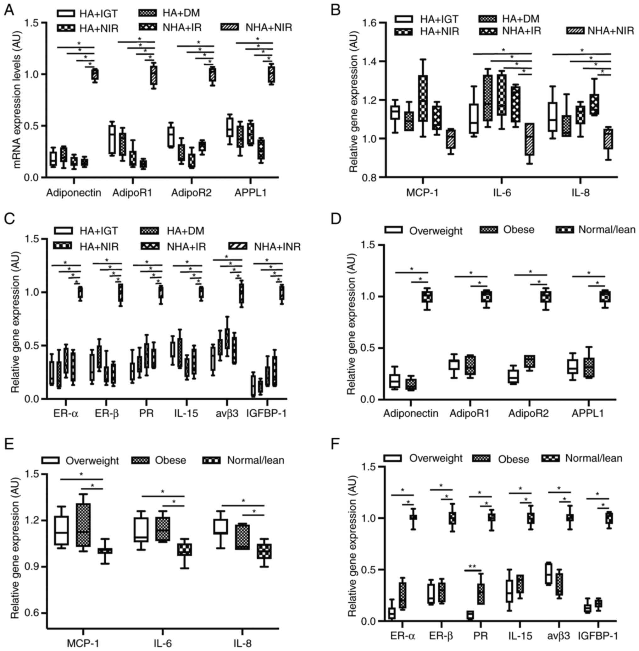 | Figure 1Relative mRNA levels of
receptivity-related factors in the endometrium across studied
groups. Relative mRNA levels of (A) adiponectin, AdipoR1, AdipoR2
and APPL1, (B) MCP-1, IL-6 and IL-8 and (C) ERα, ERβ, PR, IL-15,
avβ3 and IGFBP-1 of receptivity-related factors in endometrium
obtained from HA + IGT, HA + DM, HA + NIR and NHA + IR groups. For
each gene, the mRNA expression levels were normalized to the mean
value of NHA + NIR group (internal control). Relative mRNA levels
of (D) adiponectin, AdipoR1, AdipoR2 and APPL1, (E) MCP-1, IL-6 and
IL-8 and (F) ERα, ERβ, PR, IL-15, avβ3 and IGFBP-1 of
receptivity-related factors in endometrium obtained from
lean/normal, overweight and obese groups. For each gene, the mRNA
expression levels were normalized to the mean value of lean/normal
group (internal control). *P<0.05,
**P<0.01. HA, hyperandrogenemia; IGT, impaired
glucose tolerance; DM, diabetes mellitus; NIR, non-insulin
resistance; NHA, non-hyperandrogenemia androgen; IR, insulin
resistance; AdipoR, adiponectin receptor; APPL1, adapter protein
containing PH domain, PTB domain and leucine zipper motif 1; MCP-1,
monocyte chemoattractant protein-1; IL, interleukin; ER, estrogen
receptor; PR, progesterone receptor; avβ3, integrin β3; IGFBP-1,
insulin-like growth factor binding protein-1. |
For the comparison of different bodyweight groups,
the relative mRNA expression levels of adiponectin, AdipoR1,
AdipoR2, APPL1, ERα, ERβ, PR, IL-15, avβ3, and IGFBP-1 were
significantly lower, while the expression of MCP-1, IL-6 and IL-8
were significantly higher in the overweight and obese groups when
compared with the lean/normal group (all P<0.05). Furthermore,
the relative mRNA expression of PR in the obese group was
significantly higher compared with that in the overweight group
(P<0.01; Fig. 1).
Proportion of uNK cells
As presented in Fig.
2, the percentages of CD16-/CD56+ and
CD16+/CD56+ in endometrium were comparable
across HA + IGT, HA + DM, HA + NIR, NHA + IR and NHA + NIR groups
(P>0.05). In addition, no significant difference was revealed
among patients in the lean/normal, overweight and obese groups
(P>0.05).
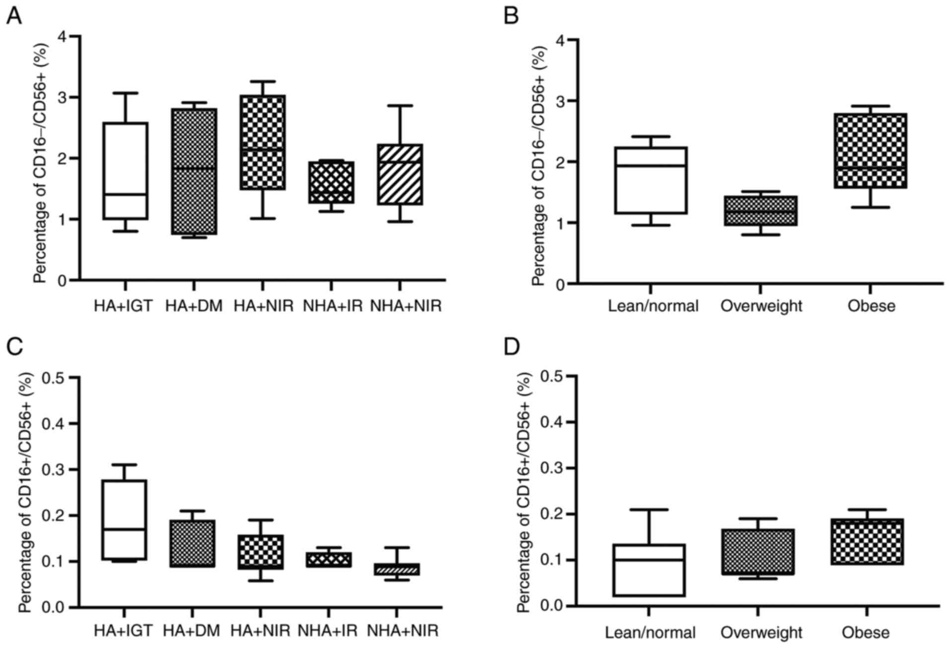 | Figure 2Percentage of subgroups of uNK cells
in the endometrium across studied groups. Percentage of CD16-/CD56+
in endometrium obtained from (A) HA + IGT, HA + DM, HA + NIR and
NHA + IR groups and (B) from lean/normal, overweight, and obese
groups. (C) Percentage of CD16+/CD56+ in endometrium obtained from
(C) HA + IGT, HA + DM, HA + NIR and NHA + IR groups and (D) from
lean/normal, overweight and obese groups. HA, hyperandrogenemia;
IGT, impaired glucose tolerance; DM, diabetes mellitus; NIR,
non-insulin resistance; NHA, non-hyperandrogenemia androgen; IR,
insulin resistance; uNK cells, uterine natural killer cells. |
Discussion
Wickham et al (42) hypothesized that IR can reduce serum
HMW adiponectin, while O'Connor et al (43) claimed that serum HMW adiponectin is
not dependent on IR. On the other hand, studies have indicated that
high androgen status may indirectly reduce serum HMW adiponectin in
patients with PCOS (44,45), but the evidence was still
insufficient. Garcia et al (41) revealed that the serum adiponectin
level in obese patients with PCOS was lower compared with that in
the lean or obese patient groups. Nevertheless, other studies
revealed no differences in the expression of adiponectin (46) or HMW adiponectin (47) among overweight/obese or
normal-weight patients between PCOS and control groups. So far,
there is no consensus on whether there are differences in serum
adiponectin in different types of patients with PCOS (42-47).
The present study revealed no significant difference in serum HMW
adiponectin levels among different high androgen statuses,
insulin-resistant levels or BMI levels in patients with PCOS. The
various patient group settings and the detection method used for
HMW adiponectin may explain the inconsistency in results compared
with previous studies. Although the present study revealed no
differences in HMW adiponectin, this does not mean that there was
no difference in other forms of adiponectin. The alteration in
other molecular forms of adiponectin should also be considered, and
this needs further exploration.
The endometrium undergoes three different phases
during the normal menstrual cycle, which is induced by the steroid
hormones fluctuation (48).
Estrogen (E2) promotes the expression of estrogen receptor α (Erα)
and estrogen receptor β (Erβ); the expression levels of these
receptors are highest in the late stage of proliferation. In
addition, E2, together with ER, promotes progesterone receptor (PR)
expression in the endometrium. After ovulation, progesterone
inhibits the expression of ER in the endometrium, thus inducing
decidualization. Decidualization is a necessary transformation of
endometrium for successful embryo implantation, including
endometrial stromal cells proliferation, the increase of glandular
epithelial secretion and natural killer cells aggregation (24). Decidualization usually happens at
5-6 days after ovulation (49).
The process of decidualization is accompanied by the alteration of
ER, PR and inhibition of pro-inflammatory cytokines (interleukin
and MCP-1) and endometrial pathological and physiological-related
factors (avβ3, IGFBP-1) (24,50).
Some researchers hypothesize that the decreased expression of ERα,
ERβ, PR, IL-15, avβ3 and IGFBP-1 may indicate the decline of embryo
implantation rate (51-54);
however, the mechanism of how these factors affect endometrial
receptivity remains unclear. Furthermore, previous studies have
revealed increased expression of Erα (37,55,56)
and PR (56), decreased expression
of IL-15(39), avβ3(57) and IGFBP-1(58) and unchanged expression of Erβ
(58) in secretory phase
endometrium in patients with PCOS when compared with non-PCOS
patients.
The present study demonstrated that the expression
levels of ERα, ERβ, PR, IL-15, avβ3 and IGFBP-1 were significantly
decreased in the presence of HA and/or IR and/or obesity (BMI ≥24
kg/m2), which were consistent with previous studies by
Matteo et al (39), Cermik
et al (57) and Piltonen
et al (58). Nevertheless,
the results were different from the findings by Margarit et
al (56) and Quezada et
al (37). The difference may
be due to the diversity in studied population. Apart from the
distinction of the hyperandrogenemia states, the IR levels and BMI,
the control group used in previous studies were different. All
previous studies were carried out with in untreated patients with
PCOS (with spontaneous ovulation) and non-PCOS patients. By
contrast, patients in the present study were treated with HRT to
guarantee the endometrial transformation.
During the menstrual cycle, adiponectin and its
receptors can be detected, the mRNA expression of adiponectin
increased significantly in the early stage of proliferation;
however, the mRNA expression levels of AdipoR1 and AdipoR2
increased significantly in the peri-implantation period (25). These changes have been confirmed in
the artificial decidua model as well (59). Adiponectin has been indicated to be
reduced in the endometrium of obese and PCOS patients (60). Garcia et al (41) demonstrated that the obese group
(compared with the lean group) have an increased expression of
endometrial AdipoR1 and a decreased expression of endometrial
APPL1, while the AdipoR2 expression is similar (41). In addition, they demonstrated that
when treating immortalized human endometrial stromal cell lines
with testosterone and insulin, the mRNA expression levels of
adiponectin, AdipoR1, AdipoR2 and APPL1 are significantly decreased
(41).
Similarly, partly consistent with previous studies
(37,41), the present study revealed that the
mRNA expression levels of adiponectin, AdipoR1, AdipoR2 and APPL1
were significantly decreased in the presence of HA and/or IR and/or
obesity (BMI ≥24 kg/m2) when compared with the NHA + NIR
group and the lean/normal group, respectively. These findings were
different from the results of Garcia et al (41), and this may be explained by the
variance in sample collection. Garcia et al (41) collected samples in the
proliferative phase and set untreated patients with PCOS (with
spontaneous ovulation) as the control group. By contrast, all the
patients with PCOS in the present study were treated with HRT, and
endometrium samples were collected during the window of
implantation in the HRT cycle.
MCP-1, IL-6 and IL-8 are pro-inflammatory factors
which are involved in the morphological and pathological changes in
the process of endometrial decidualization (61). It has been demonstrated that
adiponectin exerts anti-inflammatory effects in the endometrium by
inhibiting the production of pro-inflammatory cytokines (IL-6, IL-8
and MCP-1) (5). Compared with the
NHA + NIR group and the lean/normal group, the expression levels of
MCP-1, IL-6 and IL-8 were significantly increased in the
endometrium in the presence of HA and/or IR and/or obesity (BMI ≥24
kg/m2) of patients with PCOS. It was hypothesized that
the reduction of adiponectin may explain the increased levels of
IL-6, IL-8 and MCP-1 in the endometrium.
Rosenbaum et al (30) revealed that obesity can affect
endometrial decidualization in the mouse model and human embryonic
stem cells. In addition, Comstock et al (62) claimed that obesity can change the
expression of genes involved in implantation-related chemokine
signaling pathways during implantation, especially in obese
patients with metabolic syndrome (63). Notably, the present study revealed
that the mRNA expression level of PR in the obese group was
significantly higher compared with that in the overweight group. We
hypothesized that obesity increased the expression of PR as the
response to progesterone resistance (53). However, the underlying mechanism
remains elusive and further studies are needed.
As a factor associated with endometrial receptivity
(39,64), the percentage of uNK cells
fluctuates along with the hormones changing during the menstrual
cycle (65,66), which increases during the secretory
phase (39). However, increases in
the number of peripheral blood and endometrial NK cells
(CD56+) have been used as an indicator to assess the
risk of infertility or recurrent miscarriage (39). Piltonen et al (67) and Matteo et al (39) revealed that uNK cells decrease in
the late menstrual secretion period, and the percentage of uNK
cells CD16+/CD56+ is similar, while the
percentage of CD16-/CD56+ is lower in the
secretion phase in patients with PCOS when compared with the
control group. Nevertheless, no significant difference was revealed
in the present study in uNK cells among patients with PCOS with
different BMI, androgen status and IR levels. Considering the
different phases of the menstrual cycle, the percentage of
CD16-/CD56+ was observed to decrease after
implantation, while a previous study revealed the percentage of
CD16-/CD56+ was lower in the late menstrual
secretion period (67). But this
research needs to be explored further.
To avoid the impact of actual human embryo
implantation on the endometrium, the present investigation was not
performed in a conception cycle but in the HRT cycles. The HRT
treatment was performed in all the patients with PCOS to promote
endometrial transformation and cause implantation window-related
changes. Although a previous study indicated that ER and PR
expression is significantly decreased in the endometrium in the
early luteal phase of HRT cycles (68), all the studied patients in the
present study were treated with HRT; therefore avoiding the impact
of internal hormone alteration caused by ovulation, and ensuring
all patients were in the same endometrial phase. In the majority of
previous studies (24,37,41)
the expression of receptivity markers are measured from mRNA level.
However, to clarify the differences in the expression level of
theses markers, the detection from protein level is important; thus
further exploration is needed to validate the present findings.
In conclusion, evidence regarding secretory
endometrial receptivity factors in patients with PCOS is limited,
so consistent conclusions cannot yet be made. The present study
revealed that the IR status, hyperandrogenemia and obesity would
impact the endometrial receptivity in patients with PCOS, which may
explain the damaged embryo implantation and pregnancy outcomes. To
develop the targeted therapies and improve pregnancy outcomes in
patients with PCOS, further studies are needed to investigate the
underlying mechanism of the impaired endometrium receptivity caused
by metabolic disorders.
Supplementary Material
uNK cell subtype percentages of
studied groups. Images represent flow cytometry plots of
endometrial cells, including lymphocyte cells, single cells, and
CD3- cells. (A) HA + IGT group; (B) HA + DM group; (C)
HA + NIR group; (D) NHA + IR group; (E) NHA + NIR group. HA,
hyperandrogenemia; IGT, impaired glucose tolerance; DM, diabetes
mellitus; NIR, noninsulin resistance; NHA, non-hyperandrogenemia
androgen; IR, insulin resistance; uNK cells, uterine natural killer
cells.
uNK cell subtype percentages of
studied groups. Images represent flow cytometry plots of
endometrial cells, including lymphocytes cells, single cells, and
CD3- cells. (A) lean/normal group; (B) overweight group;
(C) obese group. HA, hyperandrogenemia; IGT, impaired glucose
tolerance; DM, diabetes mellitus; NIR, non-insulin resistance; NHA,
non-hyperandrogenemia androgen; IR, insulin resistance; uNK cells,
uterine natural killer cells.
Acknowledgements
Not applicable.
Funding
Funding: No funding was received.
Availability of data and materials
The datasets used and/or analyzed during the current
study are available from the corresponding author on reasonable
request.
Authors' contributions
CW and QYM conceptualized the study and analyzed and
interpreted the data. CW and YXW performed experiments and wrote
the manuscript. CW and QYM confirm the authenticity of all the raw
data. All authors have read and approved the final manuscript.
Ethics approval and consent to
participate
The present study was approved by the Clinical
Scientific Research and Experimental Animal Ethics Committee of the
First Affiliated Hospital of Sun Yat-sen University [approval no.
Ethics (2020) no. 422-1]. Written informed consent was obtained
from all participants.
Patient consent for publication
Not applicable.
Competing interests
The authors declare that they have no competing
interests.
References
|
1
|
Dabadghao P, Roberts BJ, Wang J, Davies MJ
and Norman RJ: Glucose tolerance abnormalities in Australian women
with polycystic ovary syndrome. Med J Aust. 187:328–331.
2007.PubMed/NCBI View Article : Google Scholar
|
|
2
|
Li R, Zhang Q, Yang D, Li S, Lu S, Wu X,
Wei Z, Song X, Wang X, Fu S, et al: Prevalence of polycystic ovary
syndrome in women in China: A large community-based study. Hum
Reprod. 28:2562–9256. 2013.PubMed/NCBI View Article : Google Scholar
|
|
3
|
Ovalle F and Azziz R: Insulin resistance,
polycystic ovary syndrome, and type 2 diabetes mellitus. Fertil
Steril. 77:1095–1105. 2002.PubMed/NCBI View Article : Google Scholar
|
|
4
|
DeUgarte CM, Bartolucci AA and Azziz R:
Prevalence of insulin resistance in the polycystic ovary syndrome
using the homeostasis model assessment. Fertil Steril.
83:1454–1460. 2005.PubMed/NCBI View Article : Google Scholar
|
|
5
|
Carmina E and Lobo RA: Use of fasting
blood to assess the prevalence of insulin resistance in women with
polycystic ovary syndrome. Fertil Steril. 82:661–665.
2004.PubMed/NCBI View Article : Google Scholar
|
|
6
|
Moran LJ, Pasquali R, Teede HJ, Hoeger KM
and Norman RJ: Treatment of obesity in polycystic ovary syndrome: A
position statement of the androgen excess and polycystic ovary
syndrome society. Fertil Steril. 92:1966–1982. 2009.PubMed/NCBI View Article : Google Scholar
|
|
7
|
Greenwood EA and Huddleston HG: Insulin
resistance in polycystic ovary syndrome: Concept versus cutoff.
Fertil Steril. 112:827–828. 2019.PubMed/NCBI View Article : Google Scholar
|
|
8
|
Zeng X, Xie YJ, Liu YT, Long SL and Mo ZC:
Polycystic ovarian syndrome: Correlation between hyperandrogenism,
insulin resistance and obesity. Clin Chim Acta. 502:214–221.
2020.PubMed/NCBI View Article : Google Scholar
|
|
9
|
Xiao XU and Xue-Lian LI: The role of
androgens in the pathogenesis and treatment of female reproductive
endocrine diseases. J Int Obstet Gynecol. 46:229–232. 2019.
|
|
10
|
Patlolla S, Vaikkakara S, Sachan A,
Venkatanarasu A, Bachimanchi B, Bitla A, Settipalli S, Pathiputturu
S, Sugali RN and Chiri S: Heterogenous origins of hyperandrogenism
in the polycystic ovary syndrome in relation to body mass index and
insulin resistance. Gynecol Endocrinol. 34:238–242. 2018.PubMed/NCBI View Article : Google Scholar
|
|
11
|
Naver KV, Grinsted J, Larsen SO, Hedley
PL, Jørgensen FS, Christiansen M and Nilas L: Increased risk of
preterm delivery and pre-eclampsia in women with polycystic ovary
syndrome and hyperandrogenaemia. BJOG. 121:575–581. 2014.PubMed/NCBI View Article : Google Scholar
|
|
12
|
Elenis E, Desroziers E, Persson S,
Sundström Poromaa I and Campbell RE: Early initiation of
anti-androgen treatment is associated with increased probability of
spontaneous conception leading to childbirth in women with
polycystic ovary syndrome: A population-based multiregistry cohort
study in Sweden. Hum Reprod. 36:1427–1435. 2021.PubMed/NCBI View Article : Google Scholar
|
|
13
|
Chang EM, Han JE, Seok HH, Lee DR, Yoon TK
and Lee WS: Insulin resistance does not affect early embryo
development but lowers implantation rate in in vitro maturation-in
vitro fertilization-embryo transfer cycle. Clin Endocrinol.
79:93–99. 2013.PubMed/NCBI View Article : Google Scholar
|
|
14
|
Tian L, Shen H, Lu Q, Norman RJ and Wang
J: Insulin resistance increases the risk of spontaneous abortion
after assisted reproduction technology treatment. J Clin Endocrinol
Metab. 92:1430–1433. 2007.PubMed/NCBI View Article : Google Scholar
|
|
15
|
Khattab S, Mohsen IA, Foutouh IA, Ramadan
A, Moaz M and Al-Inany H: Metformin reduces abortion in pregnant
women with polycystic ovary syndrome. Gynecol Endocrinol.
22:680–684. 2006.PubMed/NCBI View Article : Google Scholar
|
|
16
|
Huang K, Liao X, Dong X and Zhang H:
Effect of overweight/obesity on IVF-ET outcomes in Chinese patients
with polycystic ovary syndrome. Int J Clin Exp Med. 7:5872–5876.
2014.PubMed/NCBI
|
|
17
|
Veleva Z, Tiitinen A, Vilska S,
Hydén-Granskog C, Tomás C, Martikainen H and Tapanainen JS: High
and low BMI increase the risk of miscarriage after IVF/ICSI and
FET. Hum Reprod. 23:878–884. 2008.PubMed/NCBI View Article : Google Scholar
|
|
18
|
Fedorcsák P, Storeng R, Dale PO, Tanbo T
and Abyholm T: Obesity is a risk factor for early pregnancy loss
after IVF or ICSI. Acta Obstet Gynecol Scand. 79:43–48.
2000.PubMed/NCBI
|
|
19
|
Bu Z, Dai W, Guo Y, Su Y, Zhai J and Sun
Y: Overweight and obesity adversely affect outcomes of assisted
reproductive technologies in polycystic ovary syndrome patients.
Int J Clin Exp Med. 6:991–995. 2013.PubMed/NCBI
|
|
20
|
Patel SS and Carr BR: Oocyte quality in
adult polycystic ovary syndrome. Semin Reprod Med. 26:196–203.
2008.PubMed/NCBI View Article : Google Scholar
|
|
21
|
Jungheim ES, Schon SB, Schulte MB,
DeUgarte DA, Fowler SA and Tuuli MG: IVF outcomes in obese donor
oocyte recipients: A systematic review and meta-analysis. Hum
Reprod. 28:2720–2727. 2013.PubMed/NCBI View Article : Google Scholar
|
|
22
|
Wattanakumtornkul S, Damario MA, Stevens
HS, Thornhill AR and Tummon IS: Body mass index and uterine
receptivity in the oocyte donation model. Fertil Steril.
80:336–340. 2003.PubMed/NCBI View Article : Google Scholar
|
|
23
|
Bellver J, Rossal LP, Bosch E, Zúñiga A,
Corona JT, Meléndez F, Gómez E, Simón C, Remohí J and Pellicer A:
Obesity and the risk of spontaneous abortion after oocyte donation.
Fertil Steril. 79:1136–1140. 2003.PubMed/NCBI View Article : Google Scholar
|
|
24
|
Piltonen TT: Polycystic ovary syndrome:
Endometrial markers. Best Pract Res Clin Obstet Gynaecol. 37:66–79.
2016.PubMed/NCBI View Article : Google Scholar
|
|
25
|
Takemura Y, Osuga Y, Yamauchi T, Kobayashi
M, Harada M, Hirata T, Morimoto C, Hirota Y, Yoshino O, Koga K, et
al: Expression of adiponectin receptors and its possible
implication in the human endometrium. Endocrinology. 147:3203–3210.
2006.PubMed/NCBI View Article : Google Scholar
|
|
26
|
Wang Y, Xie X and Zhu W: Serum adiponectin
and resistin levels in patients with polycystic ovarian syndrome
and their clinical implications. J Huazhong Univ Sci Technolog Med
Sci. 30:638–642. 2010.PubMed/NCBI View Article : Google Scholar
|
|
27
|
Barbe A, Bongrani A, Mellouk N, Estienne
A, Kurowska P, Grandhaye J, Elfassy Y, Levy R, Rak A, Froment P and
Dupont J: Mechanisms of adiponectin action in fertility: An
overview from gametogenesis to gestation in humans and animal
models in normal and pathological conditions. Int J Mol Sci.
20(1526)2019.PubMed/NCBI View Article : Google Scholar
|
|
28
|
Fisher FM, Trujillo ME, Hanif W, Barnett
AH, McTernan PG, Scherer PE and Kumar S: Serum high molecular
weight complex of adiponectin correlates better with glucose
tolerance than total serum adiponectin in Indo-Asian males.
Diabetologia. 48:1084–1087. 2005.PubMed/NCBI View Article : Google Scholar
|
|
29
|
Yamauchi T, Iwabu M, Okada-Iwabu M and
Kadowaki T: Adiponectin receptors: A review of their structure,
function and how they work. Best Pract Res Clin Endocrinol Metab.
28:15–23. 2014.PubMed/NCBI View Article : Google Scholar
|
|
30
|
Rosenbaum D, Haber RS and Dunaif A:
Insulin resistance in polycystic ovary syndrome: Decreased
expression of GLUT-4 glucose transporters in adipocytes. Am J
Physiol. 264:E197–E202. 1993.PubMed/NCBI View Article : Google Scholar
|
|
31
|
Rotterdam ESHRE/ASRM-Sponsored PCOS
Consensus Workshop Group. Revised 2003 consensus on diagnostic
criteria and long-term health risks related to polycystic ovary
syndrome. Fertil Steril. 81:19–25. 2004.PubMed/NCBI View Article : Google Scholar
|
|
32
|
Chang WY, Knochenhauer ES, Bartolucci AA
and Azziz R: Phenotypic spectrum of polycystic ovary syndrome:
Clinical and biochemical characterization of the three major
clinical subgroups. Fertil Steril. 83:1717–1723. 2005.PubMed/NCBI View Article : Google Scholar
|
|
33
|
Soonthornpun S, Setasuban W, Thamprasit A,
Chayanunnukul W, Rattarasarn C and Geater A: Novel insulin
sensitivity index derived from oral glucose tolerance test. J Clin
Endocrinol Metab. 88:1019–1023. 2003.PubMed/NCBI View Article : Google Scholar
|
|
34
|
Salley KE, Wickham EP, Cheang KI, Essah
PA, Karjane NW and Nestler JE: Glucose intolerance in polycystic
ovary syndrome-a position statement of the androgen excess society.
J Clin Endocrinol Metab. 92:4546–4556. 2007.PubMed/NCBI View Article : Google Scholar
|
|
35
|
Finucane MM, Stevens GA, Cowan MJ, Danaei
G, Lin JK, Paciorek CJ, Singh GM, Gutierrez HR, Lu Y, Bahalim AN,
et al: National, regional, and global trends in body-mass index
since 1980: Systematic analysis of health examination surveys and
epidemiological studies with 960 country-years and 9.1 million
participants. Lancet. 377:557–567. 2011.PubMed/NCBI View Article : Google Scholar
|
|
36
|
Noyes RW, Hertig AT and Rock J: Reprint
of: Dating the endometrial biopsy. Fertil Steril. 112 (4 Suppl
1):e93–e115. 2019.PubMed/NCBI View Article : Google Scholar
|
|
37
|
Quezada S, Avellaira C, Johnson MC, Gabler
F, Fuentes A and Vega M: Evaluation of steroid receptors,
coregulators, and molecules associated with uterine receptivity in
secretory endometria from untreated women with polycystic ovary
syndrome. Fertil Steril. 85:1017–1026. 2006.PubMed/NCBI View Article : Google Scholar
|
|
38
|
Livak KJ and Schmittgen TDL: Analysis of
relative gene expression data using real-time quantitative PCR and
the 2(-Delta Delta C(T)) method. Methods. 25:402–408.
2001.PubMed/NCBI View Article : Google Scholar
|
|
39
|
Matteo M, Serviddio G, Massenzio F,
Scillitani G, Castellana L, Picca G, Sanguedolce F, Cignarelli M,
Altomare E, Bufo P, et al: Reduced percentage of natural killer
cells associated with impaired cytokine network in the secretory
endometrium of infertile women with polycystic ovary syndrome.
Fertil Steril. 94:2222–2227.e1-e3. 2010.PubMed/NCBI View Article : Google Scholar
|
|
40
|
Sinclair JC and Haynes RB: Selecting
participants that raise a clinical trial's population attributable
fraction can increase the treatment effect within the trial and
reduce the required sample size. J Clin Epidemiol. 64:893–902.
2011.PubMed/NCBI View Article : Google Scholar
|
|
41
|
Garcia V, Oróstica L, Poblete C, Rosas C,
Astorga I, Romero C and Vega M: Endometria from obese PCOS women
with hyperinsulinemia exhibit altered adiponectin signaling. Horm
Metab Res. 47:901–909. 2015.PubMed/NCBI View Article : Google Scholar
|
|
42
|
Wickham ER III, Cheang KI, Clore JN,
Baillargeon JP and Nestler JE: Total and high-molecular weight
adiponectin in women with the polycystic ovary syndrome.
Metabolism. 60:366–372. 2011.PubMed/NCBI View Article : Google Scholar
|
|
43
|
O'Connor A, Phelan N, Tun TK, Boran G,
Gibney J and Roche HM: High-molecular-weight adiponectin is
selectively reduced in women with polycystic ovary syndrome
independent of body mass index and severity of insulin resistance.
J Clin Endocrinol Metab. 95:1378–1385. 2010.PubMed/NCBI View Article : Google Scholar
|
|
44
|
Panidis D, Kourtis A, Farmakiotis D,
Mouslech T, Rousso D and Koliakos G: Serum adiponectin levels in
women with polycystic ovary syndrome. Hum Reprod. 18:1790–1796.
2003.PubMed/NCBI View Article : Google Scholar
|
|
45
|
Xu A, Chan KW, Hoo RL, Wang Y, Tan KC,
Zhang J, Chen B, Lam MC, Tse C, Cooper GJ and Lam KS: Testosterone
selectively reduces the high molecular weight form of adiponectin
by inhibiting its secretion from adipocytes. J Biol Chem.
280:18073–18080. 2005.PubMed/NCBI View Article : Google Scholar
|
|
46
|
Lecke SB, Mattei F, Morsch DM and Spritzer
PM: Abdominal subcutaneous fat gene expression and circulating
levels of leptin and adiponectin in polycystic ovary syndrome.
Fertil Steril. 95:2044–2049. 2011.PubMed/NCBI View Article : Google Scholar
|
|
47
|
Barber TM, Hazell M, Christodoulides C,
Golding SJ, Alvey C, Burling K, Vidal-Puig A, Groome NP, Wass JA,
Franks S and McCarthy MI: Serum levels of retinol-binding protein 4
and adiponectin in women with polycystic ovary syndrome:
Associations with visceral fat but no evidence for fat
mass-independent effects on pathogenesis in this condition. J Clin
Endocrinol Metab. 93:2859–2865. 2008.PubMed/NCBI View Article : Google Scholar
|
|
48
|
Snijders MP, de Goeij AF, Debets-Te Baerts
MJ, Rousch MJ, Koudstaal J and Bosman FT: Immunocytochemical
analysis of oestrogen receptors and progesterone receptors in the
human uterus throughout the menstrual cycle and after the
menopause. J Reprod Fertil. 94:363–371. 1992.PubMed/NCBI View Article : Google Scholar
|
|
49
|
Gellersen B and Brosens JJ: Cyclic
decidualization of the human endometrium in reproductive health and
failure. Endocr Rev. 35:851–905. 2014.PubMed/NCBI View Article : Google Scholar
|
|
50
|
Weyer C, Tataranni PA, Bogardus C and
Pratley RE: Insulin resistance and insulin secretory dysfunction
are independent predictors of worsening of glucose tolerance during
each stage of type 2 diabetes development. Diabetes Care. 24:89–94.
2001.PubMed/NCBI View Article : Google Scholar
|
|
51
|
Benkhalifa M, Madkour A, Louanjli N,
Bouamoud N, Saadani B, Kaarouch I, Chahine H, Sefrioui O, Merviel P
and Copin H: From global proteome profiling to single targeted
molecules of follicular fluid and oocyte: Contribution to embryo
development and IVF outcome. Expert Rev Proteomics. 12:407–423.
2015.PubMed/NCBI View Article : Google Scholar
|
|
52
|
Lessey BA, Palomino WA, Apparao KB, Young
SL and Lininger RA: Estrogen receptor-alpha (ER-alpha) and defects
in uterine receptivity in women. Reprod Biol Endocrinol. 4 (Suppl
1)(S9)2006.PubMed/NCBI View Article : Google Scholar
|
|
53
|
Young SL and Lessey BA: Progesterone
function in human endometrium: Clinical perspectives. Semin Reprod
Med. 28:5–16. 2010.PubMed/NCBI View Article : Google Scholar
|
|
54
|
Gnainsky Y, Granot I, Aldo PB, Barash A,
Or Y, Schechtman E, Mor G and Dekel N: Local injury of the
endometrium induces an inflammatory response that promotes
successful implantation. Fertil Steril. 94:2030–2036.
2010.PubMed/NCBI View Article : Google Scholar
|
|
55
|
Gregory CW, Wilson EM, Apparao KB,
Lininger RA, Meyer WR, Kowalik A, Fritz MA and Lessey BA: Steroid
receptor coactivator expression throughout the menstrual cycle in
normal and abnormal endometrium. J Clin Endocrinol Metab.
87:2960–2966. 2002.PubMed/NCBI View Article : Google Scholar
|
|
56
|
Margarit L, Taylor A, Roberts MH, Hopkins
L, Davies C, Brenton AG, Conlan RS, Bunkheila A, Joels L, White JO
and Gonzalez D: MUC1 as a discriminator between endometrium from
fertile and infertile patients with PCOS and endometriosis. J Clin
Endocrinol Metab. 95:5320–5329. 2010.PubMed/NCBI View Article : Google Scholar
|
|
57
|
Cermik D, Selam B and Taylor HS:
Regulation of HOXA-10 expression by testosterone in vitro and in
the endometrium of patients with polycystic ovary syndrome. J Clin
Endocrinol Metab. 88:238–243. 2003.PubMed/NCBI View Article : Google Scholar
|
|
58
|
Piltonen TT, Chen JC, Khatun M,
Kangasniemi M, Liakka A, Spitzer T, Tran N, Huddleston H, Irwin JC
and Giudice LC: Endometrial stromal fibroblasts from women with
polycystic ovary syndrome have impaired progesterone-mediated
decidualization, aberrant cytokine profiles and promote enhanced
immune cell migration in vitro. Hum Reprod. 30:1203–1215.
2015.PubMed/NCBI View Article : Google Scholar
|
|
59
|
Gamundi-Segura S, Serna J, Oehninger S,
Horcajadas JA and Arbones-Mainar JM: Effects of adipocyte-secreted
factors on decidualized endometrial cells: Modulation of
endometrial receptivity in vitro. J Physiol Biochem. 71:537–546.
2015.PubMed/NCBI View Article : Google Scholar
|
|
60
|
Palin MF, Bordignon VV and Murphy BD:
Adiponectin and the control of female reproductive functions. Vitam
Horm. 90:239–287. 2012.PubMed/NCBI View Article : Google Scholar
|
|
61
|
Yoshino O, Osuga Y, Hirota Y, Koga K,
Hirata T, Yano T, Ayabe T, Tsutsumi O and Taketani Y: Endometrial
stromal cells undergoing decidualization down-regulate their
properties to produce proinflammatory cytokines in response to
interleukin-1 beta via reduced p38 mitogen-activated protein kinase
phosphorylation. J Clin Endocrinol Metab. 88:2236–2241.
2003.PubMed/NCBI View Article : Google Scholar
|
|
62
|
Comstock IA, Diaz-Gimeno P, Cabanillas S,
Bellver J, Sebastian-Leon P, Shah M, Schutt A, Valdes CT,
Ruiz-Alonso M, Valbuena D, et al: Does an increased body mass index
affect endometrial gene expression patterns in infertile patients?
A functional genomics analysis. Fertil Steril. 107:740–748.e2.
2017.PubMed/NCBI View Article : Google Scholar
|
|
63
|
Tierney EP, Tulac S, Huang ST and Giudice
LC: Activation of the protein kinase A pathway in human endometrial
stromal cells reveals sequential categorical gene regulation.
Physiol Genomics. 16:47–66. 2003.PubMed/NCBI View Article : Google Scholar
|
|
64
|
Gellersen B, Brosens IA and Brosens JJ:
Decidualization of the human endometrium: Mechanisms, functions,
and clinical perspectives. Semin Reprod Med. 25:445–453.
2007.PubMed/NCBI View Article : Google Scholar
|
|
65
|
Giudice LC: Endometrium in PCOS:
Implantation and predisposition to endocrine CA. Best Pract Res
Clin Endocrinol Metab. 20:235–244. 2006.PubMed/NCBI View Article : Google Scholar
|
|
66
|
Laird SM, Tuckerman EM, Cork BA, Linjawi
S, Blakemore AI and Li TC: A review of immune cells and molecules
in women with recurrent miscarriage. Hum Reprod Update. 9:163–174.
2003.PubMed/NCBI View Article : Google Scholar
|
|
67
|
Piltonen TT, Chen J, Erikson DW, Spitzer
TL, Barragan F, Rabban JT, Huddleston H, Irwin JC and Giudice LC:
Mesenchymal stem/progenitors and other endometrial cell types from
women with polycystic ovary syndrome (PCOS) display inflammatory
and oncogenic potential. J Clin Endocrinol Metab. 98:3765–3775.
2013.PubMed/NCBI View Article : Google Scholar
|
|
68
|
Habiba MA, Bell SC and Al-Azzawi F: The
effect of hormone replacement therapy on the immunoreactive
concentrations in the endometrium of oestrogen and progesterone
receptor, heat shock protein 27, and human beta-lactoglobulin. Hum
Reprod. 15:36–42. 2000.PubMed/NCBI View Article : Google Scholar
|


