Introduction
Hepatoma is a common malignant digestive system
tumor with high morbidity (854,000) and mortality (810,000) (2015
global incidence and deaths for hepatoma) (1), and seriously threatens the health and
lives of individuals. The treatment of hepatoma includes surgery,
interventional therapy and radiotherapy (2).
Chemotherapy is the main treatment for patients with
hepatoma who have lost the opportunity for surgical treatment. The
side effects and drug resistance during chemotherapy seriously
affect the outcome of and may lead to the failure of chemotherapy.
The aim of the present study was to explore the mechanism of drug
resistance in hepatoma and targeted interventions that may
effectively improve the outcome of chemotherapy.
It has been found that the adenosine triphosphate
(ATP) binding transporter family has an important role in the
development of multidrug resistance by reducing the concentration
of chemotherapeutic drugs in tumor cells (3-5),
among which ATP binding cassette (ABC)B1 and ABCG2 are most closely
associated with multidrug resistance (6-9).
ABCG2 is involved in the formation of multidrug
resistance in numerous types of tumors and is the main marker of
side population cells (10), which
are involved in the formation of cancer stem cells (11). Chen (12) reported that ABCG2 protein is a
marker of glioma stem cells and side population cells, and found
that ABCG2 is related to drug resistance in glioma. Wang et
al (13) reported that ABCG2
is a marker of breast cancer stem cells and that ABCG2 is related
to the chemoresistance, tumor recurrence, metastasis and invasion
of breast cancer. However, to the best of our knowledge, the
association between ABCG2 and the multidrug resistance of hepatoma
has rarely been reported. In the present study, drug-resistant
cells of hepatoma were established and the expression of ABCG2 in
such drug-resistant cells was detected. The association between the
multidrug resistance of ABCG2 and hepatoma was then discussed.
ABCG2 may cause a decrease of the effective drug
concentration in tumor cells by the efflux of chemotherapeutic
drugs, which leads to drug resistance (13). Therefore, the aim of the present
study was to discuss the mechanism of ADM resistance and provide an
ADM-resistant cell model for the clinical study of ADM resistance
in hepatoma cells. To this end, drug-resistant hepatoma cells were
established and the expression of ABCG2 in these drug-resistant
cells was detected. Flow cytometry (FCM) was used to detect the
concentration of Adriamycin (ADM) in HepG2/ADM (drug-resistant
hepatoma cells), HepG2/ABCG2, HepG2/PCDNA3.1 and their parental
cells (HepG2) and to analyze the efflux function of HepG2/ADM and
HepG2/ABCG2 cells. The association between the multidrug resistance
of ABCG2 and hepatoma was then discussed.
Materials and methods
Chemicals and reagents
ABCG2 antibody (cat. no. sc-18841) was purchased
from Santa Cruz Biotechnology, Inc. IgG-FITC antibody (cat. no.
115-095-003) was purchased from Jackson ImmunoResearch
Laboratories, Inc. Propidium iodide/RNase (cat. no. 550825) and
Annexin V-FITC/propidium iodide (cat. no. 556547) were purchased
from Becton, Dickinson and Company. ADM (cat. no. H33021980) was
purchased from Hanhui Pharmaceuticals Co., Ltd.
Establishment of the ADM-resistant
hepatoma cell line
The HepG2 cell line (cat. no. CL-0103) was obtained
from Procell Life Science & Technology Co., Ltd., and was
authenticated by STR profiling and analyzed for mycoplasma
contamination. The HepG2 cell line was cultured in MEM containing
10% FBS (Gibco; Thermo Fisher Scientific, Inc.), 1% penicillin and
streptomycin. Cells were sustained in an incubator at 37˚C with 5%
CO2. An ADM-resistant hepatoma cell line was established
from the HepG2 cells by continuous exposure to increasing
concentrations of ADM, from 0.01 to 0.1 µg/ml for 3 months. One of
the surviving clones was isolated and designated as HepG2/ADM
cells.
ABCG2 gene transfection
pcDNA3.1-ABCG2 plasmid [sequences: CDS of ABCG2
(BC021281) gene (494…2,461)] containing ABCG2 cDNA and empty
pcDNA3.1 plasmid were purchased from Wuhan Genesil Biotechnology
Co., Ltd. Lipofectamine® 2000 (Invitrogen; Thermo Fisher
Scientific, Inc.) was used as the transfection reagent, according
to the manufacturer's instructions, and stably transfected cell
clones were selected using 600 mg/l G418 (without penicillin and
streptomycin) subsequent to transfection for 72 h. The transfection
reagents used were as follows. i) Reagent 1: 5 µl of
Lipofectamine® 2000 was mixed with 245 µl of MEM culture
medium (FBS- and antibiotic-free), and then placed at room
temperature for 5 min; ii) reagent 2: 4 µl of pcDNA3.1-ABCG2 or
pcDNA3.1 plasmid (containing 2 µg plasmid) was mixed with 246 µl of
MEM culture medium (FBS- and antibiotic-free) and then placed at
room temperature for 5 min. Reagents 1 and 2 were mixed gently,
placed at room temperature for 20 min and then added into the
wells. The HepG2 cells transfected with pcDNA3.1-ABCG2 or pcDNA3.1
and selected with 600 mg/l G418 for 10 days were termed HepG2/ABCG2
or HepG2/pcDNA3.1 cells, respectively. ABCG2 mRNA and protein
expression levels were monitored to ascertain the efficacy and
specificity of the transfection using reverse
transcription-quantitative PCR and FCM, respectively.
Cytotoxicity assay
The ADM anticancer drug sensitivity of the HepG2,
HepG2/pcDNA3.1, HepG2/ADM and HepG2/ABCG2 cells was determined
using the MTT assay. This method is based on the capacity of viable
cells to metabolize a yellow tetrazolium salt, MTT, using
mitochondrial succinate dehydrogenase, into purple formazan
crystals when dissolved in acidified propan-2-ol; the resulting
purple solution is then spectrophotometrically measured at 490 nm
(14). The cells were seeded into
96-well culture plates at a density of 1x10⁴ cells/ml.
The serial concentrations of ADM (0, 0.001, 0.005, 0.01, 0.05, 0.1,
0.5, 1, 5, 10 and 50 µg/ml) were added in a final volume of 200
µl/well. Normal saline (vehicle; containing 0 µg/ml ADM) was used
for the control group. Following drug treatment in an incubator at
37˚C with 5% CO2 for 24 h, the medium was replaced with
an equal volume of complete culture medium containing 0.5 mg/ml MTT
and incubated at 37˚C with 5% CO2 for 4 h. The medium
was removed, 180 µl DMSO was added to each well and plates were
continuously shaken for 10 min at room temperature. The cytotoxic
effects of the drugs were determined using the optical density (OD)
values from a microplate reader at an absorption wavelength of 490
nm (OD490). The inhibitory rate (IR) was calculated
using the following formula in order to calculate the half-maximal
inhibitory concentration (IC50) of the cells: IR
(%)=(1-OD490 treated cells/OD490 control
cells) x100%. The resistance index was determined as the
IC50 of the resistant cells (HepG2/ADM or HepG2/ABCG2
cells)/IC50 of the parental cells (HepG2 cells). Each
experiment was performed in triplicate wells.
Detection of ABCG2 mRNA expression in
cells
Total RNA of cells was extracted by RNA isolater
(Vazyme Biotech) according to the manufacturer's instructions.
Following extraction of total RNA from HepG2, HepG2/pcDNA3.1,
HepG2/ADM and HepG2/ABCG2 cells, total RNA was reversely
transcribed into cDNA using the HiScript II 1st Strand cDNA
Synthesis Kit (Vazyme Biotech) according to the manufacturer's
instructions, and PCR amplification was then performed using the
cDNA as a template. The thermocycling conditions were as follows:
95˚C for 5 min (pre-denaturation), followed by 40 cycles of 95˚C
for 15 sec, 58˚C for 30 sec and 72˚C for 25 sec, and a final cycle
of 95˚C for 15 sec, 58˚C for 1 min and 95˚C for 30 sec. SYBR-Green
I was used as the fluorescent dye. Real-time PCR was performed using
AceQ qPCR SYBR Green Master Mix kit (Vazyme Biotech) according to
the manufacturer's instructions. Human GAPDH was used as the
internal reference for standardization. The primers used were as
follows: ABCG2 forward, 5'-GGTCAGAGTGTGGTTTCTGTAGCA-3' and reverse,
5'-GTGAGAGATCGATGCCCTGCTTTA-3'; and GAPDH forward,
5'-ACCACAGTCCATGCCATCAC-3' and reverse, 5'-TCCACCACCCTGTTGCTGTA-3'.
The relative expression level of ABCG2 mRNA was calculated using
the 2-ΔΔCq method
(ΔCq=CqABCG2-CqGAPDH) (15). Each experiment was performed three
independent replicates.
Assessment of cell apoptosis and cell
cycle distribution using FCM
HepG2/ADM, HepG2/ABCG2, HepG2/pcDNA3.1 and HepG2
cells were collected during the logarithmic growth phase. A total
of 1 ml of the single-cell suspension (containing 1x10⁶
cells) was prepared, washed with cold PBS once and suspended in 100
µl of 1X binding buffer. Subsequently, 10 µl Annexin V-FITC was
added and the mixture was placed on ice for 15 min in the dark.
Next, 380 µl of 1X binding buffer and 10 µl propidium iodide were
added and the cell mixture was incubated on ice for 15 min in the
dark, washed with cold PBS once and suspended with 1 ml PBS. The
apoptosis of the cells was measured using an FC500 flow cytometer
(Beckman Coulter, Inc.). EXPO32 ADC software (version 1.2; Beckman
Coulter, Inc.) was used to analyze the immunofluorescence data and
evaluate the apoptotic rate. Each experiment was performed as three
independent replicates.
A total of 1 ml of the single-cell suspension
(containing 1x10⁶ cells) was prepared, washed with cold
PBS twice and fixed with 70% ethanol at 4˚C for 6 h. Following
this, the single-cell suspension was washed with PBS twice. The
cells were then suspended in 100 µl PBS and 1 ml propidium iodide
was added to the suspension for staining at 4˚C for 30 min.
Subsequently, the cells were detected using the FC500 flow
cytometer. MultiCycle AV software (version 275; Phoenix Flow
Systems, Inc.) was used to analyze the cell cycle. The
proliferative index (PI) was calculated according to the following
formula: PI=(S+G2/M)/(G0/1+S+G2/M)
x100%. Each experiment was performed as three independent
replicates.
Assessment of ABCG2 protein expression
in cells using FCM
HepG2/ADM, HepG2/ABCG2, HepG2/pcDNA3.1 and HepG2
cells were collected during the logarithmic growth phase. A total
of 1 ml of the single-cell suspension (containing 1x10⁶
cells) was first prepared, then washed with cold PBS once and
resuspended in 100 µl PBS. A total of 10 µl of ABCG2 antibody
(dilution, 1:100; cat. no. sc-18841; Santa Cruz Biotechnology,
Inc.) was added to each sample. The suspension was then incubated
for 30 min at room temperature, washed with cold PBS once and
resuspended in 100 µl of PBS. IgG-FITC secondary antibodies
(dilution, 1:100; cat. no. 115-095-003; Jackson ImmunoResearch
Laboratories, Inc.) were added to each sample. The suspension was
then incubated for 30 min in the dark, washed with cold PBS once
and resuspended in 1 ml PBS. The stained cells were analyzed using
the FC500 flow cytometer and the data were analyzed using EXPO32
ADC software, with the mean fluorescence intensity representing the
expression of ABCG2 protein. Each experiment was performed three
independent replicates.
ADM efflux by HepG2/ADM and HepG2/ABCG2
cells
The cellular accumulation and efflux of ADM were
analyzed by flow cytometry. HepG2/ADM, HepG2/ABCG2, HepG2/pcDNA3.1
and HepG2 cells (4x10⁵ cells) were incubated with 0.1
µg/ml ADM at 37˚C for 2 h and washed twice with ice-cold PBS. The
cells were resuspended in ADM-free complete culture medium and
incubated in an incubator at 37˚C with 5% CO2 for 1 h.
The cells were washed with ice-cold PBS and the ADM (the ADM has an
intrinsic fluorescence peak at 595 nm) (16) retained in the cells was detected
using the FC500 flow cytometer and the data were analyzed using
EXPO32 ADC software, with the mean fluorescence intensity
representing the ADM content. Each experiment was performed three
independent replicates.
Statistical analysis
All data are presented as the mean ± the SD (n=3)
and were statistically analyzed using one-way ANOVA or two-way
ANOVA (MTT assay: Two independent variables-ADM concentration and
group) followed by Tukey's or Bonferroni's test (SPSS software
version 21; IBM Corp.). P<0.05 was considered to indicate a
statistically significant difference.
Results
Drug resistance of HepG2/ADM and
HepG2/ABCG2 cells to ADM
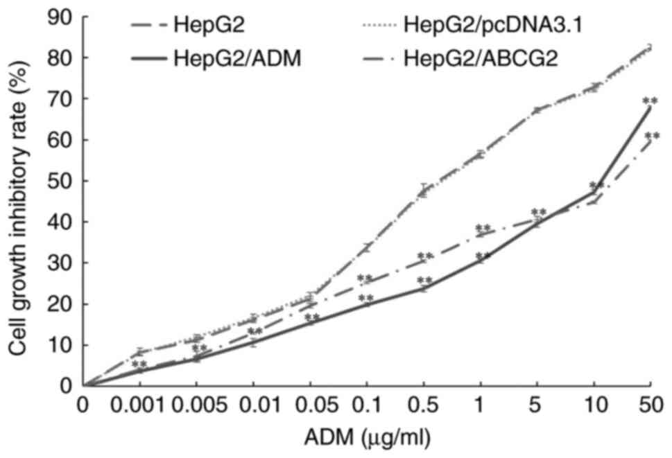
Following anticancer drug (ADM) treatment for 24 h,
the cell growth inhibitory rate was detected by an MTT assay. As
presented in Fig. 1, compared with
the control group (0 µg/ml ADM), the HepG2, HepG2/pcDNA3.1,
HepG2/ADM and HepG2/ABCG2 cell survival decreased in a
dose-dependent manner after treatment with concentrations of ADM
ranging from 0.001 to 50 µg/ml. The cell growth inhibitory rate in
HepG2/ADM and HepG2/ABCG2 cells was significantly lower than that
in HepG2 and HepG2/pcDNA3.1 cells (P<0.01). The IC50
was calculated through the MTT assay. The IC50 of
HepG2/ADM, HepG2/ABCG2, HepG2/pcDNA3.1 and HepG2 cells was
11.17±0.59, 12.75±0.47, 0.74±0.01 and 0.72±0.03 µg/ml,
respectively. The resistance index of HepG2/ADM and HepG2/ABCG2
cells to ADM was 15.51 and 17.71, respectively. HepG2/ADM and
HepG2/ABCG2 cells were resistant to ADM.
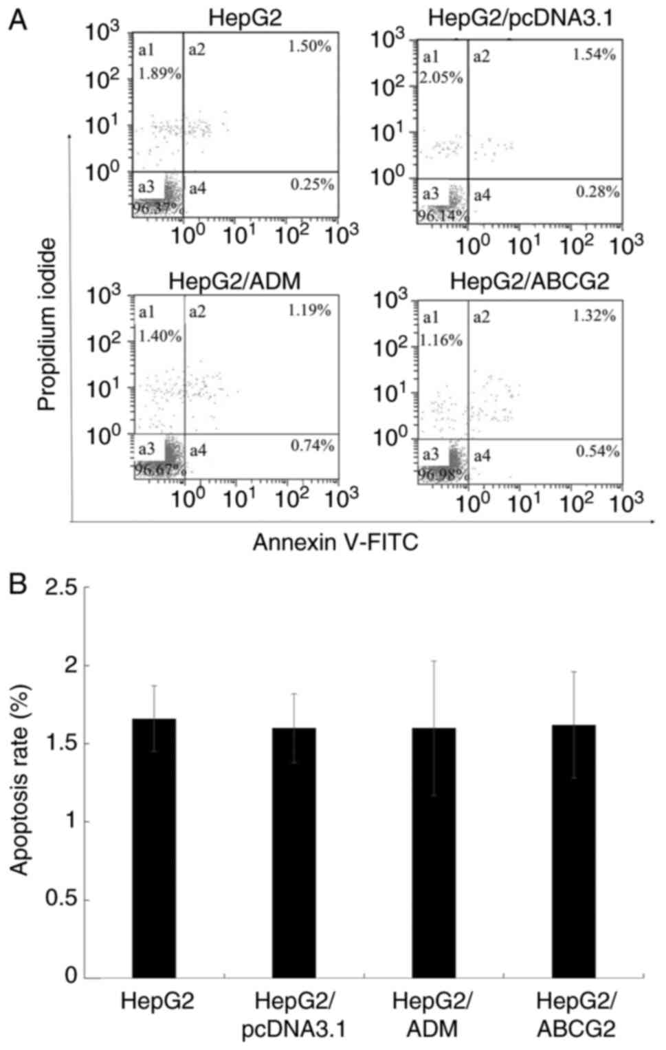
Apoptotic rate of cells
The results of the FCM analysis indicated no
significant difference in the apoptotic rate of HepG2/ADM,
HepG2/ABCG2, HepG2/pcDNA3.1 cells and their parental HepG2 cells
(P>0.05; Fig. 2).
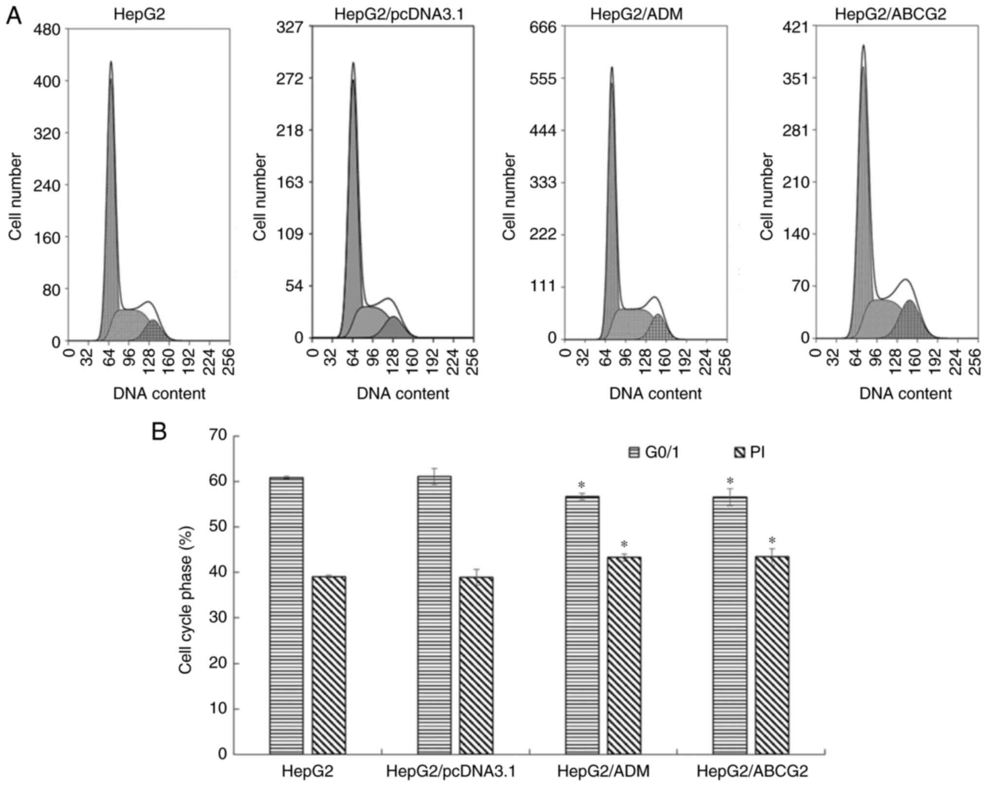
Evaluation of the cell cycle using
FCM
The results of the FCM analysis (Fig. 3) indicated that the
G0/G1 phase population of HepG2/ADM and
HepG2/ABCG2 cells decreased significantly compared with that of
their parental HepG2 cells and HepG2/pcDNA3.1 cells (P<0.05).
The PI of HepG2/ADM and HepG2/ABCG2 cells increased significantly
compared with their parental HepG2 cells and HepG2/pcDNA3.1 cells
(P<0.05). Compared with HepG2/ABCG2 cells, HepG2/ADM exhibited
no significant difference in G0/G1 phase and
PI (P>0.05); compared with HepG2 cells, HepG2/pcDNA3.1 had no
significant difference in G0/G1 phase and PI
(P>0.05).
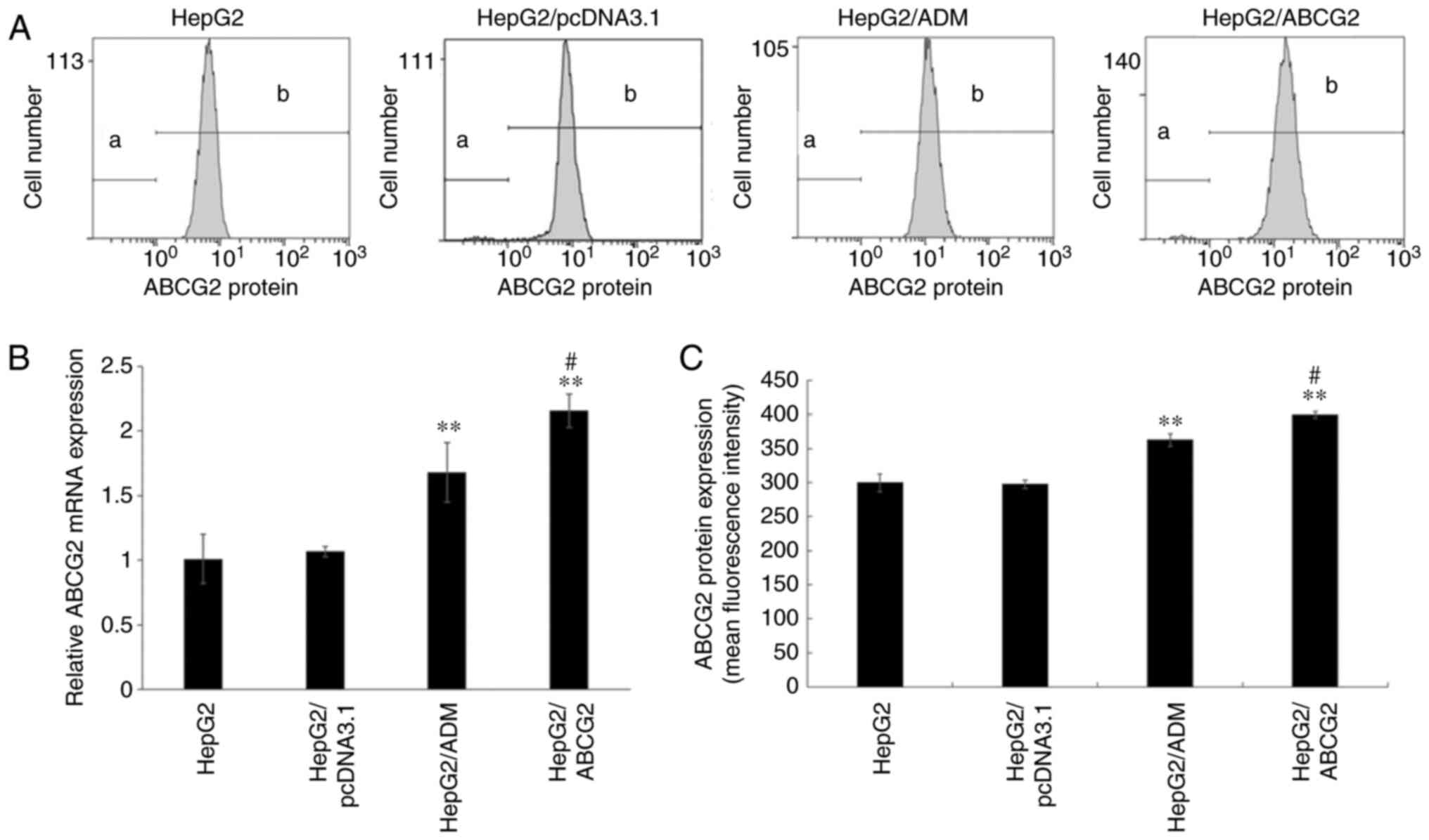
Expression of ABCG2 gene and protein
in drug-resistant hepatoma cells and their parental cells
The results indicated that the gene and protein
expression levels of ABCG2 in the HepG2/ADM and HepG2/ABCG2 cells
were significantly higher than those in HepG2 and HepG2/pcDNA3.1
cells (P<0.01), but there was no significant difference between
HepG2/pcDNA3.1 and HepG2 cells (P>0.05). Compared with HepG2/ADM
cells, the expression levels of ABCG2 gene and protein in
HepG2/ABCG2 cells increased significantly (P<0.05), as indicated
in Fig. 4.
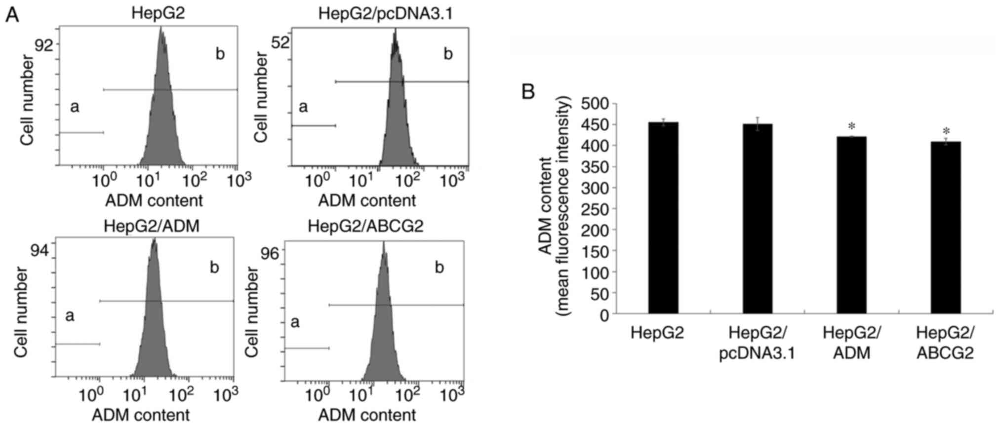
ADM efflux effect of HepG2/ADM and
HepG2/ABCG2 cells assayed by FCM
An ADM efflux effect assay was used to investigate
how the HepG2/ADM and HepG2/ABCG2 cells resisted the anticancer
agent using FCM (Fig. 5). The
cells were incubated at 37˚C with 0.1 µg/ml ADM for 2 h and then
without ADM for 1 h. The level of ADM decreased in HepG2/ADM and
HepG2/ABCG2 cells compared with HepG2 and HepG2/pcDNA3.1 cells
(P<0.05). There was no significant difference between
HepG2/pcDNA3.1 and HepG2 cells (P>0.05). Thus, the results
showed that the ADM efflux effect of the HepG2/ADM and HepG2/ABCG2
cells was more pronounced than that of the HepG2 and HepG2/pcDNA3.1
cells.
Discussion
Hepatoma is a common malignant tumor of the
digestive system, and is characterized by high morbidity and
mortality (1,2). Chemotherapy is one of the
conventional treatments for hepatoma, particularly for patients
unable to undergo surgery at an advanced stage. The factors that
affect the efficacy of chemotherapy include the side effects of the
chemotherapeutic drugs and the multidrug resistance of tumor cells
to chemotherapeutic drugs (17).
Exploring the mechanism and finding the target of multidrug
resistance is important for reversing multidrug resistance in the
clinic.
The most widely studied and clinically significant
mechanisms of multidrug resistance in tumor cells include the
following: i) The membrane transporter-mediated drug efflux pump
mechanism (18), consisting of
drug delivery through transmembrane transporters to reduce the
level of antitumor drugs in multidrug-resistant cells; ii) the
enzyme-mediated mechanism, which consists of the activation of cell
oxidation and the glutathione-related detoxification enzyme system,
to inactivate drugs or accelerate the excretion of drugs directly
(19); and iii) the regulating
mechanism through apoptotic genes that mediate the increase of the
expression of anti-apoptotic factors, such as Bcl-2 expression, and
the decrease of the gene and protein expression of apoptotic
factors, so that multidrug-resistant cells prevent apoptosis
induced by antitumor drugs (20,21).
The drug efflux pump mechanism mediated by membrane
transporters is one of the most widely studied multidrug resistance
mechanisms (11,22,23).
Cell transmembrane transporters may pump intracellular antitumor
drugs out of cells and reduce the concentration of chemotherapeutic
drugs in cells, thus producing multidrug resistance to tumors.
Several studies have suggested that the majority of transmembrane
transporters belong to the ABC transporter family (3,24-27).
To date, >100 types have been found, of which ≥48 types are in
humans. The most widely studied ABC proteins are ABCB1 and ABCG2
(6,7,28-30).
ABCG2 is associated with multidrug resistance in a variety of tumor
cells (31-38),
and is involved in forming side population cells and tumor stem
cells (10,39,40).
However, studies on the relationship between ABCG2 and multidrug
resistance of hepatoma cells are rare (41). The present study established
drug-resistant hepatoma cells by increasing the drug concentration
in cell culture and gene transfection. The role of ABCG2 in the
formation of multidrug resistance in hepatoma cells was then
discussed.
The IC50 value of ADM in HepG2 cells was
calculated using the MTT assay. The ADM concentration in the
culture of drug-resistant hepatoma cells was set up according to
the IC50 value of ADM in HepG2 cells. The ADM
concentration in the culture was increased from 0.01 to 0.1 µg/ml.
Eventually, HepG2 cells grew steadily in the culture containing 0.1
µg/ml ADM and these cells were designated HepG2/ADM cells. The
morphology of HepG2/ADM cells was more irregular than that of the
others and the cell volume was increased (data not shown). The
resistance index of HepG2/ADM cells to ADM was 15.51. The
expression levels of ABCG2 protein and gene in HepG2/ADM cells were
significantly higher than those in parental HepG2 cells. In order
to verify that ABCG2 is involved in the drug resistance of hepatoma
cells, HepG2/ABCG2 cells overexpressing the ABCG2 gene were
established by gene transfection. The results indicated that
HepG2/ABCG2 cells were also resistant to ADM and the drug
resistance index was 17.71. These results suggested that ABCG2 is
involved in the drug resistance of hepatoma cells (HepG2/ADM and
HepG2/ABCG2).
The apoptosis and cell cycle of HepG2/ADM,
HepG2/ABCG2, HepG2/pcDNA3.1 and HepG2 cells were detected by FCM.
The results indicated no difference in the apoptotic rate of
HepG2/ADM and HepG2/ABCG2 cells compared with HepG2 and
HepG2/pcDNA3.1 cells, but the G0/G1 phase
decreased and the PI increased significantly. The proliferation of
HepG2/ADM and HepG2/ABCG2 cells was higher than that of their
parental cells.
ABCG2 is a member of the ABC superfamily. The
multidrug resistance mechanism of the ABC superfamily involves drug
efflux (42-45),
which decreases the intracellular drug concentration. The ADM
efflux effect of HepG2/ADM and HepG2/ABCG2 cells was significantly
higher than that of HepG2 and HepG2/pcDNA3.1 cells, which suggested
that the drug resistance of hepatoma cells is related to the efflux
of chemotherapeutics regulated by ABCG2. These results are
consistent with the mechanism of drug resistance induced by ABCG2
in the literature (42-45).
In conclusion, in the present study, it was
demonstrated that HepG2/ADM and HepG2/ABCG2 cells were resistant to
ADM. The mechanism of drug resistance was related to high
expression of ABCG2 in cells and the resulting increase of ADM
efflux, which led to a decreased concentration of ADM in the cells,
resulting in drug resistance. However, assessing the drug
resistance of only one chemotherapeutic agent (ADM) was a
limitation of the present study. The molecular mechanisms
underlying the drug resistance of hepatoma cells are complex,
necessitating further study in the future.
Acknowledgements
The present study is based on a previously published
meeting abstract presented at the 17th International Congress of
Immunology (October 19-23, 2019, Beijing, China) and published as
an abstract entitled ‘Study on the drug resistance of HepG2 cells
induced by adenosine triphosphate (ATP)-binding cassette
transporter G2’ in the European Journal of Immunology, vol. 49,
Supplement S3, p3073.
Funding
Funding: No funding was received.
Availability of data and materials
The datasets used and/or analyzed during the current
study are available from the corresponding author on reasonable
request.
Authors' contributions
YL wrote the manuscript. YL, BD, JL, XL, CH, JW and
LL performed the experiments. BD and JL conducted the statistical
analysis. LL designed the study and revised the manuscript. All
authors have read and approved the final manuscript. YL and LL
confirm the authenticity of all the raw data.
Ethics approval and consent to
participate
Not applicable.
Patient consent for publication
Not applicable.
Competing interests
The authors declare that they have no competing
interests.
References
|
1
|
Global Burden of Disease Cancer
Collaboration. Fitzmaurice C, Allen C, Barber RM, Barregard L,
Bhutta ZA, Brenner H, Dicker DJ, Chimed-Orchir O, Dandona R, et al:
Global, regional, and national cancer incidence, motality, years of
life lost, yeas lived with disability, and disability-adjusted
life-years for 32 cancer groups, 1990 to 2015: A systematic
analysis for the global burden of disease study. JAMA Oncol.
3:524–548. 2017.PubMed/NCBI View Article : Google Scholar
|
|
2
|
Ju DY, Hainaut P, Gores GJ, Amadou A,
Plymoth A and Roberts LR: A global view of hepatocellular
carcinoma: trends, risk, prevention and management. Nat Rev
Gastroenterol Hepatol. 16:589–604. 2019.PubMed/NCBI View Article : Google Scholar
|
|
3
|
Choi YH and Yu AM: ABC transporters in
multidrug resistance and pharmacokinetics, and strategies for drug
development. Curr Pharm Des. 20:793–807. 2014.PubMed/NCBI View Article : Google Scholar
|
|
4
|
Thomas C and Tampé R: Structural and
mechanistic principles of ABC transporters. Ann Rev Biochem.
89:605–636. 2020.PubMed/NCBI View Article : Google Scholar
|
|
5
|
Wang Y, Qin Z, Cai S, Yu L, Hu H and Zeng
S: The role of non-coding RNAs in ABC transporters regulation and
their clinical implications of multidrug resistance in cancer.
Expert Opin Drug Metab Toxicol. 17:291–306. 2021.PubMed/NCBI View Article : Google Scholar
|
|
6
|
Hegedus C, Telbisz A, Hegedus T, Sarkadi B
and Ozvegy-Laczka C: Lipid regulation of the ABCB1 and ABCG2
multidrug transporters. Adv Cancer Res. 125:97–137. 2015.PubMed/NCBI View Article : Google Scholar
|
|
7
|
To KK, Poon DC, Wei Y, Wang F, Lin G and
Fu LW: Vatalanib sensitizes ABCB1 and ABCG2-overexpressing
multidrug resistant colon cancer cells to chemotherapy under
hypoxia. Biochem Pharmacol. 97:27–37. 2015.PubMed/NCBI View Article : Google Scholar
|
|
8
|
Limniatis G and Georges E: Down-regulation
of ABCB1 by collateral sensitivity drugs reverses multidrug
resistance and up-regulates enolase I. J Biochem. 172:37–48.
2022.PubMed/NCBI View Article : Google Scholar
|
|
9
|
Kukal S, Guin D, Rawat C, Bora S, Mishra
MK, Sharma P, Paul PR, Kanojia N, Grewal GK, Kukreti S, et al:
Multidrug efflux transporter ABCG2: expression and regulation. Cell
Mol Life Sci. 78:6887–6939. 2021.PubMed/NCBI View Article : Google Scholar
|
|
10
|
Xie ZY, Lv K, Xiong Y and Guo WH:
ABCG2-meditated multidrug resistance and tumor-initiating capacity
of side population cells from colon cancer. Oncol Res Treat.
37:666–668. 2014.PubMed/NCBI View Article : Google Scholar
|
|
11
|
Kozovska Z, Gabrisova V and Kucerova L:
Colon cancer: Cancer stem cells markers, drug resistance and
treatment. Biomed Pharmacother. 68:911–916. 2014.PubMed/NCBI View Article : Google Scholar
|
|
12
|
Chen D: Tumor formation and drug
resistance properties of human glioblastoma side population cells.
Mol Med Rep. 11:4309–4314. 2015.PubMed/NCBI View Article : Google Scholar
|
|
13
|
Wang M, Wang Y and Zhong J: Side
population cells and drug resistance in breast cancer. Mol Med Rep.
11:4297–4302. 2015.PubMed/NCBI View Article : Google Scholar
|
|
14
|
Kumar P, Nagarajan A and Uchil PD:
Analysis of cell viability by the MTT assay. Cold Spring Harb
Protoc. 2018.PubMed/NCBI View Article : Google Scholar
|
|
15
|
Livak KJ and Schmittgen TD: Analysis of
relative gene expression data using real-time quantitative PCR and
the 2(-Delta Delta C(T)) method. Method. 25:402–408.
2001.PubMed/NCBI View Article : Google Scholar
|
|
16
|
Motlagh NS, Parvin P, Ghasemi F and Atyabi
F: . Fluorescence properties of several chemotherapy drugs:
Doxorubicin, paclitaxel and bleomycin. Biomed Opt Express.
25:2400–2406. 2016.PubMed/NCBI View Article : Google Scholar
|
|
17
|
Wu Q, Yang Z, Nie Y, Shi Y and Fan D:
Multi-drug resistance in cancer chemotherapeutics: mechanisms and
lab approaches. Cancer Lett. 347:159–166. 2014.PubMed/NCBI View Article : Google Scholar
|
|
18
|
Lu JF, Pokharel D and Bebawy M: MRP1 and
its role in anticancer drug resistance. Drug Metab Rev. 47:406–419.
2015.PubMed/NCBI View Article : Google Scholar
|
|
19
|
Masanek U, Stammler G and Volm M:
Modulation of multidrug resistance in human ovarian cancer cell
lines by inhibition of P-glycoprotein 170 and PKC isoenzymes with
antisense oligonucleotides. J Exp Ther Oncol. 2:37–41.
2002.PubMed/NCBI View Article : Google Scholar
|
|
20
|
Zhang YF, Li XH, Shi YQ, Wu YY, Li N, He
Q, Ji Q, Wang RQ, Yang SM and Fang DC: CIAPIN1 confers multidrug
resistance through up-regulation of MDR-1 and Bcl-L in LoVo/Adr
cells and is independent of p53. Oncol Rep. 25:1091–1098.
2011.PubMed/NCBI View Article : Google Scholar
|
|
21
|
Lo YL, Wang W and Ho CT:
7,3',4'-Trihydroxyisoflavone modulates multidrug resistance
transporters and induces apoptosis via production of reactive
oxygen species. Toxicology. 302:221–232. 2012.PubMed/NCBI View Article : Google Scholar
|
|
22
|
Kunická T and Souček P: Importance of
ABCC1 for cancer therapy and prognosis. Drug Metab Rev. 46:325–342.
2014.PubMed/NCBI View Article : Google Scholar
|
|
23
|
Misra R, Das M, Sahoo BS and Sahoo SK:
Reversal of multidrug resistance in vitro by co-delivery of MDR1
targeting siRNA and doxorubicin using a novel cationic
poly(lactide-co-glycolide) nanoformulation. Int J Pharm.
475:372–384. 2014.PubMed/NCBI View Article : Google Scholar
|
|
24
|
Hsieh MJ, Chen MK, Yu YY, Sheu GT and
Chiou HL: Psoralen reverses docetaxel-induced multidrug resistance
in A549/D16 human lung cancer cells lines. Phytomedicine.
21:970–977. 2014.PubMed/NCBI View Article : Google Scholar
|
|
25
|
Wu CP, Hsiao SH, Murakami M, Lu YJ, Li YQ,
Huang YH, Hung TH, Ambudkar SV and Wu YS: Alpha-mangostin reverses
multidrug resistance by attenuating the function of the multidrug
resistance-linked ABCG2 transporter. Mol Pharm. 14:2805–2814.
2017.PubMed/NCBI View Article : Google Scholar
|
|
26
|
Murakami M, Ohnuma S, Fukuda M, Chufan EE,
Kudoh K, Kanehara K, Sugisawa N, Ishida M, Naitoh T, Shibata H, et
al: Synthetic analogs of curcumin modulate the function of
multidrug resistance-linked ATP-binding cassette transporter ABCG2.
Drug Metab Dispos. 45:1166–1177. 2017.PubMed/NCBI View Article : Google Scholar
|
|
27
|
Fan YF, Zhang W, Zeng L, Lei ZN, Cai CY,
Gupta P, Yang DH, Cui Q, Qin ZD, Chen ZS and Trombetta LD:
Dacomitinib antagonizes multidrug resistance (MDR) in cancer cells
by inhibiting the efflux activity of ABCB1 and ABCG2 transporters.
Cancer Lett. 421:186–198. 2018.PubMed/NCBI View Article : Google Scholar
|
|
28
|
Chen Y, Bieber MM and Teng NN: Hedgehog
signaling regulates drug sensitivity by targeting ABC transporters
ABCB1 and ABCG2 in epithelial ovarian cancer. Mol Carcinog.
53:625–634. 2014.PubMed/NCBI View
Article : Google Scholar
|
|
29
|
Sodani K, Tiwari AK, Singh S, Patel A,
Xiao ZJ, Chen JJ, Sun YL, Talele TT and Chen ZS: GW583340 and
GW2974, human EGFR and HER-2 inhibitors, reverse ABCG2- and
ABCB1-mediated drug resistance. Biochem Pharmacol. 83:1613–1622.
2012.PubMed/NCBI View Article : Google Scholar
|
|
30
|
Lepper ER, Nooter K, Verweij J, Acharya
MR, Figg WD and Sparreboom A: Mechanisms of resistance to
anticancer drugs: The role of the polymorphic ABC transporters
ABCB1 and ABCG2. Pharmacogenomics. 6:115–138. 2005.PubMed/NCBI View Article : Google Scholar
|
|
31
|
Sodani K, Patel A, Anreddy N, Singh S,
Yang DH, Kathawala RJ, Kumar P, Talele TT and Chen ZS: Telatinib
reverses chemotherapeutic multidrug resistance mediated by ABCG2
efflux transporter in vitro and in vivo. Biochem Pharmacol.
89:52–61. 2014.PubMed/NCBI View Article : Google Scholar
|
|
32
|
Rosenfeldt MT, Bell LA, Long JS, O'Prey J,
Nixon C, Roberts F, Dufès C and Ryan KM: E2F1 drives
chemotherapeutic drug resistance via ABCG2. Oncogene. 33:4164–4172.
2014.PubMed/NCBI View Article : Google Scholar
|
|
33
|
Nakanishi T and Ross DD: Breast cancer
resistance protein (BCRP/ABCG2): Its role in multidrug resistance
and regulation of its gene expression. Chin J Cancer. 31:73–99.
2012.PubMed/NCBI View Article : Google Scholar
|
|
34
|
Yousaf M and Ali M: Modulation of ABCG2
surface expression by Rab5 and Rab21 to overcome multidrug
resistance in cancer cells. Xenobiotica. 50:988–996.
2020.PubMed/NCBI View Article : Google Scholar
|
|
35
|
Kokubo S, Ohnuma S, Murakami M, Kikuchi H,
Funayama S, Suzuki H, Kajiwara T, Yamamura A, Karasawa H, Sugisawa
N, et al: A phenylfurocoumarin derivative reverses ABCG2-mediated
multidrug resistance in vitro and in vivo. Int J Mol Sci.
22(12502)2021.PubMed/NCBI View Article : Google Scholar
|
|
36
|
Mioč M, Telbisz Á, Radman K, Bertoša B,
Šumanovac T, Sarkadi B and Kralj M: Interaction of crown ethers
with the ABCG2 transporter and their implication for multidrug
resistance reversal. Histochem Cell Biol. 158:261–277.
2022.PubMed/NCBI View Article : Google Scholar
|
|
37
|
Ma Y, Guo Z, Fan C, Chen J, Xu S and Liu
Z: Rationally screened and designed ABCG2-binding aptamers for
targeting cancer stem cells and reversing multidrug resistance.
Anal Chem. 94:7375–7382. 2022.PubMed/NCBI View Article : Google Scholar
|
|
38
|
Zattoni IF, Delabio LC, Dutra JP, Kita DH,
Scheiffer G, Hembecker M, Pereira GDS, Moure VR and Valdameri G:
Targeting breast cancer resistance protein (BCRP/ABCG2): Functional
inhibitors and expression modulators. Eur J Med Chem.
237(114346)2022.PubMed/NCBI View Article : Google Scholar
|
|
39
|
Zhang QH, Dou HT, Xu P, Zhuang SC and Liu
PS: Tumor recurrence and drug resistance properties of side
population cells in high grade ovary cancer. Drug Res (Stuttg).
65:153–157. 2015.PubMed/NCBI View Article : Google Scholar
|
|
40
|
Britton KM, Eyre R, Harvey IJ, Stemke-Hale
K, Browell D, Lennard TWJ and Meeson AP: . Breast cancer, side
population cells and ABCG2 expression. Cancer Lett. 323:97–105.
2012.PubMed/NCBI View Article : Google Scholar
|
|
41
|
Tandia M, Mhiri A, Paule B, Saffroy R,
Cailliez V, Noé G, Farinotti R and Bonhomme-Faivre L: Correlation
between clinical response to sorafenib in hepatocellular carcinoma
treatment and polymorphisms of P-glycoprotein (ABCB1) and of breast
cancer resistance protein (ABCG2): Monocentric study. Cancer
Chemother Pharmacol. 79:759–766. 2017.PubMed/NCBI View Article : Google Scholar
|
|
42
|
Zhang H, Zhang YK, Wang YJ, Kathawala RJ,
Patel A, Zhu H, Sodani K, Talele TT, Ambudkar SV, Chen ZS and Fu
LW: WHI-P154 enhances the chemotherapeutic effect of anticancer
agents in ABCG2-overexpressing cells. Cancer Sci. 105:1071–1078.
2014.PubMed/NCBI View Article : Google Scholar
|
|
43
|
Wang XK, To KK, Huang LY, Xu JH, Yang K,
Wang F, Huang ZC, Ye S and Fu LW: Afatinib circumvents multidrug
resistance via dually inhibiting ATP binding cassette subfamily G
member 2 in vitro and in vivo. Oncotarget. 5:11971–11985.
2014.PubMed/NCBI View Article : Google Scholar
|
|
44
|
Dai CL, Liang YJ, Wang YS, Tiwari AK, Yan
YY, Wang F, Chen ZS, Tong XZ and Fu LW: Sensitization of
ABCG2-overexpressing cells to conventional chemotherapeutic agent
by sunitinib was associated with inhibiting the function of ABCG2.
Cancer Lett. 279:74–83. 2009.PubMed/NCBI View Article : Google Scholar
|
|
45
|
Jing W, Zhou M, Chen R, Ye X, Li W, Su X,
Luo J, Wang Z and Peng S: In vitro and ex vivo
anti-tumor effect and mechanism of Tucatinib in leukemia stem cells
and ABCG2-overexpressing leukemia cells. Oncol Rep. 45:1142–1152.
2021.PubMed/NCBI View Article : Google Scholar
|