Introduction
Rheumatoid arthritis (RA) is a chronic inflammatory
disease characterized by high levels of synovitis leading to joint
damage, and is associated with notable morbidity and mortality
(1). Clinical epidemiological data
have confirmed that RA occurs in 0.5-1% of the global population,
and that it occurs 2-3 times more frequently in women than in men.
In addition, RA can affect people of any age, but the peak onset is
between 50 and 59 years (2). If RA
occurs owing to a family history of inheritance, then the risk of
disease development can increase by 3-5 times (3). Currently, there is no clear
understanding about the pathogenesis of RA, but there is a
widespread belief it is related to immune abnormalities, probably
influenced by genetic and environmental factors (4). Rheumatoid factor (RF) testing is an
important indicator of an RA diagnosis and prognosis and indicates
disease severity. However, RF testing yields a negative result in
the early stages of RA, similar to that obtained for other
autoimmune and inflammatory diseases (5). Therefore, more effective and
sensitive auxiliary biomarkers of RA are needed. Based on the
genetic predisposition, guanylate-binding protein 1 (GBP1),
C-X-C motif chemokine ligand 10 (CXCL10),
granulocyte-macrophage colony-stimulating factor (GM-CSF)
and its receptor colony-stimulating factor 2 receptor β
(CSF2RB) have been reported to be highly expressed in RA,
and therefore may play an important role in the early diagnosis and
treatment of RA (6,7). Genetic testing may provide
opportunities for the early detection of effective therapeutic
responses to RA, even a few months before the preclinical phase
(8). Therefore, identifying novel
and feasible biomarkers is critical for the early diagnosis and
treatment of RA.
In the present study, bioinformatics analysis was
performed to determine the potential biomarkers of RA and identify
specific targets for therapeutic intervention, with the aim of
improving the understanding of the mechanism of RA and the clinical
treatment of RA. Initially, raw data of RA from the NCBI Gene
Expression Omnibus (GEO) database were obtained and four hub genes
were selected. Furthermore, the results of bioinformatics analysis
were verified using a rat model of Freund's complete adjuvant
(FCA)-induced RA. These results may provide insights into the
underlying molecular mechanisms of RA and potential biomarkers and
target genes for treatment.
Materials and methods
Selection of GEO datasets
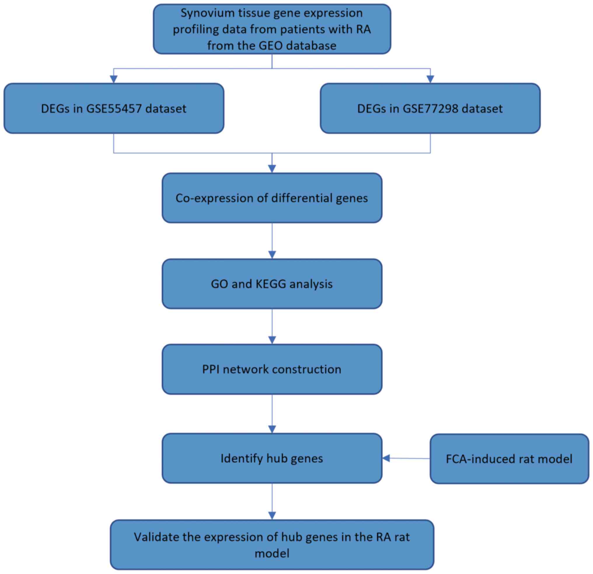
A flowchart of the overall experimental design is
shown in Fig. 1. The mRNA datasets
were downloaded from the GEO database (www.ncbi.nlm.nih.gov/geo/). To gain an improved
understanding of the underlying mechanism of RA, the independent
datasets of GSE55457(6) and
GSE77298(7) were selected, which
included synovial tissues from 13 and 16 patients with RA and 10
and 7 healthy subjects, respectively.
Differential expression analysis
To identify differentially expressed mRNAs
(DE-mRNAs) using the GSE55457 and GSE77298 datasets, the mRNA
expression levels of the tissue samples from patients with RA and
healthy subjects were compared. DE-mRNAs were defined using the
‘limma’ package (version 3.52.2) (9), based on the criteria of
log2 fold change >1 and P<0.05(8).
Functional enrichment analysis
Database for Annotation, Visualization and
Integrated Discover (DAVID) (https://david.ncifcrf.gov/) analysis was performed on
Kyoto Encyclopedia of Genes and Genomes (KEGG) and Gene Ontology
(GO) terms (10). In the present
study, the ‘enrichGO’ function of the clusterProfiler package
(10) was used to perform GO
pathway enrichment analysis based on the differentially expressed
genes (DEGs). Based on their function, these genes were divided
into three categories: Biological process (BP), molecular function
(MF) and cellular component (CC). A significant enrichment of GO
terms was observed for DEGs based on the false discovery
rate-adjusted P<0.05. The DAVID webtools were used to analyze
KEGG enrichment of the integrated DEGs in the present study.
Adjusted P<0.05 was considered to indicate a statistically
significant difference. The software packages R (version 4.2.2;
https://www.R-project.org/) (11), MCODE plug-in (version 2.0.2)
(12,13) and CytoHubba plug-in (https://apps.cytoscape.org/apps/cytohubba) in
Cytoscape (version 3.9.1) (14,15)
were used to obtain the results.
Building a protein-protein interaction
(PPI) network and identifying hub genes
Based on the STRING database (version 11.5; using
confidence score >0.9) (16), a
set of high-confidence PPI networks for humans were constructed. To
screen for hub genes, topological algorithms of the cytoHubba
plug-in application in Cytoscape (version 3.9.1) were applied to
predict and explore important nodes and hubs. The core module was
extracted from the PPI network using the MCODE plug-in. The
parameters were set as follows: Degree cut-off=2, K-Core=2 and node
score cut-off=0.2(17). It was
hypothesized that the potential hub genes could be found in the
core module. Thus, the genes selected were considered the hub
genes.
Animals
A total of 30 male Sprague Dawley rats (6 weeks old;
180±20 g body weight) were obtained from Chengdu Dashuo Laboratory
Animal Co., Ltd. (permit no. SCXK 2015-030). The animals were fed
and maintained under laboratory conditions of 22-24˚C and 50-65%
humidity, with natural light and dark cycles, and had free access
to food and drinking water. The rats were randomized into the
control and RA groups (n=15/group). All animal procedures were
approved by The Ethics Committee of Chengdu University of
Traditional Chinese Medicine (Chengdu, China; approval no.
2018-11).
Establishment of the RA model
On the first day of the experiment, to establish the
RA model, the rats were in the RA group subjected to the induction
process, wherein they received an injection of 0.5 ml/kg of FCA
(Sigma-Aldrich; Merck KGaA) in the footpad. The control group was
injected with the same amount of normal saline. RA progression was
evaluated based on the swelling and redness of the foot pad. The
overall condition and bipedal lesions of all rats were observed
daily. A waterproof marker was used to mark the middle of the
footpad of each rat and all future measurements were taken from
this point. Calipers were used to measure the footpad thickness
before and 1, 2 and 3 weeks after establishment of the RA model
(18).
At the end of the experiment, the rats were
anesthetized with 7% chloral hydrate (400 mg/kg, intraperitoneally;
Sinopharm Chemical Reagent Co., Ltd.). It was confirmed that the
rats showed no signs of peritonitis, pain or discomfort following
this anesthesia method. Blood was subsequently collected from the
abdominal aorta of the live anesthetized rats. After the 5 ml of
blood was collected, the rats were subsequently sacrificed by
cervical dislocation. Ankle joints, synovial tissue and the spleen
were also collected from each animal.
Hematoxylin and eosin (H&E)
staining
The ankle joint was fixed with 4% paraformaldehyde
(Wuhan Servicebio Technology Co., Ltd.) for >24 h at room
temperature. After fixation, the paraffin-embedded tissue sections
were sliced into 5-mm sections using a paraffin microtome (Leica
Microsystems, Ltd.). The sections were then stained with the
H&E staining solution kit (Wuhan Servicebio Technology Co.,
Ltd.), which included staining with hematoxylin and eosin
individually (5 min at room temperature for each staining step).
The stained sections were dehydrated with ethanol, cleared with
xylene, mounted onto slides with neutral balsam and covered with
coverslips. Histopathological examination was performed on the
mounted sections and images were captured under a light microscope
(Nikon Corporation). The slides were graded according to the degree
of inflammatory changes. The rat synovial pathological score
included synovial tissue hyperplasia, inflammatory cell
infiltration and macrophage hyperplasia. The tissue was scored
based on the following: 0, normal; 1, sparse or scattered; 2,
diffuse increase; 3, large increase with respect to inflammatory
cell infiltration, macrophage hyperplasia and fibroplasia.
Enzyme-linked immunosorbent assay
(ELISA)
The blood samples collected from each group were
centrifuged at 1,200 x g for 20 min at room temperature (Heal Force
Bio-meditech Holdings Ltd.) and the supernatant was collected. The
expression levels of tumor necrosis factor α (TNF-α) (cat. no.
JM-01587R1) and interleukin (IL-) 12 (cat. no. JM10372R1) in the
supernatant were detected using an ELISA kit (all Jiangsu Jingmei
Biotechnology Co., Ltd.), according to the manufacturer's
guidelines. Standards (50 µl) and samples (10 µl of serum and 40 µl
of diluent, supplied with the kit) were added to predetermined
microplate wells. Horseradish peroxidase (HRP)-labeled conjugate
(100 µl; supplied with the kit) was added to the wells, which were
then sealed and incubated at 37˚C for 1 h. The plate was washed
five times with PBS and substrates A (50 µl) and B (50 µl) were
added to each well. After incubation for 15 min at 37˚C, stop
solution (50 µl) was added. A microplate reader (BioTek
Instruments, Inc.) was used to measure the optical density (OD)
values in all wells at a wavelength of 450 nm. TNF-α and IL-12
concentrations were calculated using the obtained OD values of each
group of samples, according to the linear regression equation of
the standard curve.
Reverse transcription-quantitative
polymerase chain reaction (RT-qPCR)
Spleen tissue (100 mg) from the RA animal model and
control group was homogenized using a homogenization tube.
Following the manufacturer's instructions, total RNA was extracted
using the TRIzol™ method (Thermo Fisher Scientific, Inc.). The
concentration and purity of the isolated RNA were determined using
a microvolume spectrophotometer (Thermo Fisher Scientific, Inc.).
According to the manufacturer's protocol, RNA was subsequently
converted into cDNA using a RT kit (Thermo Fisher Scientific, Inc.)
and a PCR machine (Eastwin Life Sciences Inc.). For the qPCR step,
the reaction mixture constituted the following components: 2 µl
cDNA, 2.5 µl forward and reverse primers, 7.5 µl 2X SYBR Green qPCR
Master Mix (Wuhan Servicebio Technology Co., Ltd.) and ultrapure
water to reach a 15 µl total volume. Amplification was performed
using a fluorescent qPCR instrument (Bio-Rad Laboratories, Inc.).
The reaction parameters were as follows: Initial denaturation (95˚C
for 30 sec), 40 cycles of denaturation (95˚C for 15 sec) and
annealing/extension (60˚C for 30 sec). Data were collected and
analyzed using SDS software (version 1.4; Applied Biosystems;
Thermo Fisher Scientific, Inc.). GAPDH was used as the internal
reference gene. Furthermore, 2-ΔΔCq (19) was used to calculate the relative
expression level of discs large homolog-associated protein 5
(DLGAP5), kinesin family member 20A (KIF20A),
maternal embryonic leucine zipper kinase (MELK) and nuclear
division cycle 80 (NDC80). The primer sequences used were as
follows: GAPDH-forward (F), 5'-CTGGAGAAACCTGCCAAGTATG-3';
GAPDH-reverse (R), 5'-GGTGGAAGAATGGGAGTTGCT-3'; MELK-F,
5'-TCCCATTGAGTGGCAAAGCA-3'; MELK-R, 5'-CCTGCTGTTGCGGTGATGTA-3';
NDC80-F, 5'-CAAAGACCTGGAAGCCGAACAA-3'; NDC80-R,
5'-CTAGCCAACTTGTGGTACTCTGC-3'; DLGAP5-F,
5'-GAAGTGCCAACGCTTTCCTGA-3'; DLGAP5-R,
5'-CGTTCTTTCCTAGAGTCTGTTTGTG-3'; KIF20A-F,
5'-GAACCACCTAGCCATCAGCATA-3'; KIF20A-R,
5'-ACTGTGACTGCGACTAGAGTGCT-3'.
Western blotting
The synovial tissue was washed 2-3 times with cold
PBS to remove blood, after which the tissue was cut into small
pieces and placed in a homogenizer tube. Protease inhibitors and 10
times the tissue volume (10 ml buffer/g tissue) of radio
immunoprecipitation assay lysis buffer (Wuhan Servicebio
Technology, Co., Ltd.) were added to the tube and the contents were
homogenized thoroughly on ice. The homogenate was transferred to a
1.5 ml centrifuge tube and the contents were shaken well. The
homogenate was pipetted repeatedly for 30 min in an ice bath to
ensure complete cell lysis, after which the contents were
centrifuged at 12,000 x g for 10 min at 4˚C and the supernatant
(containing total protein) was collected. Protein concentration was
determined using the bicinchoninic acid method. Subsequently,
sodium dodecyl sulfate-polyacrylamide gel electrophoresis was
performed using 10% gels to separate the isolated proteins (30
µg/lane). The separated proteins were then transferred onto
polyvinylidene difluoride membranes. After blocking the membranes
with tris-buffered saline containing 5% skimmed milk powder at room
temperature for 1 h, the membranes were incubated with the
following primary antibodies overnight at 4˚C: KIF20A (cat. no.
ab70791; 1:1,000; Abcam), NDC80 (cat. no. ab109496; 1:1,000;
Abcam), MELK (cat. no. ab108529; 1:1,000; Abcam), DLGAP5 (cat. no.
ab70744; 1:1,000; Abcam) and GAPDH (cat. no. gb15002; 1:2,000;
Wuhan Servicebio Technology, Co., Ltd.). After repeated washing,
the membranes were incubated with HRP-conjugated goat anti-rabbit
(cat. no. gb23303; 1:5,000; Wuhan Servicebio Technology, Co., Ltd.)
secondary antibody for 2 h at 37˚C. Images were collected using a
chemiluminescent imaging system (Wuhan Servicebio Technology, Co.,
Ltd.) and grayscale analysis was performed using the system's
ChemiScope analysis software (version ChemiScope 6100; Clinx
Science Instrument Co., Ltd.).
Statistical analysis
All data had to pass the Shapiro-Wilk normality test
and the Levene's test for homogeneity of variance. Then the results
were analyzed by pairwise test. For correlation analyses, when the
data distribution was normal, the Pearson correlation analysis was
used, whereas when the distribution was not normal, the Spearman
correlation analysis was used. Data were presented as the mean ±
standard deviation. P<0.05 was considered to indicate a
statistically significant difference. All data were assessed using
SPSS 26.0 software (IBM Corp.). The relevant boxplots were compiled
using Xiantao Academic (http://www.xiantao.love).
Results
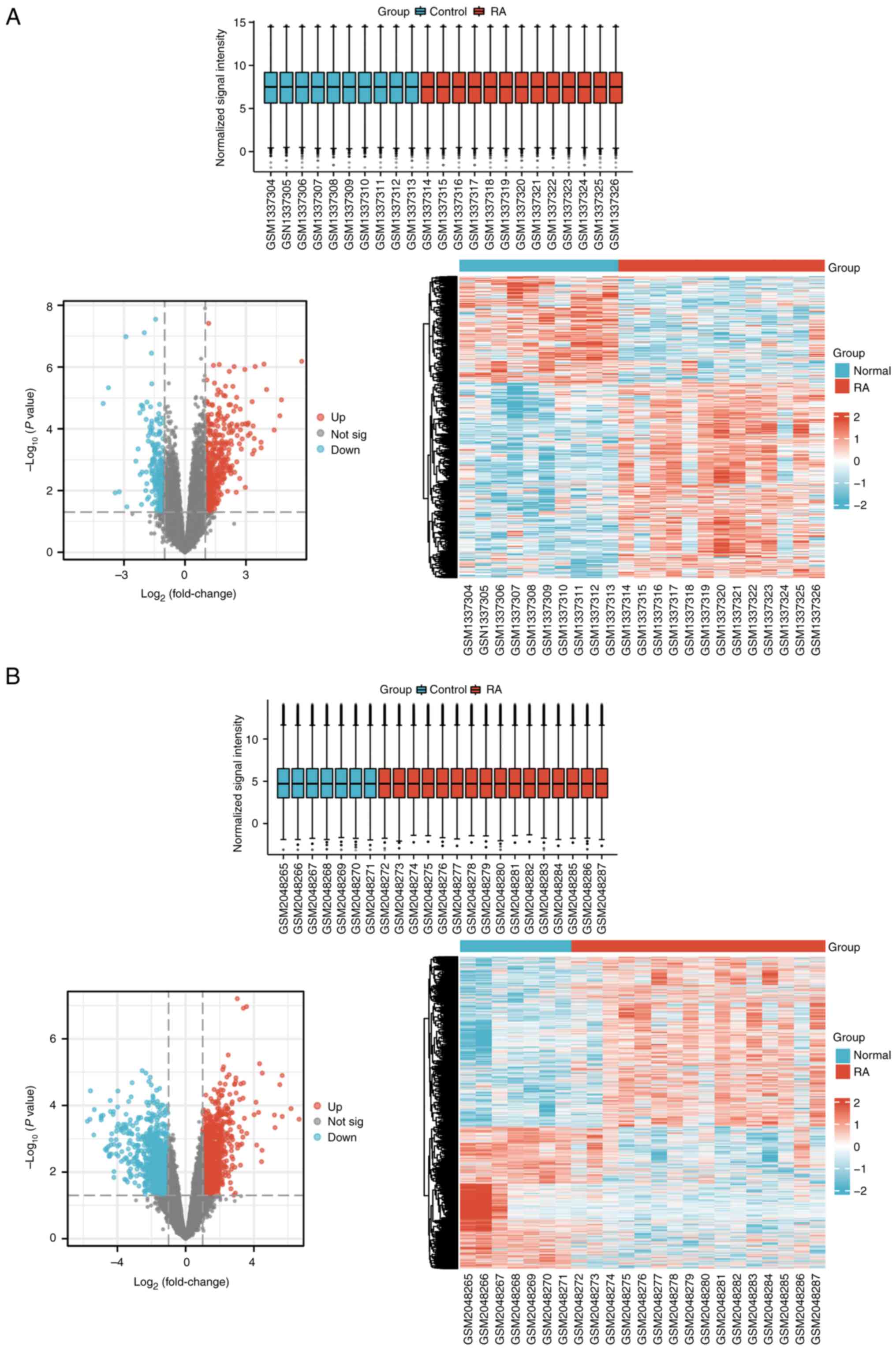
Identification of DE-mRNAs
The mRNA expression levels from the GSE55457 and
GSE77298 datasets from the GEO database were investigated and the
results were determined through boxplots, volcano plots and
heatmaps (Fig. 2). There were 13
samples from patients with RA and 10 samples from healthy
individuals in the GSE55457 dataset, whereas there were 16 samples
from patients with RA and 7 samples from healthy individuals in the
GSE77298 dataset. In the GSE55457 dataset, 664 DE-mRNAs were
identified (429 with upregulated expression and 235 with
downregulated expression), while in the GSE77298 dataset, 2,150
DE-mRNAs were detected (1,170 with upregulated expression and 980
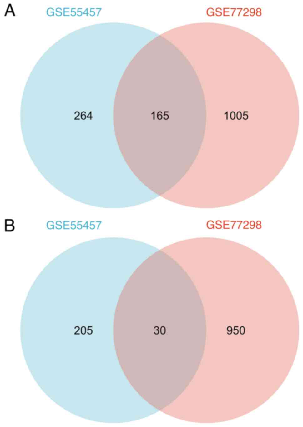
with downregulated expression). According to Venn diagram analyses,
165 DE-mRNAs that had upregulated expression and 30 DE-mRNAs that
had downregulated expression were shared between the two datasets
(Fig. 3 and Table SI).
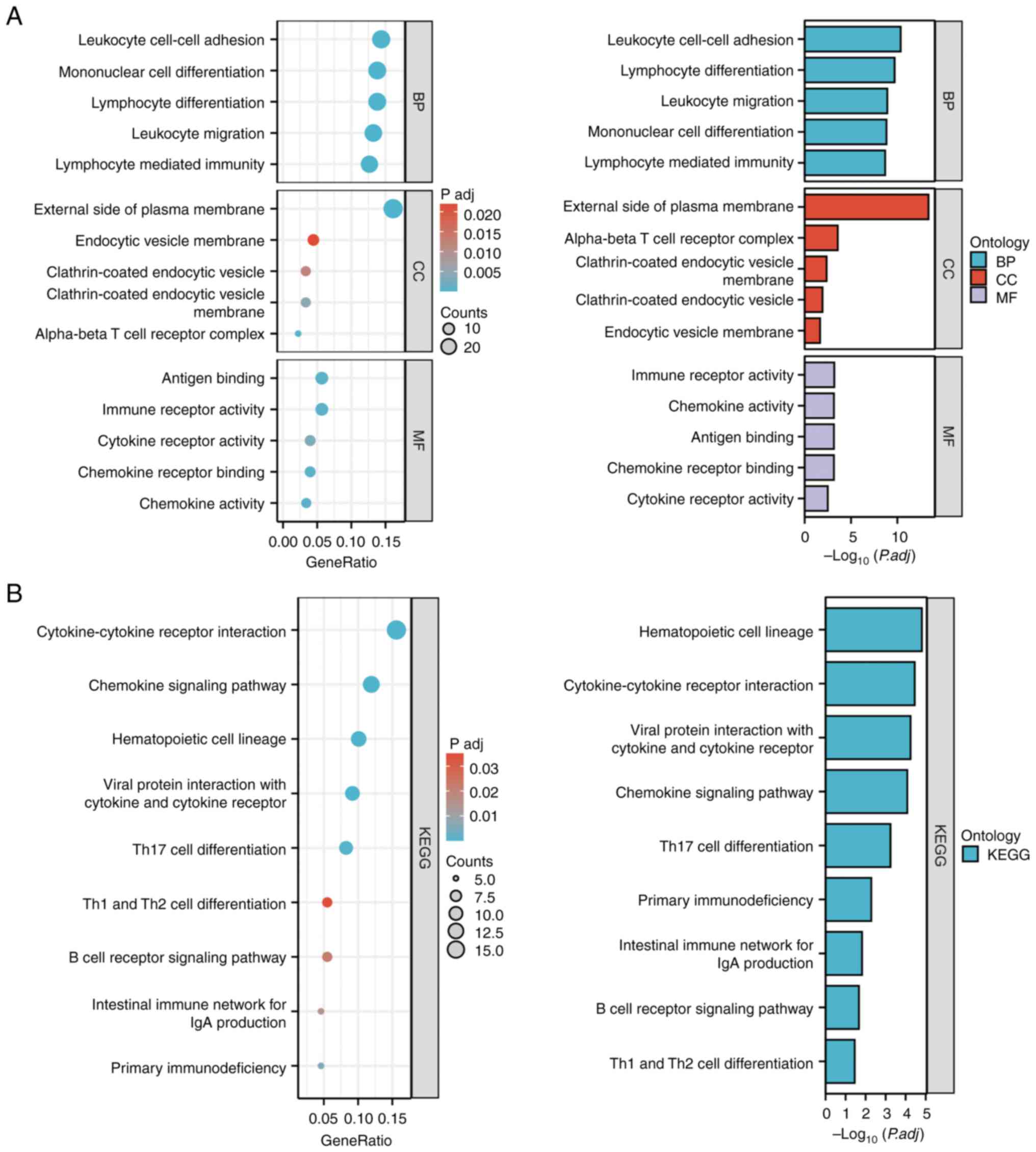
Enrichment analysis of DE-mRNAs
To explore the potential functions of the identified
DE-mRNAs, functional enrichment analysis was performed using GO and
KEGG analyses. Among them, the top five BP, top five CC and top
five MF GO terms are shown in Fig.
4A and Table SII. In BP,
‘leukocyte cell-cell adhesion,’ ‘mononuclear cell differentiation,’
‘lymphocyte differentiation,’ ‘leukocyte migration’ and ‘lymphocyte
mediated immunity’ were the top five most enriched terms.
Additionally, ‘external side of plasma membrane,’ ‘endocytic
vesicle membrane,’ ‘clathrin-coated endocytic vesicle,’
‘clathrin-coated endocytic vesicle membrane’ and ‘alpha-β T-cell
receptor complex’ were the top five GO terms on CC. Furthermore,
‘antigen binding,’ ‘immune receptor activity,’ ‘cytokine receptor
activity,’ ‘chemokine receptor binding’ and ‘chemokine activity’
were the top five GO terms on MF. Based on the KEGG analysis,
significant enrichments were observed in nine KEGG pathways for the
apparent DE-mRNAs, such as ‘hematopoietic cell lineage,’
‘cytokine-cytokine receptor interaction,’ ‘viral protein
interaction with cytokine and cytokine receptor,’ ‘chemokine
signaling pathway’ and ‘Th17 cell differentiation’ (Fig. 4B and Table SII).
Analysis of DE-mRNAs and screening of
hub mRNAs
To further explore the functions of these mRNAs, the
STRING database was analyzed using a PPI network analysis and mRNAs
with the highest confidence levels were selected. Overall, 195
DE-mRNAs were filtered into a PPI network complex containing 181
nodes and 781 edges. Then, MCODE was used to identify the most
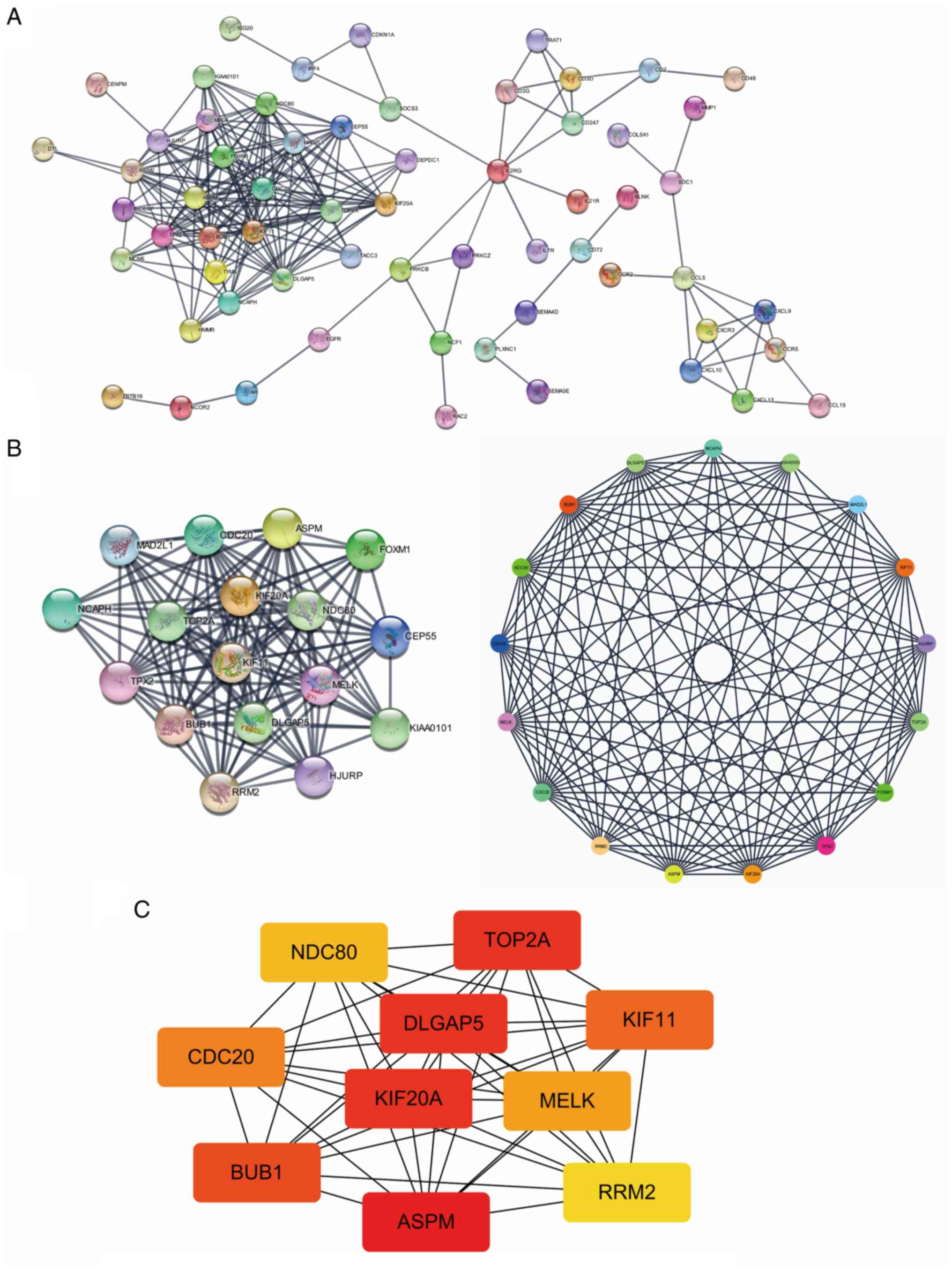
connected hub mRNAs in the PPI network (Fig. 5A and B). Then, cytoHubba was used to analyze
the top 10 most interacting central mRNAs: Assembly factor for
spindle microtubules (ASPM), DLGAP5, DNA
topoisomerase IIα (TOP2A), KIF20A, BUB1,
KIF11, CDC20, MELK, NDC80 and
ribonucleotide reductase regulatory subunit M2 (RRM2)
(Fig. 5C).
Validation of the FCA-induced
arthritis rat model
A schematic diagram of the flowchart after the
establishment of the FCA-induced arthritis rat model is shown in
Fig. S1A. The rat footpad
thickness was measured and the thickness in the model group was
significantly increased compared with the control group (P<0.01;
Fig. S1B). H&E staining of
the joint tissue demonstrated that, compared with the control
group, a greater amount of inflammatory cell infiltration,
macrophage hyperplasia and fibrous tissue hyperplasia was present
in the model group (Fig. S1C).
The groups were then scored blindly with all criteria tested by at
least two investigators and the average scores were obtained. The
scores represent the degree of tissue damage with a higher score
indicating a higher degree of tissue damage. Compared with the
control group, the joints of the rats in the model group showed
apparent inflammatory cell infiltration, fibrous tissue hyperplasia
and macrophage proliferation (P<0.01; Fig. S1D-F). Therefore, the results
demonstrated that, compared with the control group, the RA group
had a higher degree of bone tissue destruction and inflammation,
indicating that the FCA-induced RA rat model was successfully
established.
Validation of four DE-mRNAs in the
FCA-induced arthritis rat model
The mRNA expression levels of four previously
unreported genes (DLGAP5, KIF20A, MELK and
NDC80) among the top 10 central DE-mRNAs (or hub genes) from
the PPI network analysis were examined in RA rat spleen tissues
using qPCR, to validate the reliability of the dataset. Compared
with the control group, the expression levels of DLGAP5,
KIF20A, MELK and NDC80 in the RA group were
significantly increased (P<0.01; Fig. 6A-D). In addition, western blotting
was performed to detect the protein expression levels of DLGAP5,
KIF20A, MELK and NDC80. Compared with the control group, the
protein expression levels of DLGAP5, KIF20A, MELK and NDC80 in the
RA group were significantly increased (P<0.01; Fig. S2). Furthermore, ELISA results
demonstrated that, compared with the control group, IL-12 and TNF-α
levels in the RA group were significantly increased (P<0.01;
Fig. 6E and F). In addition, correlation analysis was
performed to analyze the correlation between inflammatory factors
and hub genes, and the results demonstrated that the inflammatory
factors were highly correlated with the hub genes (Fig. S3).
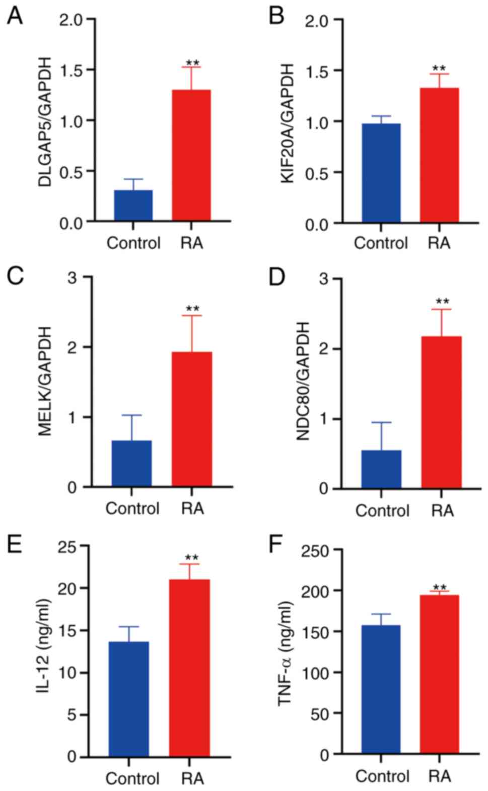 | Figure 6Validation of relative gene
expression in the RA model using qPCR and ELISA. Relative
expression of (A) DLGAP5, (B) KIF20A, (C) MELK
and (D) NDC80 in the FCA-induced and control rats was
determined using qPCR. Levels of the inflammatory cytokines, (E)
IL-12 and (F) TNF-α, in the FCA-induced and control rats were
determined using ELISA. Data are presented as the mean ± standard
deviation (n=6). **P<0.01 vs. the control group. FCA,
Freund's complete adjuvant; RA, rheumatoid arthritis; IL-12,
interleukin 12; TNF-α, tumor necrosis factor-α; DLGAP5, discs large
homolog-associated protein 5; KIF20A, kinesin family member 20A;
MELK, maternal embryonic leucine zipper kinase; NDC80, nuclear
division cycle 80; qPCR, quantitative PCR; ELISA, enzyme-linked
immunosorbent assay. |
Discussion
RA is a chronic and systemic inflammatory disease
that particularly affects the joints, leading to decreased physical
function and even disability. Since the complex pathogenesis of RA
is still unclear, it has become one of the important frontier
research topics in the field of immunity. Previously, studies have
focused on the role of inflammation in the occurrence of RA or the
regulation of RA development and notable progress has been made in
this research area (20-23).
Moxibustion treatment. can effectively reduce synovial inflammation
in RA rats by regulating the macrophage migration inhibitory
factor/glucocorticoid signaling pathway (24). In addition, it can also improve RA
symptoms by regulating the polarization of macrophage signaling
pathways and exert a potentially bone protective effect (25,26).
Chen et al (27) reported
that receptor activator of NF-κB ligand (RANKL) plays an essential
role in the orchestration of osteoclast maturation, activation,
migration and survival. However, osteoprotegerin can inhibit RANKL
activity to achieve bone homeostasis by inhibiting
osteoclastogenesis and subsequent bone resorption. Thus, cartilage
degeneration and bone destruction can be alleviated through the
osteoprotegerin/RANKL signaling pathway. These findings suggested
that pharmacological treatment or moxibustion treatment can
effectively improve the joint swelling, inflammation level and bone
destruction symptoms of RA. Targeted treatment of key genes is
becoming increasingly important in RA. It has been reported that
using (+)-JQ-1 as an inhibitor can displace bromodomain-containing
protein 2 (BRD2) and bromodomain-containing protein 4
(BRD4) from upstream regions of key super-enhancers, a
unique group of cis-regulatory elements, associated with key genes
controlling intracellular state, cell identity and
cell-type-specific functions. Loss of BRD2 and BRD4 leads to
downregulation of the expression of genes that are critically
involved in the pathogenesis and progression of RA (28). Upon activation or high expression
of four hub genes identified in the present study (DLGAP5,
KIF20A, MELK and NDC80), growth, proliferation
and migration of cells, including pro-inflammatory cells and
macrophages, is promoted (29-39).
Under the action of these genes, pro-inflammatory cells and
macrophages then migrate to the lesion site and the inflammatory
cells begin to release a number of inflammatory factors (such as
IL-12 and TNF-α), which causes systemic inflammation and macrophage
infiltration (29,40,41).
The balance between M1 and M2 macrophages is crucially involved in
the pathogenesis of RA. M1 macrophages act as pro-inflammatory
mediators in the synovium, whereas M2 macrophages suppress
inflammation and promote tissue repair. Notably, an imbalance in
the ratio of M1/M2 macrophages can induce the activation of
pro-inflammatory cytokines, the production and release of
chemokines, and promote the proliferation and survival of
macrophages, eventually leading to the occurrence of RA (42-44).
Abnormal gene expression is closely associated with
a variety of pathological conditions, such as RA. However, it
remains unclear which genes play a key role in the onset and
progression of this disease. In the present study, 429 RA-related
DEGs with upregulated expression and 235 with downregulated
expression were identified in the GSE55457 dataset and 1,170
RA-related DEGs with upregulated expression and 980 with
downregulated expression were identified in the GSE77298 dataset.
Meanwhile, 165 RA-related DEGs with upregulated expression and 30
with downregulated expression were confirmed in the intersection of
DEGs between the two datasets. These genes were associated with T
helper (Th)1 and Th2 cell differentiation, chemokine signaling
pathways, Th17 cell differentiation and the B-cell receptor
signaling pathway. Among the DEGs, 10 hub genes associated with RA
(ASPM, DLGAP5, TOP2A, KIF20A,
BUB1, KIF11, CDC20, MELK, NDC80
and RRM2) were identified. To the best of our knowledge,
there is no clear understanding of the specific target gene or
molecular biomarker responsible for the changes in the expression
of the hub genes in the tissues of patients with RA. Biomarkers for
RA and associated diseases include changes in the mRNA expression
levels of genes, such as GBP1, CXCL10, GM-CSF and its receptor
CSF2RB (6,7). In the present study, four new genes
were selected as candidates from the identified hub genes, namely,
DLGAP5, KIF20A, MELK and NDC80.
DLGAP5 was initially identified as a cell
cycle-regulating protein. However, it is now regarded as a
microtubule-associated protein, associated with the regulation of
KIF18A located at the plus end of centromere microtubules, to
stabilize K fibers and promote chromosome aggregation (32). Previous studies analyzed the
structure and function of DLGAP5 in different species from
physiological and clinicopathological perspectives, and reported
that DLGAP5 plays an important role in promoting cell growth,
proliferation and migration (30,31).
The KIF20A gene encodes a protein with 890 amino acid
residues, which belongs to the KIF superfamily and is mainly
distributed in the central region of the mitotic spindle.
Therefore, it participates in the process of driving mitosis and
promoting cell proliferation (32,33).
MELK is involved in various physiological and pathological
processes of cells, such as cell proliferation, apoptosis and tumor
development. In the last few decades, numerous studies have shown
that MELK is significantly upregulated in a number of cancer
types and plays an important role in promoting cell proliferation
and maintaining cancer cell stemness (34,35).
Silencing the MELK gene can slow down or even inhibit the
proliferation, invasion and tumorigenicity of cancer cells
(36,37). NDC80/Hec1, a subunit of the
kinetochore complex (also called the NDC80 complex), constitutes
and stabilizes microtubule-kinetochore attachment during the
segregation of mitotic chromosomes, and plays an important role in
mitosis and cell proliferation (38). NDC80 upregulation is also an
important biomarker for cancer; high levels of NDC80 have been
reported to indicate a poor prognosis in various types of cancer,
such as gastric cancer, ovarian cancer and blood cancer (39). Although DLGAP5,
KIF20A, MELK and NDC80 have been studied
separately in different tumors, to the best of our knowledge, there
is still a lack of evidence for the correlation between
DLGAP5, KIF20A, MELK and NDC80 and RA
development.
Previous studies have confirmed that RA can be
treated in a number of different ways, such as by targeting
signaling pathways, key proteins and circadian rhythms (18,22,24,26).
Therefore, to further explore the potential biomarkers and target
genes in RA, validation with FCA-induced RA rats was performed in
the present study. As FCA-induced RA resembles RA in humans in
terms of pathogenesis and clinical symptoms, it is widely used for
research owing to its simplicity, reliability and reproducibility
for RA modeling. As an immune organ, the spleen serves a regulatory
role in systemic immunity. Therefore, in the present study, an RA
rat model was established and the expression of DLGAP5,
KIF20A, MELK and NDC80 mRNA in the spleen of
this model was examined. As expected, the results demonstrated that
the expression levels of these genes were significantly increased
in the RA model, consistent with the results obtained by
bioinformatics analysis. In addition, the expression of
representative inflammatory factors of RA (IL-12 and TNF-α) were
verified in this model and these factors were correlated with the
expression of the key genes (DLGAP5, KIF20A,
MELK and NDC80). Thus, the present study demonstrated
that these genes may play a crucial role in eliciting immune
responses in RA.
In conclusion, four possible RA-related hub genes
were identified in the present study, namely DLGAP5,
KIF20A, MELK and NDC80, using the GEO
database, and the expression of these four genes in an RA model
were confirmed in validation experiments. These findings provide
new insights into the mechanism of RA development, and the
upregulated expression of these four hub genes is closely related
to inflammation in RA. The present study provides an improved
understanding of RA mechanisms and may lead to the identification
of potential biomarkers in the future.
Supplementary Material
Experimental validation of the
FCA-induced arthritis rat model. (A) Experimental timeline. (B)
Changes in foot pad thickness. (C) Hematoxylin and eosin staining
of the joint tissue (magnification x20 and x200). (D) Inflammatory
cell infiltration score. (E) Macrophage hyperplasia. (F)
Fibroplasia score. Data are presented as the mean ± standard
deviation (n=6). **P<0.01 vs. the control group. FCA,
Freund's complete adjuvant; RA, rheumatoid arthritis.
Validation of relative gene expression
in the RA model using western blotting. (A) Protein expression of
the hub genes were verified using western blotting. Quantification
of (B) DLGAP5, (C) KIF20A, (D) MELK and (E) NDC80 protein
expression in rats with FCA-induced RA and control rats. Data are
presented the as mean ± standard deviation (n=3).
**P<0.01 vs. the control group. FCA, Freund's
complete adjuvant; RA, rheumatoid arthritis; DLGAP5, discs large
homolog-associated protein 5; KIF20A, kinesin family member 20A;
MELK, maternal embryonic leucine zipper kinase; NDC80, nuclear
division cycle 80.
Correlation analysis. Correlation
analysis of (A) DLGAP5, (B) KIF20A, (C) MELK and (D) NDC80 with
IL-12 and TNF-α. IL-12, interleukin 12; TNF-α, tumor necrosis
factor-α; DLGAP5, discs large homolog-associated protein 5; KIF20A,
kinesin family member 20A; MELK, maternal embryonic leucine zipper
kinase; NDC80, nuclear division cycle 80.
Screening for Co-DEGs in patients with
RA using the GSE55457 and GSE77298 datasets.
Top five GO (BP, CC and MF) and KEGG
terms related to the DEGs between the control and RA groups.
Acknowledgements
The authors wish to thank the Animal Experiment
Center of Chengdu University of Traditional Chinese Medicine
(Chengdu, China) for the housing of the animals in this study.
Funding
Funding: This work was supported by The National Natural Science
Foundation of China (grant no. 81973959), The National Key R&D
Program of China (grant no. 2019YFC1709001), The Science and
Technology Innovation Seedling Project of Sichuan Province (grant
no. 2022037) and The Foundation of Sichuan Provincial
Administration of Traditional Chinese Medicine (grant no.
2018JC007).
Availability of data and materials
All data generated or analyzed during this study are
included in this published article.
Authors' contributions
KL drafted the manuscript, performed most of the
experiments and analyzed the data. YDG, YMX and JWN conducted part
of the animal experiments. HYZ and YMZ contributed to the design of
the study, and were involved in proofreading and editing. HYZ and
YMZ confirm the authenticity of all the raw data. All authors
contributed considerably to this study. All authors read and
approved the final version of the manuscript.
Ethics approval and consent to
participate
All animal procedures were approved by The Ethics
Committee of Chengdu University of Traditional Chinese Medicine
(Chengdu, China; approval no. 2018-11). The authors observed the
National Institutes of Health Guide for the Care and Use of
Laboratory Animals.
Patient consent for publication
Not applicable.
Competing interests
The authors declare that they have no competing
interests.
References
|
1
|
Giraud EL, Jessurun NT, van Hunsel FPAM,
van Puijenbroek EP, van Tubergen A, Ten Klooster PM and Vonkeman
HE: Frequency of real-world reported adverse drug reactions in
rheumatoid arthritis patients. Expert Opin Drug Saf. 19:1617–1624.
2020.PubMed/NCBI View Article : Google Scholar
|
|
2
|
Smith MH and Berman JR: What is rheumatoid
arthritis? JAMA. 327(1194)2022.PubMed/NCBI View Article : Google Scholar
|
|
3
|
Smolen JS, Aletaha D and McInnes IB:
Rheumatoid arthritis. Lancet. 388:2023–2038. 2016.PubMed/NCBI View Article : Google Scholar
|
|
4
|
Gravallese EM and Firestein GS: Rheumatoid
arthritis-common origins, divergent mechanisms. N Engl J Med.
388:529–542. 2023.PubMed/NCBI View Article : Google Scholar
|
|
5
|
Aletaha D and Smolen JS: Diagnosis and
management of rheumatoid arthritis: A review. JAMA. 320:1360–1372.
2018.PubMed/NCBI View Article : Google Scholar
|
|
6
|
Woetzel D, Huber R, Kupfer P, Pohlers D,
Pfaff M, Driesch D, Häupl T, Koczan D, Stiehl P, Guthke R and Kinne
RW: Identification of rheumatoid arthritis and osteoarthritis
patients by transcriptome-based rule set generation. Arthritis Res
Ther. 16(R84)2014.PubMed/NCBI View
Article : Google Scholar
|
|
7
|
Broeren MG, de Vries M, Bennink MB, Arntz
OJ, Blom AB, Koenders MI, van Lent PL, van der Kraan PM, van den
Berg WB and van de Loo FA: Disease-regulated gene therapy with
anti-inflammatory interleukin-10 under the control of the CXCL10
promoter for the treatment of rheumatoid arthritis. Hum Gene Ther.
27:244–254. 2016.PubMed/NCBI View Article : Google Scholar
|
|
8
|
Davis S and Meltzer PS: GEOquery: A bridge
between the gene expression omnibus (GEO) and BioConductor.
Bioinformatics. 23:1846–1847. 2007.PubMed/NCBI View Article : Google Scholar
|
|
9
|
Smyth GK: Limma: Linear models for
microarray data. In: Bioinformatics and Computational Biology
Solutions using R and Bioconductor. Springer, New York, NY,
397-420, 2005.
|
|
10
|
Yu G, Wang LG, Han Y and He QY:
clusterProfiler: An R package for comparing biological themes among
gene clusters. OMICS. 16:284–287. 2012.PubMed/NCBI View Article : Google Scholar
|
|
11
|
R Core Team: R: A language and environment
for statistical computing. R Foundation for Statistical Computing,
Vienna, Austria, 2023. https://www.R-project.org/.
|
|
12
|
Lu S, Yadav AK and Qiao X: Identification
of potential miRNA-mRNA interaction network in bone marrow T cells
of acquired aplastic anemia. Hematology. 25:168–175.
2020.PubMed/NCBI View Article : Google Scholar
|
|
13
|
Liu S, Chen H, Ma W, Zhong Y, Liang Y, Gu
L, Lu X and Li J: Non-coding RNAs and related molecules associated
with form-deprivation myopia in mice. J Cell Mol Med. 26:186–194.
2022.PubMed/NCBI View Article : Google Scholar
|
|
14
|
Chin CH, Chen SH, Wu HH, Ho CW, Ko MT and
Lin CY: cytoHubba: Identifying hub objects and sub-networks from
complex interactome. BMC Syst Biol. 8 (Suppl 4)(S11)2014.PubMed/NCBI View Article : Google Scholar
|
|
15
|
Otasek D, Morris JH, Bouças J, Pico AR and
Demchak B: Cytoscape automation: Empowering workflow-based network
analysis. Genome Biol. 20(185)2019.PubMed/NCBI View Article : Google Scholar
|
|
16
|
Szklarczyk D, Gable AL, Nastou KC, Lyon D,
Kirsch R, Pyysalo S, Doncheva NT, Legeay M, Fang T, Bork P, et al:
Correction to ‘The STRING database in 2021: Customizable
protein-protein networks, and functional characterization of
user-uploaded gene/measurement sets.’. Nucleic Acids Res.
49(10800)2021.PubMed/NCBI View Article : Google Scholar
|
|
17
|
Li A, Zhang Z, Ru X, Yi Y, Li X, Qian J,
Wang J, Yang X and Yao Y: Identification of SLAMF1 as an
immune-related key gene associated with rheumatoid arthritis and
verified in mice collagen-induced arthritis model. Front Immunol.
13(961129)2022.PubMed/NCBI View Article : Google Scholar
|
|
18
|
Zhong YM, Zhang LL, Lu WT, Shang YN and
Zhou HY: Moxibustion regulates the polarization of macrophages
through the IL-4/STAT6 pathway in rheumatoid arthritis. Cytokine.
152(155835)2022.PubMed/NCBI View Article : Google Scholar
|
|
19
|
Livak KJ and Schmittgen TD: Analysis of
relative gene expression data using real-time quantitative PCR and
the 2(-Delta Delta C(T)) Method. Methods. 25:402–408.
2001.PubMed/NCBI View Article : Google Scholar
|
|
20
|
Chen Y, Liu K, Qin Y, Chen S, Guan G,
Huang Y, Chen Y and Mo Z: Effects of Pereskia aculeate Miller
petroleum ether extract on complete Freund's adjuvant-induced
rheumatoid arthritis in rats and its potential molecular
mechanisms. Front Pharmacol. 13(869810)2022.PubMed/NCBI View Article : Google Scholar
|
|
21
|
Pan H, Guo R, Ju Y, Wang Q, Zhu J, Xie Y,
Zheng Y, Li T, Liu Z, Lu L, et al: A single bacterium restores the
microbiome dysbiosis to protect bones from destruction in a rat
model of rheumatoid arthritis. Microbiome. 7(107)2019.PubMed/NCBI View Article : Google Scholar
|
|
22
|
Han C, Yang Y, Sheng Y, Wang J, Zhou X, Li
W, Guo L, Zhang C and Ye Q: Glaucocalyxin B inhibits cartilage
inflammatory injury in rheumatoid arthritis by regulating M1
polarization of synovial macrophages through NF-κB pathway. Aging
(Albany NY). 13:22544–22555. 2021.PubMed/NCBI View Article : Google Scholar
|
|
23
|
Kumar H and Bot A: In this issue: Role of
immune cells and molecules in rheumatoid arthritis pathogenesis and
cancer immunotherapy. Int Rev Immunol. 37:127–128. 2018.PubMed/NCBI View Article : Google Scholar
|
|
24
|
Zhang L, Zhong Y, Lu W, Shang Y, Guo Y,
Luo X, Chen Y, Luo K, Hu D, Yu H, et al: Moxibustion of Zusanli
(ST36) and Shenshu (BL23) alleviates the inflammation of rheumatoid
arthritis in rats through regulating macrophage migration
inhibitory factor/glucocorticoids signaling. J Tradit Chin Med (In
Chinese).
|
|
25
|
Su H, Su SY, Yang P, Guo YJ and Li J:
Progress of research on mechanism of acupuncture and moxibustion in
the treatment of rheumatoid arthritis. Zhen Ci Yan Jiu. 48:500–507.
2023.PubMed/NCBI View Article : Google Scholar : (In Chinese).
|
|
26
|
Zhong YM, Wu F, Luo XC, Chen Y, Ren JG,
Yang X, Ma WB and Zhou HY: Mechanism on moxibustion for rheumatoid
arthritis based on PD-1/PD-L1 signaling pathway. Zhongguo Zhen Jiu.
40:976–982. 2020.PubMed/NCBI View Article : Google Scholar : (In Chinese).
|
|
27
|
Chen Y, Li H, Luo X, Liu H, Zhong Y, Wu X
and Liu X: Moxibustion of Zusanli (ST36) and Shenshu (BL23)
alleviates cartilage degradation through RANKL/OPG signaling in a
rabbit model of rheumatoid arthritis. Evid Based Complement
Alternat Med. 2019(6436420)2019.PubMed/NCBI View Article : Google Scholar
|
|
28
|
Krishna V, Yin X, Song Q, Walsh A,
Pocalyko D, Bachman K, Anderson I, Madakamutil L and Nagpal S:
Integration of the transcriptome and genome-wide landscape of BRD2
and BRD4 binding motifs identifies key superenhancer genes and
reveals the mechanism of bet inhibitor action in rheumatoid
arthritis synovial fibroblasts. J Immunol. 206:422–431.
2021.PubMed/NCBI View Article : Google Scholar
|
|
29
|
Tang N, Dou X, You X, Shi Q, Ke M and Liu
G: Pan-cancer analysis of the oncogenic role of discs large homolog
associated protein 5 (DLGAP5) in human tumors. Cancer Cell Int.
21(457)2021.PubMed/NCBI View Article : Google Scholar
|
|
30
|
Li K, Fu X, Wu P, Zhaxi B, Luo H and Li Q:
DLG7/DLGAP5 as a potential therapeutic target in gastric cancer.
Chin Med J. 135:1616–1618. 2022.PubMed/NCBI View Article : Google Scholar
|
|
31
|
Zhang H, Liu Y, Tang S, Qin X, Li L, Zhou
J, Zhang J and Liu B: Knockdown of DLGAP5 suppresses cell
proliferation, induces G2/M phase arrest and apoptosis in ovarian
cancer. Exp Ther Med. 22(1245)2021.PubMed/NCBI View Article : Google Scholar
|
|
32
|
Ren X, Chen X, Ji Y, Li L, Li Y, Qin C and
Fang K: Upregulation of KIF20A promotes tumor proliferation and
invasion in renal clear cell carcinoma and is associated with
adverse clinical outcome. Aging (Albany, NY). 12:25878–25894.
2020.PubMed/NCBI View Article : Google Scholar
|
|
33
|
Wu C, Qi X, Qiu Z, Deng G and Zhong L: Low
expression of KIF20A suppresses cell proliferation, promotes
chemosensitivity and is associated with better prognosis in HCC.
Aging (Albany, NY). 13:22148–22163. 2021.PubMed/NCBI View Article : Google Scholar
|
|
34
|
Zhang X, Wang J, Wang Y, Liu G, Li H, Yu
J, Wu R, Liang J, Yu R and Liu X: MELK inhibition effectively
suppresses growth of glioblastoma and cancer stem-like cells by
blocking AKT and FOXM1 pathways. Front Oncol.
10(608082)2020.PubMed/NCBI View Article : Google Scholar
|
|
35
|
Ye J, Deng W, Zhong Y, Liu H, Guo B, Qin
Z, Li P, Zhong X and Wang L: MELK predicts poor prognosis and
promotes metastasis in esophageal squamous cell carcinoma via
activating the NF-κB pathway. Int J Oncol. 61(94)2022.PubMed/NCBI View Article : Google Scholar
|
|
36
|
Tang B, Zhu J, Liu F, Ding J, Wang Y, Fang
S, Zheng L, Qiu R, Chen M, Shu G, et al: xCT contributes to
colorectal cancer tumorigenesis through upregulation of the MELK
oncogene and activation of the AKT/mTOR cascade. Cell Death Dis.
13(373)2022.PubMed/NCBI View Article : Google Scholar
|
|
37
|
Xu Q, Ge Q, Zhou Y, Yang B, Yang Q, Jiang
S, Jiang R, Ai Z, Zhang Z and Teng Y: MELK promotes endometrial
carcinoma progression via activating mTOR signaling pathway.
EBioMedicine. 51(102609)2020.PubMed/NCBI View Article : Google Scholar
|
|
38
|
Chen X, He Q, Zeng S and Xu Z:
Upregulation of nuclear division cycle 80 contributes to
therapeutic resistance via the promotion of autophagy-related
protein-7-dependent autophagy in lung cancer. Front Pharmacol.
13(985601)2022.PubMed/NCBI View Article : Google Scholar
|
|
39
|
Chen J and Ünal E: Meiotic regulation of
the Ndc80 complex composition and function. Curr Genet. 67:511–518.
2021.PubMed/NCBI View Article : Google Scholar
|
|
40
|
Huang J, Zheng M, Li Y, Xu D and Tian D:
DLGAP5 promotes gallbladder cancer migration and tumor-associated
macrophage M2 polarization by activating cAMP. Cancer Immunol
Immunother: Jul 8, 2023 (Epub ahead of print).
|
|
41
|
Zhang Z, Sun C, Li C, Jiao X, Griffin BB,
Dongol S, Wu H, Zhang C, Cao W, Dong R, et al: Upregulated MELK
leads to doxorubicin chemoresistance and M2 macrophage polarization
via the miR-34a/JAK2/STAT3 pathway in uterine leiomyosarcoma. Front
Oncol. 10(453)2020.PubMed/NCBI View Article : Google Scholar
|
|
42
|
Chung SJ, Yoon HJ, Youn H, Kim MJ, Lee YS,
Jeong JM, Chung JK, Kang KW, Xie L, Zhang MR and Cheon GJ:
18F-FEDAC as a targeting agent for activated macrophages in DBA/1
mice with collagen-induced arthritis: Comparison with
18F-FDG. J Nucl Med. 59:839–845. 2018.PubMed/NCBI View Article : Google Scholar
|
|
43
|
Park SY, Lee SW, Lee SY, Hong KW, Bae SS,
Kim K and Kim CD: SIRT1/adenosine monophosphate-activated protein
kinase α signaling enhances macrophage polarization to an
anti-inflammatory phenotype in rheumatoid arthritis. Front Immunol.
8(1135)2017.PubMed/NCBI View Article : Google Scholar
|
|
44
|
Cutolo M, Campitiello R, Gotelli E and
Soldano S: The role of M1/M2 macrophage polarization in rheumatoid
arthritis synovitis. Front Immunol. 13(867260)2022.PubMed/NCBI View Article : Google Scholar
|