Introduction
Nanobodies (Nbs) originate from the variable region
fragments of heavy chain-only antibodies [variable domain of heavy
chain-only antibody (VHH)] originally found in camelid mammals
(1). They represent the smallest
class of natural antibody fragments, possessing high degrees of
antigen-binding specificity and affinity (1,2). Nbs
typically have a molecular weight of ~15 kDa and can recognize
hidden antigen epitopes that are challenging for conventional
antibodies to detect. This capability has driven their increasingly
popular application in diagnosing and treating various diseases,
including breast cancer, brain tumours, lung diseases and
infectious diseases (3-6).
However, due to their small molecular size, Nbs have a short serum
half-life and exhibit 102-103 times lower
binding affinity to antigens compared with polyclonal antibodies,
limiting their effectiveness in highly sensitive bioassays
(7-9).
Multimerization is an attractive approach for
enhancing the performance of Nbs, which is garnering interest in
this field (10). A number of
methods (2,5,11,12)
for creating multimers have been proposed, with the most common
being the formation of multimers through the use of self-assembling
peptides (12,13). At present, the most commonly used
Nb multimeric peptides include right-handed coiled coil (RHCC),
verotoxin (VTB), C4-binding protein (C4bp) and cartilage oligomeric
matrix protein (COMP) (14,15).
Wang et al (12) previously
produced single-domain antibodies derived from camel antibody
variable domains fused with three different polypeptides (RHCC,
COMP and C4bp), which in turn generated tetramers, pentamers and
heptamers. These multimeric constructs were successfully expressed
in prokaryotic expression systems (BL21) (12), with all recombinant polymers
maintaining their antigen-specific recognition. Another previous
study demonstrated that C4bp can enhance the multimerization of
Nbs. This multimerization can further improve Nb resistance to
hydrolysis, stability, affinity and sensitivity (16).
Fusing alkaline phosphatase (AP) or HRP to Nbs can
streamline the detection process because the time is reduced as
only one incubation step is required, eliminating intermediate
washing steps, shortening the overall detection time and enhancing
detection sensitivity (15,17).
To date, various Nbs fused with AP, HRP or other tag proteins have
been tested for food safety analysis and human health diagnosis
(17), such as Nb-AP, Nb with
streptavidin binding peptide, Nb-HRP, Nb-nanosized luciferase and
super folder green fluorescent protein (GFP)-Nb (1,18-28).
Notably, AP is widely used in fluorescence immunoassays. This
enzyme catalyzes the dephosphorylation of fluorogenic substrates,
yielding strong and detectable fluorescent signals that can be
quantitatively measured (29,30).
C-reactive protein (CRP) is a key biomarker for
inflammation, the accurate quantification of which is crucial
during infections and healing processes (31). The present study provides a
comprehensive account of the construction, expression, purification
and evaluation of Nbs specific to CRP (CRPNb)-VTB-AP, CRPNb-C4bp-AP
and CRPNb-RHCC-AP multimers. These constructs are based on
self-assembling peptide domains, namely RHCC, VTB and C4bp, using
CRPNb as the VHH derived from a non-immune llama VHH library and AP
as the detection tag (an example with pentamers is shown in
Fig. S1). The aim of the present
study was to enhance the performance of Nb multimers for improved
functionality.
Materials and methods
Reagents
The cDNA sequence encoding the Nb specific to human
CRP (31) and the cDNA sequence
for ALP of Escherichia coli, which exhibited a 35-fold
increase in specific activity after substituting the serine for the
completely invariant Asp101 using site-directed mutagenesis, were
previously prepared (32). Sangon
Biotech Co., Ltd., provided the E. coli strain BL21 (DE3)
used in the present study. The PCL expression plasmids were
synthesized based on the sequence of pIG6 (33,34),
with the Amp resistance gene replaced by the kanamycin resistance
gene in the new vector. Our laboratory (Dalian Key Laboratory of
Oligosaccharide Recombination and Recombinant Protein Modification,
Medical College, Dalian University, Dalian, Liaoning, China)
maintained the PCL-PelB-anti VHHs plasmid.
The cDNA sequences for anti-CRPNb-VTB-AP-histidine
(H), CRPNb-C4bp-H, CRPNb-H and CRPNb-RHCC-H were obtained from
Sangon Biotech Co., Ltd., and inserted into the PCL-PelB-antiGFP
VHHs vector using the restriction enzymes BamHI and
XhoI. The cDNA sequences for VTB (35), RHCC self-assembled peptide
(36) and C4bp (17) were synthesized based on the
published peptides sequences (Table
SI). Restriction enzymes, DNA ligation kit reagents and various
enzymes (such as LA Taq and T4 Polynucleotide Kinase) were
purchased from Takara Biotechnology Co., Ltd., whilst isopropyl
thio-β-D-galactoside (IPTG), kanamycin and additional reagents
(such as NaCl, NaH2PO4 and
KH2PO4) were sourced from Sangon Biotech Co.,
Ltd.
Construction of plasmids
The PCL-PelB-CRPNb-VTB-AP-H and PCL-PelB-CRPNb-H
vectors were subsequently digested with the restriction enzymes
BamHI and EcoRI, yielding the fragments
PCL-PelB-CRPNb, AP-H and PCL-PelB-CRPNb-H, respectively. T4 DNA
Ligase was used to ligate the CRPNb and AP-H fragments, forming
CRPNb-AP-H. Finally, the CRPNb-AP-H fragment was inserted into the
PCL-PelB-anti-GFP VHHs vector using the restriction enzymes
BamHI and XhoI to form PCL-PelB-CRPNb-AP-H. The
recombinant plasmid was then transformed into E. coli strain
BL21 (DE3) and positive clones were selected on Luria-Bertani (LB)
plates containing 50 µg/ml kanamycin from Sangon Biotech Co., Ltd.
Monoclonal clones were picked for digestion analysis, before the
positive clones were sent to Sangon Biotech Co., Ltd., for DNA
sequencing identification using Sanger sequencing.
Expression and purification
The protocols for the expression and purification of
CRPNb-H, CRPNb-AP-H, CRPNb-RHCC-AP-H, CRPNb-VTB-AP-H and
CRPNb-C4bp-AP-H were largely identical. BL21 (DE3) cells harboring
the PCL-PelB vectors containing CRPNb-H, PCL-PelB-CRPNb-RHCC-AP-H,
PCL-PelB-CRPNb-VTB-AP-H, PCL-PelB-CRPNb-C4bp-AP-H and
PCL-PelB-CRPNb-AP-H were grown overnight in LB medium (Sangon
Biotech Co., Ltd.), supplemented with the appropriate antibiotics
such as kanamycin (50 µg/ml), at 37˚C with shaking at 220 rpm. The
following day, the culture was inoculated at a ratio of 1:100 into
a medium containing 500 ml LB liquid medium and 50 µg/ml kanamycin
and incubated at 37˚C with shaking until the cell optical density
(OD) detected using the multifunctional microplate detector
SpectraMax® Plus 384 (Molecular Devices, LLC) at 600 nm
reached ~0.6. IPTG was then added to a final concentration of 0.2
mM, before 200 ml culture was harvested after 6 h of induction at
25˚C. The pellet was resuspended in PBS (0.02 M
KH2PO4 and 0.02 M
Na2HPO4; pH=7.4) and then lysed by sonication
on ice for 15 min of 3-sec pulses with 2-sec intervals at 36 W. The
lysate was collected and centrifuged at 12,000 x g at 4˚C for 10
min. The supernatant was recovered as the ‘soluble’ fraction. After
washing once with ice-cold PBS, the pellets were re-suspended in 80
µl ice-cold PBS and designated as the ‘insoluble’ fraction. An
equal volume of 40 mM imidazole was added to the supernatant to a
final concentration of 20 mM before filter sterilization. Tween-20
was then added into the supernatant to a final concentration of 3%.
The HisTrap Excel (Sigma-Aldrich; Merck KGaA) and injection tubing
were washed with ultrapure water filtered through a 0.45-µm
membrane, and then equilibrated with binding buffer (0.02 M PBS,
0.5 M NaCl; pH 7.4) at a flow rate of 1 ml/min for a total volume
of 20 ml. The supernatant was loaded onto the column, followed by
washing with 20 mM imidazole at a flow rate of 2 ml/min for a
volume of 20 ml. An elution buffer with different concentrations of
imidazole (20, 40 and 80 mM) was then added to the column to elute
the target proteins. Protein concentrations were measured using the
BCA assay with BSA (Thermo Fisher Scientific, Inc.) as standard.
Western blotting
The samples corresponding to purified CRPNb-H,
CRPNb-AP-H, CRPNb-RHCC-AP-H, CRPNb-VTB-AP-H and CRPNb-C4bp-AP-H
were mixed with 20 µl 5X loading buffer separately and boiled for
10 min. After cooling to room temperature, 8-µl samples were
separated on a 15% polyacrylamide gel and transferred onto a PVDF
membrane. For the control (CRPNb-VTB-AP-H), 20 µl (240 µg/ml) was
loaded. Following blocking of non-specific binding sites with 5%
skim milk in TBS with Tween-20 (TBST) buffer (20 mM Tris-HCl pH
8.0, 150 mM NaCl, 0.05% Tween-20) for 2 h at room temperature, the
PVDF membrane was further incubated with anti-His antibody
(MA1-21315; Thermo Fisher Scientific, Inc.) diluted 1:4,000 in TBST
buffer for 1 h at room temperature. The membrane was then washed
eight times (5 min each) in TBST buffer prior to incubation with
the secondary antibody conjugated to horseradish peroxidase (31430;
Thermo Fisher Scientific, Inc.) diluted 1:4,000 in TBST buffer for
40 min at room temperature. An enhanced chemiluminescence kit
(Shanghai Epizyme Biopharmaceutical Technology Co., Ltd.; Ipsen
Pharma) and a system with Image Lab™ image capture
software (version 5.2.1; Bio-Rad Laboratories, Inc.) were used for
signal detection. Semi-quantitative gray analysis was performed
with ImageJ software (v1.8.0; National Institutes of Health). These
data are derived from at least three replicate experiments.
Nb activity
A direct ELISA was used to assess the activity of
the CRPNb-RHCC-AP tetramers, CRPNb-C4bp-AP heptamers and
CRPNb-VTB-AP pentamers. CRP antigen at 5 µg/ml was added to 96-well
microplates and the plates were incubated at 4˚C for 24 h.
Following this, serial dilutions of the multimers (100 µl/well)
were added and the plates were incubated for 2 h at 37˚C. After
washing the plates with PBS containing 0.05% Tween 20 (PBST), the
microplates were incubated with a polyclonal anti-His-tag antibody
(100 µl/well; diluted 1:3,000 in PBS; MA1-21315; Thermo Fisher
Scientific, Inc.) at 37˚C for 1 h. Subsequently, the plates were
washed again with PBST, before an HRP-conjugated goat anti-mouse
secondary antibody (diluted 1:5,000 in PBST; 31430; Thermo Fisher
Scientific, Inc.) was added and plates were incubated at 37˚C for 1
h.
After rinsing the plates with PBST, 100 µl
3,3',5,5'-tetramethylbenzidine substrate solution was added to each
well and plates were incubated at 37˚C for 10 min. The chromogenic
reaction was immediately stopped by adding 50 µl 2 M
H2SO4 to each well. The OD of the final
solution was measured at 450 nm using a multifunctional microplate
reader (SpectraMax®Plus 384; Molecular Devices,
LLC).
AP activity
The AP activity of the CRPNb-RHCC-AP tetramers,
CRPNb-VTB-AP pentamers and CRPNb-C4bp-AP heptamers was assessed
using a calorimetric assay with p-nitrophenyl phosphate (pNPP;
Dalian Meilun Biology Technology Co., Ltd.). Briefly, various
concentrations (0, 0.312, 0.625, 1.25, 2.5, 5.0 and 10 µM) of the
CRPNb-RHCC-AP tetramers, CRPNb-VTB-AP pentamers and CRPNb-C4bp-AP
heptamers were added to 96-well microplates at 100 µl/well.
Subsequently, 50 µl/well of a 2.7-mM pNPP solution (pH 9.8),
containing 1 M diethanolamine and 0.5 mM MgCl2, was
added. After incubation at 37˚C for 10 min, the chromogenic
reaction was terminated by adding 3 M NaOH to each well at 50
µl/well. The OD of the solution was then measured at 405 nm using a
multifunctional microplate reader.
Thermal stability
The thermal stability of the CRPNb-RHCC-AP
tetramers, CRPNb-VTB-AP pentamers and CRPNb-C4bp-AP heptamers was
evaluated using two different heat treatment methods. Initially,
eight aliquots of the CRPNb-RHCC-AP tetramers, CRPNb-VTB-AP
pentamers and CRPNb-C4bp-AP heptamers were subjected to heating at
25, 30, 40, 50, 60, 70, 80 and 90˚C for 5 min each. In total, 10
aliquots of the CRPNb-RHCC-AP tetramers, CRPNb-VTB-AP pentamers and
CRPNb-C4bp-AP heptamers were heated at 80˚C for 0, 5, 15, 25, 35,
45, 55 and 65 min. Subsequently, the activity of the Nb was
assessed as aforementioned. The remaining Nb activity was
calculated using the following equation: Remaining Nb activity
(%)=[(OD450 of heat-treated multimers)/(OD450 of untreated
multimers)] x100%.
Effect of pH and PBS on the Nb
activity of multimers
A direct ELISA was conducted to evaluate the impact
of pH and PBS concentration on the Nb activity. A 5 µg/ml solution
of CRP antigen was added to a 96-well microplate (442404; Thermo
Fisher Scientific, Inc.), which was then incubated at 4˚C for 24 h.
Following this, serial concentrations of multimers (100 µl/well)
were added, with varying pH (4, 5, 6, 7, 7.4, 8, 9 and 10) and PBS
concentrations (2X, 1X, 0.5X and 0.25X). The procedure outlined in
the Nb activity subsection was followed for the remaining steps.
The remaining Nb activity was calculated using the following
equation: Remaining Nb activity (%)=[(OD450 of treated
multimers)/(OD450 of untreated multimers)] x100%.
Detection efficiency of multimers
A competitive ELISA was conducted according to
previously established methods (15). A checkerboard titration was
utilized to determine the optimal concentration of the coating
antigen and multimers. CRP (0, 0.25, 0.50, 1.0, 2.0, 5.0 and 10.0
µg/ml) and multimers (0.25, 0.5, 1.0, 2.0 and 5.0 µg/ml) were
diluted at different concentrations for ELISAs to determine the
optimal concentration of the coating antigen and multimers.
Subsequently, the reaction conditions were optimized, including the
antigen and antibody concentration (5, 2.5 or 1.25 µg/ml CRP; 3,
1.5 or 0.75 µg/ml pentamers), blocking solution (3% BSA, 1.5% BSA,
5% skim milk powder or 2.5% skim milk powder), competitive reaction
duration (30, 60 or 120 min), pH value (6.0, 7.0, 7.4, 8.0 or 9.0)
and PBS concentration (2X, 1X or 0.5X).
After overnight coating of the plates (442404;
Thermo Fisher Scientific, Inc.) at 4˚C with 100 µl CRP coating
antigen (2.5 µg/ml), the plate was blocked with 5% skimmed milk at
37˚C for 2 h. The plates were washed five times with PBST.
Subsequently, 50 µl CRP and 50 µl fusion proteins, namely CRPNb-AP,
CRPNb-C4bp-AP, CRPNb-VTB-AP and CRPNb-RHCC-AP, were added to each
well. After incubation at room temperature for 1 h, the plates were
rinsed three times with PBST, and 100 µl pNPP substrate (5 µg/ml in
ethanol) was added and plates were incubated at 37˚C for 15 min.
The reaction was stopped by adding 50 µl NaOH, and the absorbance
was measured at 405 nm using a SpectraMax®Plus 384
microplate reader (Molecular Devices, LLC).
Results and Discussion
Construction and expression of
multimers
The cDNA sequences for anti-CRPNb-VTB-AP-H,
CRPNb-C4bp-H, CRPNb-H and CRPNb-RHCC-H were obtained from Sangon
Biotech Co., Ltd., and inserted into the PCL-PelB-antiGFP VHHs
vector using the restriction enzymes BamHI and XhoI.
The recombinant plasmids PCL-CRPNb-RHCC-AP, PCL-CRPNb-VTB-AP and
PCL-CRPNb-C4bp-AP were successfully introduced into E. coli
BL21 (DE3) for the expression of tetramers, pentamers and heptamers
of CRPNb-RHCC-AP, CRPNb-VTB-AP and CRPNb-C4bp-AP, respectively. The
expression and purification of these multimers were then analyzed
using western blotting. In the present study, CRPNb, which was
derived from llama and specifically targets CRP, was used (24). The linear schematic of the proposed
CRPNb and CRPNb-AP peptides is shown in Fig. 1A. Using self-assembling peptides
RHCC, VTB or C4bp, CRPNb and AP were then fused to obtain
recombinant constructs CRPNb-RHCC-AP, CRPNb-VTB-AP and
CRPNb-C4bp-AP, respectively. The linear peptide schematics of these
recombinant multimers are shown in Fig. 1B. In addition, linker 1
(APKADNKGGGGS) and linker 2 (APKADNKEFGGGGS) are derived from
protein A, and can separate the different domains. Based on their
amino acid composition, the calculated molecular weights of CRPNb,
CRPNb-AP, CRPNb-RHCC-AP, CRPNb-VTB-AP and CRPNb-C4bp-AP are 12.85,
65.29, 72.38, 74.47 and 73.15 kDa, respectively.
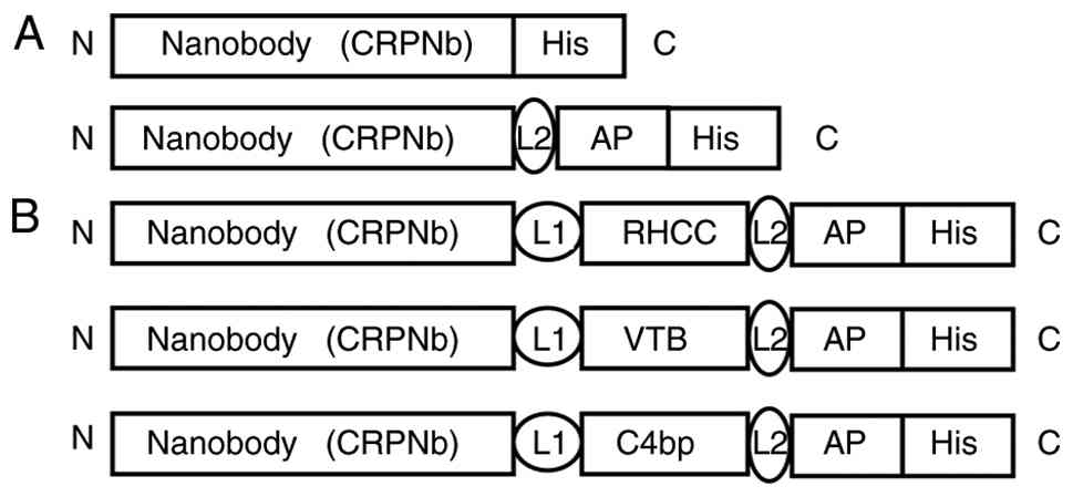 | Figure 1(A) Primary structure of the CRPNb
and CRPNb-AP nanobodies. (B) Primary structure of the recombinant
multimers (top, CRPNb-RHCC-AP; middle, CRPNb-VTB-AP; bottom,
CRPNb-C4bp-AP). CRPNb, C-reactive protein nanobody; AP, alkaline
phosphatase; RHCC, right-handed coiled coil; VTB, verotoxin; C4bp,
C4-binding protein; N, N-terminus; C, C-terminus; L, linker; L1,
APKADNKGGGGS; L2, APKADNKEFGGGGS; His, polyhistidine purification
tag. |
Western blotting was performed using the mouse
anti-His tag antibody and a precipitated ECL substrate solution.
The predicted thick bands, corresponding to ~12, 65, 72, 74 and 73
kDa, matching the molecular weights of CRPNb, CRPNb-AP,
CRPNb-RHCC-AP tetramers, CRPNb-VTB-AP pentamers and CRPNb-C4bp-AP
heptamers, respectively, could be observed (Fig. 2A). Based on a previous study, it
can be inferred that β-mercaptoethanol and heating lead to the
complete degradation of the CRPNb-RHCC-AP multimers, CRPNb-VTB-AP
pentamers and CRPNb-C4bp-AP heptamers into monomers (37). Although native PAGE was not
performed in the present study, previous studies have confirmed
that these recombinant constructs can efficiently self-assemble
into specific oligomers: Tetramers mediated by RHCC, pentamers
mediated by VTB and heptamers mediated by C4bp (15-18,35,37-41).
Correspondingly, the genes fused with RHCC, VTB and C4bp can
successfully express the tetrameric, pentameric and heptameric
target proteins, respectively.
![(A) Western blot analysis was
conducted to examine the fusion proteins of CRPNb, CRPNb-AP,
CRPNb-RHCC-AP, CRPNb-VTB-AP and CRPNb-C4bp-AP using purified
CRPNb-VTB-AP as a molecular weight standard for reference. (B)
Relative expression levels of CRPNb-AP, CRPNb-RHCC-AP, CRPNb-VTB-AP
and CRPNb-C4bp-AP. Proteins were quantified using purified
CRPNb-VTB-AP [lane P in (A)] as a control. The concentration of the
control protein CRPNb-VTB-AP was 240 µg/ml, as determined by a BCA
assay. Image Lab software was used for the intensity analysis.
CRPNb, C-reactive protein nanobody; AP, alkaline phosphatase; RHCC,
right-handed coiled coil; VTB, verotoxin; C4bp, C4-binding protein;
S, soluble; IS, insoluble; P, positive control.](/article_images/etm/32/4/etm-32-04-13279-g01.jpg) | Figure 2(A) Western blot analysis was
conducted to examine the fusion proteins of CRPNb, CRPNb-AP,
CRPNb-RHCC-AP, CRPNb-VTB-AP and CRPNb-C4bp-AP using purified
CRPNb-VTB-AP as a molecular weight standard for reference. (B)
Relative expression levels of CRPNb-AP, CRPNb-RHCC-AP, CRPNb-VTB-AP
and CRPNb-C4bp-AP. Proteins were quantified using purified
CRPNb-VTB-AP [lane P in (A)] as a control. The concentration of the
control protein CRPNb-VTB-AP was 240 µg/ml, as determined by a BCA
assay. Image Lab software was used for the intensity analysis.
CRPNb, C-reactive protein nanobody; AP, alkaline phosphatase; RHCC,
right-handed coiled coil; VTB, verotoxin; C4bp, C4-binding protein;
S, soluble; IS, insoluble; P, positive control. |
The recombinant proteins (CRPNb-AP, CRPNb-VTB-AP and
CRPNb-C4bp-AP) were found to be expressed in both soluble and
insoluble forms (Fig. 2B).
CRPNb-RHCC-AP was found to be expressed in the soluble form
(Fig. 2B). The soluble fraction
was sonicated and the supernatant was subsequently purified. The
purification process involved elution with imidazole using a
HisTrap Excel column, followed by BCA protein quantification. In
the present study, the relative expression levels of the multimers
were determined using purified CRPNb-VTB-AP as a control (Fig. 2B). The relative protein expression
levels for the soluble fraction normalized to the control were as
follows: 2.1 for the CRPNb-RHCC-AP tetramers, 3.0 for the
CRPNb-VTB-AP pentamers, 0.5 for the CRPNb-C4bp-AP heptamers and 2.7
for CRPNb-AP. The proportions of soluble to insoluble components
for CRPNb-VTB-AP, CRPNb-C4bp-AP and CRPNb-AP were found to be 3.06,
0.63 and 2.16, respectively. The proportion of soluble to insoluble
components could not be calculated for CRPNb-RHCC-AP because the
level of the insoluble components was too low.
In the present study, CRPNb-VTB-AP exhibited the
highest level of soluble components among the CRPNb-RHCC-AP
tetramers, CRPNb-VTB-AP pentamers and CRPNb-C4bp-AP heptamers.
The protein levels of CRPNb-RHCC-AP tetramers and
CRPNb-C4bp-AP heptamers aligned with previous findings, which
reported successful expression and proper folding of tetrameric EG2
[an anti-epidermal growth factor receptor single-domain antibody
(sdAb)]-RHCC and heptameric EG2-C4bp in prokaryotic expression
systems such as BL21 and E. coli TG1 (12,38).
Zhang et al (39)
previously developed a multimerization strategy and used
size-exclusion chromatography to characterize the formation of
pentamers.
Characterization of CRPNb-RHCC-AP
tetramers, CRPNb-C4bp-AP heptamers and CRPNb-VTB-AP pentamers
The antibody activities of the CRPNb-RHCC-AP
tetramers, CRPNb-VTB-AP pentamers and CRPNb-C4bp-AP heptamers were
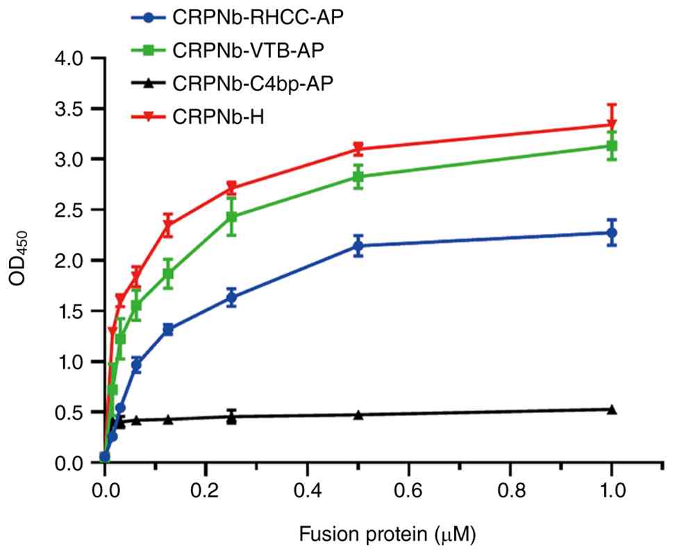
next examined and compared with those of CRPNb-H. Fig. 3 shows that CRPNb-H exhibited a
markedly higher OD450 signal compared with CRPNb-VTB-AP,
CRPNb-RHCC-AP tetramers and CRPNb-C4bp-AP heptamers, suggesting a
superior antigen-binding ability. Additionally, the OD450 for
CRPNb-VTB-AP was slightly higher compared with that of
CRPNb-RHCC-AP, suggesting that CRPNb-VTB-AP had a stronger
antigen-binding capacity compared with CRPNb-RHCC-AP. By contrast,
CRPNb-C4bp-AP exhibited no antigen-binding capacity, which is
inconsistent with previous studies (15-17).
The enzymatic activities of the CRPNb-RHCC-AP tetramers,
CRPNb-VTB-AP pentamers and CRPNb-C4bp-AP heptamers were next
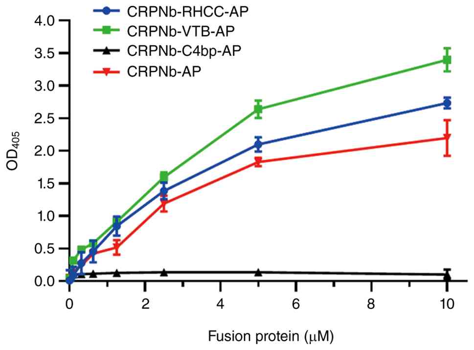
examined and compared with those of CRPNb-AP. Fig. 4 shows that CRPNb-RHCC-AP tetramers
and CRPNb-VTB-AP pentamers exhibited higher enzyme activity
compared with CRPNb-AP. The CRPNb-C4bp-AP heptamers exhibited no AP
activity, contradicting previous studies (15,17).
Due to experimental constraints, the control group in Fig. 3 was CRPNb-H. However CRPNb-AP
should have been employed as the control to align with Fig. 4, which would have made the
experiment more scientifically sound and reasonable. Therefore, the
results, such as Fig. 3 showing
that CRPNb-H exhibited a markedly higher OD450 signal compared with
CRPNb-VTB-AP, CRPNb-RHCC-AP tetramers and CRPNb-C4bp-AP heptamers,
have certain limitations. If the control group were replaced with
CRPNb-AP, the results might have been different.
As shown in Fig. 4,
the AP activity of CRPNb-RHCC-AP tetramers and CRPNb-VTB-AP
pentamers was increased compared with that of CRPNb-AP. This
indicated that AP fusion with the multimeric domain did not impair
AP activity in the CRPNb-RHCC-AP tetramers and CRPNb-VTB-AP
pentamers. Compared with findings from previous studies (29,36),
the present study revealed that CRPNb-C4bp-AP heptamers exhibited
both a lack of antigen-binding capacity and no AP activity. There
are a number of possible reasons for these observations. The
position of CRPNb and AP at either the N- or C-terminus of C4bp may
have impacted their activity (18). In addition, the spatial structure
of the CRPNb-C4bp-AP fusion protein may have affected the key amino
acid sites responsible for CRP recognition in the heptamers.
Furthermore, the flexible linker may not have adequately separated
the functional regions of CRPNb and AP, potentially impacting the
key amino acid sites involved in CRP recognition due to
interference from the fusion expression with AP. In summary, the
pentameric CRPNb-VTB-AP demonstrated superior AP activity and CRP
binding ability compared with the other multimers tested.
Stability of multimers. Thermal
stability
Two distinct heat treatment methods were used for
the thermal stability testing of CRPNb-RHCC-AP tetramers,
CRPNb-VTB-AP pentamers and CRPNb-C4bp-AP heptamers. As shown in
Fig. 5A, the Nb activity of the
CRPNb-RHCC-AP tetramers, CRPNb-VTB-AP pentamers and CRPNb-C4bp-AP
heptamers decreased with increasing temperature. As the temperature
increased, ~85% of the activity of the CRPNb-VTB-AP pentamers, ~75%
of the activity of CRPNb-AP and ~60% of the activity of the
CRPNb-RHCC-AP tetramers was maintained even at 80˚C, whilst the
activity of CRPNb-H dropped to ~40%. Based on these results, the
impact of incubation time at 80˚C on the binding activity between
the antigen and antibody was further examined. Fig. 5B shows that after 55 min of
incubation at 80˚C, ~40% of the activity was retained for the
CRPNb-RHCC-AP tetramers and CRPNb-VTB-AP pentamers. This suggested
that the multimeric Nbs were thermostable, suggesting that they may
be suitable for long-term storage at room temperature.
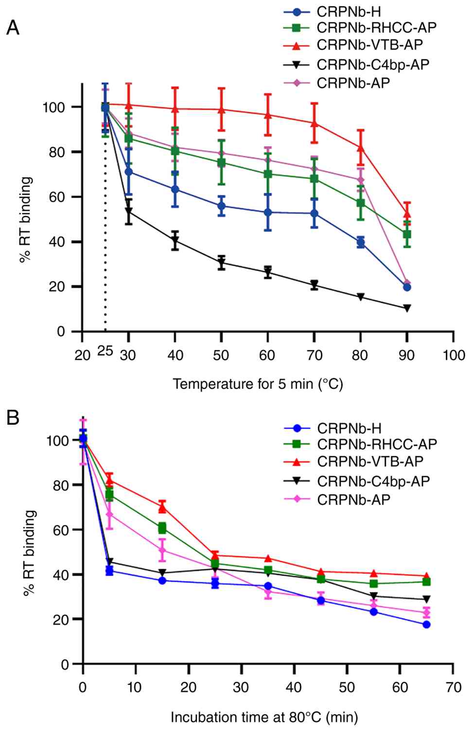 | Figure 5(A) Each of the five fusion proteins
was incubated for 5 min at various temperatures, before the
temperature stability of different Nbs was compared. The binding
value obtained after treatment at room temperature (25˚C) was used
as the 100% binding control. % RT binding refers to the binding
value relative to that at room temperature (25˚C). (B) Each of the
five fusion proteins was incubated for the indicated durations at
80˚C, before the temperature stability of different Nbs was
compared. The binding value at 0 min after treatment at 80˚C was
used as the 100% binding control. % RT binding refers to the
binding value relative to that at 0 min after treatment at 80˚C.
Error bars indicate the standard deviation of three replicates of
the experiment. Nb, nanobodies; CRPNb, C-reactive protein nanobody;
AP, alkaline phosphatase; RHCC, right-handed coiled coil; VTB,
verotoxin; C4bp, C4-binding protein; H, histidine; RT, room
temperature. |
Effect of pH and PBS on Nb activity. To
evaluate the influence of pH on the analysis performance, the
effect of pH on the assay was evaluated over a range of pH 4 to 10
using the optimized assay buffer by direct ELISA. The binding
signal of the antibody at pH 7.4 was defined as 100%, and the
binding activity at other pH conditions was expressed as the
percentage relative to this value, denoted as % binding activity at
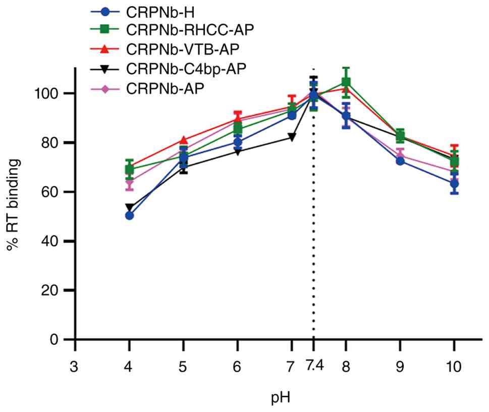
room temperature (RT). As shown in Fig. 6, the % RT binding of the
CRPNb-RHCC-AP tetramers and CRPNb-VTB-AP pentamers was increased
from 69 to 105% and 70.5 to 102% as the pH of the assay buffer
increased from pH 4 to 8, before decreasing from pH 9 onwards. By
contrast, the binding of CRPNb-H, CRPNb-C4bp-AP and CRPNb-AP was
increased from 50, 53 and 62 to 100% as the pH of the assay buffer
increased from 4 to 7.4, before decreasing from pH 7.4 onwards,
suggesting that pH affected Nb binding activity. Therefore, pH 7.4
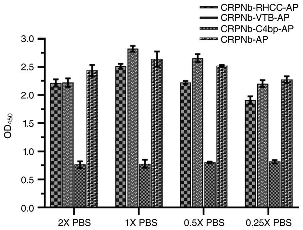
was chosen as the optimal pH for the assay buffer. Similarly,
different concentrations of PBS were next optimized (Fig. 7). The OD450 absorbance of the
CRPNb-RHCC-AP tetramers, CRPNb-VTB-AP pentamers and CRPNb-AP was
slightly increased as the concentration of PBS decreased from 2X to
1X PBS, then decreased from 1X to 0.25X PBS. Therefore, 1X PBS was
selected as the optimal PBS concentration.
Comparative analysis of the detection
performance of monomers and multimers
To assess the detection performance of the monomers
and multimers, a competitive ELISA was used to evaluate the
inhibition curves of the CRPNb-RHCC-AP tetramers, CRPNb-VTB-AP
pentamers and CRPNb-C4bp-AP heptamers. The optimal concentrations
of the coating antigen and antibody were determined using
checkerboard titration (Fig. S2).
The optimal antigen concentration was 2 µg/ml and the optimal
antibody concentration was 1 µg/ml. At these concentrations, the
absorbance value approached 1. Fig.
S3 presents an experimental study on conditional optimization
using CRPN-VTB-AP pentamers as a case study. Appropriate
experimental conditions were selected for subsequent competitive
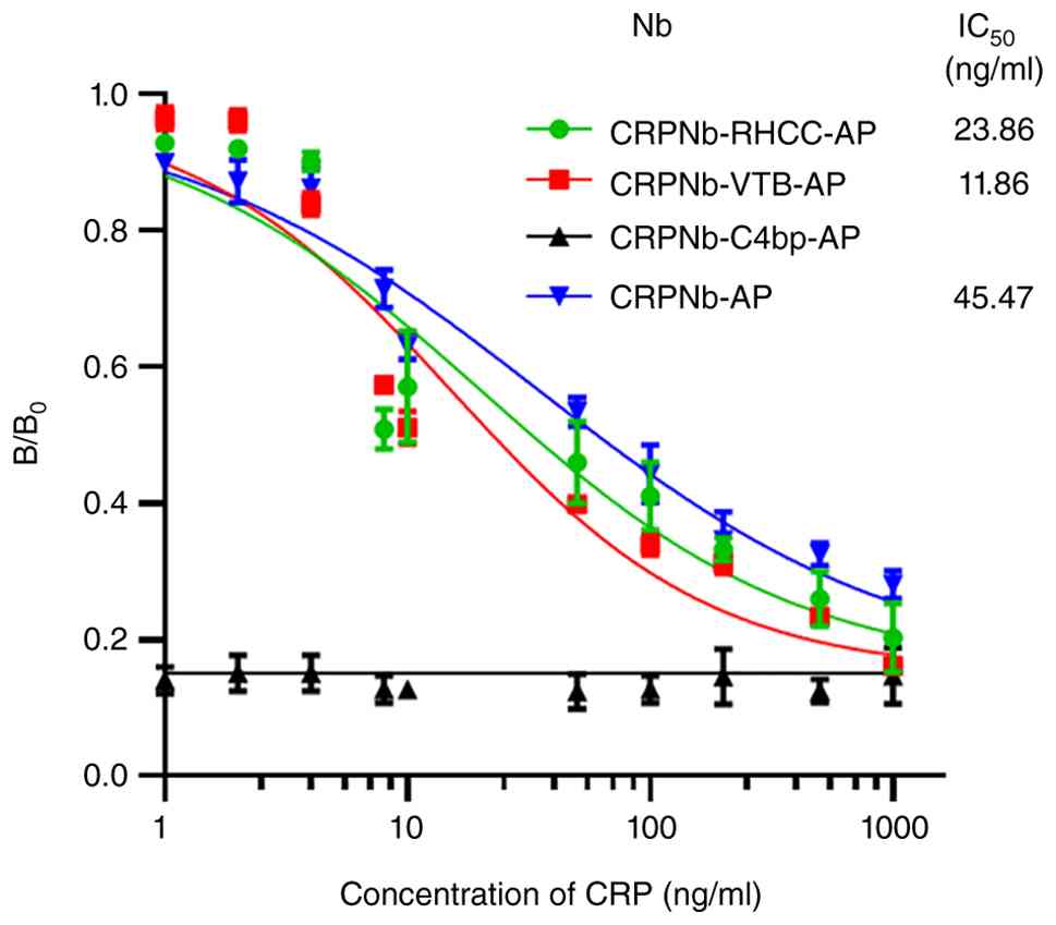
ELISAs. As shown in Fig. 8, the
CRPNb-VTB-AP pentamers exhibited the highest sensitivity, with an
IC50 of 11.86 ng/ml, compared with CRPNb-AP and
CRPNb-RHCC-AP, which had IC50 values of 45.47 and 23.86
ng/ml, respectively. Compared with the CRPNb-AP monomer, these
values were 4X lower for CRPNb-VTB-AP and 2X lower for
CRPNb-RHCC-AP. This suggested that multimerization strategies could
effectively enhance the affinity of Nbs to antigens. The increased
affinity of Nbs is essential for improving the sensitivity of
immunodetection. Nb multimers from previous studies have
demonstrated improved antigen affinity and detection sensitivity
compared with monomers (40,41),
which is consistent with the results observed in the present
study.
In the present study, recombinant CRPNb-RHCC-AP
tetramers, CRPNb-VTB-AP pentamers and CRPNb-C4bp-AP heptamers were
successfully constructed using the three self-associating peptides
RHCC, VTB and C4bp. Within the cytoplasm, these peptides are likely
to adopt the right conformation and are purified as soluble
proteins, thus avoiding laborious denaturation and refolding
procedures, making the process both cost-effective and
environmentally friendly (10,33).
In future studies, non-denaturing PAGE should be
used to verify the multimer products. Additionally, the
CRPNb-VTB-AP pentamers and CRPNb-RHCC tetramers demonstrated
antigen-specific recognition and enhanced affinity compared with
the CRP-AP monomer. Furthermore, all three multimers exhibited
thermal stability, making them suitable for storage and
transportation at ambient temperatures. This platform enables the
fusion of Nbs with various detection tags, allowing for the
generation of multimers with different combinations of Nbs and
different tags. By selecting compatible Nbs and self-assembling
peptides, Nb activity, enzyme activity, binding affinity and
detection performance can be optimized. This approach may lead to
the development of bio-functional immune reagents that serve as
both high-affinity binders and sensitive tracers.
Although previous studies have studied tetramers,
pentamers and heptamers based on RHCC, VTB and C4bp using other Nbs
such as EG2-sdAb (13) with AP or
other tags, to the best of our knowledge, the present study is the
first to form tetramers, pentamers and heptamers using CRPNb and AP
based on the three self-assembled peptides and to assess the
activity of CRP and AP. The present study aimed to establish a
platform for identifying the most effective multimers with superior
antigen-binding capabilities and increased AP activity. This will
facilitate the development of sensitive and reliable detection
methods for CRP.
Supplementary Material
Reference diagram of the competitive
ELISA (one-step) procedure, using pentamers as an example. CRP,
C-reactive protein; AP, alkaline phosphatase; VTB, verotoxin; KAN,
kanamycin.
Checkerboard optimization of coating
antigen and antibody concentration. Antibodies refers to C-reactive
protein nanobody-verotoxin-alkaline phosphatase.
Optimization of competitive ELISA
conditions, using pentamers as an example. (A) Coating antigen
concentration. (B) Incubating antibody concentration. (C) Blocking
solution. (D) Different competition times. (E) pH. (F) Ionic
strength. The y-axis represents B/B0, which is the ratio
of the bound signal intensity at a given CRP concentration (B) to
the maximum bound signal intensity in the absence of competing free
CRP (B0, normalized to 1.0). CRP, C-reactive
protein.
Sequences of self-assembly
peptides.
Acknowledgements
Not applicable.
Funding
Funding: Funding for the present study was provided by the
National Natural Science Foundation of China (grant nos. 32070936,
32270985 and 32201050), the Dalian Science and Technology
Innovation Fund of China (grant no. 2022JJ13S N07 0), the Yantai
Science and Technology Innovation Development Plan (Policy
Guidance) Project (grant no. 2023YD025), the Educational Department
of Liaoning Province (grant no. JYTQN2023103) and the Department of
Science & Technology of Liaoning Province (grant no.
2025-MSLH-026).
Availability of data and materials
The data generated in the present study may be
requested from the corresponding author.
Authors' contributions
XZ, XH, HL and CZ were involved in the design of the
study. XZ and XH were involved in the conception of the study. ND,
QW, JL and YZ were involved in all the biological experiments and
in data analysis. HL, CZ and XZ were involved in the writing of the
manuscript. XZ and XH supervised all experiments and confirmed the
authenticity of all the raw data. All authors have read and
approved the final version of the manuscript.
Ethics approval and consent to
participate
Not applicable.
Patient consent for publication
Not applicable.
Competing interests
The authors declare that they have no competing
interests.
References
|
1
|
Liu X, Xu Y, Wan DB, Xiong YH, He ZY, Wang
XX, Gee SJ, Ryu D and Hammock BD: Development of a
nanobody-alkaline phosphatase fusion protein and its application in
a highly sensitive direct competitive fluorescence enzyme
immunoassay for detection of ochratoxin A in cereal. Anal Chem.
87:1387–1394. 2015.PubMed/NCBI View Article : Google Scholar
|
|
2
|
Els Conrath K, Lauwereys M, Wyns L and
Muyldermans S: Camel single-domain antibodies as modular building
units in bispecific and bivalent antibody constructs. J Biol Chem.
276:7346–7350. 2001.PubMed/NCBI View Article : Google Scholar
|
|
3
|
Huo J, Li Z, Wan D, Li D, Qi M, Barnych B,
Vasylieva N, Zhang J and Hammock BD: Development of a highly
sensitive direct competitive fluorescence enzyme immunoassay based
on a nanobody-alkaline phosphatase fusion protein for detection of
3-phenoxybenzoic acid in urine. J Agric Food Chem. 66:11284–11290.
2018.PubMed/NCBI View Article : Google Scholar
|
|
4
|
Jovčevska I and Muyldermans S: The
therapeutic potential of nanobodies. BioDrugs. 34:11–26.
2020.PubMed/NCBI View Article : Google Scholar
|
|
5
|
Chanier T and Chames P: Nanobody
engineering: Toward next generation immunotherapies and
immunoimaging of cancer. Antibodies (Basel). 8(13)2019.PubMed/NCBI View Article : Google Scholar
|
|
6
|
Naidoo DB and Chuturgoon AA: The potential
of nanobodies for COVID-19 diagnostics and therapeutics. Mol Diagn
Ther. 27:193–226. 2023.PubMed/NCBI View Article : Google Scholar
|
|
7
|
Sun Z, Wang X, Chen Q, Yun Y, Tang Z and
Liu X: Nanobody-alkaline phosphatase fusion protein-based
enzyme-linked immunosorbent assay for one-step detection of
ochratoxin A in rice. Sensors (Basel). 18(4044)2018.PubMed/NCBI View Article : Google Scholar
|
|
8
|
Nuñez-Prado N, Compte M, Harwood S,
Álvarez-Méndez A, Lykkemark S, Sanz L and Álvarez-Vallina L: The
coming of age of engineered multivalent antibodies. Drug Discov
Today. 20:588–594. 2015.PubMed/NCBI View Article : Google Scholar
|
|
9
|
Deyev SM and Lebedenko EN: Multivalency:
The hallmark of antibodies used for optimization of tumor targeting
by design. Bioessays. 30:904–918. 2008.PubMed/NCBI View Article : Google Scholar
|
|
10
|
Arbabi-Ghahroudi M: Camelid single-domain
antibodies: Historical perspective and future outlook. Front
Immunol. 8(1589)2017.PubMed/NCBI View Article : Google Scholar
|
|
11
|
Bathula NV, Bommadevara H and Hayes JM:
Nanobodies: The future of antibody-based immune therapeutics.
Cancer Biother Radiopharm. 36:109–122. 2021.PubMed/NCBI View Article : Google Scholar
|
|
12
|
Wang L, Liu X, Zhu X, Wang L, Wang W, Liu
C, Cui H, Sun M and Gao B: Generation of single-domain antibody
multimers with three different self-associating peptides. Protein
Eng Des Sel. 26:417–423. 2013.PubMed/NCBI View Article : Google Scholar
|
|
13
|
Asano R, Koyama N, Hagiwara Y, Masakari Y,
Orimo R, Arai K, Ogata H, Furumoto S, Umetsu M and Kumagai I:
Anti-EGFR scFv tetramer (tetrabody) with a stable monodisperse
structure, strong anticancer effect, and a long in vivo half-life.
FEBS Open Bio. 6:594–602. 2016.PubMed/NCBI View Article : Google Scholar
|
|
14
|
Li C, Feng H, Xia X, Wang L, Gao B, Zhang
Y and Lan X: (99m) Tc-labeled tetramer and pentamer of
single-domain antibody for targeting epidermal growth factor
receptor in xenografted tumors. J Labelled Comp Radiopharm.
59:305–312. 2016.PubMed/NCBI View Article : Google Scholar
|
|
15
|
Bao K, Liu X, Xu Q, Su B, Liu Z, Cao H and
Chen Q: Nanobody multimerization strategy to enhance the
sensitivity of competitive ELISA for detection of ochratoxin A in
coffee samples. Food Control. 127(108167)2021.
|
|
16
|
Li Z, Wang Y, Vasylieva N, Wan D, Yin Z,
Dong J and Hammock BD: An ultrasensitive bioluminescent enzyme
immunoassay based on nanobody/nanoluciferase heptamer fusion for
the detection of tetrabromobisphenol A in sediment. Anal Chem.
92:10083–10090. 2020.PubMed/NCBI View Article : Google Scholar
|
|
17
|
Su B, Bei Z, Pei H, Xie X, Sun Z, Chen Q,
Cao H and Liu X: Generation of a nanobody-alkaline phosphatase
heptamer fusion for ratiometric fluorescence immunodetection of
trace alpha fetoprotein in serum. Int J Biol Macromol. 201:507–515.
2022.PubMed/NCBI View Article : Google Scholar
|
|
18
|
Su B, Xu H, Xie G, Chen Q, Sun Z, Cao H
and Liu X: Generation of a nanobody-alkaline phosphatase fusion and
its application in an enzyme cascade-amplified immunoassay for
colorimetric detection of alpha fetoprotein in human serum.
Spectrochim Acta A Mol Biomol Spectrosc. 262(120088)2021.PubMed/NCBI View Article : Google Scholar
|
|
19
|
Wang J, Majkova Z, Bever CRS, Yang J, Gee
SJ, Li J, Xu T and Hammock BD: One-step immunoassay for
tetrabromobisphenol a using a camelid single domain
antibody-alkaline phosphatase fusion protein. Anal Chem.
87:4741–4748. 2015.PubMed/NCBI View Article : Google Scholar
|
|
20
|
Sheng Y, Wang K, Lu Q, Ji P, Liu B, Zhu J,
Liu Q, Sun Y, Zhang J, Zhou EM and Zhao Q: Nanobody-horseradish
peroxidase fusion protein as an ultrasensitive probe to detect
antibodies against Newcastle disease virus in the immunoassay. J
Nanobiotechnology. 17(35)2019.PubMed/NCBI View Article : Google Scholar
|
|
21
|
Mu Y, Jia C, Zheng X, Zhu H, Zhang X, Xu
H, Liu B, Zhao Q and Zhou EM: Correction to: A nanobody-horseradish
peroxidase fusion protein-based competitive ELISA for rapid
detection of antibodies against porcine circovirus type 2. J
Nanobiotechnology. 19(66)2021.PubMed/NCBI View Article : Google Scholar
|
|
22
|
Sun Z, Lv J, Liu X, Tang Z, Wang X, Xu Y
and Hammock BD: Development of a nanobody-AviTag fusion protein and
its application in a streptavidin-biotin-amplified enzyme-linked
immunosorbent assay for ochratoxin A in cereal. Anal Chem.
90:10628–10634. 2018.PubMed/NCBI View Article : Google Scholar
|
|
23
|
Chen Q, Sun D, Pei H, Su B, Bao K, Cao H,
Zhang C, Hammock BD and Liu X: Development of a nanobody tagged
with streptavidin-binding peptide and its application in a Luminex
fluoroimmunoassay for alpha fetal protein in serum. RSC Adv.
10:23767–23774. 2020.PubMed/NCBI View Article : Google Scholar
|
|
24
|
Chen Q, Wang Y, Mao F, Su B, Bao K, Zhang
Z, Xie G and Liu X: Development of a horseradish
peroxidase-nanobody fusion protein for visual detection of
ochratoxin A by dot immunoassay. RSC Adv. 10:33700–33705.
2020.PubMed/NCBI View Article : Google Scholar
|
|
25
|
Su B, Zhang Z, Sun Z, Tang Z, Xie X, Chen
Q, Cao H, Yu X, Xu Y, Liu X and Hammock BD: Fluonanobody-based
nanosensor via fluorescence resonance energy transfer for
ultrasensitive detection of ochratoxin A. J Hazard Mater.
422(126838)2022.PubMed/NCBI View Article : Google Scholar
|
|
26
|
Yu S, Li Z, Li J, Zhao S, Wu S, Liu H, Bi
X, Li D, Dong J, Duan S and Hammock BD: Generation of dual
functional nanobody-nanoluciferase fusion and its potential in
bioluminescence enzyme immunoassay for trace glypican-3 in serum.
Sens Actuators B Chem. 336(129717)2021.PubMed/NCBI View Article : Google Scholar
|
|
27
|
Aydın EB, Aydın M and Sezgintürk MK:
Highly selective and sensitive sandwich immunosensor platform
modified with MUA-capped GNPs for detection of spike Receptor
Binding Domain protein: A precious marker of COVID 19 infection.
Sens Actuators B Chem. 345(130355)2021.PubMed/NCBI View Article : Google Scholar
|
|
28
|
Wang X, Wang Y, Wang Y, Chen Q and Liu X:
Nanobody-alkaline phosphatase fusion-mediated phosphate-triggered
fluorescence immunoassay for ochratoxin a detection. Spectrochim
Acta A Mol Biomol Spectrosc. 226(117617)2020.PubMed/NCBI View Article : Google Scholar
|
|
29
|
Xiao T, Sun J, Zhao J, Wang S, Liu G and
Yang X: FRET effect between fluorescent polydopamine nanoparticles
and MnO2 nanosheets and its application for sensitive
sensing of alkaline phosphatase. ACS Appl Mater Interfaces.
10:6560–6569. 2018.PubMed/NCBI View Article : Google Scholar
|
|
30
|
Zhang Z, Su B, Xu H, He Z, Zhou Y, Chen Q,
Sun Z, Cao H and Liu X: Enzyme cascade-amplified immunoassay based
on the nanobody-alkaline phosphatase fusion and MnO2
nanosheets for the detection of ochratoxin A in coffee. RSC Adv.
11:21760–21766. 2021.PubMed/NCBI View Article : Google Scholar
|
|
31
|
Oloketuyi S, Bernedo R, Christmann A,
Borkowska J, Cazzaniga G, Schuchmann HW, Niedziółka-Jönsson J,
Szot-Karpińska K, Kolmar H and de Marco A: Native llama nanobody
library panning performed by phage and yeast display provides
binders suitable for C-reactive protein detection. Biosensors
(Basel). 11(496)2021.PubMed/NCBI View Article : Google Scholar
|
|
32
|
Mandecki W, Shallcross MA, Sowadski J and
Tomazic-Allen S: Mutagenesis of conserved residues within the
active site of Escherichia coli alkaline phosphatase yields
enzymes with increased kcat. Protein Eng. 4:801–804.
1991.PubMed/NCBI View Article : Google Scholar
|
|
33
|
Chao S, Liu Y, Ding N, Lin Y, Wang Q, Tan
J, Li W, Zheng Y, Hu X and Li J: Highly expressed soluble
recombinant anti-GFP VHHs in Escherichia coli via optimized
signal peptides, strains, and inducers. Front Mol Biosci.
9(848829)2022.PubMed/NCBI View Article : Google Scholar
|
|
34
|
Zhu J, Ruan Y, Fu X, Zhang L, Ge G, Wall
JG, Zou T, Zheng Y, Ding N and Hu X: An engineered pathway for
production of terminally sialylated N-glycoproteins in the
periplasm of Escherichia coli. Front Bioeng Biotechnol.
8(313)2020.PubMed/NCBI View Article : Google Scholar
|
|
35
|
Zhang J and Mackenzie CR: Multivalent
display of single-domain antibodies. Methods Mol Biol. 911:445–456.
2012.PubMed/NCBI View Article : Google Scholar
|
|
36
|
Mayr J, Lupas A, Kellermann J, Eckerskorn
C, Baumeister W and Peters J: A hyperthermostable protease of the
subtilisin family bound to the surface layer of the archaeon
Staphylothermus marinus. Curr Biol. 6:739–749.
1996.PubMed/NCBI View Article : Google Scholar
|
|
37
|
Kielkopf CL, Bauer W and Urbatsch IL:
Sodium dodecyl sulfate-polyacrylamide gel electrophoresis of
proteins. Cold Spring Harb Protoc. 2021:494–504. 2021.PubMed/NCBI View Article : Google Scholar
|
|
38
|
Stone E, Hirama T, Chen W, Soltyk AL,
Brunton J, MacKenzie CR and Zhang J: A novel pentamer versus
pentamer approach to generating neutralizers of verotoxin 1. Mol
Immunol. 44:2487–2491. 2007.PubMed/NCBI View Article : Google Scholar
|
|
39
|
Zhang J, Li Q, Nguyen TD, Tremblay TL,
Stone E, To R, Kelly J and Roger MacKenzie C: A pentavalent
single-domain antibody approach to tumor antigen discovery and the
development of novel proteomics reagents. J Mol Biol. 341:161–169.
2004.PubMed/NCBI View Article : Google Scholar
|
|
40
|
Wang F, Li ZF, Wan DB, Vasylieva N, Shen
YD, Xu ZL, Yang JY, Gettemans J, Wang H, Hammock BD and Sun YM:
Enhanced non-toxic immunodetection of Alternaria mycotoxin
tenuazonic acid based on ferritin-displayed anti-idiotypic
nanobody-nanoluciferase multimers. J Agric Food Chem. 69:4911–4917.
2021.PubMed/NCBI View Article : Google Scholar
|
|
41
|
Fan K, Jiang B, Guan Z, He J, Yang D, Xie
N, Xie C and Yan X: Fenobody: A ferritin-displayed nanobody with
high apparent affinity and half-life extension. Anal Chem.
90:5671–5677. 2018.PubMed/NCBI View Article : Google Scholar
|