Introduction
Cadmium (Cd) is an environmental pollutant which
poses significant health risks to humans (1). After entering into human body
through ingestion, inhalation or skin absorption, Cd is not
biodegradable and persists in many human organs (2). The kidney is one of the primary
organs targeted by Cd (3). With
chronic exposure, Cd accumulates in the epithelial cells of the
proximal tubule, resulting in tubular impairment with a loss of
reabsorptive capacity and the development of proteinuria (4). Previous findings suggest that Cd
also affects the vasculature in the kidney (3). In the human body, Cd ions enter the
blood circulation and bind to plasma proteins (5,6).
Through the circulation, Cd reaches the targeted organs and passes
through the endothelium of each vascular bed (5). In the kidney, Cd directly interacts
with glomerular endothelial cells (GECs), which comprise the inner
layer of the glomerular filtration barrier. However, the effects of
Cd on GECs remain largely unknown.
Cd induces cell death in a variety of cell types
(7,8). Cell death is an active process that
crucially maintains homeostasis in multicellular organisms
(9). There are three major types
of cell death: apoptosis, autophagy and necrosis. Apoptosis occurs
primarily through two well known pathways, the intrinsic or
mitochondrial-mediated pathway and the extrinsic or death
receptor-mediated pathway (10).
Both pathways are mediated by cell membrane receptors, such as
tumor necrosis factor receptor 1/2 (TNFR1/2), Fas/CD95, death
receptor 3 (DR3) and TRAIL-receptor (TRAIL-R)1 and TRAIL-R2
(11,12), and activate the caspase family of
proteases, which ultimately dismantle the cell. Autophagy is a
survival mechanism in which parts of the cytosol and specific
organelles are engulfed by a double-membrane autophagosome and
degraded (13). Necrosis is
characterized by the rapid loss of membrane integrity (13). All three may be activated through
distinct or overlapping signaling pathways in response to Cd
(9). These signaling pathways
include reactive oxygen species (ROS), nuclear factor-κB (NF-κB),
B-cell lymphoma 2 (Bcl-2) and mitogen-activated protein kinas
(MAPK) signaling (9).
NF-κB is a heterodimeric protein composed of
different combinations of members of the Rel family of
transcription factors including RelA (p65), RelB (p50), c-Rel,
NF-κB1 (p59/p105), and NF-κB2 (p52/p100) (14). Usually, NF-κB dimers reside in the
cytosol, bound to the inhibitory protein inhibitor of κB (IκB), and
are activated by stimuli capable of inducing phosphorylation and
proteolysis of IκB (15).
Following the removal of IκB, NF-κB enters the nucleus to induce
the expression of coordinate sets of targets genes for cell
survival (15). Previous findings
have reported that the activation of the NF-κB pathway antagonizes
apoptosis by the ligand engagement of 'death receptors' such as
TNFR1 (12). Other evidence
suggests that the NF-κB pathway is required for cell survival
(16). The NF-κB pathway
regulates the expression of several inhibitors of apoptosis (IAPs),
including c-IAP1, c-IAP2 and X chromosome-linked IAP (XIAP), which
inhibits the caspase pathway (17,18). In addition, it has been
demonstrated that the negative regulation of c-Jun N-terminal
kinase (JNK) activation by NF-κB contributes to the inhibition of
apoptosis (19).
JNK, a member of the MAPK superfamily, also mediates
apoptosis-related pathways. The persistent activation of JNK
correlates with apoptosis, whereas suppression of JNK correlates
with cell survival (20). In the
nuclear apoptosis pathway, activated JNK translocates to the
nucleus and activates c-Jun (21), leading to the formation of
activator protein 1 (AP-1) (22).
The JNK-AP-1 pathway is involved in the increased expression of
pro-apoptotic genes such as tumor necrosis factor-α (TNF-α), Fas-L
and Bak (23). In the
mitochondrial signaling of apoptosis, JNK is required for the
release of cytochrome c from the inner membrane space of
mitochondria and the activation of the caspase-9 cascade (24). JNK also promotes the activity of
pro-apoptotic BH3, which is the only subgroup of the Bcl-2 family
of proteins such as Bim and Bmf capable of activating Bax and/or
Bak to initiate apoptosis (25).
In the present study, we evaluated the effects of
low dose Cd on the apoptosis of human renal glomerular endothelial
cells (HRGECs) and explored the underlying mechanisms. Although 4
µM Cd does not affect the survival of HRGECs, it
significantly activates the NF-κB pathway. Treatment of the cells
with pyrrolidine dithiocarbamate (PDTC), a potent NF-κB inhibitor,
followed by Cd exposure induced extensive apoptosis of HRGECs.
Inhibition of the NF-κB pathway significantly increased Cd-induced
JNK phosphorylation. The addition of SP600125, a JNK pathway
inhibitor, partially reversed the apoptosis of HRGECs induced by
the combination treatment of Cd and PDTC. Our results indicate that
Cd maintains the survival of Cd-exposed HRGECs through the
activation of the NF-κB pathway.
Materials and methods
Cell culture
HRGECs were purchased from ScienCell Research
Laboratories (Carlsbad, CA, USA). HRGECs were cultured in
Dulbecco's modified Eagle's medium (DMEM)/F12, supplemented with
10% fetal bovine serum (Lonza, Basel, Switzerland), 100 IU/ml
penicillin (Sigma-Aldrich,St. Louis, MO, USA) and 100 µg/ml
streptomycin (Sigma-Aldrich). The cells were cultured in humidified
air at 37°C with 5% CO2. Cadmium chloride
(CdCl2) was purchased from Sigma-Aldrich and dissolved
in phosphate-buffered saline (PBS) at a stock concentration of 1
mM. PDTC and SP600125 were purchased from Cell Signaling Technology
(Danvers, MA, USA) and dissolved in dimethyl sulfoxide (DMSO).
During the experiments, the cells were treated with various
combinations of 4 µM CdCl2, 10 µM PDTC
(for 1 h prior to Cd exposure) and 10 µM SP600125.
Cell viability assay
The viability of HRGECs was assessed following
exposure to CdCl2, PDTC or combination treatment. The
cell cultures were washed with PBS and incubated in 0.05% trypsin
for 2 min at 37°C. After disaggregation with pipettes, the single
cell suspension was diluted 1:1 in 0.4% trypan blue (w/v in 0.9%
NaCl) (Santa Cruz Biotechnology, Inc., Santa Cruz, CA, USA). The
number and percentage of dye-free cells was calculated.
Detection of apoptosis by Annexin
V-fluorescein isothiocyanate (FITC) and propidium iodide (PI)
analysis
The apoptosis of HRGECs following Cd exposure was
detected by Annexin V-FITC and propidium iodide (PI) staining using
an assay kit (Neobiosciences, Shenzhen, China) according to the
manufacturer's instructions. Briefly, the cells were trypsinized,
pelleted, washed twice with PBS and resuspended into single cells.
Then, 1×10⁶ cells were stained with Annexin V-FITC
(0.25%) and PI (1 µg/ml) in 1X binding buffer (10 mM HEPES,
pH 7.4, 140 mM NaOH, 2.5 mM CaCl2) for 15 min at room
temperature in the dark. Positive staining of the cells was
detected using a FACSAria II flow cytometer, and analyzed using the
FACSDiva acquisition and analysis software (both from BD
Biosciences, San Jose, CA, USA).
Western blot analysis
After treatment, HRGECs were lysed using RIPA buffer
(20 mM Tris, pH 7.5, 150 mM NaCl, 50 mM NaF, 1% NP-40, 0.1%
deoxycholate, 0.1% sodium dodecyl sulfate, 1 mM EDTA) supplemented
with protease inhibitors aprotonin (1 µg/ml), leupeptin (10
µg/ml) and PMSF (1 mM). The protein concentration was
determined using the bicinchoninic acid (BCA) assay (Bio-Rad
Laboratories, Inc., Berkeley, CA, USA). Equal amounts of protein
(40 µg) were separated by sodium dodecyl
sulfate-polyacrylamide gel electrophoresis (SDS-PAGE) (10%
acrylamide gel) and transferred to a PVDF membrane. The membrane
was blocked in TBST (20 mM Tris, 150 mM NaCl, 0.1% Tween-20) with
2.5% non-fat milk at 37°C for 1.5 h prior to incubation with
primary antibodies overnight at 4°C. After washing three times with
TBST, the membrane was incubated with secondary antibody at 37°C
for 2 h. The following primary antibodies were used: rabbit
anti-SAPK/JNK (9258), rabbit anti-phospho-SAPK/JNK (4668), mouse
anti-IκBα (L35A5), rabbit anti-NF-κB p65 (6956), and rabbit
anti-GAPDH (2118) (all from Cell Signaling Technology). The
secondary antibody was HRP-linked anti-rabbit IgG (7074) and
HRP-linked anti-mouse IgG (7076) (Cell Signaling Technology). The
blots were developed with enhanced chemiluminescence reagents
(Millipore, Billerica, MA, USA), and the relative intensities of
the blots were quantified using ImageJ software (National
Institutes of Health, Bethesda, MD, USA).
Immunofluorescence
HRGECs were allowed to grow to confluence on
fibronectin-coated glass chamber slides and exposed to Cd for 24 h.
The monolayers of HRGECs were then washed with PBS containing 100
mM glycine, fixed with 4% paraformaldehyde for 5 min, and washed
three times with PBS for 10 min. Immunoreactivity was examined by
staining with a rabbit polyclonal anti NF-κB p65 antibody (1:400;
Cell Signaling Technology) overnight at 4°C and incubation with an
Alexa 546-labeled anti-rabbit secondary antibody (1:200; Molecular
Probes, Eugene, OR, USA) for 2 h. Nuclear staining was achieved
using DAPI (1:1,000; Molecular Probes) and images of the the
samples were captured using an Olympus LCX100 Imaging system
(Olympus Corp., Tokyo, Japan) with an excitation wavelength of 546
nm.
Statistical analysis
The data are presented as the means ± standard
error. All experiments were performed using at least three separate
cell preparations. The differences between the groups were
evaluated using a Student's t-test (two-tailed). A p-value <0.05
was considered to indicate a statistically significant
difference.
Results
Cd activates NF-κB signaling in
HRGECs
NF-κB signaling regulates apoptosis in response to
oxidative stress, which may be induced by Cd (26). The activation of NF-κB requires
the degradation of IκBa, which forms a cytoplasmic and inactive
complex with the p65-p50 heterodimer (27). We examined the effects of Cd on
NF-κB signaling in HRGECs. Western blot analysis revealed that the
protein level of IκBα in the cytoplasm of HRGECs was significantly
decreased by Cd exposure, whereas levels of the internal control
GAPDH remained unchanged (Fig. 1A and
B). Using immunofluorescence, we were able to demonstrate the
nuclear translocation of the NF-κB/p65 subunit in HRGECs (Fig. 1C). Following exposure to Cd for 24
h, p65 in the nucleus markedly increased compared with that in the
untreated cells. These results indicate that NF-κB signaling
pathway is activated by Cd in HRGECs.
Effects of Cd exposure on the apoptosis
of HRGECs
Cd induces cytotoxicity in different types of cell
depending on the concentration and the exposure time (28,29). We examined the effect of low dose
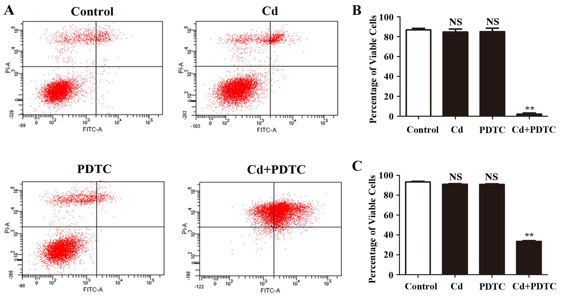
Cd on the apoptosis of HRGECs. Using PI/Annexin V flow cytometric
analysis, 4 µM Cd did not change the percentage of viable
cells of HRGECs (86.8±1.6 vs. 84.7±2.9%, p=0.34). PDTC prevents
IκBα degradation and the dissociation of NF-κB from IκBα thus
inhibits the NF-κB signaling pathway (30). We pre-treated HRGECs with 10
µM PDTC for 1 h prior to Cd exposure and found that
combination treatment with PDTC and Cd significantly decreased the
percentage of viable cells of HRGECs compared to that of PDTC
treatment alone (6.5±0.4 vs. 85.1±3.2%, p<0.01) (Fig. 2A and B). At a concentration of 4
µM, Cd did not significantly affect cell viability after 24
h in a trypan blue exclusion assay (93.4±0.6 vs. 91.1±0.7%, p=0.26)
(Fig. 2C); however, combination
treatment with PDTC and Cd significantly reduced the viability of
HRGECs (91.1±0.7 vs. 33.4±0.8%, p<0.01) (Fig. 2C). Taken together, these findings
demonstrated that NF-κB signaling pathway was required for the
survival of Cd-stimulated HRGECs.
Activation of NF-κB signaling inhibits
JNK signaling in Cd-stimulated HRGECs
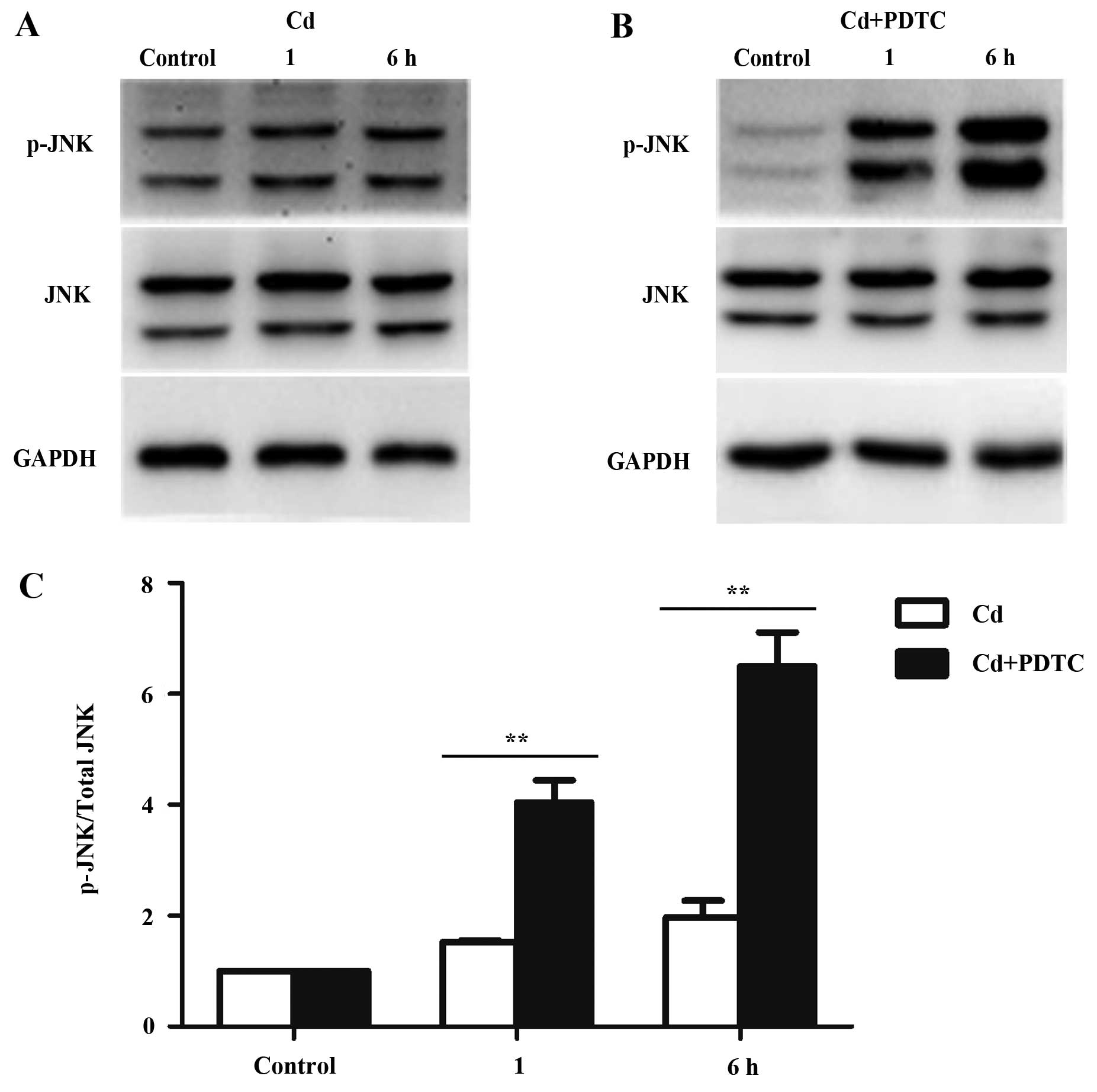
The JNK pathway mediates apoptosis and is directly
activated by Cd in several cell lines (7,8).
It has been reported that NF-κB signaling pathway negatively
regulates the JNK pathway (31).
We examined JNK activation in HRGECs exposed to Cd and PDTC.
Fig. 3A showed that Cd increased
phosphorylated (p-)JNK in HRGECs, whereas the total levels of JNK
protein and the internal control GAPDH remain unchanged. In the
presence of PDTC, Cd markedly increased the phosphorylation of JNK
(Fig. 3B). Densitometry analyses
showed that the combination of PDTC and Cd significantly elevated
p-JNK compared with exposure to Cd alone (p<0.01) (Fig. 3C). Thus, the JNK pathway is
suppressed by Cd-activated NF-κB signaling pathway.
SP600125 partially reverses the apoptosis
of HRGECs induced by Cd and PDTC
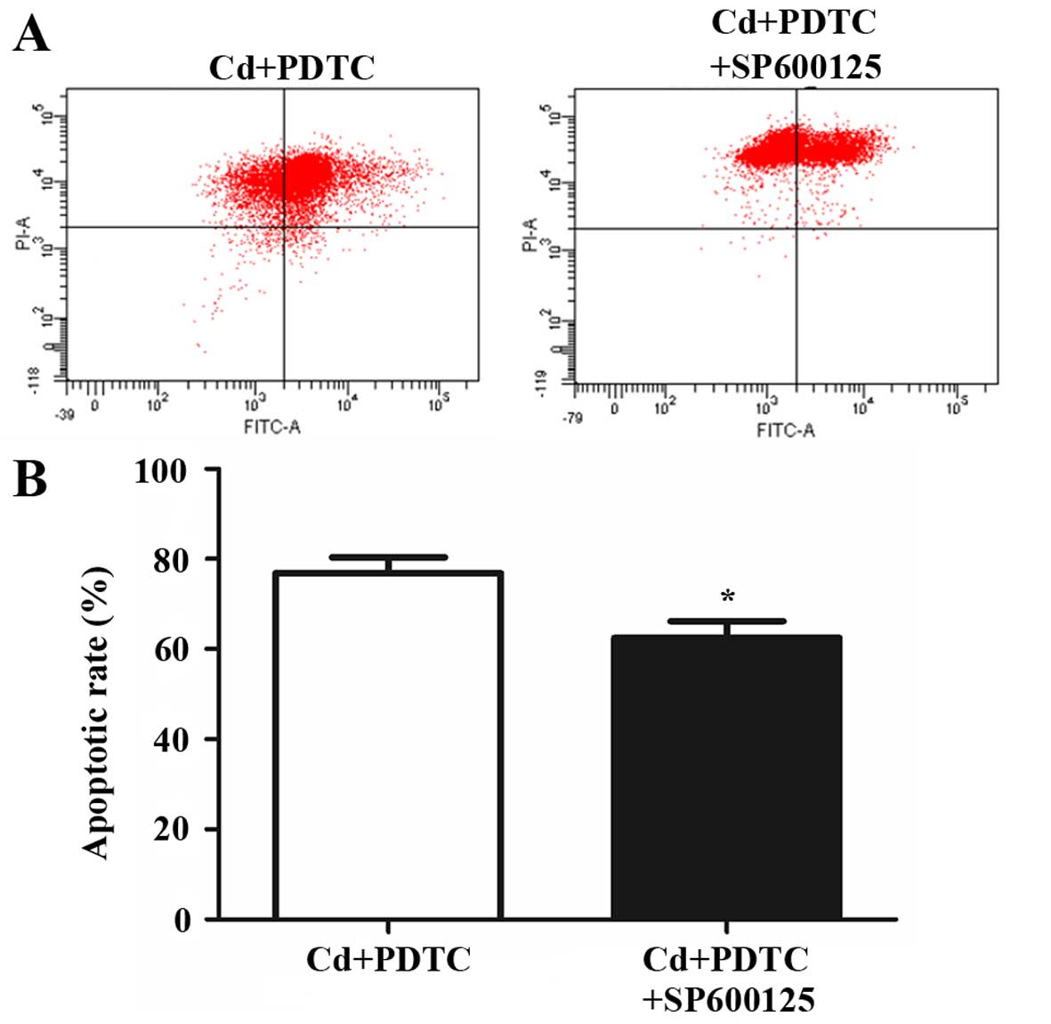
To further confirm the role of the JNK pathway in
Cd-stimulated HRGECs, 10 µM SP600125, an inhibitor of JNK
pathway, was applied to cells in addition to Cd and PDTC. The
addition of SP600125 partially reversed the apoptosis of HRGECs
(76.8±3.5 vs. 62.4±3.7%, p<0.05) (Fig. 4). This suggests that NF-κB
partially inhibits the Cd-induced apoptosis of HRGECs through the
negative regulation of the JNK pathway.
Discussion
Cd, one of the most widespread pollutants, induces a
number of clinical symptoms of kidney damage (3). In the present study, we evaluated
the effects of low dose Cd on the apoptosis of HRGECs, which
comprise the primary layer of glomerular filtration. Our data
indicated that 4 µM Cd does not induce the apotosis of
HRGECs. Cd activates the NF-κB pathway and inhibition of NF-κB
pathway significantly induces cell apoptosis in Cd-stimulated
HRGECs. In addition, the NF-κB pathway maintains cell survival
partially through the suppression of the JNK pathway. The present
study indicates that the NF-κB pathway is essential for maintaining
the survival of HRGECs exposed to Cd.
GECs are a specialized type of microvascular cell
which form the inner layer of the glomerular filter barrier
(32). Injury to GECs leads to
nephrotoxicity characterized by proteinuria, polyuria and
glucosuria (3). We have recently
reported that 1 µM and 1 h exposure of Cd increases the
permeability in HRGECs monolayers and redistributes the adherens
junction proteins vascular endothelial-cadherin and β-catenin;
however, this short-term, low dose of Cd does not induce the
apoptosis of HRGECs (33).
Morales et al have shown that after a 9-week sub-chronic
exposure to Cd in rats, renal endothelial nitric oxide synthase
(eNOS) was upregulated by 10–20-fold (34). It is recognized that Cd
potentially causes damage to the glomeruli endothelium; however,
the detailed mechanism responsible for thse effects remains
unclear. In the present study, we determined that 4 µM and
24 h Cd exposure does not induce the apoptosis of HRGECs, by
balancing the activation of pro-apoptotic and pro-survival
signaling pathways.
The NF-κB pathway is closely associated with cell
survival (35). Mice with a
genetic disruption of RelA, a major activating subunit of NF-κB,
died from massive apoptosis of hepatocytes in the liver (36). In the present study, we found that
low dose Cd activates the NF-κB pathway in HRGECs through the
degradation of IκBα and the translocation of the NF-κB p65 subunit
from the cytosol to the nucleus. Cd induces the activation of NF-κB
in several types of cell. Jeong et al proved that 3
µM Cd activates the NF-κB pathway in cerebrovascular
endothelial cells (37).
Moreover, high concentrations of Cd (20 or 30 µM)
significantly upregulated the protein levels of p65 in the nuclei
of bronchial epithelial cells (38). We also found that the inhibition
of the NF-κB pathway by PDTC induces marked apoptosis of
Cd-stimulated HRGECs. In another study, the inhibitory protein IκBα
was modified to resist ubiquitin-mediated degradation; it
consistently prevented NF-κB from entering into the nucleus,
leading to the sensitization of the cells to apoptotic stimuli
(39). Taken together, these
findings suggest that Cd activates the NF-κB pathway, which
maintains the survival of HRGECs.
To maintain the survival of HRGECs, it is likely
that Cd activates pro-apoptotic pathways together with the NF-κB
pathway. The JNK pathway is one of the initiators of apoptosis, and
is regulated by the NF-κB pathway (20). Tang et al (19) and De Smaele et al (41) have independently demonstrated that
TNF-α induces prolonged JNK activation in NF-κB
activation-deficient cells, such as RelA and IKKβ knockouts. In the
present study, we examined the role of the JNK pathway in
Cd-induced apoptosis. We found that Cd activates the JNK pathway in
HRGECs and Cd induced the activation of the NF-κB pathway which
inhibits the JNK pathway. In Cd- and PDTC-treated HRGECs, the
addition of a JNK inhibitor partially reversed the Cd-induced
apoptosis of HRGECs. Thus, NF-κB promotes cell survival partially
by inhibiting the JNK pathway. In addition to the JNK pathway,
NF-κB is involved in multiple mechanisms which suppress apoptosis.
The NF-κB pathway upregulates the Bcl-2 family members Bcl-xL and
A1/Bfl-1, which reduce the effect of mitochondrial depolarization
and inhibit cytochrome c release from mitochondria (42). NF-κB also increases the expression
of TRAF1/2, c-IAP 1/2 and XIAP, which suppress apoptosis at the
level of caspase-8 (40).
Therefore, the downstream effectors of the NF-κB pathway which
mediate the survival of HRGECs warrant further investigation.
In conclusion, we found that low dose Cd activates
the NF-κB pathway through the degradation of IκBα, and promotes the
translocation of NF-κB/p65 into the nucleus of HRGECs. The
activation of NF-κB is required for the survival of Cd-stimulated
HRGECs. In addition, the Cd-activated NF-κB pathway prevents cell
death partially by inhibiting the JNK pathway. The present study
provides important information for improving our understanding of
the molecular mechanisms underlying Cd-induced nephrotoxicity.
Acknowledgments
The present study was supported by grants from the
Science and Technology Development Plan of Shandong Province (no.
2013GSF11805), the National Natural Science Foundation of China
(no. 81370269) and the Shandong Taishan Scholarship (Ju Liu).
References
|
1
|
Järup L, Berglund M, Elinder CG, Nordberg
G and Vahter M: Health effects of cadmium exposure - a review of
the literature and a risk estimate. Scand J Work Environ Health.
24(Suppl 1): 1–51. 1998.
|
|
2
|
Thévenod F and Lee WK: Toxicology of
cadmium and its damage to mammalian organs. Met Ions Life Sci.
11:415–490. 2013. View Article : Google Scholar : PubMed/NCBI
|
|
3
|
Prozialeck WC and Edwards JR: Early
biomarkers of cadmium exposure and nephrotoxicity. Biometals.
23:793–809. 2010. View Article : Google Scholar : PubMed/NCBI
|
|
4
|
Chakraborty PK, Lee WK, Molitor M, Wolff
NA and Thévenod F: Cadmium induces Wnt signaling to upregulate
proliferation and survival genes in sub-confluent kidney proximal
tubule cells. Mol Cancer. 9:1022010. View Article : Google Scholar : PubMed/NCBI
|
|
5
|
Prozialeck WC, Edwards JR and Woods JM:
The vascular endothelium as a target of cadmium toxicity. Life Sci.
79:1493–1506. 2006. View Article : Google Scholar : PubMed/NCBI
|
|
6
|
Dong F, Guo F, Li L, Guo L, Hou Y, Hao E,
Yan S, Allen TD and Liu J: Cadmium induces vascular permeability
via activation of the p38 MAPK pathway. Biochem Biophys Res Commun.
450:447–452. 2014. View Article : Google Scholar : PubMed/NCBI
|
|
7
|
Liu J and Kapron CM: Differential
induction of MAP kinase signalling pathways by cadmium in primary
cultures of mouse embryo limb bud cells. Reprod Toxicol.
29:286–291. 2010. View Article : Google Scholar : PubMed/NCBI
|
|
8
|
Yuan Y, Jiang C, Hu F, Wang Q, Zhang K,
Wang Y, Gu J, Liu X, Bian J and Liu Z: The role of
mitogen-activated protein kinase in cadmium-induced primary rat
cerebral cortical neurons apoptosis via a mitochondrial apoptotic
pathway. J Trace Elem Med Biol. 29:275–283. 2015. View Article : Google Scholar
|
|
9
|
Green DR and Llambi F: Cell Death
Signaling. Cold Spring Harb Perspect Biol. 7:72015. View Article : Google Scholar
|
|
10
|
Wyllie AH, Kerr JF and Currie AR: Cell
death: The significance of apoptosis. Int Rev Cytol. 68:251–306.
1980. View Article : Google Scholar : PubMed/NCBI
|
|
11
|
Kucharczak J, Simmons MJ, Fan Y and
Gélinas C: To be, or not to be: NF-kappaB is the answer - role of
Rel/NF-kappaB in the regulation of apoptosis. Oncogene.
22:8961–8982. 2003. View Article : Google Scholar : PubMed/NCBI
|
|
12
|
Wajant H, Pfizenmaier K and Scheurich P:
Tumor necrosis factor signaling. Cell Death Differ. 10:45–65. 2003.
View Article : Google Scholar : PubMed/NCBI
|
|
13
|
Ashkenazi A and Salvesen G: Regulated cell
death: signaling and mechanisms. Annu Rev Cell Dev Biol.
30:337–356. 2014. View Article : Google Scholar : PubMed/NCBI
|
|
14
|
Dutta J, Fan Y, Gupta N, Fan G and Gélinas
C: Current insights into the regulation of programmed cell death by
NF-kappaB. Oncogene. 25:6800–6816. 2006. View Article : Google Scholar : PubMed/NCBI
|
|
15
|
Huxford T, Huang DB, Malek S and Ghosh G:
The crystal structure of the IkappaBalpha/NF-kappaB complex reveals
mechanisms of NF-kappaB inactivation. Cell. 95:759–770. 1998.
View Article : Google Scholar : PubMed/NCBI
|
|
16
|
Karin M and Ben-Neriah Y: Phosphorylation
meets ubiquitination: the control of NF-[kappa]B activity. Annu Rev
Immunol. 18:621–663. 2000. View Article : Google Scholar
|
|
17
|
Irmler M, Thome M, Hahne M, Schneider P,
Hofmann K, Steiner V, Bodmer JL, Schröter M, Burns K, Mattmann C,
et al: Inhibition of death receptor signals by cellular FLIP.
Nature. 388:190–195. 1997. View
Article : Google Scholar : PubMed/NCBI
|
|
18
|
Chu ZL, McKinsey TA, Liu L, Gentry JJ,
Malim MH and Ballard DW: Suppression of tumor necrosis
factor-induced cell death by inhibitor of apoptosis c-IAP2 is under
NF-kappaB control. Proc Natl Acad Sci USA. 94:10057–10062. 1997.
View Article : Google Scholar : PubMed/NCBI
|
|
19
|
Tang G, Minemoto Y, Dibling B, Purcell NH,
Li Z, Karin M and Lin A: Inhibition of JNK activation through
NF-kappaB target genes. Nature. 414:313–317. 2001. View Article : Google Scholar : PubMed/NCBI
|
|
20
|
Chen YR and Tan TH: The c-Jun N-terminal
kinase pathway and apoptotic signaling (Review). Int J Oncol.
16:651–662. 2000.PubMed/NCBI
|
|
21
|
Davis RJ: Signal transduction by the JNK
group of MAP kinases. Cell. 103:239–252. 2000. View Article : Google Scholar : PubMed/NCBI
|
|
22
|
Dhanasekaran DN and Johnson GL: MAPKs:
function, regulation, role in cancer and therapeutic targeting.
Oncogene. 26:3097–3099. 2007. View Article : Google Scholar : PubMed/NCBI
|
|
23
|
Fan M and Chambers TC: Role of
mitogen-activated protein kinases in the response of tumor cells to
chemotherapy. Drug Resist Updat. 4:253–267. 2001. View Article : Google Scholar
|
|
24
|
Tournier C, Hess P, Yang DD, Xu J, Turner
TK, Nimnual A, Bar-Sagi D, Jones SN, Flavell RA and Davis RJ:
Requirement of JNK for stress-induced activation of the cytochrome
c-mediated death pathway. Science. 288:870–874. 2000. View Article : Google Scholar : PubMed/NCBI
|
|
25
|
Lei K and Davis RJ: JNK phosphorylation of
Bim-related members of the Bcl2 family induces Bax-dependent
apoptosis. Proc Natl Acad Sci USA. 100:2432–2437. 2003. View Article : Google Scholar : PubMed/NCBI
|
|
26
|
Chen LF and Greene WC: Shaping the nuclear
action of NF-kappaB. Nat Rev Mol Cell Biol. 5:392–401. 2004.
View Article : Google Scholar : PubMed/NCBI
|
|
27
|
Jacobs MD and Harrison SC: Structure of an
IkappaBalpha/NF-kappaB complex. Cell. 95:749–758. 1998. View Article : Google Scholar : PubMed/NCBI
|
|
28
|
Yokouchi M, Hiramatsu N, Hayakawa K,
Okamura M, Du S, Kasai A, Takano Y, Shitamura A, Shimada T, Yao J
and Kitamura M: Involvement of selective reactive oxygen species
upstream of proapoptotic branches of unfolded protein response. J
Biol Chem. 283:4252–4260. 2008. View Article : Google Scholar
|
|
29
|
Liu F, Wang B, Li L, Dong F, Chen X, Li Y,
Dong X, Wada Y, Kapron CM and Liu J: Low-dose cadmium upregulates
VEGF expression in lung adenocarcinoma cells. Int J Environ Res
Public Health. 12:10508–10521. 2015. View Article : Google Scholar : PubMed/NCBI
|
|
30
|
Dong F, Zhou X, Li C, Yan S, Deng X, Cao
Z, Li L, Tang B, Allen TD and Liu J: Dihydroartemisinin targets
VEGFR2 via the NF-κB pathway in endothelial cells to inhibit
angiogenesis. Cancer Biol Ther. 15:1479–1488. 2014. View Article : Google Scholar
|
|
31
|
Javelaud D and Besançon F: NF-kappa B
activation results in rapid inactivation of JNK in TNF
alpha-treated Ewing sarcoma cells: a mechanism for the
anti-apoptotic effect of NF-kappa B. Oncogene. 20:4365–4372. 2001.
View Article : Google Scholar : PubMed/NCBI
|
|
32
|
Du L, Dong F, Guo L, Hou Y, Yi F, Liu J
and Xu D: Interleukin-1β increases permeability and upregulates the
expression of vascular endothelial-cadherin in human renal
glomerular endothelial cells. Mol Med Rep. 11:3708–3714.
2015.PubMed/NCBI
|
|
33
|
Li L, Dong F, Xu D, Du L, Yan S, Hu H,
Lobe CG, Yi F, Kapron CM and Liu J: Short-term, low-dose cadmium
exposure induces hyperpermeability in human renal glomerular
endothelial cells. J Appl Toxicol. 36:257–265. 2016. View Article : Google Scholar
|
|
34
|
Morales AI, Vicente-Sánchez C, Jerkic M,
Santiago JM, Sánchez-González PD, Pérez-Barriocanal F and
López-Novoa JM: Effect of quercetin on metallothionein, nitric
oxide synthases and cyclooxygenase-2 expression on experimental
chronic cadmium nephrotoxicity in rats. Toxicol Appl Pharmacol.
210:128–135. 2006. View Article : Google Scholar
|
|
35
|
Bubici C, Papa S, Pham CG, Zazzeroni F and
Franzoso G: NF-kappaB and JNK: an intricate affair. Cell Cycle.
3:1524–1529. 2004. View Article : Google Scholar : PubMed/NCBI
|
|
36
|
Beg AA and Baltimore D: An essential role
for NF-kappaB in preventing TNF-alpha-induced cell death. Science.
274:782–784. 1996. View Article : Google Scholar : PubMed/NCBI
|
|
37
|
Jeong EM, Moon CH, Kim CS, Lee SH, Baik
EJ, Moon CK and Jung YS: Cadmium stimulates the expression of
ICAM-1 via NF-kappaB activation in cerebrovascular endothelial
cells. Biochem Biophys Res Commun. 320:887–892. 2004. View Article : Google Scholar : PubMed/NCBI
|
|
38
|
Chen DJ, Xu YM, Du JY, Huang DY and Lau
AT: Cadmium induces cytotoxicity in human bronchial epithelial
cells through upregulation of eIF5A1 and NF-kappaB. Biochem Biophys
Res Commun. 445:95–99. 2014. View Article : Google Scholar : PubMed/NCBI
|
|
39
|
Huang TT, Wuerzberger-Davis SM, Wu ZH and
Miyamoto S: Sequential modification of NEMO/IKKgamma by SUMO-1 and
ubiquitin mediates NF-kappaB activation by genotoxic stress. Cell.
115:565–576. 2003. View Article : Google Scholar : PubMed/NCBI
|
|
40
|
Wang CY, Mayo MW, Korneluk RG, Goeddel DV
and Baldwin AS Jr: NF-kappaB antiapoptosis: induction of TRAF1 and
TRAF2 and c-IAP1 and c-IAP2 to suppress caspase-8 activation.
Science. 281:1680–1683. 1998. View Article : Google Scholar : PubMed/NCBI
|
|
41
|
De Smaele E, Zazzeroni F, Papa S, Nguyen
DU, Jin R, Jones J, Cong R and Franzoso G: Induction of gadd45beta
by NF-kappaB downregulates pro-apoptotic JNK signalling. Nature.
414:308–313. 2001. View Article : Google Scholar : PubMed/NCBI
|
|
42
|
Zong WX, Edelstein LC, Chen C, Bash J and
Gélinas C: The prosurvival Bcl-2 homolog Bfl-1/A1 is a direct
transcriptional target of NF-kappaB that blocks TNFalpha-induced
apoptosis. Genes Dev. 13:382–387. 1999. View Article : Google Scholar : PubMed/NCBI
|