1. Introduction
The extracellular matrix (ECM) derived from
organs/tissues is a complex, highly organized assembly of
macromolecules with an adequate three-dimensional (3D) organization
(1). Dry ECM powders can be
further processed to generate various injectable hydrogels by
enzymatical digestion. To match tissue defects and improve
therapeutic outcomes, these ECM-hydrogels have been combined with
synthetic materials to fabricate electrospun nanofibers or
3D-printed scaffolds/conduits via electrospinning technology or 3D
printing. The prepared hydrogels or solid scaffolds can seed with
stem cells and/or incorporate with growth factors (GFs) to further
enhance their bioactivity and repair function. These extracellular
matrices and their final products are all termed as the
decellularized matrix grafts (ECM-G) or the ECM preparations
(ECM-P). In addition to regulating intracellular signaling pathways
for inducing cell adhesion, these biologic ECM-P also provide a
permissive environment for cell growth, proliferation, migration
and differentiation, which have widely applied for the therapeutic
reconstruction in heterologous tissue disorder (2). Commonly, ECM-P consists of a complex
mixture of structural and functional proteins, including collagen,
fibronectin, laminin, glycosaminoglycans, and growth factors (GFs).
Besides abundant bioactive factors, its inherent cross-linked
polymeric network and suitable mechanical property, not only
provides physical support for tissue integrity and elasticity, but
also modulates the wound healing response towards tissue remodeling
(3,4). Additionally, ECM-P has been used in
various forms, such as scaffold incorporated with stem cells and/or
GFs, and even as a bioink for constructing 3D-printed conduits,
which have been implanted in virtually every body system (5).
Injectable materials prepared from untreated raw ECM
frequently invoke chronic inflammatory response and host foreign
body reaction in a variety of body systems, due to residual
immunogenicity components, such as Galactose-α(1,3)-galactose (α-gal), major
histocompatibility complex class I (MHC I), endotoxins and
cell-derived nucleic acids (6).
Additionally, some pathogenic contaminations contained in the
biological ECM material may also provoke severe immune rejection
and foreign body response in preliminary xenografts (7). They are divided into two major
categories: Viral particles/elements and salmonella (8). The former is particularly
problematic in terms of xenozoonoses, including brucellosis,
leptospirosis, and tularellosis. The latter mainly refers to
prions, which are derived from xenogeneic and allogeneic tissue
sources (9). Thus, the objective
on any decellularization process is maximizing the removal of these
residual pathogenic contaminants or extracellular antigen
molecules, while retaining the functional performance of
non-immunogenic ECM and maintaining its native ultrastructure and
mechanical strength. Currently, the most commonly utilized methods
for decellularization of xenogeneic and allogeneic tissues involve
physical, chemical, and enzymatic approaches (10). The choice of these
decellularization approaches (single or combined method) depends on
the complex intrinsic structure, composition, and mechanical
properties of the raw ECM (11).
Following the decellularization and solubilization of raw ECM, the
resulting ECM-P should still retain the tissue-specific composition
and nanostructure that are essential in contributing to ischemic
injury repair, tissue regeneration or organ replacement.
The use of ECM grafts (ECM-G) for tissue engineering
and drug delivery has already been broadly investigated (3). The ideal ECM-G for regenerative
medicine should be clear of cell residues from the tissue source,
and comprises a loosely organized nanofibrous architecture with
interconnecting pores, which are essential for nutrient and gas
exchange for healthy cell migration and growth (12). In addition to having
biocompatible, biodegradable, and adequate biomechanical
properties, the ECM-G should also display appropriate
visco-elasticity and match the permeability property of the
autologous homologous tissue (13). Moreover, the proteolytic turnover
of grafted ECM should match the rate of new tissue formation in
order to withstand mechanical stress from neighboring tissues
during the regeneration period (14,15). Furthermore, ECM-G has been
developed as a delivery vehicle for incorporating GFs and/or cells
to enhance the repair and regeneration of the damaged tissues and
holds promising potential for improving the therapy of traumatic
diseases.
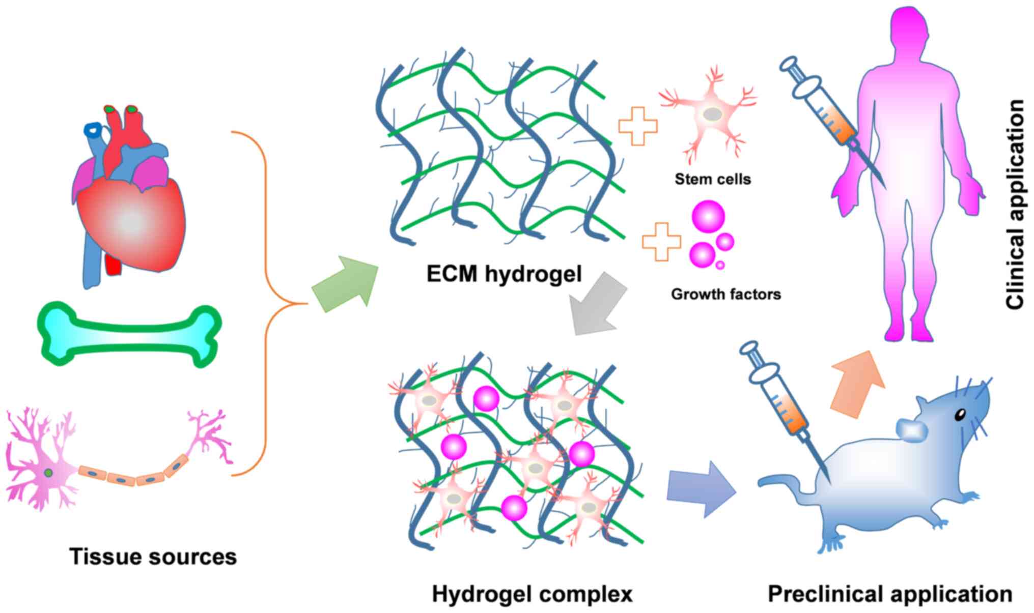
In this review, we firstly provide an overview of
the unique properties of ECM-G and different decellularization
methods for achieving sufficient cell removal from source tissues.
Then, we discuss the effect of undesired residual cellular material
invoking the degree of immune response. Finally, applying ECM-G for
tissue engineering and regenerative medicine will be discussed,
including current limitations and future directions (Fig. 1).
2. ECM-G characterization
ECM-G is a class of naturally derived proteinaceous
biomaterials, with excellent biophysical, biomechanical, and
biochemical properties, which can provide biological signals and
maintain tissue microarchitecture for guiding on cell growth,
differentiation, neovascularization and functional improvement
(16). It has been shown that
collagen and elastin, both of which are the most abundant proteins
in the ECM-G, played a critical role in controlling tissue osmotic
pressure and regulating intracellular signaling cascades that
direct stem cell differentiation and function (17,18). Glycosaminoglycans (GAGs) are also
regarded as important associated macromolecules found in the ECM-G,
as they generally served as crosslinkers for carrying GFs because
their binding sites are highly negatively charge, leading to high
affinity to cationic GFs (19).
Thus, ECM-G also serve as a drug delivery vehicle for the
controlled release of GFs in a spatial and temporal manner when
applied in pre-clinical research. Moreover, their thermo-responsive
feature is suitable for injecting a cavity site of damage via a
catheter or syringe. In addition, the three-dimensional
cross-linked network of fibers is another feature that renders them
capable of holding large amounts of water. Although the pore size,
fiber diameter and fiber alignment of ECM-G vary from different
source tissue, its typical nano-scale topography is enough to be
sensed and manipulated by infiltrating cells (20). The viscoelastic property of ECM-G
is another important parameter for evaluating stiffness and
solid-like behavior, which can be accurately determined by
turbidimetric gelation kinetics and rheology. A suitable viscosity
of the pre-gel solution is favorable for supporting stem cell
differentiation and proliferation for in vitro culture and
promoting the constructive and functional outcome of tissues and
organs. For example, the 3D meniscus-derived hydrogel with storage
modulus (a typical index for reflecting viscosity) of 838±296 Pa
(12 mg/ml) showed good cellular compatibility by facilitating the
differentiation of bone marrow mesenchymal stem cells into nucleus
pulposus-like cells after culturing for 2 weeks (21). In vivo examination of a low
viscosity ECM hydrogel derived from porcine spinal cord showed that
it remained within the defect site at body temperature (37°C)
condition which stimulates neovascularization and axonal outgrowth
into the cavity site of the acute model of spinal cord injury (SCI)
(22).
The different matrix density of various hydrogels,
including water content and macromolecular density, is mainly
dependent on tissue sources and status (23). Generally, hydrogel composition and
density play important roles in regulating cell activity and
phenotype (24). The abundant
water content filling the space between hydrogel crosslinks allows
for the diffusion of solute molecules. Bio-activate molecules, such
as GFs, proteoglycans and collagens, are necessary for the
activation of intracellular signaling cascades through integrin
receptors to induced cell adhesion, migration, proliferation and
differentiation (1). Thus, an
ideal natural hydrogel should retain several distinct ECM
macromoleculars as much as possible and contain water with proper
proportion in case of reduced mechanical force and viscoelastic
property for the prepared hydrogel products. Preclinical rodent
studies using porcine-derived urinary bladder matrix (UBM)-ECM
hydrogel with the concentration of 8 mg/ml implanted into a
14-day-old stroke cavity induced a robust invasion of endothelial
cells with neovascularization for brain regeneration (25). Further research will focus on
optimizing the matrix density of various hydrogels to open new
therapeutic avenues for tissue engineering and regenerative
medicine.
Overall, these topological, biochemical and
mechanical properties of the ECM-G are essential for modulating
diverse fundamental aspects of cell biology and functional outcomes
in disease models. Generally, the intrinsic property of a specific
ECM-G is mostly determined by the source of tissue type and
species. However, the optimal decellularization method is critical
for the resulting ECM quality concerning ultrastructure and
molecular composition (26).
Thus, successful procedures for ECM preparations must relate to the
tissue of origin, comprehensively utilizing physical, chemical and
biological methods to remove cellular material as much as possible,
while retaining the ECM biopolymer components.
There are extensive molecular changes occurring in
the ECM including post-translational modification (e.g.,
glycosylation), proteolytic processing, crosslinking, assembling
into polymers and higher complex structures. All these processes
are crucial for ECM properties and function, turnover and
stability, as well as cellular interactions. These modifications
solve the specific issue of different diseases. For instance, it
has been shown that ECM proteins, such as collagen, contained in
the subendothelial basement membrane could activate platelets,
leading to thrombosis at the site of anastomosis during vascular
surgery (27). To overcome this
shortcoming, chemically modified vascular ECM was developed via
covalently immobilizing anticoagulant heparin onto the ECM using
collagen binding peptide (CBP) as an intermediate linker that
selectively binds collagen within ECM. This heparin-modified ECM
exhibits beneficial effects on reducing long-term thromboresistance
and targeting VEGF to facilitate the adhesion and growth of
endothelial cells (28). Another
representative study by Li et al identified
nanofiber-hydrogel modification for repairing SCI (29). They engineered an injectable
nanofiber-hydrogel composite (NHC) by covalently conjugating
hyaluronic acid (HA) hydrogel with electrospun polycaprolactone
(PCL) fibers. This unique bonding resulted in a structure that
possessed mechanical strength and porosity to prevent contused
spinal cord collapse and induce cellular migration within the
injury site. After injecting this NHC into the cavity region of an
adult rat with spinal cord contusion, macrophage polarization,
vascularization, neurogenesis and axonal growth became
significantly ameliorated at 28 days treatment.
3. Methods of decellularization
treatments
Decellularization has become a popular technique for
transforming different organs, such as the skin, heart, liver,
kidneys, muscle, sis mucosa, nerves, tendons, ligaments and blood
vessel, into bioactive ECM-G through physical and/or chemical
processing (30). Since different
organs or tissues have their unique compositions and mechanical
behavior which are closely associated with regulating cell behavior
and tissue regeneration, these unique compositions and inherent
property must be retained as much as possible during
decellularization to obtain the biologic ECM-G (10). Several popular methods have been
examined for performing decellularization, which can be mainly
classified as physical, chemical and biological approaches
(31). These decellularization
methods require various decellularization agents that involve
specific purpose, extent, influence factor and effect on ECM
(Table I). A complete
decellularization process should combine these approaches together,
that is, firstly destroying the cell membrane via physical shaking
or ionic detergents, followed by solubilization of cytoplasmic and
nuclear cellular components using sodium dodecyl sulphate (SDS) and
sodium deoxycholate, and finally digestion of the extracellular
matrix into a homogeneous gel by trypsin, dispase and
phospholipase. Furthermore, this effective removal of cellular
components is able to achieve further improvement through coupling
with mechanical agitation. Nevertheless, it should be highlighted
that the entire removal of cytoplasmic and nuclear components while
preserving the entire/native extracellular matrix entities and
structure is an extremely difficult task. The optimal recipe of
decellularization agents is dependent upon different specific
tissues, as well as the intended clinical application (32). Several decellularization methods
have been developed for a variety of tissues (Table II) and these will be reviewed in
the following sections.
 | Table ISelected agents and techniques for
decellularizing tissue. |
Table I
Selected agents and techniques for
decellularizing tissue.
| Methods/Refs. |
Characteristics | Effects on ECM |
|---|
| Snap freezing
(103-105) | Decellularization
of tendinous, ligamentous tissue and nerve tissue
Usually combined with complement chemical and enzymatic
techniques
Affected by temperature | Disruption of
cellular membranes and inducing cell lysis |
| Mechanical
sonication (106,107) | Tissues with hard
structures
Usually combined with complement chemical and enzymatic
techniques
Affected by mechanical frequency and amplitude | Largely damage the
ECM structure |
| Mechanical
agitation (44,108) | Removal of cellular
contents
Usually combined with complement chemical and enzymatic
techniques
Affected by the speed and length of mechanical agitation | Direct damage to
ECM |
| Triton X-100
(109-111) | Removing nuclear
and cytoplasmic waste
Mixed results with efficacy
Dependent on tissue
Affected by exposure time, temperature, and concentration | More effective cell
removal from thin tissues
Mild disruption of ultrastructure and removal of GAG |
| SDS (45,112,113) | Applying for
retaining the overall matrix structure
Very effective for removal of cellular components from
tissue
Affected by exposure time, temperature, and concentration | Removes nuclear
remnants and cytoplasmic proteins |
| Sodium deoxycholate
(114-116) | Very effective for
removing cellular remnants
Causing disruption to the native tissue architecture
Affected by exposure time, PH and concentration | Damages the matrix,
similar to the SDS |
| CHAPS (44,117) | Cell removal from
thinner tissues, such as blood vessels and lung
95% removal efficiency of nuclear materials
Affected by pH and concentration | Effectively removes
cells in thin tissues and mildly disrupts ultrastructure in thin
tissues |
| Trypsin (47,48) | Specifically target
ECM proteins
Strongly damage the ECM proteins collagen laminin, and
fibronectin
Affected by exposure time, temperature, pH and concentration | Digestion of the
proteins in the ECM, in particular collagen laminin, and
fibronectin |
| Pepsin (118,119) | Generally target
ECM proteins
Milld damage the ECM proteins collagen laminin, and
fibronectin
Affected by exposure time, temperature, pH and concentration | Damage ECM proteins
if digested too long |
| Lipase (120,121) | Specifically
targets lipids
Strongly efficiency
Affected by exposure time, temperature, pH and concentration | Hydrolyzing fat to
derive adipose derived ECM |
| Collagenases
(123) | Specifically
targets collagen at early step
Strongly efficiency
Affected by exposure time, temperature, pH and concentration | Effectively removes
collagen in ECM; Prolonged expose will disrupt ECM
ultrastructure |
| Nucleases (123,124) | Specifically break
down DNA or RNA sequences Highly efficiency
Affected by temperature, pH and concentration | No function on ECM
proteins; Only removal of nucleotides |
| Table IIApplications of different organ
decellularization techniques to various organs. |
Table II
Applications of different organ
decellularization techniques to various organs.
| Organ | Decellularization
agent | Solubilization
protocol | Species | (Refs.) |
|---|
| Heart | 10 U/ml heparinized
water
5.0% SDS
1% (v/v) Triton X-100 | 10X PBS
RT, 48 h | Porcine | (125) |
| Lung | 0.0035% Triton-X
100
0.1% SDS
0.1% potassium laurate | Perfusion
1.5 mg/ml pepsin | Rat | (126) |
| Liver | 4% Triton
X-100
10 mM Tris-HCl
0.25% trypsin |
Voytik-Harbin
10 mg pepsin | Rat | (127) |
| Kidney | Gradient of SDS
(0.5%-1.0%)
0.1% Triton X-100 | Perfusion
0.1 M HCl | Rat | (128) |
| Skin | 1% SDS and 0.5%
pen/strep
Isopropyl alcohol
0.001% Triton X-100 | Perfusion
0.1 M HCl | Murine | (30) |
| Nerve | 3.0% Triton
X-100
4.0% SDS | 0.01 M
HCl
0.1 M NaOH | Porcine | (84) |
| Skeletal
muscle | 0.7% NaCl
1% Triton X-100
70% ethanol | Perfusion
0.02 M HCl
24 h | Mouse | (129) |
Physical methods
A variety of physical methods, such as freezing,
mechanical agitation and sonication, have been frequently applied
to facilitate tissue decellularization. Snap freezing has been used
to disrupt cell-cell and cell-matrix bonds via the formation of
intracellular ice crystals (33).
After rapidly reducing the temperature of a tissue to the freezing
point, cell lysis occurred immediately, facilitating the removal of
immunogenic material from the tissue. However, it should be noted
that the rate of temperature change for special tissue must be
carefully controlled to protect matrix integrity from ice crystals
disruption. Similarly, mechanical agitation is another effective
method for conducting cell lysis using a magnetic stir plate or an
orbital shaker (34). However,
for tissue from the small intestine and the urinary bladder,
mechanical agitation alone is not sufficient to completely remove
intracellular contents and immunogenic macromolecules due to the
fragility of the organs and their internal structural complexity.
Thus, this technology is only used at the beginning of the
decellularization protocol to enhance the efficacy of further
efforts to clear cellular debris from the tissue. Sonication is
also commonly used to destroy cell membranes to achieve the goal of
removing nuclear remnants and cytoplasmic proteins (35). Moreover, the optimal magnitude or
frequency of sonication for breaking down cells is dependent on the
composition, volume, and density of the tissue. Along with chemical
or enzymatic methods, these mechanical methods have been used
successfully in assistance of cell lysis and removal of cellular
debris.
Chemical methods
Chemical methods involve the use of a variety of
detergents to disrupt cell-cell and cell-matrix bonds, which have
been regarded as the most extensive and robust method for
decellularization (36,37). These detergents can be classified
as four categories: Ionic (sodium dodecyl sulfate: SDS), nonionic
(Triton X-100), zwitterionic (CHAPS), alkaline and acid. The
mechanism of these detergents for decellularization includes
facilitating cell lysis and solubilizing the released cellular
components through the formation of micelles (38). The choice of decellularized
detergents depends on tissue characteristics, such as cellular
density, lipid content, and thickness. The following section will
summarize the optimal recipe of decellularization agents for
removing cellular components efficiently from the entire organ
system.
Ionic detergents are subdivided into cationic and
anionic solutions. Among them, sodium dodecyl sulfate (SDS) and
sodium deoxycholate are representative examples for the removal of
cellular debris from tissues (39). SDS is commonly used in the removal
of nuclear remnants and cytoplasmic proteins, while sodium
deoxycholate proved to be superior for solubilizing cytoplasmic and
nuclear membranes. Thus, they are generally combined together to
effectively eliminate cellular content in the medullary regions of
dense organs, such as the kidney (40). However, there are some limitations
when fusing ionic detergents for decellularization, such as the
denaturation of ECM proteins and disruption of native tissue
structure (41). Conversely,
Triton X-100, a nonionic detergent, has the least negative impact
on the protein structure and is therefore commonly used for
decellularization protocols (42). When Triton X-100 is applied to
decellularize a heart valve, a complete removal of nuclear remnants
and maintenance of the native ECM structure and composition after
24 h immersion is observed (43).
CHAPS is a zwitterionic detergent that has been confirmed to have a
mild ability to retain mechanical strength when used for the
decellularization of lungs (44).
CHAPS-treated artery tissue is presented as an intact structure
with native collagen and elastin morphology and the collagen
content is approximately the same as the native artery (45). Acidic and alkaline solutions,
including HCl and NaOH, are commonly used to disrupt cell membranes
and solubilize the cytoplasmic component at low concentrations.
Moreover, it has also been shown that pH change in ECM digestible
solution to prepare porcine spinal cord tissue via sequentially
adding NaOH and HCl, increased the rate of gelation (46). Regarding the types and
concentrations of chemicals employed in the decellularization
process, it is generally more advantageous to use different
chemicals and form a proper combination to exert the optimum
decellularization efficiency.
Enzymatic methods
Enzymatic technology for decellularization is
frequently utilized to disrupt the interactions between the cells
and the ECM, or to remove antigenic material to decrease
immunogenicity (47). Generally,
proteases (e.g., trypsin, pepsin), nucleases (e.g., DNase, RNase),
lipase, heparinases and hyaluronidase are the most widely applied
proteolytic enzymes in decellularization protocols for a variety of
tissues. The advantages of using enzymatic treatments for efficient
decellularization are listed as follows (48,49): i) Efficient decellularization via
combining with other detergents; ii) maintaining the structural
integrity of the ECM for complex organs; iii) targeting specific
target molecules removal in tissues, such as Gal epitope and DNA.
It has been shown that enzymatic methods for the removal of cell
debris are through specifically targeting the proteins to disrupt
cell-ECM adhesions. As one of the most commonly used proteolytic
enzymes, trypsin inactivates cell surface receptors, apart from
adhesion complexes. Moreover, it exerts the maximal enzymatic
activity to disrupt cell-matrix interactions in tissues at the
condition of 37°C and pH=8.0 (50). Although trypsin alone is able to
decellularize a soft tissue entirely, efficiency in the removal of
complex tissues is shown to be greater when it combines with other
detergents, including EDTA, and NaCl (51). Pepsin in weak acetic acid
increases the yield of highly crosslinked fibrillar collagen (e.g.,
type I from skin, bone or tendon) but decreases the stability of
reconstituted gels at neutral pH. Nucleases, including DNases and
RNases, are used to cleave nucleic acid sequences after cell lysis
in tissues. Recent findings have shown that intervertebral discs
subjected to treatment with 0.02 mg/ml DNase and 20 mg/ml RNAse,
not only removed DNA residual at acceptable levels of less than 50
ng/mg dry weight, but also markedly reduced the total processing
time of ECM digestion (52).
Lipase specifically targets ester bond of triglycerides to
hydrolyze lipase into glycerol and fatty acids. Thus, it is widely
used for digesting lipase from fatty tissues, including intestinal
mucosa, human nerve and heart. In addition, heparinases and
hyaluronidase aid in releasing growth factors, exposing proteins
such as surface receptors and decreasing the water binding capacity
(happening also in arthritic conditions). Overall, adding enzyme as
the final step for solubilizing decellularized tissue may be
desirable or even necessary, particularly for complete removal of
cell residues or undesirable ECM constituents from dense
tissues.
4. Removal of residual cellular components
and chemicals
It is well known that to prepare proper ECM hydrogel
after decellularization and solubilization, it is required to
ensure the removal of undesired materials and decellularization
agents as much as possible, while mostly retaining the desired ECM
components and native architectures as well (53). The undesirable residual cellular
materials include cellular-derived DNA, endotoxins, xenoantigens
and pathogenic contaminations, as well as the decellularization
agents mentioned in the above chapter. The residual
cellular-derived DNA can be regarded as undesirable remnants of
decellularization rather than an accurate and reliable
representative universal reporter of cellular contamination
(54). Current reports of minimal
criteria for acceptable amounts of residual DNA in biologic ECM
hydrogel is less than 50 ng/mg of dry product with fragment length
of less than 300 bp (55), which
can be detected simply through commercial dye-based optometric
assays or other histologic staining techniques. As contaminants in
biologically derived materials, endotoxins have the potent ability
to stimulate acute inflammatory responses for different cell types
with varying threshold levels of contamination. Presently, the US
FDA has stipulated that the detection limit of endotoxins in all
medical devices, including hydrogels made from decellularized
tissues, need to meet the requirement of less than 0.5 EU/ml
(56). Based on the fact that
endotoxin determinations are required for ECM-derived materials,
the use of commercialized limulus amebocyte lysate test has been
accepted as a highly sensitive and accurate method for assessing
the safety of a wider range of ECM-P (57). Xenoantigens, including α-gal and
MHC-I, are the two major extracellular components presented in the
purified ECM-G. When applied in clinical studies, these two
antigens could promote recruitment and activation of immune cells,
such as T-cells and B-cells, to secrete a large number of cytokines
and chemokines that strongly invoked implant rejection and a host
response (58). Thus, these
xenoantigens should be eliminated from the prepared ECM-G as much
as possible. Besides xenogenic cellular antigens, residual
chemicals in the decellularized materials is also an important
concern.
The decellularization steps involve the utilization
of a wide variety of chemical agents. These residual chemicals
within the ECM-G are mainly various non-ionic and ionic solutions,
including Triton X-100 and SDS (10). A high concentration of these
residual chemicals within the ECM-G will most likely provoke an
adverse host tissue response and lead to cytotoxicity (59). Thus, care must be taken to flush
residual chemicals away from ECM-G after decellularization. As
these residual chemicals have high affinity with ECM-related
proteins, there is no optimal method for the complete removal of
these residual chemicals, except for persistent washing steps with
sterile water (59). As such, we
need to create a useful detergent that has the capability of
absorbing these residual chemicals and develop a standardized
analytical technique that can accurately detect the presence of
chemicals after decellularization.
5. Application of ECM-P in regenerative
medicine
In recent years, the use of ECM-P for surgical
applications has become increasingly prevalent, especially for the
field of nerve regeneration and bone repair (60). It is well known that ECM-P contain
a complex meshwork of proteins and polysaccharides, which provides
biochemical support to the surrounding cells for promoting their
survival, proliferation and differentiation (61). Moreover, they also possess an
intact three-dimensional structure and a certain intrinsic
mechanical property, which contribute to creating an optical
microenvironment for wound healing and tissue remodeling (62). Additionally, they are used as
in vitrocell culture platforms for seeding and
differentiating stem cells into tissue- and organ-specific cells,
or regarded as a bio-therapeutic vehicle capable of delivering GFs
or cytokines to control their release in a steady manner at the
local site of action (3,63). Thus, ECM-P have been used in
different ways and combinations for guiding cell regrowth and
tissue repair. The applications of ECM-P for numerous pre-clinical
and clinical restoration of dysfunctional cells/tissues are
described in the following subsections.
Cellular response to ECM-P
The ECM-P are composed of various distinct
components that create a permissive environment for cell spreading,
migration, proliferation, and differentiation. They also regulate
cellular phenotype and behavior in various forms. Emerging
researches and preliminary clinical studies have used this matrix
for 3D cell culture. For instance, when human mesenchymal stem
cells were encapsulated into a hydrogel with interpenetrating
network to form a 3D culture model, the components of collagen and
fibrillar could interact with the stem cell surface receptors, CD44
and RHAMM15, to support their spreading and focal adhesion
formation (64). Furthermore, the
combination of human umbilical cord mesenchymal stem cells and
umbilical vein endothelial cells in a 3D co-culture system formed
by photocrosslinking GelMA hydrogel efficiently stimulated cell
proliferation and differentiation as well as vascularization
(65). Besides acting as the 3D
culture platforms, ECM-P are also proposed for the construction of
bioinks for tissue 3D printing. Lee and colleagues constructed a
highly accurate human heart model which enabled rapid cellular
infiltration and microvascularization using the freeform reversible
embedding of suspended hydrogels via 3D-bioprinting technique
(66). Spatial organization of
cardiac progenitor cells into porcine left ventricle tissue-derived
decellularized extracellular matrix bioink using 3D cell printing
method could effectively facilitate cell survival and
differentiation, and improve cell-to-cell interactions, resulting
in beneficial effects on reducing cardiac hypertrophy and fibrosis
along with improving cardiac function after patch transplantation
(67). ECM hydrogels incorporated
with stem cells hold great promise for the formation and growth of
human organoids which can be applied as a therapeutic tool for
various disease models. The earliest report identified intestinal
organoid formation through expansion of mouse and human intestinal
stem cell matrices in an appropriate 3D matrix hydrogel (68). Subsequently, the use of ECM
hydrogels derived from decellularized porcine small intestine
mucosa endodermal organoid has the advantage for providing a
structural support and biochemical signals to enable formation and
growth of endoderm-derived human organoids, including hepatic,
pancreatic, and small intestine (2). Similarly, Saheli et al
reported that a 3D sheep liver-derived ECM hydrogel has the
capability to tailor the biochemical and biophysical
microenvironment for inducing a functional liver organoids
generation by co-culturing human hepatocarcinoma cells, human
mesenchymal and endothelial cells at a 3:2:1 ratio (69). Therefore, hydrogel-based organoid
morphogenesis has been employed for the construction of 3D tissue
models in vitro to revolutionize biomedical research and
drug development.
Although 3D organotypic construct has provided a
suitable platform for potential applications in imitating disease
modeling and organ development, as well as regenerative medicine,
there are some obstacles that need to be over-come. One major issue
is low reproducibility of organoids and limited capability of
differentiation into special tissue and organ types (70). It is well known that cell
expansion, differentiation and self-organization are mainly
dependent on inherent genetic reprogramming and external
microenvironmental cues, such as distinct biochemical and
biophysical factors (71,72). To reproducibly and accurately
recapitulate the expansion and differentiation of specific
organoids, emerging solutions adopted gene reprogramming technology
to directly alter specific gene of DNA in stem cells or utilized
engineering approaches to precisely control cell-matrix
inter-actions, nutrient supply and the local stiffness of the
organoids formation (73,74). Another issue is the lack of
vascular system during the generation of organoids (72). Neovascularization is of great
importance for maintaining tissue oxygenation and fluid
homeostasis. This problem may be solved by utilizing
prevascularized scaffolds from matrix hydrogels modules via
sacrificial printing (75).
It should be noted that ECM-P sourced from different
tissues/organs contained some specific molecules that play an
important role in cell phenotype and behavior (21). Logically, the native ECM-P of the
homologous tissue or organ sources have superior biological
property for inducing cell survival, proliferation and
differentiation, as well as exerting multiple regenerative medicine
therapies (3). It has been
reported that the canine sciatic nerve-specific extracellular
matrix-based hydrogel had the inherent ability to increase the M2
macro-phage ratio and enhance Schwann cell migration, leading to
functional recovery and nerve repair in a rodent nerve gap defect
model (76). In addition, results
of a study by Keane et al showed that a homologous
esophageal ECM-gel derived from small intestinal submucosa had more
biological advantage in enhancing the migration of esophageal stem
cells and the formation of 3D organoids than that of the
non-homologous ECM-gel isolated from urinary bladder (77). These outcomes indicated that the
site-specific or homologous ECM hydrogel could provide a set of
tissue-specific matrix and cell-secreted molecules for promoting
site-appropriate differentiation of stem cells and maintaining site
appropriate phenotype in vitro.
ECM-P for preclinical applications
The decellularized tissue materials inherit various
biochemical components that are favorable for organ development,
tissue repair, and wound healing. Currently, ECM-P have been
successfully used in a variety of pre-clinical animal model
studies, such as spinal cord injury (SCI), peripheral nerve
regeneration, myocardial repair, and so on (1,78).
The reason for ECM-P serving as a suitable substitute for damaged
tissue restoration is their ability to provide a native tissue
microenvironment for coexisting and interacting with specific body
tissues or physiological systems without provoking strong immune
and toxicity responses. Besides these unique properties, numerous
proteins including collagen, elastin, fibrillin, and fibulin in the
ECM-P also activate a series of downstream signals of PI3K/AKT,
MEK/ERK and/or Rho A/ROCK to exert their biological effect via
binding to cell surface receptors (79-81). In additional, associated
macromolecular non-protein glycosaminoglycans found in the ECM-P
reversibly adsorb GFs and cytokines, expanding their application
for tissue morphogenesis and organ development. Presently, we will
discuss ECM-P for preclinical applications through two main
aspects: The nerves and the heart.
The adult nervous system, classified into the
central (CNS) and peripheral (PNS) regions, initiates a biological
response via receiving internal and external stimuli on the
neuronal membrane. As the longest and thickest nerve in the PNS,
sciatic nerve arranges the movement and sensation of leg and foot
muscle. If sciatic nerve suffered from traumatic injury, surgery or
compression, the partial or total loss of motor, sensory, and
autonomic functions are bound to happen, leading to restricted
activity and affecting the quality of life for clinical patients
(82). An established strategy
for therapeutic interventions is using ECM-P, such as ECM-based
conduit or scaffold incorporated with/without GFs or
macromolecules, to implant into the lesion region (83). This technique, not only provides
mechanical support for cell adhesion, but also produces insoluble
microenvironmental cues for improving nerve functional recovery.
Thus, this material is used widely for peripheral nerve
regeneration. A decellularized porcine nerve matrix hydrogel could
support SC proliferation in vitro and promote axon
regeneration, myelination, and functional recovery when combined
with electrospun conduits together to repair 15-mm rat sciatic
nerve defect model in vivo (84). Shuai et al have
successfully developed a human decellularized nerve scaffold via
combining decellularized nerve matrix hydrogel and glial-derived
neurotrophic factor together and applying it to bridge a 50 mm
sciatic nerve defect in a beagle model. The result showed that this
nerve scaffold had excel-lent effects on promoting motor function
recovery and nerve tissue remodeling (85). Additionally, studies have
confirmed that alginate/hyaluronic acid 3D scaffold was used
successfully to direct the differentiation of encapsulated gingival
mesenchymal stem cells towards neurogenic tissues for nerve
regeneration therapies (86).
Overall, these biological ECM-P derived from mammal sciatic nerve
showed various structural and functional characteristics for
enhancing peripheral nerve regeneration.
The CNS trauma, including traumatic brain injury
(TBI) and SCI, initiates a cascade of changes at both cellular and
molecular level, which disturbs the microenvironmental homeostasis,
impairs axon regeneration and inhibits full functional recovery.
Injectable hydrogel with appropriate mechanical properties has been
applied most extensively for both injury models (22). It has been demonstrated that
urinary bladder matrix hydrogel injection alone decreased lesion
volume and myelin disruption, as well as improved neurobehavioral
recovery following TBI (87).
Further studies demonstrated that transplantation of proliferating
neural stem cells in bioactive urinary bladder matrix hydrogel
significantly ameliorated memory and cognitive impairments
following TBI (88). Similarly,
extensive findings also apply ECM-P for conducting SCI trial. It
has been reported that injection of thermosensitive
poly(organophosphazenes) hydrogel into the cystic cavities of
injured spinal cord could support axon growth, reduce cavity volume
and decrease locomotor deficit (89). Use of synthetic matrix materials,
seen in a study by Hong et al, included PEGDA and GelMa been
fabricated into a spinal cord scaffold via 3D printing (90). Their results showed significant
improvements in motor functional outcome and axonal elongation from
the lesion site into the distal host spinal cord.
Myocardial infarction (MI) is a term for an event of
heart attack with an increased risk of morbidity and mortality
(91). Porcine myocardial ECM
hydrogel for treating progressive heart failure following MI has
been investigated for recent regenerative therapy application,
because this ECM material is capable of assembling into a
nanofibrous network that allows cell migration and has
tissue-specific cues that are in favor for appropriate cardiac
tissue remodeling (92). For
instance, decellularized myocardial matrix hydrogel has become an
alternative option for MI treatment and achieved long-term
functional stabilization and improvement in heart function
(93). However, simple use of
solubilized porcine myocardial ECM hydrogel for MI application has
some problems, such as limited mechanical strength and rapid
degradation (94). To overcome
these limitations, Efraim et al presented a newly-developed
injectable scaffold via cross-linking decellularized porcine
cardiac extracellular matrix hydrogel with chitosan, which
exhibited significant improvement for cardiac tissue regeneration
when injected into rat hearts following acute and chronic MI
(95). Moreover, use of
nanocomposite hydrogel as a carrier for the delivery of the
mesenchymal stem cells showed an efficient improvement in capillary
density and myocardial regeneration, as well as reduction in scar
area (96). Thus, incorporation
of stem cell and/or cytokines within the myocardial ECM hydrogel
represents a viable option for the treatment of acute myocardial
infarction.
ECM-P for clinical applications
The use of allogeneic or xenogeneic ECM-P, which are
commercially available for more than 20 years, have become a
primary option for remodeling a variety of clinical tissues
defects, such as the myocardium reconstruction, bone regeneration
and nerve repair (3). Most
commercial products from various ECM sources have been reviewed in
depth elsewhere (97). Thus, we
just list some of their therapeutic outcomes. One example for
evaluating MI repair in clinical trials is myocardial ECM hydrogel
(identifier: NCT02305602). This heterologous material had the
ability to go through a cardiac injection catheter to enhance
vascular cell infiltration and cardiomyocyte survival (98). In parallel, previous findings have
shown that Avance® Nerve Graft (AxoGen Inc.) has been
used to repair human sciatic nerve defect and achieve positive axon
regrowth and motor functional recovery (99). Besides, a biocompatible hydrogel
scaffold (Geistlich Pharma AG) isolated from the decellularized and
demineralized bone has confirmed promising outcomes for repairing
early and mid-term clinical osteochondral knee defects (100). The common features of these
ECM-P for extensive applications of regenerative medicine can be
categorized as follows: i) Preserving meshwork of native
architecture and biologically active molecules; ii) excellent
mechanical and structural profiles; iii) biodegradation and
temperature-sensitive property; and iv) easily integrating with the
native tissue by filling the irregular defects. In this sense,
ECM-P have provided an efficient therapeutic approach to guide
tissue regeneration and replacement.
6. Challenges and future outlook on
ECM-P
Although ECM-P appear to have many advantages, there
are some existing issues that need to be addressed. One problem is
tissue homogeny. It has been shown that tissue sources, including
the species, age, and specificity, can significantly alter
tissue-specific cell phenotype and function (101). Therefore, selection of the
proper hydrogel product is the precondition for clinical tissue
reconstruction. Another issue is product size and shape. As the
cavity region of damaged tissue is irregular, implantation of
pre-formed scaffolds is usually inefficient (102). At this condition, injection of
gelatinous liquid is probably more suitable for treating complex
disease and injury models. Additionally, requirements may be
completely different when ECM-P were used for replacing a heart
valve or a piece of aorta, repairing wounds or defects in skin,
mucosa, joints, or bones. Thus, the design of ECM-P needs to
satisfy the specific requirements for different diseases. As ECM-P
are becoming the alternative biomaterials for the regeneration and
repair of damaged tissues, some of the current challenges can be
overcome via developing international standards and good
manufacturing practices.
7. Conclusions
Overall, this review sought to highlight the
selection of an appropriate decellularized technique for improving
biocompatibility and biomimetic properties in the ECM-P that are
suitable for applying in regenerative medicine research. With
regards to structural and compositional diversity, each kind of
ECM-P from specific tissue or organ have their unique
microenvironments and biochemical cues for inducing
site-appropriate cellular growth and tissue regeneration. In the
future, with the development of 3D bioprinting approach and
computer-aided design technology, biocompatible ECM products are
emerging as a promising artificial tissue substitutes with suitable
mechanical and morphological characteristics for restoring damaged
tissues or organ.
Funding
This study was partially supported by a research
grant from the National Natural Science Funding of China
(81802238), Zhejiang Provincial Natural Science Foundation of China
(LWQ20H170001).
Availability of data and materials
Not applicable.
Authors' contributions
YL, RL and H were involved in the conception of the
study. RL, CH and LH were involved in the literature search and
critical reviewing of the manuscript. YJ and RL were involved in
the preparation of the draft of the manuscript. All authors read
and approved the final manuscript.
Ethics approval and consent to
participate
Not applicable.
Patient consent for publication
Not applicable.
Competing interests
The authors confirm this article has no conflicts of
interest.
Acknowledgments
Not applicable.
References
|
1
|
Bonnans C, Chou J and Werb Z: Remodelling
the extracellular matrix in development and disease. Nat Rev Mol
Cell Biol. 15:786–801. 2014. View
Article : Google Scholar : PubMed/NCBI
|
|
2
|
Giobbe GG, Crowley C, Luni C, Campinoti S,
Khedr M, Kretzschmar K, De Santis MM, Zambaiti E, Michielin F,
Meran L, et al: Extracellular matrix hydrogel derived from
decellularized tissues enables endodermal organoid culture. Nat
Commun. 10:56582019. View Article : Google Scholar : PubMed/NCBI
|
|
3
|
Spang MT and Christman KL: Extracellular
matrix hydrogel therapies: In vivo applications and development.
Acta Biomater. 68:1–14. 2018. View Article : Google Scholar :
|
|
4
|
Kraehenbuehl TP, Zammaretti P, Van der
Vlies AJ, Schoenmakers RG, Lutolf MP, Jaconi ME and Hubbell JA:
Three-dimensional extracellular matrix-directed cardiopro-genitor
differentiation: Systematic modulation of a synthetic
cell-responsive PEG-hydrogel. Biomaterials. 29:2757–2766. 2008.
View Article : Google Scholar : PubMed/NCBI
|
|
5
|
Ma Y, Ji Y, Huang G, Ling K, Zhang X and
Xu F: Bioprinting 3D cell-laden hydrogel microarray for screening
human periodontal ligament stem cell response to extracellular
matrix. Biofabrication. 7:0441052015. View Article : Google Scholar : PubMed/NCBI
|
|
6
|
Aamodt JM and Grainger DW: Extracellular
matrix-based biomaterial scaffolds and the host response.
Biomaterials. 86:68–82. 2016. View Article : Google Scholar : PubMed/NCBI
|
|
7
|
Vincent AT, Schiettekatte O, Goarant C,
Neela VK, Bernet E, Thibeaux R, Ismail N, Mohd Khalid MKN, Amran F,
Masuzawa T, et al: Revisiting the taxonomy and evolution of
pathogenicity of the genus Leptospira through the prism of
genomics. PLoS Negl Trop Dis. 13:e00072702019. View Article : Google Scholar : PubMed/NCBI
|
|
8
|
Szalewski DA, Hinrichs VS, Zinniel DK and
Barletta RG: The pathogenicity of Aspergillus fumigatus, drug
resistance, and nanoparticle delivery. Can J Microbiol. 64:439–453.
2018. View Article : Google Scholar : PubMed/NCBI
|
|
9
|
Zilelidou EA and Skandamis PN: Growth,
detection and virulence of Listeria monocytogenes in the presence
of other microorganisms: Microbial interactions from species to
strain level. Int J Food Microbiol. 277:10–25. 2018. View Article : Google Scholar : PubMed/NCBI
|
|
10
|
Gilbert TW, Sellaro TL and Badylak SF:
Decellularization of tissues and organs. Biomaterials.
27:3675–3683. 2006.PubMed/NCBI
|
|
11
|
Choi JS, Yang HJ, Kim BS, Kim JD, Kim JY,
Yoo B, Park K, Lee HY and Cho YW: Human extracellular matrix (ECM)
powders for injectable cell delivery and adipose tissue
engineering. J Control Release. 139:2–7. 2009. View Article : Google Scholar : PubMed/NCBI
|
|
12
|
Sackett SD, Tremmel DM, Ma F, Feeney AK,
Maguire RM, Brown ME, Zhou Y, Li X, O'Brien C, Li L, et al:
Extracellular matrix scaffold and hydrogel derived from
decellularized and delipidized human pancreas. Sci Rep.
8:104522018. View Article : Google Scholar : PubMed/NCBI
|
|
13
|
Lv S, Bu T, Kayser J, Bausch A and Li H:
Towards constructing extracellular matrix-mimetic hydrogels: An
elastic hydrogel constructed from tandem modular proteins
containing tenascin FnIII domains. Acta Biomater. 9:6481–6491.
2013. View Article : Google Scholar : PubMed/NCBI
|
|
14
|
Rao N, Agmon G, Tierney MT, Ungerleider
JL, Braden RL, Sacco A and Christman KL: Engineering an injectable
muscle-specific microenvironment for improved cell delivery using a
nanofibrous extracellular matrix hydrogel. ACS Nano. 11:3851–3859.
2017. View Article : Google Scholar : PubMed/NCBI
|
|
15
|
Seif-Naraghi SB, Horn D, Schup-Magoffin PJ
and Christman KL: Injectable extracellular matrix derived hydrogel
provides a platform for enhanced retention and delivery of a
heparin-binding growth factor. Acta Biomater. 8:3695–3703. 2012.
View Article : Google Scholar : PubMed/NCBI
|
|
16
|
Davidov T, Efraim Y, Dahan N, Baruch L and
Machluf M: Porcine arterial ECM hydrogel: Designing an in vitro
angiogenesis model for long-term high-throughput research. FASEB J.
34:7745–7758. 2020. View Article : Google Scholar : PubMed/NCBI
|
|
17
|
Rosso F, Giordano A, Barbarisi M and
Barbarisi A: From cell-ECM interactions to tissue engineering. J
Cell Physiol. 199:174–180. 2004. View Article : Google Scholar : PubMed/NCBI
|
|
18
|
Engler AJ, Sen S, Sweeney HL and Discher
DE: Matrix elasticity directs stem cell lineage specification.
Cell. 126:677–689. 2006. View Article : Google Scholar : PubMed/NCBI
|
|
19
|
Divya P and Krishnan LK:
Glycosaminoglycans restrained in a fibrin matrix improve ECM
remodelling by endothelial cells grown for vascular tissue
engineering. J Tissue Eng Regen Med. 3:377–388. 2009. View Article : Google Scholar : PubMed/NCBI
|
|
20
|
Kim SH, Lee SH, Lee JE, Park SJ, Kim K,
Kim IS, Lee YS, Hwang NS and Kim BG: Tissue adhesive, rapid
forming, and sprayable ECM hydrogel via recombinant tyrosinase
cross-linking. Biomaterials. 178:401–412. 2018. View Article : Google Scholar : PubMed/NCBI
|
|
21
|
Wu J, Ding Q, Dutta A, Wang Y, Huang YH,
Weng H, Tang L and Hong Y: An injectable extracellular matrix
derived hydrogel for meniscus repair and regeneration. Acta
Biomater. 16:49–59. 2015. View Article : Google Scholar : PubMed/NCBI
|
|
22
|
Tukmachev D, Forostyak S, Koci Z,
Zaviskova K, Vackova I, Vyborny K, Sandvig I, Sandvig A, Medberry
CJ, Badylak SF, et al: Injectable extracellular matrix hydrogels as
scaffolds for spinal cord injury repair. Tissue Eng Part A.
22:306–317. 2016. View Article : Google Scholar : PubMed/NCBI
|
|
23
|
Ahearne M: Introduction to cell-hydrogel
mechanosensing. Interface Focus. 4:201300382014. View Article : Google Scholar : PubMed/NCBI
|
|
24
|
Vats K and Benoit DS: Dynamic manipulation
of hydrogels to control cell behavior: a review. Tissue Eng Part B
Rev. 19:455–469. 2013. View Article : Google Scholar : PubMed/NCBI
|
|
25
|
Ghuman H, Mauney C, Donnelly J, Massensini
AR, Badylak SF and Modo M: Biodegradation of ECM hydrogel promotes
endogenous brain tissue restoration in a rat model of stroke. Acta
Biomater. 80:66–84. 2018. View Article : Google Scholar : PubMed/NCBI
|
|
26
|
Black C, Kanczler JM, de Andres MC, White
LJ, Savi FM, Bas O, Saifzadeh S, Henkel J, Zannettino A, Gronthos
S, et al: Characterisation and evaluation of the regenerative
capacity of Stro-4+ enriched bone marrow mesenchymal stromal cells
using bovine extracellular matrix hydrogel and a novel
biocompatible melt electro-written medical-grade polycaprolactone
scaffold. Biomaterials. 247:1199982020. View Article : Google Scholar : PubMed/NCBI
|
|
27
|
Wang Y, Gallant RC and Ni H: Extracellular
matrix proteins in the regulation of thrombus formation. Curr Opin
Hematol. 23:280–287. 2016. View Article : Google Scholar : PubMed/NCBI
|
|
28
|
Jiang B, Suen R, Wertheim JA and Ameer GA:
Targeting heparin to collagen within extracellular matrix
significantly reduces thrombogenicity and improves
endothelialization of decellular-ized tissues. Biomacromolecules.
17:3940–3948. 2016. View Article : Google Scholar : PubMed/NCBI
|
|
29
|
Li X, Zhang C, Haggerty AE, Yan J, Lan M,
Seu M, Yang M, Marlow MM, Maldonado-Lasunció I, Cho B, et al: The
effect of a nanofiber-hydrogel composite on neural tissue repair
and regeneration in the contused spinal cord. Biomaterials.
245:1199782020. View Article : Google Scholar : PubMed/NCBI
|
|
30
|
Farrokhi A, Pakyari M, Nabai L,
Pourghadiri A, Hartwell R, Jalili R and Ghahary A: Evaluation of
detergent-free and deter-gent-based methods for decellularization
of murine skin. Tissue Eng Part A. 24:955–967. 2018. View Article : Google Scholar : PubMed/NCBI
|
|
31
|
Gupta SK, Mishra NC and Dhasmana A:
Decellularization methods for scaffold fabrication. Methods Mol
Biol. 1577:1–10. 2018.
|
|
32
|
Isidan A, Liu S, Li P, Lashmet M, Smith
LJ, Hara H, Cooper DKC and Ekser B: Decellularization methods for
developing porcine corneal xenografts and future perspectives.
Xenotransplantation. 26:e125642019. View Article : Google Scholar : PubMed/NCBI
|
|
33
|
Jackson DW, Grood ES, Arnoczky SP, Butler
DL and Simon TM: Freeze dried anterior cruciate ligament
allografts. Preliminary studies in a goat model. Am J Sports Med.
15:295–303. 1987. View Article : Google Scholar : PubMed/NCBI
|
|
34
|
Jackson DW, Grood ES, Wilcox P, Butler DL,
Simon TM and Holden JP: The effects of processing techniques on the
mechanical properties of bone-anterior cruciate ligament-bone
allografts. An experimental study in goats. Am J Sports Med.
16:101–105. 1988. View Article : Google Scholar : PubMed/NCBI
|
|
35
|
Mardhiyah A, Sha'ban M and Azhim A:
Evaluation of histological and biomechanical properties on
engineered meniscus tissues using sonication decellularization.
Annu Int Conf IEEE Eng Med Biol Soc. 2017:2064–2067.
2017.PubMed/NCBI
|
|
36
|
Hrebikova H, Diaz D and Mokry J: Chemical
decellularization: A promising approach for preparation of
extracellular matrix. Biomed Pap Med Fac Univ Palacky Olomouc Czech
Repub. 159:12–17. 2015. View Article : Google Scholar
|
|
37
|
Tchoukalova YD, Hintze JM, Hayden RE and
Lott DG: Tracheal decellularization using a combination of
chemical, physical and bioreactor methods. Int J Artif Organs. Sep
28–2017.Epub ahead of print. PubMed/NCBI
|
|
38
|
Jiang WC, Cheng YH, Yen MH, Chang Y, Yang
VW and Lee OK: Cryo-chemical decellularization of the whole liver
for mesenchymal stem cells-based functional hepatic tissue
engineering. Biomaterials. 35:3607–3617. 2014. View Article : Google Scholar : PubMed/NCBI
|
|
39
|
McCrary MW, Vaughn NE, Hlavac N, Song YH,
Wachs RA and Schmidt CE: Novel sodium deoxycholate-based chemical
decellularization method for peripheral nerve. Tissue Eng Part C
Methods. 26:23–36. 2020. View Article : Google Scholar
|
|
40
|
Tebyanian H, Karami A, Motavallian E,
Aslani J, Samadikuchaksaraei A, Arjmand B and Nourani MR:
Histologic analyses of different concentrations of tritonX-100 and
Sodium dodecyl sulfate detergent in lung decellularization. Cell
Mol Biol (Noisy-le-grand). 63:46–51. 2017. View Article : Google Scholar
|
|
41
|
Vafaee T, Thomas D, Desai A, Jennings LM,
Berry H, Rooney P, Kearney J, Fisher J and Ingham E:
Decellularization of human donor aortic and pulmonary valved
conduits using low concen-tration sodium dodecyl sulfate. J Tissue
Eng Regen Med. 12:e841–e853. 2018. View Article : Google Scholar
|
|
42
|
Yu BT, Li WT, Song BQ and Wu YL:
Comparative study of the triton X-100-sodium deoxycholate method
and detergent-enzymatic digestion method for decellularization of
porcine aortic valves. Eur Rev Med Pharmacol Sci. 17:2179–2184.
2013.PubMed/NCBI
|
|
43
|
Varhac R, Robinson NC and Musatov A:
Removal of bound triton X-100 from purified bovine heart cytochrome
bc1. Anal Biochem. 395:268–270. 2009. View Article : Google Scholar : PubMed/NCBI
|
|
44
|
Dahl SL, Koh J, Prabhakar V and Niklason
LE: Decellularized native and engineered arterial scaffolds for
transplantation. Cell Transplant. 12:659–666. 2003. View Article : Google Scholar : PubMed/NCBI
|
|
45
|
Chen RN, Ho HO, Tsai YT and Sheu MT:
Process development of an acellular dermal matrix (ADM) for
biomedical applications. Biomaterials. 25:2679–2686. 2004.
View Article : Google Scholar : PubMed/NCBI
|
|
46
|
Goissis G, Suzigan S, Parreira DR,
Maniglia JV, Braile DM and Raymundo S: Preparation and
characterization of collagen-elastin matrices from blood vessels
intended as small diameter vascular grafts. Artif Organs.
24:217–223. 2000. View Article : Google Scholar : PubMed/NCBI
|
|
47
|
Gamba PG, Conconi MT, Lo Piccolo R, Zara
G, Spinazzi R and Parnigotto PP: Experimental abdominal wall defect
repaired with acellular matrix. Pediatr Surg Int. 18:327–331. 2002.
View Article : Google Scholar : PubMed/NCBI
|
|
48
|
McFetridge PS, Daniel JW, Bodamyali T,
Horrocks M and Chaudhuri JB: Preparation of porcine carotid
arteries for vascular tissue engineering applications. J Biomed
Mater Res A. 70:224–234. 2004. View Article : Google Scholar : PubMed/NCBI
|
|
49
|
Teebken OE, Bader A, Steinhoff G and
Haverich A: Tissue engineering of vascular grafts: Human cell
seeding of decellularised porcine matrix. Eur J Vasc Endovasc Surg.
19:381–386. 2000. View Article : Google Scholar : PubMed/NCBI
|
|
50
|
Rahman S, Griffin M, Naik A, Szarko M and
Butler PEM: Optimising the decellularization of human elastic
cartilage with trypsin for future use in ear reconstruction. Sci
Rep. 8:30972018. View Article : Google Scholar : PubMed/NCBI
|
|
51
|
Warwick RM, Magee JG, Leeming JP, Graham
JC, Hannan MM, Chadwick M, Crook DW, Yearsley CP, Rayner A and
Parker R: Mycobacteria and allograft heart valve banking: An
international survey. J Hosp Infect. 68:255–261. 2008. View Article : Google Scholar : PubMed/NCBI
|
|
52
|
Hensley A, Rames J, Casler V, Rood C,
Walters J, Fernandez C, Gill S and Mercuri JJ: Decellularization
and characterization of a whole intervertebral disk xenograft
scaffold. J Biomed Mater Res A. 106:2412–2423. 2018. View Article : Google Scholar : PubMed/NCBI
|
|
53
|
Crapo PM, Gilbert TW and Badylak SF: An
overview of tissue and whole organ decellularization processes.
Biomaterials. 32:3233–3243. 2011. View Article : Google Scholar : PubMed/NCBI
|
|
54
|
Wong ML and Griffiths LG: Immunogenicity
in xenogeneic scaffold generation: Antigen removal vs.
Decellularization Acta Biomater. 10:1806–1816. 2014. View Article : Google Scholar
|
|
55
|
Nagata S, Hanayama R and Kawane K:
Autoimmunity and the clearance of dead cells. Cell. 140:619–630.
2010. View Article : Google Scholar : PubMed/NCBI
|
|
56
|
Dullah EC and Ongkudon CM: Current trends
in endotoxin detection and analysis of endotoxin-protein
interactions. Crit Rev Biotechnol. 37:251–261. 2017. View Article : Google Scholar
|
|
57
|
Ogikubo Y, Norimatsu M, Noda K, Takahashi
J, Inotsume M, Tsuchiya M and Tamura Y: Evaluation of the bacterial
endotoxin test for quantification of endotoxin contamination of
porcine vaccines. Biologicals. 32:88–93. 2004. View Article : Google Scholar : PubMed/NCBI
|
|
58
|
Yang YG and Sykes M: Xenotransplantation:
Current status and a perspective on the future. Nat Rev Immunol.
7:519–531. 2007. View Article : Google Scholar : PubMed/NCBI
|
|
59
|
Aurora A, McCarron J, Iannotti JP and
Derwin K: Commercially available extracellular matrix materials for
rotator cuff repairs: State of the art and future trends. J
Shoulder Elbow Surg. 16(Suppl 5): S171–S178. 2007. View Article : Google Scholar : PubMed/NCBI
|
|
60
|
Ercan H, Durkut S, Koc-Demir A, Elçin AE
and Elçin YM: Clinical applications of injectable biomaterials. Adv
Exp Med Biol. 1077:163–182. 2018. View Article : Google Scholar : PubMed/NCBI
|
|
61
|
Ahmadian Z, Correia A, Hasany M,
Figueiredo P, Dobakhti F, Eskandari MR, Hosseini SH, Abiri R,
Khorshid S, Hirvonen J, et al: A hydrogen-bonded extracellular
matrix-mimicking bactericidal hydrogel with radical scavenging and
hemostatic function for pH-responsive wound healing acceleration.
Adv Healthc Mater. Oct 26–2020.Epub ahead of print. View Article : Google Scholar
|
|
62
|
Ha DH, Chae S, Lee JY, Kim JY, Yoon J, Sen
T, Lee SW, Kim HJ, Cho JH and Cho DW: Therapeutic effect of
decellularized extra-cellular matrix-based hydrogel for radiation
esophagitis by 3D printed esophageal stent. Biomaterials.
266:1204772021. View Article : Google Scholar
|
|
63
|
Beachley V, Ma G, Papadimitriou C, Gibson
M, Corvelli M and Elisseeff J: Extracellular matrix
particle-glycosaminoglycan composite hydrogels for regenerative
medicine applications. J Biomed Mater Res A. 106:147–159. 2018.
View Article : Google Scholar
|
|
64
|
Lou J, Stowers R, Nam S, Xia Y and
Chaudhuri O: Stress relaxing hyaluronic acid-collagen hydrogels
promote cell spreading, fiber remodeling, and focal adhesion
formation in 3D cell culture. Biomaterials. 154:213–222. 2018.
View Article : Google Scholar
|
|
65
|
Zhang X, Li J, Ye P, Gao G, Hubbell K and
Cui X: Coculture of mesenchymal stem cells and endothelial cells
enhances host tissue integration and epidermis maturation through
AKT activation in gelatin methacryloyl hydrogel-based skin model.
Acta Biomater. 59:317–326. 2017. View Article : Google Scholar : PubMed/NCBI
|
|
66
|
Lee A, Hudson AR, Shiwarski DJ, Tashman
JW, Hinton TJ, Yerneni S, Bliley JM, Campbell PG and Feinberg AW:
3D bioprinting of collagen to rebuild components of the human
heart. Science. 365:482–487. 2019. View Article : Google Scholar : PubMed/NCBI
|
|
67
|
Jang J, Park HJ, Kim SW, Kim H, Park JY,
Na SJ, Kim HJ, Park MN, Choi SH, Park SH, et al: 3D printed complex
tissue construct using stem cell-laden decellularized extracellular
matrix bioinks for cardiac repair. Biomaterials. 112:264–274. 2017.
View Article : Google Scholar
|
|
68
|
Gjorevski N, Sachs N, Manfrin A, Giger S,
Bragina ME, Ordóñez-Morán P, Clevers H and Lutolf MP: Designer
matrices for intestinal stem cell and organoid culture. Nature.
539:560–564. 2016. View Article : Google Scholar : PubMed/NCBI
|
|
69
|
Saheli M, Sepantafar M, Pournasr B,
Farzaneh Z, Vosough M, Piryaei A and Baharvand H: Three-dimensional
liver-derived extracellular matrix hydrogel promotes liver
organoids function. J Cell Biochem. 119:4320–4333. 2018. View Article : Google Scholar
|
|
70
|
Broguiere N, Isenmann L, Hirt C, Ringel T,
Placzek S, Cavalli E, Ringnalda F, Villiger L, Züllig R, Lehmann R,
et al: Growth of epithelial organoids in a defined hydrogel. Adv
Mater. 30:e18016212018. View Article : Google Scholar : PubMed/NCBI
|
|
71
|
Augsornworawat P, Velazco-Cruz L, Song J
and Millman JR: A hydrogel platform for in vitro three dimensional
assembly of human stem cell-derived islet cells and endothelial
cells. Acta Biomater. 97:272–280. 2019. View Article : Google Scholar : PubMed/NCBI
|
|
72
|
Liu H, Wang Y, Cui K, Guo Y, Zhang X and
Qin J: Advances in hydrogels in organoids and organs-on-a-chip. Adv
Mater. 31:e19020422019. View Article : Google Scholar : PubMed/NCBI
|
|
73
|
Chuang W, Sharma A, Shukla P, Li G, Mall
M, Rajarajan K, Abilez OJ, Hamaguchi R, Wu JC, Wernig M and Wu SM:
Partial reprogramming of pluripotent stem cell-derived
cardiomyocytes into neurons. Sci Rep. 7:448402017. View Article : Google Scholar : PubMed/NCBI
|
|
74
|
Garreta E, Prado P, Tarantino C, Oria R,
Fanlo L, Martí E, Zalvidea D, Trepat X, Roca-Cusachs P,
Gavaldà-Navarro A, et al: Fine tuning the extracellular environment
accelerates the derivation of kidney organoids from human
pluripotent stem cells. Nat Mater. 18:397–405. 2019. View Article : Google Scholar : PubMed/NCBI
|
|
75
|
Gong J, Schuurmans CCL, Genderen AMV, Cao
X, Li W, Cheng F, He JJ, López A, Huerta V, Manríquez J, et al:
Complexation-induced resolution enhancement of 3D-printed hydrogel
constructs. Nat Commun. 11:12672020. View Article : Google Scholar : PubMed/NCBI
|
|
76
|
Prest TA, Yeager E, LoPresti ST, Zygelyte
E, Martin MJ, Dong L, Gibson A, Olutoye OO, Brown BN and Cheetham
J: Nerve-specific, xenogeneic extracellular matrix hydrogel
promotes recovery following peripheral nerve injury. J Biomed Mater
Res A. 106:450–459. 2018. View Article : Google Scholar
|
|
77
|
Keane TJ, DeWard A, Londono R, Saldin LT,
Castleton AA, Carey L, Nieponice A, Lagasse E and Badylak SF:
Tissue-specific effects of esophageal extracellular matrix. Tissue
Eng Part A. 21:2293–2300. 2015. View Article : Google Scholar : PubMed/NCBI
|
|
78
|
Schnellmann R and Chiquet-Ehrismann R:
Preparation and application of a decellularized extracellular
matrix for identification of ADAMTS substrates. Methods Mol Biol.
2043:275–284. 2020. View Article : Google Scholar
|
|
79
|
Li R, Li Y, Wu Y, Chen H, Yuan Y, Xu K,
Zhang H, Lu Y, Wang J, Li X, et al: Heparin-poloxamer
thermosensitive hydrogel loaded with bFGF and NGF enhances
peripheral nerve regeneration in diabetic rats. Biomaterials.
168:24–37. 2018. View Article : Google Scholar : PubMed/NCBI
|
|
80
|
Slivka PF, Dearth CL, Keane TJ, Meng FW,
Medberry CJ, Riggio RT, Reing JE and Badylak SF: Fractionation of
an ECM hydrogel into structural and soluble components reveals
distinc-tive roles in regulating macrophage behavior. Biomater Sci.
2:1521–1534. 2014. View Article : Google Scholar : PubMed/NCBI
|
|
81
|
Panorchan P, Lee JS, Kole TP, Tseng Y and
Wirtz D: Microrheology and ROCK signaling of human endothelial
cells embedded in a 3D matrix. Biophys J. 91:3499–3507. 2006.
View Article : Google Scholar : PubMed/NCBI
|
|
82
|
Sjöberg J and Kanje M: The initial period
of peripheral nerve regeneration and the importance of the local
environment for the conditioning lesion effect. Brain Res.
529:79–84. 1990. View Article : Google Scholar : PubMed/NCBI
|
|
83
|
Grinsell D and Keating CP: Peripheral
nerve reconstruction after injury: A review of clinical and
experimental therapies. Biomed Res Int. 2014:6982562014. View Article : Google Scholar : PubMed/NCBI
|
|
84
|
Lin T, Liu S, Chen S, Qiu S, Rao Z, Liu J,
Zhu S, Yan L, Mao H, Zhu Q, et al: Hydrogel derived from porcine
decellularized nerve tissue as a promising biomaterial for
repairing peripheral nerve defects. Acta Biomater. 73:326–338.
2018. View Article : Google Scholar : PubMed/NCBI
|
|
85
|
Qiu S, Rao Z, He F, Wang T, Xu Y, Du Z,
Yao Z, Lin T, Yan L, Quan D, et al: Decellularized nerve matrix
hydrogel and glial-derived neurotrophic factor modifications
assisted nerve repair with decellularized nerve matrix scaffolds. J
Tissue Eng Regen Med. 14:931–943. 2020. View Article : Google Scholar : PubMed/NCBI
|
|
86
|
Ansari S, Diniz IM, Chen C, Sarrion P,
Tamayol A, Wu BM and Moshaverinia A: Human periodontal ligament-
and gingiva-derived mesenchymal stem cells promote nerve
regeneration when encapsulated in alginate/hyaluronic acid 3D
scaffold. Adv Healthc Mater. 6:102017.
|
|
87
|
Zhang L, Zhang F, Weng Z, Brown BN, Yan H,
Ma XM, Vosler PS, Badylak SF, Dixon CE, Cui XT and Chen J: Effect
of an inductive hydrogel composed of urinary bladder matrix upon
functional recovery following traumatic brain injury. Tissue Eng
Part A. 19:1909–1918. 2013. View Article : Google Scholar : PubMed/NCBI
|
|
88
|
Wang JY, Liou A, Ren ZH, Zhang L, Brown
BN, Cui XT, Badylak SF, Cai YN, Guan YQ, Leak RK, et al:
Neurorestorative effect of urinary bladder matrix-mediated neural
stem cell trans-plantation following traumatic brain injury in
rats. CNS Neurol Disord Drug Targets. 12:413–425. 2013. View Article : Google Scholar : PubMed/NCBI
|
|
89
|
Buckenmeyer MJ, Meder TJ, Prest TA and
Brown BN: Decellularization techniques and their applications for
the repair and regeneration of the nervous system. Methods.
171:41–61. 2020. View Article : Google Scholar
|
|
90
|
Hong LT, Kim YM, Park HH, Hwang DH, Cui Y,
Lee EM, Yahn S, Lee JK, Song SC and Kim BG: An injectable hydrogel
enhances tissue repair after spinal cord injury by promoting
extracellular matrix remodeling. Nat Commun. 8:5332017. View Article : Google Scholar : PubMed/NCBI
|
|
91
|
Jiang X, Yang Z and Dong M: Cardiac repair
in a murine model of myocardial infarction with human induced
pluripotent stem cell-derived cardiomyocytes. Stem Cell Res Ther.
11:2972020. View Article : Google Scholar : PubMed/NCBI
|
|
92
|
Farnebo S, Woon CY, Schmitt T, Joubert LM,
Kim M, Pham H and Chang J: Design and characterization of an
injectable tendon hydrogel: A novel scaffold for guided tissue
regeneration in the musculoskeletal system. Tissue Eng Part A.
20:1550–1561. 2014. View Article : Google Scholar
|
|
93
|
Curley CJ, Dolan EB, Otten M, Hinderer S,
Duffy GP and Murphy BP: An injectable alginate/extra cellular
matrix (ECM) hydrogel towards acellular treatment of heart failure.
Drug Deliv Transl Res. 9:1–13. 2019. View Article : Google Scholar
|
|
94
|
Grover GN, Rao N and Christman KL:
Myocardial matrix-poly-ethylene glycol hybrid hydrogels for tissue
engineering. Nanotechnology. 25:0140112014. View Article : Google Scholar
|
|
95
|
Efraim Y, Sarig H, Cohen Anavy N, Sarig U,
de Berardinis E, Chaw SY, Krishnamoorthi M, Kalifa J, Bogireddi H,
Duc TV, et al: Biohybrid cardiac ECM-based hydrogels improve long
term cardiac function post myocardial infarction. Acta Biomater.
50:220–233. 2017. View Article : Google Scholar
|
|
96
|
Waters R, Alam P, Pacelli S, Chakravarti
AR, Ahmed RP and Paul A: Stem cell-inspired secretome-rich
injectable hydrogel to repair injured cardiac tissue. Acta
Biomater. 69:95–106. 2018. View Article : Google Scholar :
|
|
97
|
Guruswamy Damodaran R and Vermette P:
Tissue and organ decellularization in regenerative medicine.
Biotechnol Prog. 34:1494–1505. 2018. View Article : Google Scholar : PubMed/NCBI
|
|
98
|
Seif-Naraghi SB, Salvatore MA,
Schup-Magoffin PJ, Hu DP and Christman KL: Design and
characterization of an injectable pericardial matrix gel: A
potentially autologous scaffold for cardiac tissue engineering.
Tissue Eng Part A. 16:2017–2027. 2010. View Article : Google Scholar : PubMed/NCBI
|
|
99
|
Karabekmez FE, Duymaz A and Moran SL:
Early clinical outcomes with the use of decellularized nerve
allograft for repair of sensory defects within the hand. Hand (NY).
4:245–249. 2009. View Article : Google Scholar
|
|
100
|
Pipino G, Risitano S, Alviano F, Wu EJ,
Bonsi L, Vaccarisi DC and Indelli PF: Microfractures and hydrogel
scaffolds in the treatment of osteochondral knee defects: A
clinical and histological evaluation. J Clin Orthop Trauma.
10:67–75. 2019. View Article : Google Scholar : PubMed/NCBI
|
|
101
|
Fitzpatrick LE and McDevitt TC:
Cell-derived matrices for tissue engineering and regenerative
medicine applications. Biomater Sci. 3:12–24. 2015. View Article : Google Scholar
|
|
102
|
Loh QL and Choong C: Three-dimensional
scaffolds for tissue engineering applications: Role of porosity and
pore size. Tissue Eng Part B Rev. 19:485–502. 2013. View Article : Google Scholar : PubMed/NCBI
|
|
103
|
Defrere J and Franckart A: Freeze-dried
fascia lata allografts in the reconstruction of anterior cruciate
ligament defects. A two- to seven-year follow-up study. Clin Orthop
Relat Res. 303:56–66. 1994.
|
|
104
|
Mahirogullari M, Ferguson CM, Whitlock PW,
Stabile KJ and Poehling GG: Freeze-dried allografts for anterior
cruciate ligament reconstruction. Clin Sports Med. 26:625–637.
2007. View Article : Google Scholar : PubMed/NCBI
|
|
105
|
Jackson DW, Grood ES, Cohn BT, Arnoczky
SP, Simon TM and Cummings JF: The effects of in situ freezing on
the anterior cruciate ligament. An experimental study in goats. J
Bone Joint Surg Am. 73:201–213. 1991. View Article : Google Scholar : PubMed/NCBI
|
|
106
|
Freytes DO, Badylak SF, Webster TJ, Geddes
LA and Rundell AE: Biaxial strength of multilaminated extracellular
matrix scaffolds. Biomaterials. 25:2353–2361. 2004. View Article : Google Scholar : PubMed/NCBI
|
|
107
|
Lin P, Chan WC, Badylak SF and Bhatia SN:
Assessing porcine liver-derived biomatrix for hepatic tissue
engineering. Tissue Eng. 10:1046–1053. 2004. View Article : Google Scholar : PubMed/NCBI
|
|
108
|
Schenke-Layland K, Vasilevski O, Opitz F,
Opitz F, König K, Riemann I, Halbhuber KJ, Wahlers T and Stock UA:
Impact of decellularization of xenogeneic tissue on extracellular
matrix integrity for tissue engineering of heart valves. J Struct
Biol. 143:201–208. 2003. View Article : Google Scholar : PubMed/NCBI
|
|
109
|
Ott HC, Matthiesen TS, Goh SK, Black LD,
Kren SM, Netoff TI and Taylor DA: Perfusion-decellularized matrix:
Using nature's platform to engineer a bioartificial heart. Nat Med.
14:213–221. 2008. View
Article : Google Scholar : PubMed/NCBI
|
|
110
|
Uygun BE, Soto-Gutierrez A, Yagi H, Izamis
ML, Guzzardi MA, Shulman C, Milwid J, Kobayashi N, Tilles A,
Berthiaume F, et al: Organ reengineering through development of a
transplantable recellularized liver graft using decellularized
liver matrix. Nat Med. 16:814–820. 2010. View Article : Google Scholar : PubMed/NCBI
|
|
111
|
Brown BN, Valentin JE, Stewart-Akers AM,
McCabe GP and Badylak SF: Macrophage phenotype and remodeling
outcomes in response to biologic scaffolds with and without a
cellular component. Biomaterials. 30:1482–1491. 2009. View Article : Google Scholar : PubMed/NCBI
|
|
112
|
Reing JE, Brown BN, Daly KA, Freund JM,
Gilbert TW, Hsiong SX, Huber A, Kullas KE, Tottey S, Wolf MT and
Badylak SF: The effects of processing methods upon mechanical and
biologic properties of porcine dermal extracellular matrix
scaffolds. Biomaterials. 31:8626–8633. 2010. View Article : Google Scholar : PubMed/NCBI
|
|
113
|
Elder BD, Kim DH and Athanasiou KA:
Developing an articular cartilage decellularization process toward
facet joint cartilage replacement. Neurosurgery. 66:722–727. 2010.
View Article : Google Scholar : PubMed/NCBI
|
|
114
|
Woods T and Gratzer PF: Effectiveness of
three extraction techniques in the development of a decellularized
bone-anterior cruciate ligament-bone graft. Biomaterials.
26:7339–7349. 2005. View Article : Google Scholar : PubMed/NCBI
|
|
115
|
Nakamura N, Saito K, Kimura T and Kishida
A: Recellularization of decellularized cancellous bone scaffolds
using low-temperature cell seeding. Tissue Cell. 66:1013852020.
View Article : Google Scholar : PubMed/NCBI
|
|
116
|
Hudson TW, Zawko S, Deister C, Lundy S, Hu
CY, Lee K and Schmidt CE: Optimized acellular nerve graft is
immunologically tolerated and supports regeneration. Tissue Eng.
10:1641–1651. 2004. View Article : Google Scholar
|
|
117
|
Conconi MT, De Coppi P, Bellini S, Zara G,
Sabatti M, Marzaro M, Zanon GF, Gamba PG, Parnigotto PP and
Nussdorfer GG: Homologous muscle acellular matrix seeded with
autologous myoblasts as a tissue-engineering approach to abdominal
wall-defect repair. Biomaterials. 26:2567–2574. 2005. View Article : Google Scholar
|
|
118
|
Poon CJ, Pereira E, Cotta MV, Sinha S,
Palmer JA, Woods AA, Morrison WA and Abberton KM: Preparation of an
adipogenic hydrogel from subcutaneous adipose tissue. Acta
Biomater. 9:5609–5620. 2013. View Article : Google Scholar
|
|
119
|
Mendibil U, Ruiz-Hernandez R,
Retegi-Carrion S, Garcia-Urquia N, Olalde-Graells B and Abarrategi
A: Tissue-specific decellularization methods: Rationale and
strategies to achieve regenerative compounds. Int J Mol Sci.
21:54472020. View Article : Google Scholar :
|
|
120
|
Flynn LE: The use of decellularized
adipose tissue to provide an inductive microenvironment for the
adipogenic differentiation of human adipose-derived stem cells.
Biomaterials. 31:4715–4724. 2010. View Article : Google Scholar : PubMed/NCBI
|
|
121
|
Brown BN, Freund JM, Han L, Rubin JP,
Reing JE, Jeffries EM, Wolf MT, Tottey S, Barnes CA, Ratner BD and
Badylak SF: Comparison of three methods for the derivation of a
biologic scaffold composed of adipose tissue extracellular matrix.
Tissue Eng Part C Methods. 17:411–421. 2011. View Article : Google Scholar :
|
|
122
|
Gilbert TW: Strategies for tissue and
organ decellularization. J Cell Biochem. 113:2217–2222. 2012.
View Article : Google Scholar : PubMed/NCBI
|
|
123
|
Petersen TH, Calle EA, Zhao L, Lee EJ, Gui
L, Raredon MB, Gavrilov K, Yi T, Zhuang ZW, Breuer C, et al:
Tissue-engineered lungs for in vivo implantation. Science.
329:538–541. 2010. View Article : Google Scholar : PubMed/NCBI
|
|
124
|
Dong X, Wei X, Yi W, Gu C, Kang X, Liu Y,
Li Q and Yi D: RGD-modified acellular bovine pericardium as a
bioprosthetic scaffold for tissue engineering. J Mater Sci Mater
Med. 20:2327–2336. 2009. View Article : Google Scholar : PubMed/NCBI
|
|
125
|
Hodgson MJ, Knutson CC, Momtahan N and
Cook AD: Extracellular matrix from whole porcine heart
decellularization for cardiac tissue engineering. Methods Mol Biol.
1577:95–102. 2018. View Article : Google Scholar
|
|
126
|
Obata T, Tsuchiya T, Akita S, Kawahara T,
Matsumoto K, Miyazaki T, Masumoto H, Kobayashi E, Niklason LE and
Nagayasu T: Utilization of natural detergent potassium laurate for
decellularization in lung bioengineering. Tissue Eng Part C
Methods. 25:459–471. 2019. View Article : Google Scholar : PubMed/NCBI
|
|
127
|
Shirakigawa N and Ijima H:
Decellularization of liver and organogenesis in rats. Methods Mol
Biol. 1577:271–281. 2018. View Article : Google Scholar
|
|
128
|
Song JJ, Guyette JP, Gilpin SE, Gonzalez
G, Vacanti JP and Ott HC: Regeneration and experimental orthotopic
transplantation of a bioengineered kidney. Nat Med. 19:646–651.
2013. View Article : Google Scholar : PubMed/NCBI
|
|
129
|
Piccoli M, Trevisan C, Maghin E, Franzin C
and Pozzobon M: Mouse skeletal muscle decellularization. Methods
Mol Biol. 1577:87–93. 2018. View Article : Google Scholar
|