Introduction
Diabetic retinopathy (DR) is a common and specific
micro-vascular complication of diabetes, and it remains the leading
cause of preventable blindness among the working-age population
(1,2). It is associated with diabetes and
increases the risk of life-threating systemic vascular
complications, which include stroke, coronary heart disease, and
heart failure (3,4). The main reason for the irreversible
visual impairment induced by DR is retinal neovascularization
(5). DR is usually associated
with the disruption of certain signaling pathways and the aberrant
expression of functional molecules (6). Hence, the potential molecular
mechanisms responsible for retinal neovascularization are of utmost
importance. In addition, the development of effective diagnostic
and therapeutic strategies for patients with DR is imperative.
Some studies have demonstrated that DR can cause the
activation of the polyol pathway and hexamine pathways, the
accumulation of advanced glycation end products, inflammation and
protein kinase C activation (5,7,8).
There is increasing evidence to indicate that the inflammatory
response plays an important role in the pathogenesis of DR
(7). Moreover, the high
expression levels of pro-inflammatory cytokines are observed in
retinas from animals with diabetes (5). Therefore, the inhibition of
inflammatory signaling may become an effective treatment strategy
for DR.
The inflammasome is a multiprotein scaffolding
complex which includes a member of the NOD-like receptor family,
pyrin domain containing family member (NLRP), procaspase 1 and
apoptosis-associated speck-like protein containing a CARD (9). To date, the NLR family pyrin domain
containing 3 (NLRP3) inflammasome is known to lead to the secretion
of the pro-inflammatory cytokine, IL-1β, by recognizing danger
signals and apoptosis-associated speck-like protein to activate
caspase-1 (9). Related research
has revealed that NLRP3 activation plays a crucial role in
metabolic disease, such as type 2 diabetes (10). Moreover, vitreous clinical samples
from patients with various stages of diabetes have been revealed to
exhibit an increased expression of the NLRP3 and related
inflammatory proteins, with the greatest increase observed in
patients with proliferative DR (11). Thus, it appears that the NLRP3
inflammasome may be involved in retinal disease.
Apoptosis signal-regulating kinase 1 (ASK1), a
member of the mitogen-activated protein kinase kinase kinase
(MAP3K) family, is an important stress responsive protein kinase
which plays a crucial role in the initiation of numerous diseases,
including neurodegenerative, cardiovascular, inflammatory,
autoimmunity, and metabolic disorders (12,13). The MAPK member, p38, is a
serine/threonine protein kinase, which responds to several cellular
processes and external stress signaling, such as cell
differentiation, cell proliferation, inflammation regulation and
cell death (14,15). ASK1 is the most well-studied
family member and is an upstream kinase of the JNK and p38 pathways
(16,17). It has been demonstrated that ASK1
is activated in response to a variety of stress-related stimuli via
distinct mechanisms and activates MKK4 and MKK3, which in turn
activate JNK and p38 (18).
Related research has revealed that the ASK1/2 signaling complex
contributes to pyroptotic cell death by regulating the NLRP3
inflammasome (19). Although
ASK1/p38 plays a substantial role in inflammation, its role in the
pathogenesis of DR has yet not been described, at least to the best
of our knowledge.
In the present study, an animal model of DR and a
cell model using primary human retinal microvascular endothelial
cells (HRMECs) exposed to high glucose (HG) were constructed to
investigate the association between ASK1/p38 and NLRP3 in DR.
Materials and methods
Cell culture and stimulation
The HRMECs were purchased from Cell Systems, and
were routinely cultured in M199 medium (EMD Millipore) supplemented
with 100 units of penicillin and 100 µg of streptomycin
(both from Sigma-Aldrich; Merck KGaA) per milliliter of medium. All
cells (passages 5-12) were cultured in grade plastic-ware and
maintained in an atmosphere of 5% CO2 at 37°C.
For the establishment of the HG cell models, the
HRMECs were cultured in conditioned medium with 5 mM [serving as
the normal glucose (NG) group] or 30 mM (the HG group) D-glucose
(Sigma-Aldrich; Merck KGaA) and incubated at 37°C with 5%
CO2; the HG group was then treated with or without 1
µM of the NLRP3 inhibitor, CY-09, 1 µM of the ASK1
inhibitor, GS-444217 or 10 µM of the p38 inhibitor, SB203580
(all from MedChemExpress) for 24 h, respectively. Each inhibitor
was dissolved in dimethyl sulfoxide (DMSO) to a concentration of 50
mM for use as stock solutions that were diluted to the required
concentrations for in vitro experiments.
Establishment and treatment of the animal
model of DR
Male C57/BL/J mice (n=30; 10 weeks old; 19-21 g)
were housed under specific pathogen-free conditions at 25°C with a
12-h light/dark cycle and free access to food and water at the
Animal Research Center of the Affiliated Wuxi No. 2 People's
Hospital of Nanjing Medical University, which observed animal
growth status every day. All of the animal experiments were
performed in accordance with the Guidelines for the Care and Use of
Laboratory Animals published by the US National Institutes of
Health (NIH Publication. no. 85-23, revised 1996), and were
approved by the Experimental Animal Ethics Committee of the
Affiliated Wuxi No. 2 People's Hospital of Nanjing Medical
University (Nanjing, China). Streptozotocin (STZ)-induced
hyperglycemic mice were utilized as the type I diabetic-like model
associated with retinopathy, as previously described (20). Male C57/BL/J mice received
constitutive intraperitoneal injections of 50 mg/kg STZ in a citric
buffer (pH 4.5) once/day for 5 days. After the final injection, the
4-h fasting blood glucose levels were determined, which were
between 15.0 to 20.0 nmol/l. The mice in the control group were
injected only with citric buffer. The hyperglycemic (HG) mice were
randomly divided into 5 groups as follows: The normal group (NG),
the HG-treated group, the HG + the NLRP3 inhibitor CY-09 treatment
group, the HG + ASK1 inhibitor GS-444217 treatment group and the HG
+ p38 inhibitor SB203580 treatment group. NLRP3 inhibitor CY-09 was
administered orally to the C57BL/6J mice at a dose of 2.5 mg/kg
once a day for 6 weeks. ASK1 inhibitor GS-444217 was administered
orally to the mice at a dose of 10 mg/kg once a day for 6 weeks.
The p38 inhibitor SB203580 was administered orally to the mice at a
dose of 15 mg/kg once a day for 6 weeks. The mice in the normal and
model groups received the same volume of water. All inhibitors were
first dissolved in a small quantity of DMSO, and then evenly mixed
with 0.5% CMC-Na solution. The final concentration of DMSO was 5%.
The treatment period lasted for 6 weeks. Twenty-four hours after
the last treatment, the mice from all of the groups were sacrificed
by cervical dislocation. When the breathing of the mouse stopped,
the pupils were dilated, and the heartbeat stopped, the mice were
considered euthanized. Then, the eyeballs were collected from the
sacrificed mice. Some of the retinas were enucleated and placed in
4% paraformaldehyde over-night for use in immunofluorescence
staining. The other retinas were stored at −80°C for use in RT-qPCR
or western blot analysis. No anesthetics were used in this
model.
Reverse transcription-quantitative PCR
(RT-qPCR)
Total RNA was isolated from the HRMECs and tissue
samples using TRIzol reagent (Sigma-Aldrich; Merck KGaA) and was
reverse transcribed into cDNA using Reverse Transcription Kit
(Bio-Rad Laboratories, Inc.) according to the manufacturer's
instructions. RT-qPCR was then performed on a MyiQ single-color
RT-PCR detection system with SYBR-Green Supermix (Vazyme Biotech
Co., Ltd.). The qPCR thermocycling conditions were as follows: 95°C
for 3 min, 40 cycles at 95°C for 30 sec, annealing at 60°C for 45
sec, and a final elongation step at 72°C for 20 sec. The CFX96
Real-Time PCR System (Bio-Rad Laboratories, Inc.) was used to
conduct the reaction and detection. The primer sequences for each
gene were as follows: IL-6 forward, 5′-CTT CAC AAG TCG GAG GCT TAA
T-3′ and reverse, 5′-GCA TCA TCG CTG TTC ATA CAA TC-3′; TNF-α
forward, 5′-GCC TCA GCC TCT TCT CAT TC-3′ and reverse, 5′-GGG AAC
TTC TCC TCC TTG TTG-3′; IL-1β forward, 5′-TGA CCC ATG TGA GCT GAA
AG-3′ and reverse, 5′-CGT TGC TTG TCT CTC CTT GTA-3′; GAPDH
forward, 5′-GGG AAA CCC ATC ACC ATC TT-3′ and reverse, 5′-CCA GTA
GAC TCC ACG ACA TAC T-3′. Relative mRNA expression was normalized
to that of GAPDH and was determined using the comparative Cq method
(2−ΔΔCq) (21).
Western blot analysis
For western blot analysis, the protein from the
HRMECs and tissue samples was lysed in RIPA buffer containing
protease inhibitor cocktail [Generay Biotech (Shanghai) Co., Ltd.].
Protein concentration was determined using a BCA assay kit (cat.
no. 23250; Pierce; Thermo Fisher Scientific, Inc.). Equal amounts
(30 µg each) of total proteins were subjected to
electrophoresis on sodium dodecyl sulphate polyacrylamide 10% gels
and transferred onto nitrocellulose membranes (Life Technologies;
Thermo Fisher Scientific, Inc.). After blocking using 5% BSA
(Sigma-Aldrich; Merck KGaA) at room temperature for 2 h, the NLRP3,
ASK1, p38, inter-leukin (IL)-6, tumor necrosis factor (TNF)-α,
vascular endothelial growth factor (VEGF), IL-1β and GAPDH levels
were probed with anti-NLRP3 (product code ab263899), anti-ASK1
(product code ab45178), anti-p38 (product code ab178867), anti-IL-6
(product code ab229381), anti-TNF-α (product code ab215188),
anti-VEGF (product code ab53465), anti-IL-1β (product code
ab234437) and anti-GAPDH (product code ab8245) antibodies overnight
at 4°C (all 1:1,000; Abcam). The membranes were then incubated with
appropriate secondary antibodies (goat anti-mouse IgG H&L,
1:10,000, product code ab150115; and goat anti-rabbit IgG H&L,
1:10,000, product code ab150077; both from Abcam) for 2 h at room
temperature. The blots were detected using Bio-Imaging System and
Quality One 1-D analysis software (Bio-Rad Laboratories, Inc.).
Immunofluorescence staining
The sections from the mouse retinal tissues were
fixed with ice-cold acetone for 20 min. The slides were blocked
with 5% BSA in PBS for 1 h at room temperature and subjected to
incubation at 4°C overnight with the following primary antibody
mixtures: Biotin-anti-mouse CD31 (1:100; product code ab222783;
from Abcam) or biotin-anti-mouse IB4 (1:100; product code I21411;
from Thermo Fisher Scientific, Inc.). The slides were then washed
and incubated with streptavidin-Alexa Fluor 488 conjugate (1:200;
cat. no. S32354) or streptavidin-Alexa Fluor 594 conjugate (1:200;
cat. no. S32356; both from Thermo Fisher Scientific, Inc.) for 90
min at room temperature. The slides were co-stained with DAPI and
mounted with fluorogel (Electron Microscopy Science). Confocal
images were acquired using a Leica TCS SP5 confocal microscope
system (Leica Microsystems GmbH; magnification, ×200) and
quantified using AxioVision 4.6.3.0 (Carl Zeiss AG).
Analysis of retinal microvascular
endothelial cell tube formation
A total of 100 µl Matrigel per well was
evenly spread on the bottom of a 24-well plate and allowed to
solidify at 37°C with 5% CO2 for 2 h. Differently
treated HRMECs, including those in the NG group, HG group and HG
group treated with inhibitors, were digested and centrifuged at
1,000 × g for 5 min at 4°C; the supernatant was removed and the
cells were suspended in complete medium; the cells were separately
seeded in 24-well plates at 3×10⁵ cells/well and
cultured at 37°C with 5% CO2 for 8 h. The tube formation
of retinal microvascular endothelial cells was photographed and
analyzed using the ImageJ 1.50 software Angiogenesis Analyzer
Plugin (National Institutes of Health).
Measurements of VEGF levels
Retinal tissue from the control group, the HG group
and HG-treated with inhibitor groups were homogenized in PBS; the
homogenate was centrifuged at 12,000 × g at 4°C for 10 min. Then,
the VEGF levels in the supernatant were then examined using the
mouse VEGF ELISA kit (cat. no. DVE00; R&D Systems, Inc.)
according to the manufacturer's instruction. For the cell
experiment, the cell-cultured supernatants from the different
treatment groups were harvested and examined using the mouse VEGF
ELISA kit.
Statistical analysis
The data were statistically analyzed using the
paired Student's t-test or one-way ANOVA followed by Dunnett's post
hoc test with GraphPad Prism 4.0 software (GraphPad Software,
Inc.). P<0.05 was considered to indicate a statistically
significant difference.
Results
Inflammatory response and microvascular
cell proliferation are induced by DR
It has been demonstrated that DR can activate some
signaling pathways and cause inflammatory response (7). Hence, in the present study, the mice
with STZ-induced hyperglycemia were utilized as the type I
diabetic-like model associated with retinopathy and constructed.
The levels of inflammatory cytokines associated with DR were
measured; the results revealed that the mRNA expression levels of
IL-6, TNF-α and IL-1β were enhanced in the retinas of the mice in
the DR groups compared with those of the control group (Fig. 1A). Microvascular formation in the
retina was then analyzed. The expression of the vascular marker,
CD31, was increased in DR group, as indicated by confocal detection
(Fig. 1B). Moreover, the VEGF
secretion levels in the DR group were increased compared with those
of the control group (Fig. 1C).
All of these data indicated that DR induces the inflammatory
response and microvascular cell proliferation in the retina.
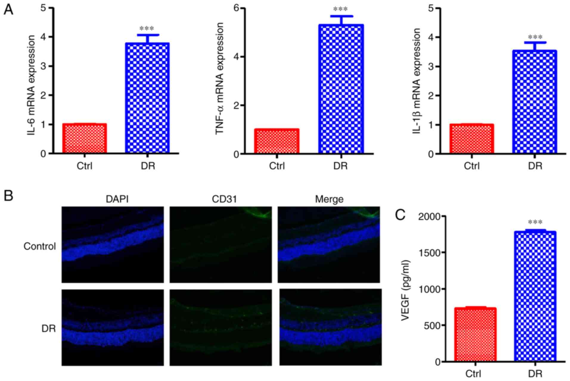 | Figure 1DR induces the inflammatory response
and microvascular cell proliferation. (A) mRNA expression levels of
the inflammatory-related cytokines, IL-6, TNF-α and IL-1β, were
enhanced in the DR group. (B) The expression of the vascular
marker, CD31, was increased in the DR group, as indicated by
confocal analysis. (C) The VEGF secretion levels in the DR group
were increased compared with the control group. Values are
expressed as the means ± SD (n=6; ***P<0.001 compared
with the control). DR, diabetic retinopathy; IL, interleukin; TNF,
tumor necrosis factor; VEGF, vascular endothelial growth factor;
Ctrl, control. |
NLRP3-mediated tissue inflammatory
response promotes microvascular cell proliferation in the
retina
The analysis of clinical samples from patients with
various stages of diabetes has demonstrated that the expression of
NLRP3 and related inflammatory proteins is increased in vitreous
samples, with the largest levels observed in patients with
proliferative DR (10). Hence, in
the present study, the role of NLRP3 in microvascular cell
proliferation in the retina was investigated. The results revealed
that the protein expression of NLRP3 was upregulated in the DR
group (Fig. 2A). Then, the DR
group was treated with NLRP3 inhibitor; following treatment with
the inhibitor, the inflammatory cytokines IL-6, TNF-α and IL-1β
mRNA expression levels were decreased compared with the DR group
not treated with the inhibitor (Fig.
2B). Moreover, the CD31 expression level was decreased in
inhibitor treatment group (Fig.
2C). In addition, the VEGF secretion level was decreased
following treatment with NLRP3 inhibitor (Fig. 2D). In order to further investigate
the role of NLRP3, the HG cell models using HRMECs were constructed
and the cells were treated with or without NLRP3 inhibitor. The
results revealed that NLRP3 protein expression levels in the HRMECs
in the HG group were upregulated, and were decreased following
treatment with the inhibitor (Fig.
2E). In the HRMECs in the HG groups, IL-6, TNF-α and IL-1β mRNA
expression levels were enhanced compared with the control, and were
suppressed by the NLRP3 inhibitor (Fig. 2F). The VEGF secretion levels in
the cells were similar to those observed in the animal model of DR
(Fig. 2G). Collectively, these
data demonstrated that NLRP3-mediated tissue inflammatory response
promoted microvascular cell proliferation in retina.
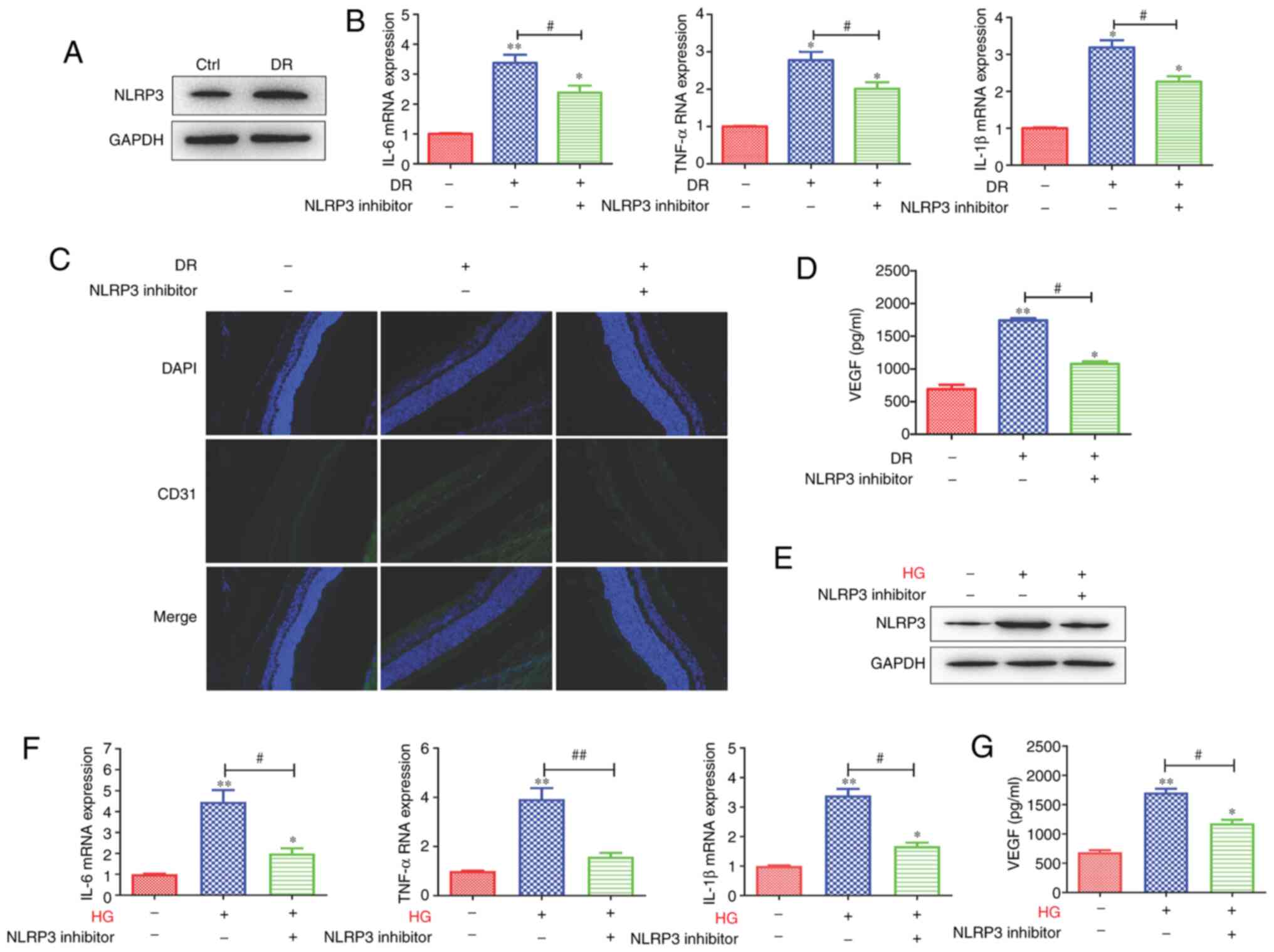 | Figure 2NLRP3-mediated tissue inflammatory
response promotes microvascular cell proliferation in the retina.
(A) The protein expression level of NLRP3 was increased in the DR
groups. (B) Following the of blocking NLRP3, the mRNA expression of
IL-6, TNF-α and IL-1β was downregulated. (C) The expression level
of the vascular marker, CD31, and (D) the VEGF secretion level were
decreased following the inhibition of NLRP3. In addition, in the
HRMEC cell model, (E) HG enhanced the protein expression of NLRP3,
which was inhibited by NLPR3 inhibitor. (F) The levels of the
inflammatory-related cytokines, IL-6, TNF-α and IL-1β, were
enhanced in the HG-induced HRMEC cell model and were inhibited by
the blocking of NLRP3. (G) The VEGF secretion level was enhanced,
and was decreased following the inhibition of NLRP3. Data are
presented as the mean ± standard deviation from triplicate wells.
*P<0.05 and **P<0.01 compared with the
control; #P<0.05 and ##P<0.01 compared
with the relative DR animal model group or HG-induced HRMEC cell
group. NLRP3, NLR family pyrin domain containing 3; DR, diabetic
retinopathy; IL, interleukin; TNF, tumor necrosis factor; VEGF,
vascular endothelial growth factor; HG, high glucose; HRMEC, human
retinal microvascular endothelial cell; Ctrl, control. |
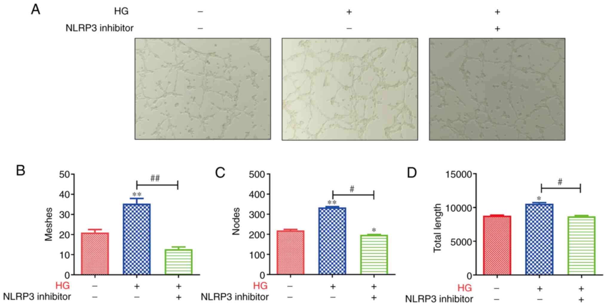
Tube formation of retinal microvascular
endothelial cells is inhibited by the blocking of NLRP3
The aforementioned data indicated that NLRP3 was
associated with inflammatory response, and promoted the expression
of the vascular markers, CD31 and VEGF. Hence, the present study
examined the tube formation of retinal microvascular endothelial
cells in the retina. As revealed in Fig. 3A, lumen formation was markedly
enhanced in HRMECs in the HG group, and was inhibited by the
blocking of NLRP3. The tube meshes, nodes and tube length were
assessed, and the results revealed that the HRMECs in the HG group
formed more meshes and nodes than the controls; moreover, the tube
total length in the HG group was higher than that in the control
group (Fig. 3B-D). However, after
blocking NLRP3, the tube meshes, nodes and tube length were reduced
compared with that in the HG group (Fig. 3B-D). These data indicated that the
tube formation of retinal microvascular endothelial cells was
inhibited by the blocking of NLRP3.
Expression level of the NLRP3
inflammasome is upregulated via the activation of the ASK1/p38
signaling axis
Data have indicated that the NLRP3-induced
inflammatory response plays an important role in DR (9). However, the potential mechanisms of
the role of NLRP3 in inflammatory response in DR remain unknown.
Thus, the present study utilized the DR model and HG HRMECs model
to investigate this matter. The results revealed that the protein
expression levels of ASK1 and p38 were upregulated in the DR and HG
HRMECs groups (Fig. 4A and B).
Subsequently, in order to investigate whether ASK1 and p38 play a
role in the NLRP3-mediated inflammatory response. ASK1 and p38
inhibitors were utilized to block the related protein expression.
The data demonstrated that the protein expression levels of NLRP3,
inflammatory cytokines (IL-6, TNF-α and IL-1β) and VEGF were
downregulated after the blocking of ASK1 and p38 (Fig. 4C and D). All these data indicated
that NLRP3-mediated tissue inflammatory signal response was
enhanced through the activation of the ASK1/p38 signaling axis.
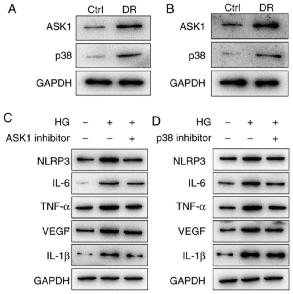 | Figure 4The expression level of inflammasome
NLRP3 is upregulated via the activation of the ASK1/p38 signaling
axis. (A) The protein expression levels of AKS1 and p38 were
upregulated in the DR animal group. (B) The results of the
HG-induced HRMEC group were similar to those of the DR animal
group. (C) The NLPR3, IL-6, TNF-α, IL-1β and VEGF protein
expression levels were inhibited by (C) ASK1 inhibitor and (D) p38
inhibitor. NLRP3, NLR family pyrin domain containing 3; ASK1,
apoptosis signal-regulating kinase 1; DR, diabetic retinopathy; HG,
high glucose; HRMEC, human retinal microvascular endothelial cell;
IL, interleukin; TNF, tumor necrosis factor; VEGF, vascular
endothelial growth factor; Ctrl, control. |
Tube formation of retinal microvascular
endothelial cells is inhibited via the blocking of the ASK1/p38
signaling axis
NLRP3 plays an important part in the tube formation
of retinal microvascular endothelial cells. Moreover, the
NLRP3-mediated tissue inflammatory response is enhanced via the
activation of the ASK1/p38 signaling axis. Hence, it was
hypothesized that the ASK1/p38 signaling axis may mediate the tube
formation of retinal microvascular endothelial cells. As revealed
in Fig. 5A and C, HG promoted
tube formation, and this formation was inhibited by ASK1 and p38
inhibitors. Moreover, the tube meshes, nodes and tube length were
assessed. The result revealed that HRMECs in the HG group formed
more meshes and nodes than the controls; in addition, the tube
total length in the HG group was higher than in the control group
(Fig. 5B and D). However, after
the blocking of ASK1 and p38, the tube meshes, nodes and tube
length were reduced compared with the HG group (Fig. 5B and D). These data indicated that
the ASK1/p38 signaling axis plays a role in the tube formation of
retinal microvascular endothelial cells.
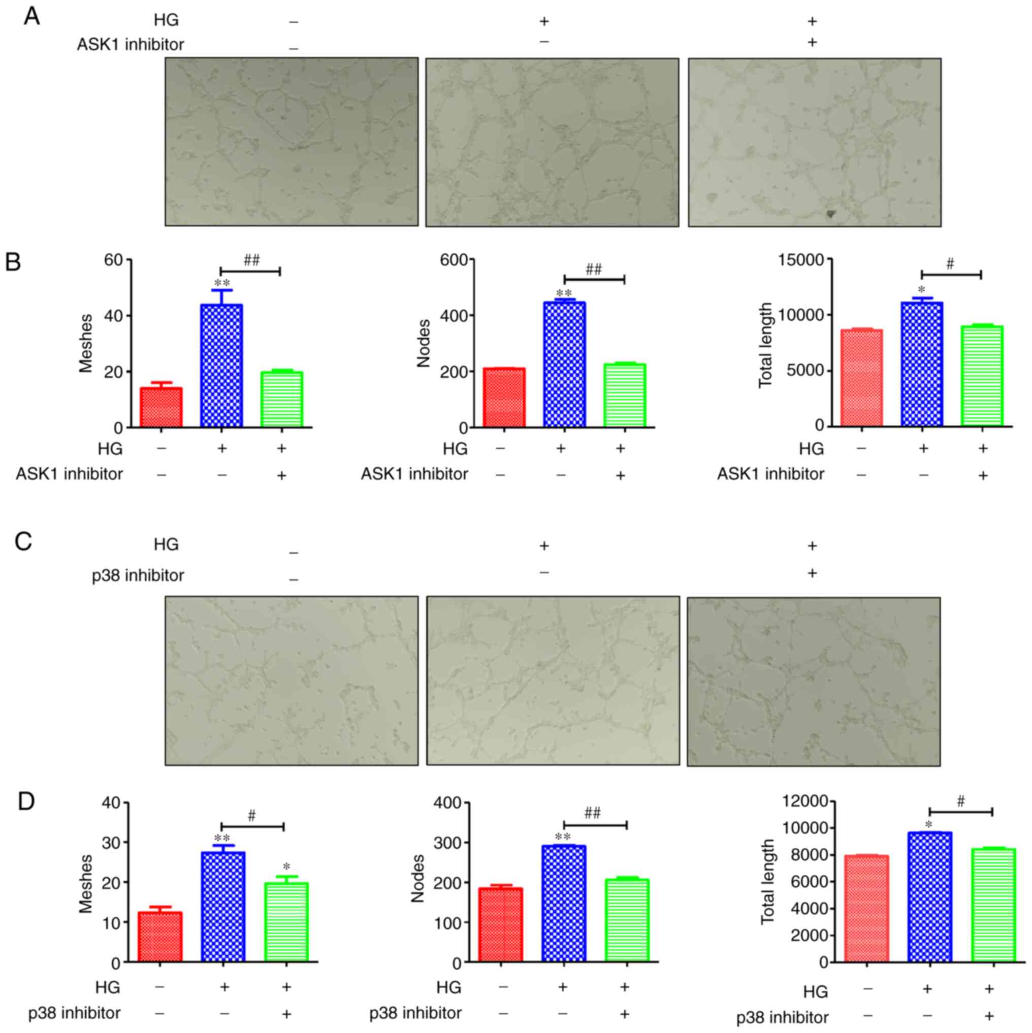 | Figure 5ASK1/p38 signaling axis regulates the
tube formation of retinal microvascular endothelial cells. (A) The
tube formation was markedly enhanced in the HG group of the HRMECs
and was inhibited by blocking of ASK1. Then, the (B) tube meshes,
nodes and tube length were counted, and the results revealed that
the HRMECs in the HG group formed more meshes and nodes than the
control; in addition, the tube total length in the HG group was
higher than that of the control group. However, after blocking of
ASK1, the tube meshes, nodes and tube length were reduced compared
with the HG group. In addition, (C) the tube formation was
inhibited by blocking of p38 and the (D) tube meshes, nodes and
tube length were counted; the results revealed that the blocking of
p38 inhibited the tube formation. *P<0.05 and
**P<0.01 compared with the control;
#P<0.05 and ##P<0.01 compared with the
relative HG-induced HRMEC cell group. ASK1, apoptosis
signal-regulating kinase 1; HG, high glucose; HRMECs, human retinal
microvascular endothelial cells. |
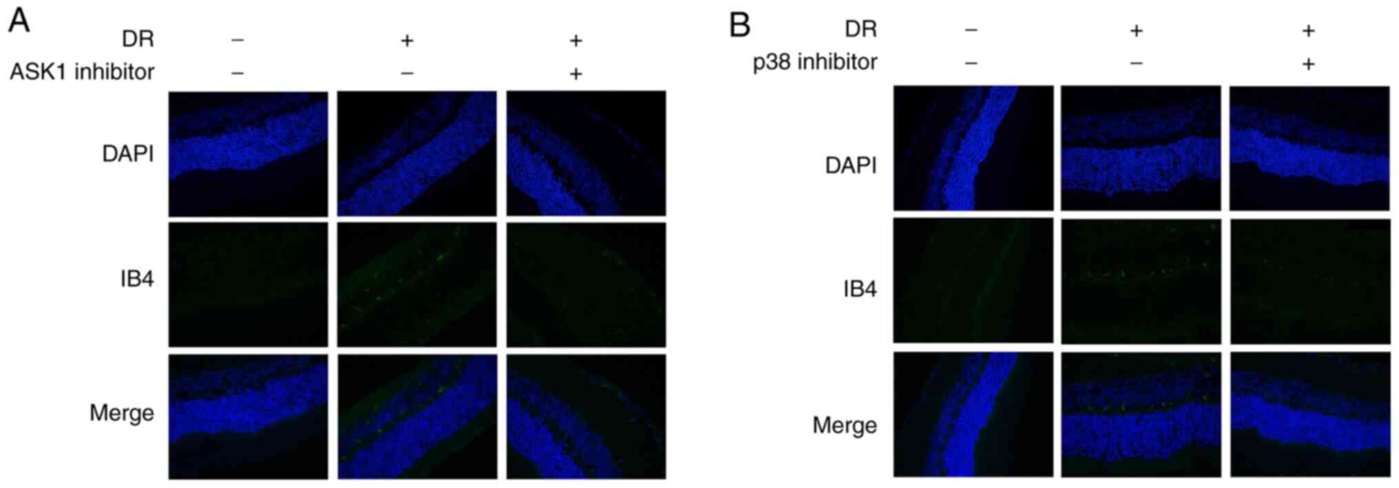
Angiogenesis is inhibited by blocking
ASK1 and p38
Subsequently, the present study investigated whether
ASK1 and p38 mediates angiogenesis. The results revealed that the
levels of the angiogenesis-related marker IB4 were markedly
increased in the DR model group (Fig.
6); However, IB4 expression was inhibited by the blocking of
ASK1 and p38 (Fig. 6). These data
suggested that ASK1 and p38 play a role in angiogenesis in DR.
Discussion
DR is a leading cause of blindness among the
working-age population worldwide (22). It is induced by diabetes, which
causes the pathological characteristics, such as, retinal
capillaries, arterioles and venules, and the subsequent effects of
leakage from or occlusion of small vessels (22-24). Laser therapy is the main effective
therapy for the preservation of sight in proliferative retinopathy
(24); however, it cannot reverse
visual loss. Hence, the development of novel and effective
therapeutic strategies for DR is imperative.
In the present study, it was revealed that NLRP3
plays an important role in aberrant retinal angiogenesis in DR. It
was firstly identified that the ASK1/p38-mediated NLRP3
inflammasome signaling pathway contributed to aberrant retinal
angiogenesis in DR. The mRNA expression levels of
inflammatory-related cytokines (IL-6, TNF-α and IL-1β) were
upregulated in DR; moreover, the levels of the vascular-related
markers, CD31 and VEGF were also increased in DR. After the
blocking of NLRP3, ASK1 or p38, the expression levels of
inflammatory cytokines were downregulated and those of the
vascular-related marker, IB4, were also decreased.
It is known that hyperglycemia can lead to a series
of inflammatory mediators in diabetes, which can further activate
the hyperinflammatory response and finally damage retinal
microvascular cells (25).
Chronic inflammation is one of the key mechanisms that are
triggered during the pathogenesis of DR, and the reduction of the
inflammation response can alleviate the development and progression
of DR (26). Related research has
indicated that NLRP3 plays a crucial key in the development of the
chronic inflammatory response through the secretion of related
cytokines, such as IL-6, TNF-α and IL-1β (27). The activation of NLRP3 contributes
to the development and progression of chronic inflammatory diseases
(28). In the present study, the
results revealed that the NLRP3-mediated tissue inflammatory
response promoted microvascular cell proliferation in the retina.
In DR animal model, the protein expression of NLRP3 was upregulated
in the DR group. The DR group was then treated with NLRP3
inhibitor; following treatment with the inhibitor, the mRNA
expression levels of the inflammatory cytokines, IL-6, TNF-α and
IL-1β were decreased compared with the DR group without treatment
with the inhibitor. In addition, the expression level of CD31 was
decreased in the inhibitor treatment group. The VEGF secretion
level was also decreased following treatment with the NLRP3
inhibitor. In addition, the NLRP3 protein expression level in
HRMECs in the HG group was upregulated, and was suppressed
following treatment with the inhibitor. In the HG groups of the
HRMECs, the mRNA expression levels of IL-6, TNF-α and IL-1β were
enhanced compared with the control, and were decreased following
treatment with the NLRP3 inhibitor. The secretion levels of VEGF in
the cells were similar to those observed in the animal model of
DR.
ASK1 is an apoptosis-related protein, which is
activated in response to a variety of stress-related stimuli via
distinct mechanisms and activates MKK4 and MKK3, which in turn
activate JNK and p38 (29).
Previous studies have demonstrated that ASK1 can contribute to the
development and progression of inflammatory response (30,31). For example, the bacterial
infection-engaged inhibition of ASK1 is responsible for regulating
Erk1/2- and p38-MAPK activation, but not JNK-MAPK signaling
(31,32). It has thus been suggested that
ASK1 and p38 have a close association in the inflammatory response.
In addition, some drugs can protect the retinal photoreceptor cells
via the activation of the p-Erk1/2/Nrf2/Trx/ASK1 signaling pathway
in diabetic mice (33). It has
been suggested that ASK1 plays an important role in
diabetic-related diseases (33).
In the present study, the data indicated that the expression level
of inflammasome NLRP3 was upregulated through the activation of the
ASK1/p38 signaling axis; moreover, the ASK1/p38 signaling axis
contributed to the tube formation of retinal microvascular
endothelial cells and development of angiogenesis in DR. It was
revealed that ASK1 and p38 protein expression levels were
upregulated in DR; however, following the blocking of ASK1 and p38,
the protein expression levels of NLRP3 and related cytokines (IL-6,
TNF-α and IL-1β) were downregulated. In addition, the tube
formation of retinal microvascular endothelial cells was inhibited
by the blocking ASK1 and p38. These data revealed that ASK1 and p38
mediated the NLRP3 inflammasome signaling pathway contributing to
aberrant retinal angiogenesis in DR.
In conclusion, the present study demonstrated that
DR induced inflammatory response and microvascular cell
proliferation. NLRP3 contributed to DR-mediated inflammatory
development and progression, which promoted the
inflammatory-related cytokine expression. Moreover, DR promoted the
tube formation of retinal microvascular endothelial cells and
angiogenesis. Further research revealed that NLRP3-mediated
aberrant retinal angiogenesis in DR was regulated by ASK1 and
p38.
Funding
This research was financially supported by National
Natural Science Foundation of China (grant no. 81700852), the Young
Talent's Subsidy Project in Science and Education of the Department
of Public Health of Jiangsu Province (grant no. QNRC2016140), the
Social Development Project of Jiangsu Provincial Science and
Technology Department (grant no. BE2017627) and the Funded Project
of the Wuxi Municipal Health and Family Planning Commission (grant
no. Q201623).
Availability of data and materials
The datasets used during the present study are
available from the corresponding author upon reasonable
request.
Authors' contributions
WZ and ZZ conceived and designed the study. ZZ, SL,
and YP performed the experiments. WZ, SL, LC, XH and ND collected,
analyzed and interpreted the data. WZ and ZW drafted and critically
revised the study for important intellectual content. All authors
read and approved the final submission.
Ethics approval and consent to
participate
All of the animal experiments were performed in
accordance with the Guidelines for the Care and Use of Laboratory
Animals published by the US National Institutes of Health (NIH
Publication no. 85-23, revised 1996), and were approved by the
Experimental Animal Ethics Committee of the Affiliated Wuxi No. 2
People's Hospital of Nanjing Medical University (Nanjing,
China).
Patient consent for publication
Not applicable.
Competing interests
The authors declare that they have no competing
interests.
Acknowledgments
Not applicable.
References
|
1
|
Congdon NG, Friedman DS and Lietman T:
Important causes of visual impairment in the world today. Jama.
290:2057–2060. 2003. View Article : Google Scholar : PubMed/NCBI
|
|
2
|
Laiginhas R, Madeira C, Lopes M, Neves JN,
Barbosa M, Rosas V, Carvalho D, Falcão-Reis F and Falco M: Risk
factors for prevalent diabetic retinopathy and proliferative
diabetic retinopathy in type 1 diabetes. Endocrine. 66:201–209.
2019. View Article : Google Scholar : PubMed/NCBI
|
|
3
|
Dong C, Liu P, Wang H, Dong M, Li G and Li
Y: Ginsenoside Rb1 attenuates diabetic retinopathy in
streptozotocin-induced diabetic rats1. Acta Cir Bras.
34:e2019002012019. View Article : Google Scholar : PubMed/NCBI
|
|
4
|
Jeganathan VS, Wang JJ and Wong TY: Ocular
associations of diabetes other than diabetic retinopathy. Diabetes
Care. 31:1905–1912. 2008. View Article : Google Scholar : PubMed/NCBI
|
|
5
|
Dehdashtian E, Mehrzadi S, Yousefi B,
Hosseinzadeh A, Reiter RJ, Safa M, Ghaznavi H and Naseripour M:
Diabetic retinopathy pathogenesis and the ameliorating effects of
melatonin; involvement of autophagy, inflammation and oxidative
stress. Life Sci. 193:20–33. 2018. View Article : Google Scholar
|
|
6
|
Abougalambou SS and Abougalambou AS: Risk
factors associated with diabetic retinopathy among type 2 diabetes
patients at teaching hospital in Malaysia. Diabetes Metab Syndr.
9:98–103. 2015. View Article : Google Scholar
|
|
7
|
Rubsam A, Parikh S and Fort PE: Role of
inflammation in diabetic retinopathy. Int J Mol Sci. 19:9422018.
View Article : Google Scholar :
|
|
8
|
Wang H, Wang G, Liang Y, Du X, Boor PJ,
Sun J and Khan MF: Redox regulation of hepatic NLRP3 inflammasome
activation and immune dysregulation in trichloroethene-mediated
autoimmunity. Free Rad Biol Med. 143:223–231. 2019. View Article : Google Scholar : PubMed/NCBI
|
|
9
|
Chen H, Zhang X, Liao N, Mi L, Peng Y, Liu
B, Zhang S and Wen F: Enhanced expression of NLRP3
inflammasome-related inflammation in diabetic retinopathy. Invest
Ophthalmol Vis Sci. 59:978–985. 2018. View Article : Google Scholar : PubMed/NCBI
|
|
10
|
Chaurasia SS, Lim RR, Parikh BH, Wey YS,
Tun BB, Wong TY, Luu CD, Agrawal R, Ghosh A, Mortellaro A, et al:
The NLRP3 inflammasome may contribute to pathologic
neovascularization in the advanced stages of diabetic retinopathy.
Sci Rep. 8:28472018. View Article : Google Scholar : PubMed/NCBI
|
|
11
|
Guigui S, Lifshitz T and Levy J: Diabetic
retinopathy in Africa: Advantages of screening. Postgrad Med.
123:119–125. 2011. View Article : Google Scholar : PubMed/NCBI
|
|
12
|
Nomura K, Lee M, Banks C, Lee G and Morris
BJ: An ASK1-p38 signalling pathway mediates hydrogen
peroxide-induced toxicity in NG108-15 neuronal cells. Neurosci
Lett. 549:163–167. 2013. View Article : Google Scholar : PubMed/NCBI
|
|
13
|
Sobhan PK, Zhai Q, Green LC, Hansford LM
and Funa K: ASK1 regulates the survival of neuroblastoma cells by
interacting with TLX and stabilizing HIF-1α. Cell Signal.
30:104–117. 2017. View Article : Google Scholar
|
|
14
|
Katome T, Namekata K, Guo X, Semba K,
Kittaka D, Kawamura K, Kimura A, Harada C, Ichijo H, Mitamura Y and
Harada T: Inhibition of ASK1-p38 pathway prevents neural cell death
following optic nerve injury. Cell Death Differ. 20:270–280. 2013.
View Article : Google Scholar :
|
|
15
|
Flaumenhaft R: Stressed platelets ASK1 for
a MAPK. Blood. 129:1066–1068. 2017. View Article : Google Scholar : PubMed/NCBI
|
|
16
|
Huang C, Zhu HJ, Li H, Li QX, Li FM, Cheng
L and Liu YG: P38-MAPK pathway is activated in retinopathy of
microvascular disease of STZ-induced diabetic rat model. Eur Rev
Med Pharmacol Sci. 22:5789–5796. 2018.PubMed/NCBI
|
|
17
|
Fortin J, Patenaude A, Deschesnes RG, Côté
MF, Petitclerc E and C-Gaudreault R: ASK1-P38 pathway is important
for anoikis induced by microtubule-targeting aryl chloroethylureas.
J Pharm Pharm Sci. 13:175–190. 2010. View
Article : Google Scholar : PubMed/NCBI
|
|
18
|
Barros-Miñones L, Orejana L, Goñi-Allo B,
Suquía V, Hervías I, Aguirre N and Puerta E: Modulation of the
ASK1-MKK3/6-p38/MAPK signalling pathway mediates silde-nafil
protection against chemical hypoxia caused by malonate. Br J
Pharmacol. 168:1820–1834. 2013. View Article : Google Scholar
|
|
19
|
Liu W, Gu J, Qi J, Zeng XN, Ji J, Chen ZZ
and Sun XL: Lentinan exerts synergistic apoptotic effects with
paclitaxel in A549 cells via activating ROS-TXNIP-NLRP3
inflammasome. J Cell Mol Med. 19:1949–1955. 2015. View Article : Google Scholar : PubMed/NCBI
|
|
20
|
Wang N, Zhang C, Xu Y, Li S, Tan HY, Xia W
and Feng Y: OMICs approaches-assisted identification of
macrophages-derived MIP-1γ as the therapeutic target of botanical
products TNTL in diabetic retinopathy. Cell Commun Signal.
17:812019. View Article : Google Scholar
|
|
21
|
Mengual L and Olivan M: Quantitative RNA
analysis from urine using real time PCR. Methods Mol Biol.
1655:227–237. 2018. View Article : Google Scholar
|
|
22
|
Klein R, Lee KE, Knudtson MD, Gangnon RE
and Klein BE: Changes in visual impairment prevalence by period of
diagnosis of diabetes: The wisconsin epidemiologic study of
diabetic retinopathy. Ophthalmology. 116:1937–1942. 2009.
View Article : Google Scholar : PubMed/NCBI
|
|
23
|
Wilkinson CP, Ferris FL III, Klein RE, Lee
PP, Agardh CD, Davis M, Dills D, Kampik A, Pararajasegaram R,
Verdaguer JT, Global Diabetic Retinopathy and Project Group:
Proposed international clinical diabetic retinopathy and diabetic
macular edema disease severity scales. Ophthalmology.
110:1677–1682. 2003. View Article : Google Scholar : PubMed/NCBI
|
|
24
|
Kang EY, Chen TH, Garg SJ, Sun CC, Kang
JH, Wu WC, Hung MJ, Lai CC, Cherng WJ and Hwang YS: Association of
statin therapy with prevention of vision-threatening diabetic
retinopathy. JAMA Ophthalmol. 137:363–371. 2019. View Article : Google Scholar : PubMed/NCBI
|
|
25
|
Lowry F: Compound could help diabetic
patients walk tightrope between heperglycemia, hypoglycemia. CMAJ.
154:705–707. 1996.PubMed/NCBI
|
|
26
|
Li S, Yang H and Chen X: Protective
effects of sulforaphane on diabetic retinopathy: Activation of the
nrf2 pathway and inhibition of NLRP3 inflammasome formation. Exp
Anim. 68:221–231. 2019. View Article : Google Scholar : PubMed/NCBI
|
|
27
|
Fouad AA, Abdel-Aziz AM and Hamouda AA:
Diacerein down-regulates NLRP3/Caspase-1/IL-1β and IL-6/STAT3
pathways of inflammation and apoptosis in a rat model of cadmium
testicular toxicity. Biol Trace Elem Res. 195:499–505. 2019.
View Article : Google Scholar
|
|
28
|
Tartey S and Kanneganti TD: Differential
role of the NLRP3 inflammasome in infection and tumorigenesis.
Immunology. 156:329–338. 2019. View Article : Google Scholar : PubMed/NCBI
|
|
29
|
Ju A, Cho YC, Kim BR, Park SG, Kim JH, Kim
K, Lee J, Park BC and Cho S: Scaffold role of DUSP22 in
ASK1-MKK7-JNK signaling pathway. PLoS One. 11:e01642592016.
View Article : Google Scholar : PubMed/NCBI
|
|
30
|
Iriyama T, Takeda K, Nakamura H, Morimoto
Y, Kuroiwa T, Mizukami J, Umeda T, Noguchi T, Naguro I, Nishitoh H,
et al: ASK1 and ASK2 differentially regulate the counteracting
roles of apoptosis and inflammation in tumorigenesis. EMBO J.
28:843–853. 2009. View Article : Google Scholar : PubMed/NCBI
|
|
31
|
Yang D, Liu X, Xu W, Gu Z, Yang C, Zhang
L, Tan J, Zheng X, Wang Z, Quan S, et al: The edwardsiella
piscicida thioredoxin-like protein inhibits ASK1-MAPKs signaling
cascades to promote pathogenesis during infection. PLoS Pathog.
15:e10079172019. View Article : Google Scholar : PubMed/NCBI
|
|
32
|
Gong X, Duan Y, Zheng J, Wang Y, Wang G,
Norgren S and Hei TK: Nephroprotective effects of n-acetylcysteine
amide against contrast-induced nephropathy through upregulating
thioredoxin-1, Inhibiting ASK1/p38MAPK pathway, and suppressing
oxidative stress and apoptosis in rats. Oxid Med Cell Longev.
2016:87151852016. View Article : Google Scholar
|
|
33
|
Ren X, Sun H, Zhang C, Li C, Wang J, Shen
J, Yu D and Kong L: Protective function of pyridoxamine on retinal
photoreceptor cells via activation of the pErk1/2/Nrf2/Trx/ASK1
signalling pathway in diabetic mice. Mol Med Rep. 14:420–424. 2016.
View Article : Google Scholar : PubMed/NCBI
|