Introduction
Hyperlipidemia is a common metabolic disorder
characterized by elevated serum cholesterol and triglycerides.
Beyond its well-established role in cardiovascular disease,
increasing evidence has linked hyperlipidemia to cognitive
impairment and all-cause dementia (1,2).
Patients with hyperlipidemia have a notably higher risk of
developing Alzheimer's disease (AD) (3,4).
Furthermore, elevated levels of free fatty acids, particularly
palmitic acid (PA), have been implicated as key pathogenic
mediators (2,5). PA can cross the blood-brain barrier
and accumulate in brain tissues, where it triggers lipotoxic injury
including oxidative stress, apoptosis and synaptic dysfunction
(6). However, the molecular
mechanisms through which PA induces neuronal damage remain
unclear.
The hippocampus, a brain region that is essential
for learning and memory, is particularly vulnerable to metabolic
stress. Elevated free fatty acid levels are correlated with reduced
hippocampal volume and impaired episodic memory scores (7,8).
Our previous studies have consistently demonstrated that excessive
lipids directly damage hippocampal neurons. In animal models of
non-alcoholic fatty liver disease (NAFLD) or type 2 diabetes
mellitus (T2DM), hyperlipidemia, hippocampal neuronal apoptosis,
synaptic protein loss and cognitive deficits were observed
(9-11). In vitro, it was found that
PA treatment of HT-22 cells induced lipid accumulation, insulin
resistance and the downregulation of synaptic plasticity related
proteins such as synapsin-1 and brain-derived neurotrophic factor
(BDNF) (12). These results
indicate that PA exposure leads to substantial injury to
hippocampal neurons. However, the upstream molecular mechanisms
remain largely unknown.
Long non-coding RNAs (lncRNAs) have emerged as
critical regulators of gene expression under both physiological and
pathological conditions. Through mechanisms such as acting as
competing endogenous RNAs (ceRNAs), lncRNAs can sponge microRNAs
(miRNAs/miRs) and modulate the expression of downstream target
genes (13). Dysregulated lncRNA
expression is increasingly recognized in the context of metabolic
disorders and neurodegeneration. For instance, lncRNA MEG3
attenuates hyperglycemia-induced damage by enhancing the
mitochondrial translocation of HSP90A in primary hippocampal
neurons (14), while lncRNA
MALAT1 is involved in oxidative stress responses in diabetic
encephalopathy (15). However,
the specific lncRNAs that govern PA-induced neuronal lipotoxicity
have not been systematically explored.
The taurine upregulated lncRNA TUG1 is the
first non-coding RNA identified in mouse retinal cells. In addition
to its critical role in retinal development and the maintenance of
normal photoreceptors (16),
lncRNA TUG1 serves as a potential mediator of
metabolic-neural crosstalk (17). Elevated lncRNA TUG1
expression has been detected in the serum or cerebrospinal fluid
from patients with temporal lobe epilepsy (18), Parkinson's disease (19) and AD (20), and is closely associated with
cellular apoptosis and inflammatory responses; it sponges
miR-145a-5p and aggravates myocardial fibrosis in a diabetic
cardiomyopathy mouse model by upregulating Cfl2 (21). Additionally, the knockdown of
lncRNA TUG1 can inhibit the apoptosis of hippocampal neurons
in AD by upregulating miR-15a and downregulating ROCK1
expression (22). Similarly,
beneficial effects have also been found for lncRNA TUG1
downregulation in BV2 cells against the challenge of
lipopolysaccharides/interferon-γ via an involvement in inhibiting
glycolysis and facilitating the shift of microglial glucose
metabolism from glycolysis to oxidative phosphorylation (23). Previous evidence indicates that
lncRNA TUG1 also contributes to hepatic steatosis and
pancreatic β cell dysfunction (24,25), thus suggesting a broader role in
lipotoxicity. Nevertheless, whether lncRNA TUG1 participates
in PA-induced neuronal lipotoxic injury in the central nervous
system has not yet been investigated.
The present study aimed to investigate whether
PA-induced neuronal injury involves lncRNA TUG1
dysregulation and to explore the underlying molecular mechanisms.
Specifically, the present study examined the effects of PA on
lncRNA TUG1 expression in HT-22 and SH-SY5Y cells and
assessed the functional consequences of lncRNA TUG1
knockdown on PA-induced apoptosis, synaptic dysfunction and lipid
peroxidation. To elucidate the downstream mechanism, it was further
explored whether miR-449a-5p mediates the effects of lncRNA
TUG1 by targeting caspase-3 and modulating the Akt/GSK-3β
pathway. This was addressed by gain- and loss-of-function
approaches for miR-449a-5p, combined with pharmacological
inhibition of Akt. Through these approaches, the present study
sought to characterize the lncRNA TUG1/miR-449a-5p/caspase-3
and Akt/GSK-3β axes in PA-induced neuronal lipotoxic injury.
Materials and methods
Cell culture
The murine hippocampal neuronal cell line HT-22
(cat. no. SNL-202; Wuhan Shangen Biotechnology Co., Ltd.) and the
human neuroblastoma cell line SH-SY5Y (cat. no. SNL-092; Wuhan
Shangen Biotechnology Co., Ltd.) were cultured in high-glucose
Dulbecco's Modified Eagle Medium (DMEM; cat. no. SH30022; HyClone;
Cytiva) supplemented with 10% fetal bovine serum (FBS; cat. no.
FCS500; Suzhou ExCell Biotechnology Co., Ltd.) at 37°C in a
humidified incubator with 5% CO2. The SH-SY5Y cell line
was authenticated by Wuhan Shangen Biotechnology Co., Ltd. using
short tandem repeat (STR) profiling with the PowerPlex®
18D system. The STR profile showed a 100% match with the SH-SY5Y
reference profile in the ExPASy Cellosaurus database (CVCL_0019),
and no interspecies cross-contamination was detected. The 293T
cells used in the present study were purchased from Wuhan
Servicebio Technology Co., Ltd. (cat. no. STCC10301) and were
maintained in the same culture medium and conditions as
aforementioned. The cells were passaged three to five times per
week. Exponentially growing cells were seeded into a 6-well plate
at a density of 5×10⁵ cells/well and allowed to reach
60% confluency. According to a previous study, PA (100 μM)
was used to stimulate HT-22 or SH-SY5Y cells to establish a
hyperlipidemic injury model (26).
3-(4,5-Dimethylthiazol-2-yl)-2,5-diphenyltetrazolium bromide (MTT)
and lactate dehydrogenase (LDH) assays
The MTT assay was performed according to the
manufacturer's instructions (cat. no. M8180; Beijing Solarbio
Science & Technology Co., Ltd.). Briefly, after treatment with
various concentrations of PA (25, 50, 100, 200, 400 and 800
μM) for 24 h at 37°C, cells were washed twice with 1X
phosphate-buffered saline (PBS) solution and then incubated in
complete medium supplemented with 0.5 mg/ml MTT for 4 h. After
incubation, the supernatant was removed from each well and 100
μl of Formazan Dissolution Solution (provided in the MTT
kit) was added. The cells were gently mixed and incubated in a
constant temperature incubator at 37°C until all purple crystals
had dissolved. The absorbance was measured at 570 nm using a
microplate reader.
Cytotoxicity was evaluated by quantifying the
release of LDH from cell supernatants using the Lactate
Dehydrogenase Cytotoxicity Assay Kit (cat. no. C0016; Beyotime
Biotechnology) according to the manufacturer's instructions.
Cell transfection
For transfection, HT-22 cells were seeded into
6-well plates at a density of 5×10⁵ cells/well and
randomly divided into different groups (n=3 for each group). When
the cells grew to 40-60% confluency, transfection was performed
using Lipofectamine™ 2000 Transfection Reagent (cat. no. 11668027;
Thermo Fisher Scientific, Inc.) according to the manufacturer's
instructions. Briefly, the transfection reagent and nucleotides
[small interfering RNA targeting lncRNA TUG1 (si-lncRNA
TUG1), miR-449a-5p mimics, miR-449a-5p inhibitor or the
corresponding negative controls (NCs)] were each diluted in 200
μl serum-free DMEM, mixed gently and incubated for 10 min at
room temperature. The mixtures were then added to the cells,
followed by incubation at 37°C for 6 h. At 6 h post-transfection,
the medium was replaced with complete DMEM containing 100 μM
PA and the cells were cultured at 37°C for a further 24 h prior to
subsequent analyses. Cells were subsequently collected for RNA
extraction, protein extraction, apoptosis detection or lipid
peroxidation (LPO) assays. All nucleotides were synthesized by
Shanghai GenePharma Co., Ltd. All transfections were performed with
50 nM nucleotide.
The nucleotide sequences were as follows: siRNA-1,
5'-GCCAGGUUUAUUCCAUAAATT-3'; siRNA-2, 5'-GGACUUGCAACCUGGUUAUTT-3';
siRNA-3, 5'-GCCUGUUCUUCUAGCUUAATT-3'; siNC sense,
5'-UUCUCCGAACGUGUCACGUTT-3' and antisense,
5'-ACGUGACACGUUCGGAGAATT-3'; mimics sense,
5'-UGGCAGUGUAUUGUUAGCUGGU-3' and anti-sense,
5'-CAGCUAACAAUACACUGCCAUU-3'; inhibitor,
5'-ACCAGCUAACAAUACACUGCCA-3'; miR inhibitor NC,
5'-CAGUACUUUUGUGUAGUACAA-3'; miR mimic NC sense,
5'-UUCUCCGAACGUGUCACGUTT-3' and antisense,
5'-ACGUGACACGUUCGGAGAATT-3'.
Measurement of malondialdehyde (MDA), LPO
and 4-hydroxynonenal (4-HNE)
To examine whether LPO levels were altered, the
levels of MDA (cat. no. S0131M; Beyotime Biotechnology) and LPO
(cat. no A106-1; Nanjing Jiancheng Bioengineering Institute) were
examined separately. Briefly, cells were collected after performing
the corresponding treatments. Cell samples were subjected to
repeated rapid freeze-thawing five times in liquid nitrogen and
37°C and were assayed separately according to the manufacturer's
requirements.
In addition, the concentration of 4-HNE in HT-22
cell homogenates was measured via Enzyme-Linked Immunosorbent Assay
using a commercial kit (cat. no. JYM1109Mo; Wuhan GeneBeauty
Biotechnology Co., Ltd.) according to the manufacturer's
instructions.
Cell apoptosis analysis
HT-22 cell apoptosis was detected using Annexin
V-FITC/propidium iodide (PI) double staining (cat. no. BB-4101;
Shanghai Bestbio Biotechnology Co., Ltd.). After removing the
medium, the HT-22 cells in each treatment group were stained with
Annexin V-FITC and PI staining solution in the dark at 4°C for 15
min. The cells were then washed twice with PBS. Apoptotic cells
were analyzed using a CytoFLEX flow cytometer (Beckman Coulter,
Inc.) with FlowJo v10.8.1 software (FlowJo LLC; BD
Biosciences).
Bioinformatics analysis
The sequence and basic characteristics of lncRNA
TUG1 were obtained from the National Center for
Biotechnology Information (NCBI) database (https://www.ncbi.nlm.nih.gov/). The LncLocator 2.0
tool (http://www.csbio.sjtu.edu.cn/bioinf/lncLocator2/)
was used to analyze the subcellular localization of the lncRNA
TUG1 (27). The binding
sites between lncRNA/mRNA and miRNA were predicted using miRDB
(http://mirdb.org/index.html) and RNA
Interactome (http://www.rna-society.org/raid/) database online
prediction software (28).
Dual-luciferase reporter gene assay
Both wild-type (WT) and mutant (Mut) fragments of
lncRNA TUG1, which span the predicted miR-449a-5p binding
site, were designed and synthesized by General Biotechnology
(Anhui) Co., Ltd., and subsequently cloned into the psiCHECK™-2
vector (Promega Corporation) downstream of the Renilla
luciferase coding sequence. The Mut plasmid contained nucleotide
substitutions within the core binding site to abrogate miRNA
binding. Data S1 includes the sequences used for plasmid
construction and verification.
293T cells were maintained in DMEM supplemented with
10% FBS. At 80-90% confluency, cells were co-transfected with
either TUG1-WT or TUG1-Mut reporter plasmid, along
with miR-449a-5p mimics or mimic NC, using
Lipofectamine® 2000 (Invitrogen; Thermo Fisher
Scientific, Inc.). After 5 h, the transfection mixture was replaced
with fresh complete medium. Cells were harvested 24 h
post-transfection, and luciferase activities were measured using
the Dual-Luciferase® Reporter Assay System (Promega
Corporation) on a BioTek Synergy 2 microplate reader.
Renilla luciferase activity was normalized to firefly
luciferase activity for each sample. All experiments were performed
in triplicate.
Reverse transcription-quantitative
polymerase chain reaction (RT-qPCR)
Total RNA was extracted from samples using TRIzol
reagent (cat. no. 15596018CN; Invitrogen; Thermo Fisher Scientific,
Inc.) according to the manufacturer's instructions. Subsequently, 1
μg of total RNA was reverse-transcribed into cDNA in a total
reaction volume of 10 μl using a reverse transcription kit
(cat. no. AG11706; Accurate Biology) according to the
manufacturer's instructions. The resulting cDNA was diluted 5-fold
to a final volume of 50 μl with nuclease-free water. qPCR
was performed in a 10 μl reaction system containing 3
μl of diluted cDNA, 0.3 μl of each forward and
reverse primer (10 μM) and 5 μl of 2X SYBR Green
SupTaq HS Premix (AG11762; Guangzhou Ruizhen Biotechnology Co.,
Ltd.). The thermocycling conditions were as follows: Initial
denaturation at 95°C for 30 sec, followed by 40 cycles of
denaturation at 95°C for 5 sec and annealing/extension at 60°C for
30 sec. All reactions were performed in triplicate on an ABI Prism
7000 sequence detection system (Applied Biosystems; Thermo Fisher
Scientific, Inc.). β-actin was used as the internal reference for
mRNA and lncRNA TUG1, whereas U6 was used for miR-449a-5p.
Relative expression was calculated using the 2−ΔΔCq
method (29). The primer
sequences for the mouse genes were as follows: lncRNA TUG1
forward, 5'-CAAGAAACAGCAACACCAGAAG-3' and reverse,
5'-TAAGGTCCCCATTCAAGTCAGT-3'; caspase-3 forward,
5'-GAGCTTGGAACGGTACGCTA-3' and reverse 5'-CCGTACCAGAGCGAGATGA-3';
β-actin forward, 5'-AGTGTGACGTTGACATCCGT-3', and reverse,
5'-TGCTAGGAGCCAGAGCAGTA-3'; miR-449a-5p forward,
5'-AACACGTGTGGCAGTGTATTG-3'; miR-34a-5p forward,
5'-AACCGGTGGCAGTGTCTTAG-3'; miR-6378 forward,
5'-AACAAGTGGTTCACGGGAGGA-3'; miR-421 forward
5'-CGCATCAACAGACATTAATTGGGCGC-3'; miR-199a-3p forward
5'-CGCACAGTAGTCTGCACATTGGTTA-3'; U6 forward,
5'-GGAACGATACAGAGAAGATTAGC-3' and reverse
5'-TGGAACGCTTCACGAATTTGCG-3'. The universal reverse primer for all
miR qPCR assays was 5'-GCGAGCACAGAATTAATACGAC-3'. The primer
sequences for the human genes were as follows: lncRNA TUG1
forward, 5'-TGAGCAAGCACTACCACCAG-3' and reverse,
5'-ACTCAGCAATCAGGAGGCAC-3'; β-actin forward
5'-GATGAGATTGGCATGGCTT-3' and reverse
5'-GTCACCTTCACCGTTCCAGT-3'.
Western blotting
Western blotting was performed as described
previously (30). Total protein
from HT-22 and SH-SY5Y cells was extracted using RIPA lysis buffer
(cat. no. P0013B; Beyotime Biotechnology) supplemented with a
protease and phosphatase inhibitor cocktail (50X; cat. no. P1046;
Beyotime Biotechnology) at a ratio of 50:1. After centrifugation at
13,500 × g for 30 min at 4°C, the supernatant was collected and the
total protein concentration was quantified using the bicinchoninic
acid protein concentration assay kit (cat. no. PC0020; Beyotime
Biotechnology). The protein samples were mixed with 5X SDS-PAGE
protein loading buffer (cat. no. P0015L; Beyotime Biotechnology)
and heated in boiling water for 10 min. Cooled protein samples (30
μg per lane) were separated on 10% SDS-PAGE gels and
transferred onto polyvinylidene difluoride (PVDF) membranes. PVDF
membranes were blocked with 5% skimmed milk in Tris-buffered saline
with 0.05% Tween 20 for 2 h at room temperature, then incubated
overnight at 4°C with the following antibodies: Anti-phosphorylated
(p-)GSK-3β (ser9) (1:1,000; cat. no. 9323; Cell Signaling
Technology, Inc.), anti-GSK-3β (1:1,000; cat. no. 9315; Cell
Signaling Technology, Inc.), anti-p-Akt1 (Ser473) (1:1,000; cat.
no. 44-623G; Thermo Fisher Scientific, Inc.), anti-Akt (1:1,000;
cat. no. WL0003b; Wanleibio Co., Ltd.), anti-synaptotagmin-1
(1:1,000; cat. no. 4329; Cell Signaling Technology, Inc.),
anti-BDNF (1:1,000; cat. no. ab108319; Abcam), anti-Synapsin-1
(1:1,000; cat. no. 5297; Cell Signaling Technology, Inc.),
anti-Bcl-2 (1:500; cat. no. WL01556; Wanleibio Co., Ltd.),
anti-caspase-3/cleaved-caspase-3 (1:500; cat. no. WL02117;
Wanleibio Co., Ltd.), anti-caspase-3 (1:1,000; cat. no. GB12532-50;
Wuhan Servicebio Technology Co., Ltd.) and anti-β-actin (1:1,000;
cat. no. TA-09; ZSGB-Bio; Beijing Zhongshan Jinqiao Biotechnology
Co., Ltd.). The PVDF membranes were then incubated with a
horseradish peroxidase-conjugated goat anti-rabbit (1:5,000; cat.
no. ab6721; Abcam), or goat anti-mouse secondary antibody (1:5,000;
cat. no. ab6789; Abcam) at room temperature for 1 h. Protein
signals were detected using SuperSignal West Pico PLUS
chemiluminescent substrate (cat. no. 34577; Thermo Fisher
Scientific, Inc.), and the images were processed and analyzed using
ImageJ software (version 1.52a; National Institutes of Health).
Akt inhibitor VIII treatment
Akt inhibitor VIII (cat. no. SF2784-10 mM; Beyotime
Biotechnology) was dissolved in dimethyl sulfoxide (DMSO) to
prepare a stock solution. The final concentration of DMSO in the
culture medium was maintained below 0.1% to avoid solvent-induced
cytotoxicity. For pharmacological inhibition of Akt, HT-22 cells
were treated with 5 μM Akt inhibitor VIII simultaneously
with 100 μM PA for 24 h at 37°C, with the administration
protocol set with reference to a previous study (31). The experimental groups included
control (untreated), PA only, PA plus NC, PA plus si-TUG1, PA plus
Akt inhibitor VIII alone, PA plus si-TUG1 combined with Akt
inhibitor VIII, and PA plus si-TUG1 combined with DMSO vehicle
control. The same grouping strategy was also applied to miR-449a-5p
mimics in place of si-TUG1. After the treatment, cells were
harvested for western blot analysis of p-Akt, total Akt, p-GSK-3β,
total GSK-3β, cleaved caspase-3, total caspase-3, Bcl-2,
synapsin-1, synaptotagmin-1 and BDNF.
Statistical analysis
Data are expressed as mean ± standard error of the
mean. Data were analyzed using SPSS 23.0 (IBM Corp.) and GraphPad
Prism 8.0 software (Dotmatics). All data were first tested for
normality using the Shapiro-Wilk test. Unpaired Student's t-test
was used for comparisons between two groups. One-way analysis of
variance (ANOVA) was performed to compare three or more groups.
When the result of the overall ANOVA was statistically significant,
the Bonferroni post hoc test was used for pairwise comparisons.
Correlation analyses were performed using Pearson's correlation
test. P<0.05 was considered to indicate a statistically
significant difference.
Results
lncRNA TUG1 is upregulated in PA-induced
neuronal injury and may be correlated with apoptosis and Akt/GSK-3β
pathway dysfunction in HT-22 cells
To explore the role of lncRNA TUG1 in
PA-induced neuronal injury, HT-22 cells were treated with a
concentration gradient of PA (25-800 μM) for 24 h. MTT assay
results showed that treatment with 100 μM PA for 24 h
significantly reduced HT-22 cell viability compared with the
control group, whereas higher concentrations (200-800 μM)
caused progressively greater cytotoxicity (Fig. 1A). Therefore, 100 μM PA
was selected as the optimal concentration for establishing the
neuronal injury model in subsequent experiments, as it induced
significant but submaximal injury, consistent with a previous study
(26). Thus, 100 μM PA
treatment for 24 h was selected as the experimental condition for
subsequent studies in HT-22 cells. In this model, PA exposure
significantly upregulated lncRNA TUG1 expression and
increased LDH release, indicating neuronal damage (Fig. 1B and C). Apoptosis-related
protein assays showed that PA treatment significantly increased the
cleaved caspase-3 to total caspase-3 ratio and reduced the
expression of anti-apoptotic protein Bcl-2 (Fig. 1D-F), indicating that PA activated
the apoptotic pathway in HT-22 cells. Given that Akt/GSK-3β
signaling is a core pro-survival pathway in neurons and has been
implicated in lipotoxic neuronal injury (32), it was next examined whether PA
exposure disrupts this signaling axis and its downstream synaptic
targets. PA stimulation significantly downregulated the protein
levels of synapsin-1, synaptotagmin-1 and BDNF (Fig. 1G and H) and those of the
Akt/GSK-3β signaling pathway, as reflected by the decreased ratios
of p-Akt/Akt and p-GSK-3β/GSK-3β (Fig. 1I and J). These data indicated
that PA impaired synaptic function and inhibited Akt/GSK-3β
signaling.
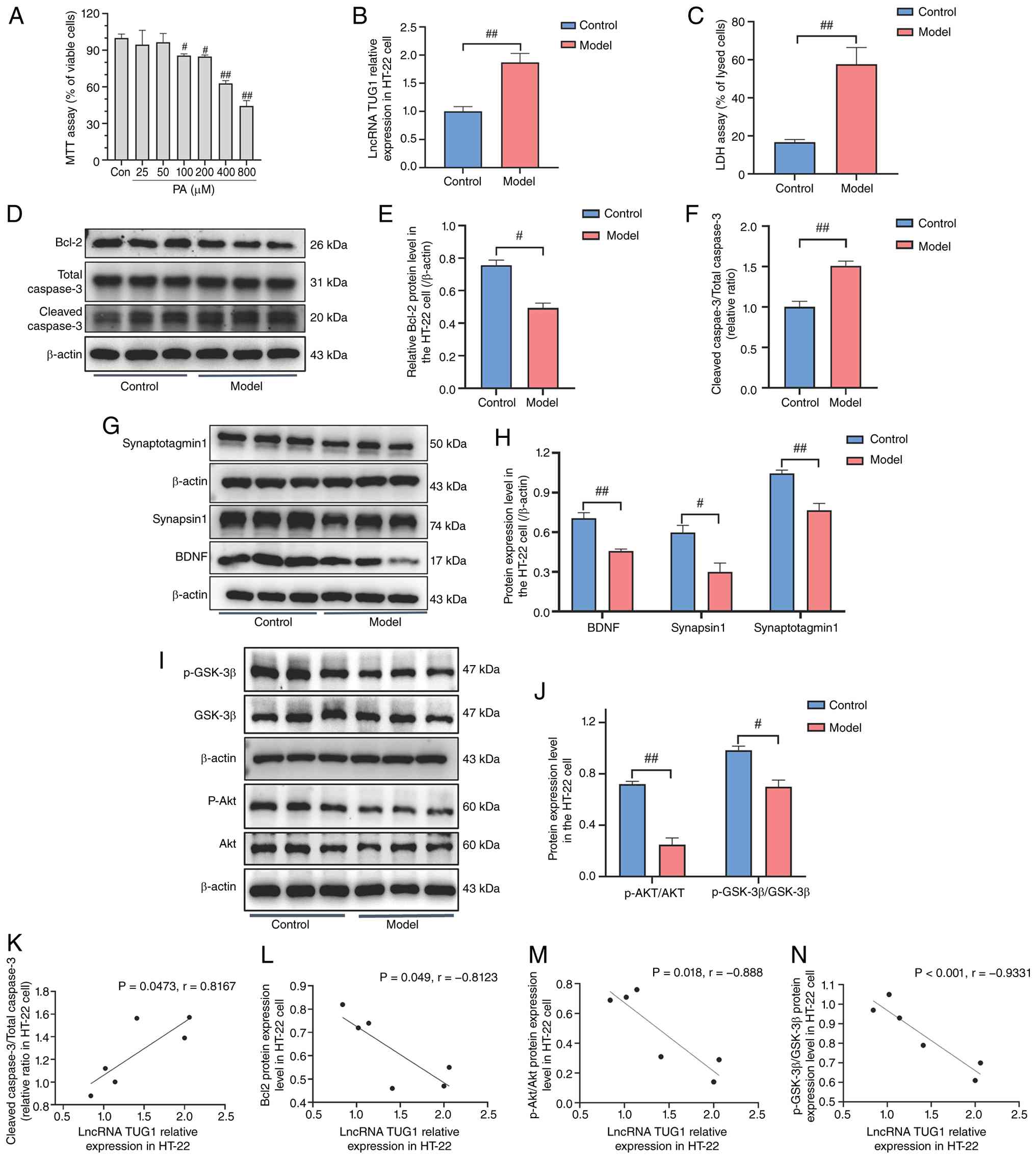 | Figure 1Effects of PA challenge on lncRNA
TUG1 expression as well as apoptosis, synaptic plasticity
and Akt/GSK-3β pathway in HT-22 cells. (A) MTT results of HT-22
cells challenged with different concentrations of PA. (B)
Expression of lncRNA TUG1 in HT-22 cells. (C) LDH rates in
HT-22 cells. (D) Representative western blotting bands of Bcl-2,
cleaved caspase-3 and total caspase-3. Semi-quantification of (E)
Bcl-2 protein levels and (F) the cleaved caspase-3 to total
caspase-3 ratio. (G) Representative western blotting bands of
Synapsin-1, Synaptotagmin-1 and BDNF. (H) Semi-quantification of
the Synapsin-1, Synaptotagmin-1 and BDNF protein levels. (I)
Representative western blotting bands of Akt, p-Akt, GSK-3β and
p-GSK-3β. (J) Semi-Quantification of the p-Akt/Akt and
p-GSK-3β/GSK-3β ratios. Correlation between lncRNA TUG1 expression
and (K) the cleaved caspase-3/total caspase-3 ratio, (L) Bcl-2
protein levels, (M) the p-Akt/Akt ratio and (N) the p-GSK-3β/GSK-3β
ratio. All treatments were performed using 100 μM PA for 24
h. Representative western blotting bands are from three independent
biological replicates per group for both the control and PA-treated
conditions. The data are presented as the mean ± SEM, with n=3 for
each group. #P<0.05, ##P<0.01 compared
with the control group. BDNF, brain-derived neurotrophic factor;
LDH, lactate dehydrogenase; lncRNA, long non-coding RNA; MTT,
3-(4,5-dimethylthiazol-2-yl)-2,5-diphenyltetrazolium bromide assay;
PA, palmitic acid; p, phosphorylated; TUG1, taurine upregulated
gene 1. |
Given that the expression levels of molecules
causally involved in disease pathogenesis often correlate
quantitatively with disease severity and key pathological
indicators (33), exploratory
Pearson correlation analysis was performed to systematically
investigate the associations between lncRNA TUG1 expression
levels and PA-induced molecular alterations. The data used for
these analyses were obtained from PA-treated HT-22 cells. LncRNA
TUG1 expression was measured as shown in Fig. 1B, and the cleaved caspase-3/total
caspase-3 ratio (Fig. 1F), Bcl-2
protein levels (Fig. 1E) and
p-Akt/Akt and p-GSK-3β/GSK-3β ratios (Fig. 1J) were determined accordingly.
Pearson correlation analysis revealed that lncRNA TUG1
expression was positively correlated with the cleaved
caspase-3/total caspase-3 ratio and negatively correlated with the
Bcl-2, p-Akt/Akt and p-GSK-3β/GSK-3β ratios (Fig. 1K-N), suggesting that lncRNA
TUG1 may participate in PA-induced neuronal injury by
regulating apoptosis and synaptic function. Similar results were
observed in SH-SY5Y cells (Fig.
S1A-L), further supporting the universal role of lncRNA
TUG1 in lipid toxicity-induced neuronal damage.
Knocking down lncRNA TUG1 in HT-22 cells
alleviates PA-induced dysfunctional changes in apoptosis, synaptic
plasticity, Akt/GSK-3β pathway and LPO
To knock down lncRNA TUG1, three specific
siRNAs were designed. Among them, siRNA3 exhibited the most
efficient knockdown effect and was selected for subsequent
transfection experiments. siRNA3 successfully reduced lncRNA
TUG1 expression in both control and PA-treated cells,
whereas the NC showed no significant effect (Fig. 2A and B). The LDH assay showed
that lncRNA TUG1 knockdown significantly reduced PA-induced
LDH release compared with the M + NC group, while it did not affect
basal LDH release in control cells (C + NC vs. C + siRNA) (Fig. 2C). This indicated that lncRNA
TUG1 knockdown attenuated PA-induced cytotoxicity. lncRNA
TUG1 knockdown also attenuated the PA-induced increase in
the cleaved caspase-3 to total caspase-3 ratio and restored Bcl-2
expression (Fig. 2D-F), leading
to a reduced apoptotic rate as measured by Annexin V/PI staining
(Fig. 2G and H). These results
suggested that lncRNA TUG1 knockdown inhibited apoptosis in
PA-treated HT-22 cells. Moreover, the PA-induced downregulation of
synaptic proteins including synaptotagmin-1, synapsin-1 and BDNF
was alleviated by lncRNA TUG1 knockdown (Fig. 2I and J), indicating that knocking
down lncRNA TUG1 protected against PA-induced synaptic
damage. Consistently, the reduced ratios of p-Akt/Akt and
p-GSK-3β/GSK-3β were also restored (Fig. 2K and L), suggesting that lncRNA
TUG1 knockdown reactivated the Akt/GSK-3β signaling pathway.
Finally, lncRNA TUG1 knockdown decreased the PA-induced
elevation of the LPO markers 4-HNE, MDA and LPO (Fig. 2M-O), indicating that lncRNA
TUG1 knockdown mitigated LPO.
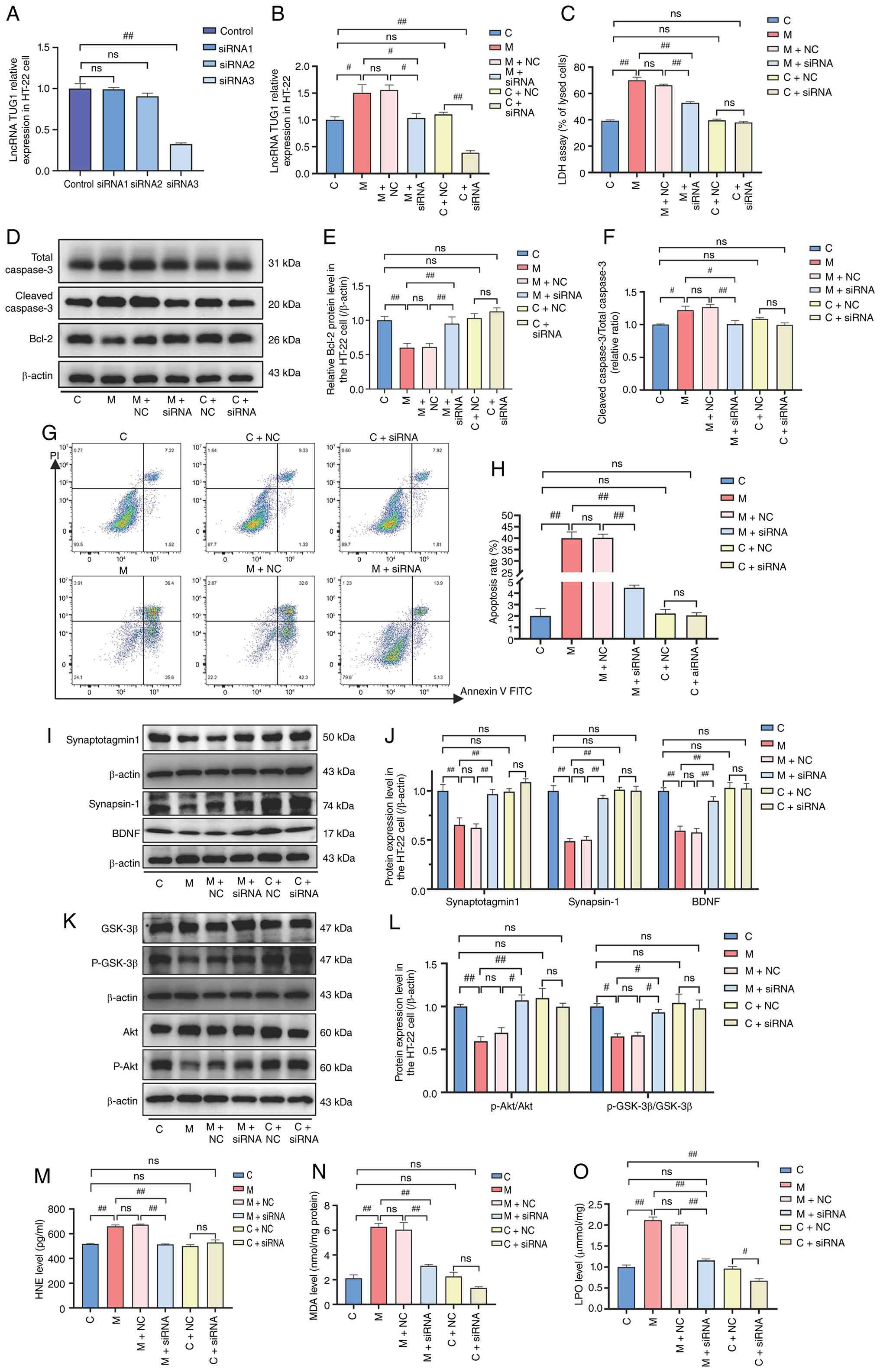 | Figure 2Effects of knocking down lncRNA
TUG1 on apoptosis, synaptic plasticity, the Akt/GSK-3β
pathway and LPO levels in PA-challenged HT-22 cells. (A)
Determination of lncRNA TUG1 expression levels in HT-22
cells using different siRNA sequences. (B) Effect of siRNA for
lncRNA TUG1 on the expression level of lncRNA TUG1 in
HT-22 cells with or without PA challenge. (C) LDH release rate
assay in si-lncRNA TUG1 pretreated PA-challenged HT-22
cells. (D) Representative western blotting bands of cleaved
caspase-3, total caspase-3 and Bcl-2. Semi-quantification of (E)
Bcl-2 protein levels and (F) the cleaved caspase-3 to total
caspase-3 ratio. (G) Flow cytometry determination of apoptosis
levels in si-lncRNA TUG1 pretreated PA-challenged HT-22
cells and (H) the statistical analysis results. (I) Representative
western blotting bands of Synapsin-1, Synaptotagmin-1 and BDNF. (J)
Semi-quantification of Synapsin-1, Synaptotagmin-1 and BDNF protein
levels. (K) Representative western blotting bands of Akt, p-Akt,
GSK-3β and p-GSK-3β. (L) Semi-quantification of p-Akt/Akt and
p-GSK-3β/GSK-3β ratios. Determination of the (M) 4-HNE, (N) MDA
levels and (O) LPO levels. The data are presented as the mean ±
SEM, with n=3 for each group, except for (H) where n=4 per group.
#P<0.05, ##P<0.01. 4-HNE,
4-hydroxynonenal; BDNF, brain-derived neurotrophic factor; C,
control; LDH, lactate dehydrogenase; lncRNA, long non-coding RNA;
LPO, lipid peroxidation; M, model (PA); MDA, malondialdehyde; NC,
negative control; ns, not significant; p-, phosphorylated; PA,
palmitic acid; si, small interfering (RNA); TUG1, taurine
upregulated gene 1. |
miR-449a-5p is a direct target of lncRNA
TUG1
To investigate the function of lncRNA TUG1,
detailed characteristics from the NCBI database were obtained. As
shown in Fig. 3A, this lncRNA is
located on chromosome 11. Thereafter, to explore the underlying
functional mechanism of lncRNA TUG1, the subcellular
localization of lncRNA TUG1 was analyzed using LncLocator
2.0, which showed that it is primarily located in the cytoplasm
rather than in the nucleus (Fig.
3B). Given that the ceRNA mechanism is a common regulatory mode
for cytoplasmic lncRNAs, this framework was adopted for the further
investigation of lncRNA TUG1. Caspase-3 is a key executioner
of apoptosis and has been reported to be targeted by miRNAs
(34). To investigate whether it
could participate in the ceRNA regulatory network involving lncRNA
TUG1 under PA-induced HT-22 cell injury, a target prediction
search using the miRDB and RNA Interactome databases was performed
to identify the target miRNAs for lncRNA TUG1 and caspase-3.
As shown in Fig. 3C, miR-449a-5p
had the potential to bind to lncRNA TUG1 or caspase-3. The
potential binding sites between lncRNA TUG1 and miR-449a-5p,
and between caspase-3 and miR-449a-5p were further predicted
(Fig. 3D). RT-qPCR was performed
to assess the effect of si-lncRNA TUG1 on the expression of
miR-449a-5p in PA-stimulated HT-22 cells. The results showed that
the PA-induced reduction in miR-449a-5p expression was alleviated
by lncRNA TUG1 knockdown (P<0.01; Fig. 3E), indicating that lncRNA
TUG1 may negatively regulate miR-449a-5p levels under PA
stress. In addition, miR-421 and miR-199a-3p, which have been
previously reported to potentially exhibit sponge relationships
with lncRNA TUG1, as well as miR-6378 and miR-34a-5p, which
were predicted to have binding interactions with lncRNA
TUG1, were detected (Fig.
S1M). We hypothesized that lncRNA TUG1 may repress
miR-449a-5p levels through direct binding and molecular
sequestration by acting as a sponge that limits miRNA availability
or promotes its degradation. To determine whether this regulatory
effect is dependent on PA treatment, the effect of TUG1
knockdown on miR-449a-5p expression under non-PA conditions was
examined. Notably, TUG1 knockdown significantly increased
miR-449a-5p expression even in the absence of PA stimulation, thus
indicating a basal repressive effect of TUG1 on this miRNA
(Fig. 3F).
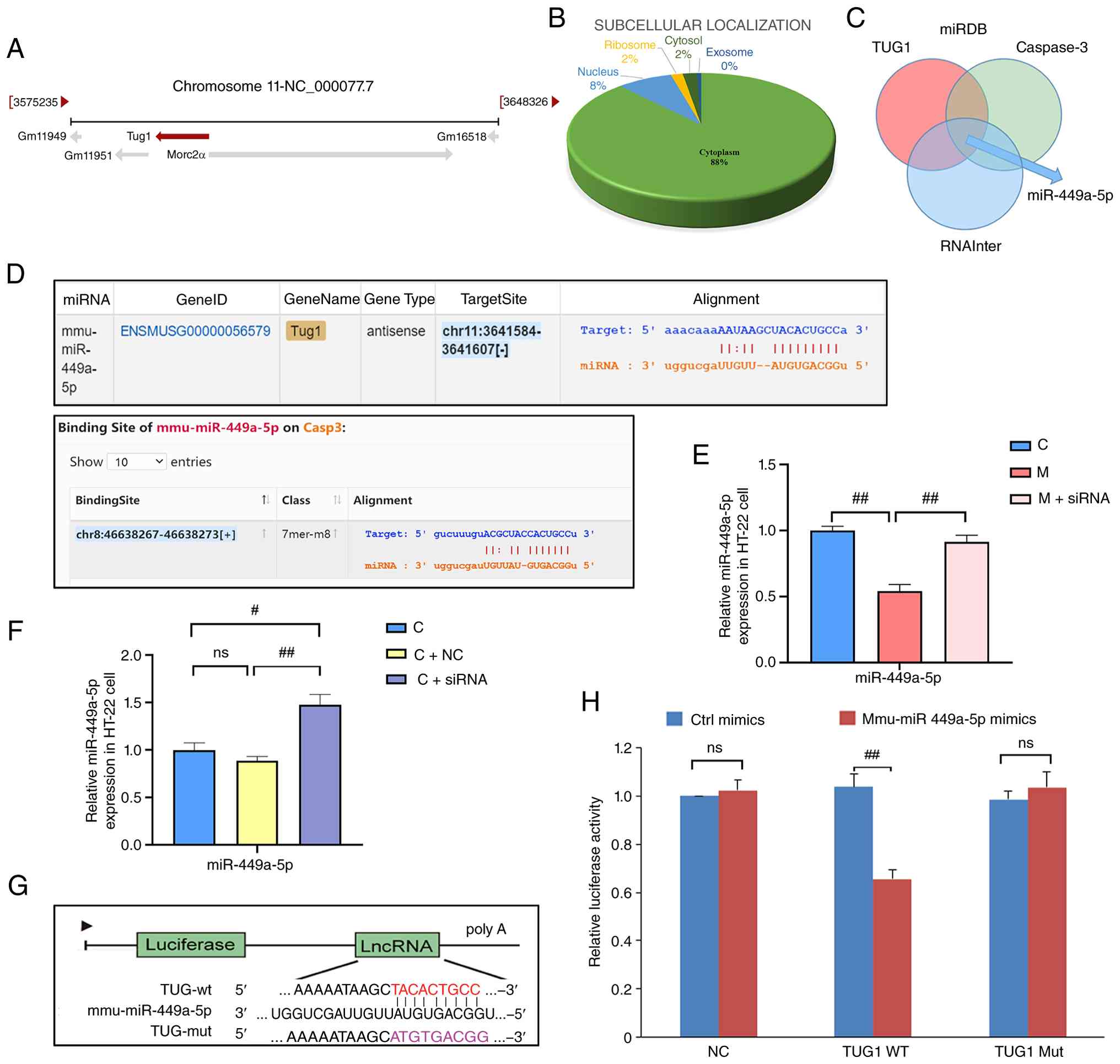 | Figure 3Target relationship between lncRNA
TUG1 and miR-449a-5p. (A) Informational characterization of
lncRNA TUG1 found through the National Center for
Biotechnology Information website. (B) Subcellular localization of
lncRNA TUG1 obtained from an online lncLocator predictor.
(C) Venn diagram of predicted miRNAs targeting lncRNA and Caspase
using miRDB and RNA Interactome databases. (D) Predicted
miR-449a-5p binding sites in lncRNA TUG1 and caspase-3. (E)
Relative miR-449a-5p expression level in PA-challenged HT-22 cells
pretreated with si-lncRNA TUG1. The groups include untreated
control cells 'C', the PA-stimulated group 'M', and the PA plus
si-lncRNA TUG1 group 'M + siRNA'. (F) Relative miR-449a-5p
expression level in HT-22 cells (without PA-challenged) pretreated
with si-lncRNA TUG1. The groups include untreated control
cells 'C', control cells transfected with negative control siRNA 'C
+ NC' and control cells transfected with si-lncRNA TUG1 'C +
siRNA'. (G) Schematic representation of the dual luciferase
reporter gene assay of lncRNA TUG1 with miR-449a-5p. (H)
Results of the dual-luciferase reporter assay containing lncRNA
TUG1-WT/Mut with miR-449a-5p mimic or miR-449a-5p mimic NC.
Ctrl mimics are the negative control miRNA mimics (scrambled
sequence). The data are presented as the mean ± SEM, with n=3 for
each group. #P<0.05, ##P<0.01. C,
control; Ctrl, control; lncRNA, long non-coding RNA; M, model (PA);
miR, microRNA; Mut, mutant; NC, negative control; ns, not
significant; PA, palmitic acid; si, small interfering; TUG1,
taurine upregulated gene 1; WT, wild-type. |
To confirm the binding between lncRNA TUG1
and miR-449a-5p, a dual-luciferase reporter gene assay was
performed (Fig. 3G). The results
showed that the relative luciferase activity of the lncRNA
TUG1-WT + miR-449a-5p mimics group was lower than that of
the lncRNA TUG1-WT + Ctrl mimics group (P<0.01). However,
no significant difference was observed when miR-449a-5p mimics or
Ctrl mimics were co-transfected with lncRNA TUG1-Mut
(P>0.05) (Fig. 3H). These
findings confirmed that miR-449a-5p may directly bind to the
predicted site on lncRNA TUG1. On the basis of these
results, we hypothesized that miR-449a-5p may serve as a direct
target of lncRNA TUG1 in PA-induced HT-22 cells.
Overexpression of miR-449a-5p in
PA-challenged HT-22 cells alleviates the dysfunctional changes in
apoptosis, synaptic plasticity, Akt/GSK-3β pathway and LPO
To investigate whether miR-449a-5p directly
regulates PA-induced neuronal injury, cells were transfected with
miR-449a-5p mimics. Efficient transfection was confirmed in
Fig. 4A, indicating successful
overexpression of miR-449a-5p. The overexpression of miR-449a-5p
phenocopied the protective effects of lncRNA TUG1 knockdown.
Specifically, miR-449a-5p mimics significantly reduced PA induced
LDH release (Fig. 4B),
indicating that miR-449a-5p overexpression attenuated PA-induced
cytotoxicity. Furthermore, miR-449a-5p mimics restored Bcl-2
expression and reversed the PA-induced increase in the cleaved
caspase-3 to total caspase-3 ratio (Fig. 4C-E). miR-449a-5p mimics also
lowered the apoptotic rate, as measured by flow cytometry (Fig. 4F and G), suggesting that
miR-449a-5p mimics inhibited apoptosis in PA-challenged HT-22
cells. The PA induced reductions in synapsin-1, synaptotagmin-1 and
BDNF were also reversed (Fig.
4K-N), indicating that miR-449a-5p protected against PA-induced
synaptic damage, and the p-Akt/Akt and p-GSK-3β/GSK-3β ratios were
restored (Fig. 4H-J), suggesting
that miR-449a-5p mimics reactivated the Akt/GSK-3β signaling
pathway. Moreover, miR-449a-5p overexpression alleviated PA induced
LPO, as indicated by the reduced 4-HNE, MDA and LPO levels
(Fig. 4O-R), demonstrating that
miR-449a-5p mitigated LPO.
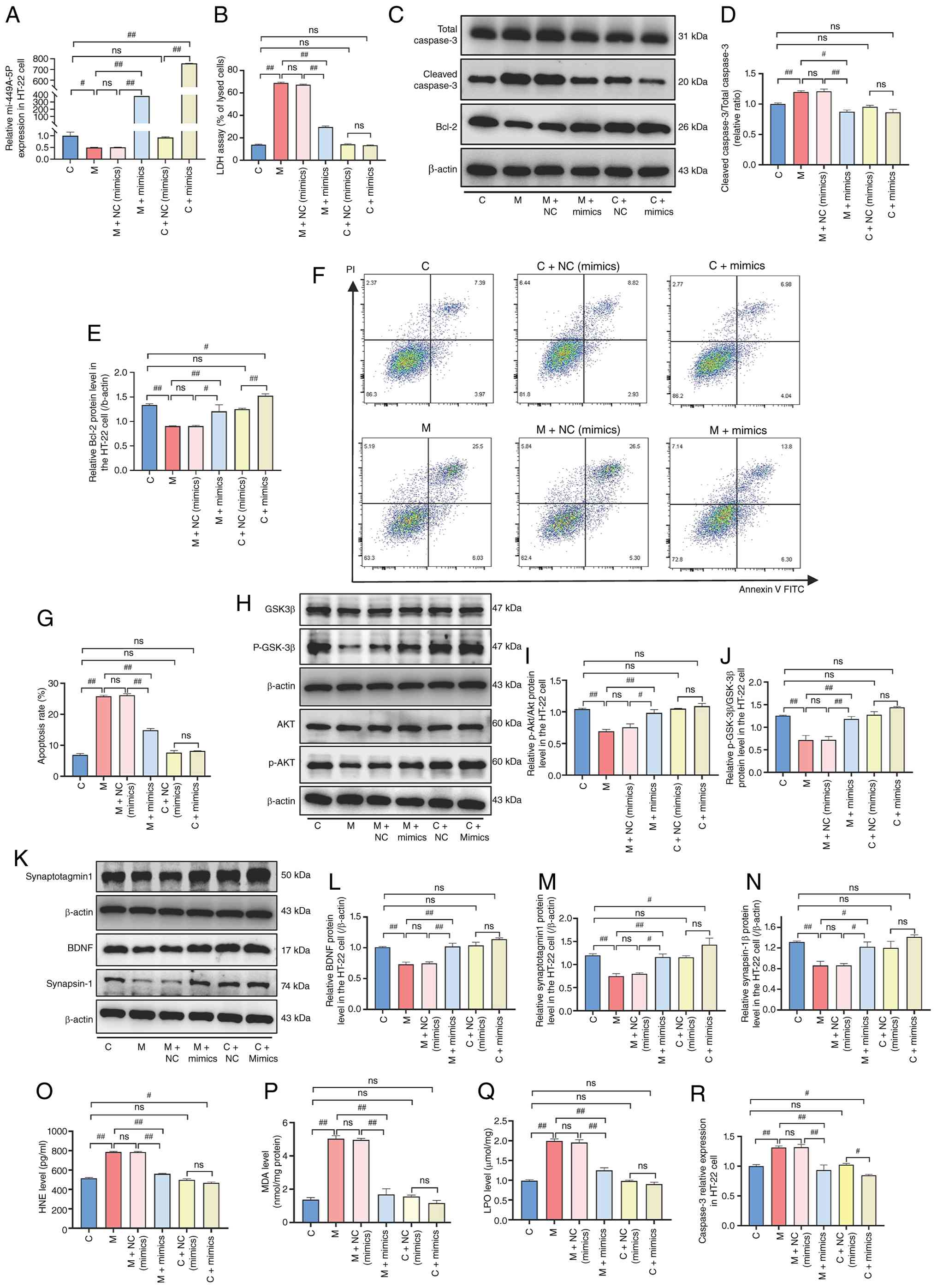 | Figure 4Effects of overexpression of
miR-449a-5p on apoptosis, synaptic plasticity, the Akt/GSK-3β
pathway and LPO levels in PA-challenged HT-22 cells. (A) Effect of
pretreatment with miR-449a-5p mimics on miR-449a-5p expression
levels in PA-challenged HT-22 cells. (B) Detection of LDH release
rate in PA-challenged HT-22 cells by pretreatment with miR-449a-5p
mimics. (C) Representative western blotting bands of Bcl-2, cleaved
caspase-3 and total caspase-3. Semi-quantification of (D) the
cleaved caspase-3 to total caspase-3 ratio and (E) Bcl-2 protein
levels. (F) Flow cytometry determination of apoptosis levels in
miR-449a-5p mimic pretreated PA-challenged HT-22 cells. (G)
Statistical analysis of apoptosis rates. (H) Representative western
blotting bands of Akt, p-Akt, GSK-3β and p-GSK-3β.
Semi-quantification of (I) p-Akt/Akt ratio and (J) p-GSK-3β/GSK-3β
ratio. (K) Representative western blotting bands of Synapsin-1,
Synaptotagmin-1 and BDNF. Semi-quantification of (L) BDNF, (M)
Synaptotagmin-1 and (N) Synapsin-1 protein levels. Determination of
(O) 4-HNE, (P) MDA and (Q) LPO levels. (R) Effect of pretreatment
with miR-449a-5p mimics on caspase-3 expression levels in
PA-challenged HT-22 cells. The data are presented as the mean ±
SEM, with n=3 for each group, except for (G) where n=4 per group.
#P<0.05, ##P<0.01. 4-HNE,
4-hydroxynonenal; BDNF, brain-derived neurotrophic factor; C,
control; LDH, lactate dehydrogenase; LPO, lipid peroxidation; M,
model (PA); MDA, malondialdehyde; miR, microRNA; NC, negative
control; ns, not significant; p, phosphorylated; PA, palmitic
acid. |
Inhibition of miR-449a-5p in
PA-challenged HT-22 cells promotes the dysfunctional changes in
apoptosis, synaptic plasticity, Akt/GSK-3β pathway and LPO
In contrast to the protective effects observed with
miR-449a-5p overexpression, the inhibition of miR-449a-5p using a
specific inhibitor exacerbated PA-induced neuronal injury. The
inhibitor was efficiently transfected and the negative control
showed no significant effect on miR-449a-5p expression (Fig. 5A). LDH release was further
increased in PA challenged cells following miR-449a-5p inhibition
compared with that in the PA-only or NC groups (Fig. 5B), indicating that miR-449a-5p
inhibition aggravated PA-induced cytotoxicity. Transfection with
the miR-449a-5p inhibitor increased the cleaved caspase-3 to total
caspase-3 ratio in the absence of PA stimulation. In the presence
of PA, it did not further alter this ratio but led to a further
reduction in Bcl-2 expression (Fig.
5C-E), thus resulting in an increased apoptotic rate, as
determined by flow cytometry (Fig.
5F and G). These results suggested that miR-449a-5p inhibition
exacerbated apoptosis in PA-challenged HT-22 cells. Moreover, the
reductions in the p-Akt/Akt and p-GSK-3β/GSK-3β ratios were further
pronounced (Fig. 5H-J),
suggesting that miR-449a-5p inhibition further suppressed the
Akt/GSK-3β signaling pathway, and the PA-induced downregulation of
synaptotagmin-1, synapsin-1 and BDNF was aggravated by miR-449a-5p
inhibition (Fig. 5K-N),
indicating that loss of miR-449a-5p function worsened PA-induced
synaptic damage. Additionally, the inhibition of miR-449a-5p
promoted PA-induced elevations in the LPO markers 4-HNE, MDA and
LPO (Fig. 5O-Q), indicating that
miR-449a-5p deficiency exacerbated LPO.
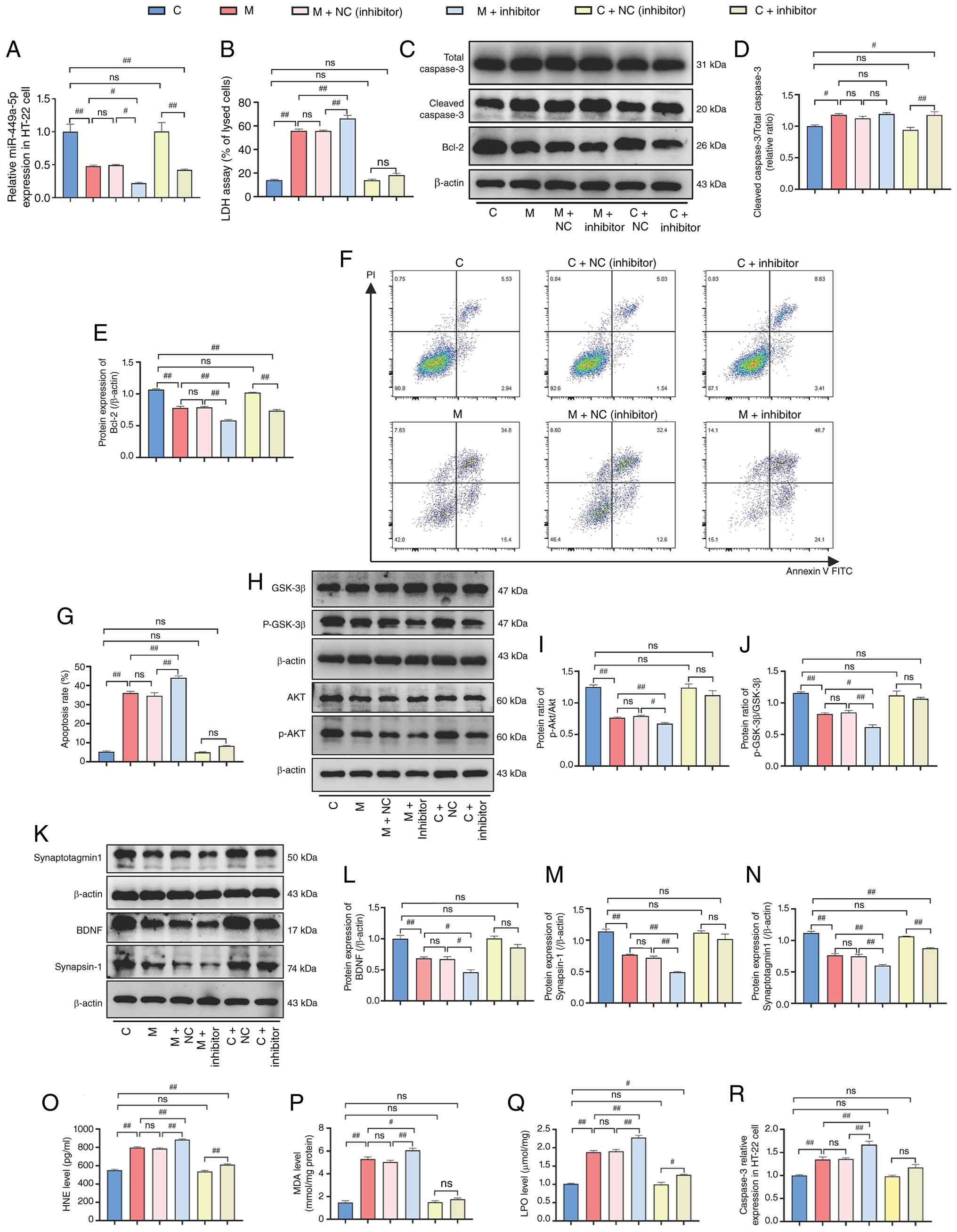 | Figure 5Effects of miR-449a-5p inhibition on
apoptosis, synaptic plasticity, Akt/GSK-3β pathway and LPO levels
in PA-challenged HT-22 cells. (A) Effect of pretreatment with
miR-449a-5p inhibitor on miR-449a-5p expression levels in
PA-challenged HT-22 cells. (B) Detection of LDH release rate in
PA-challenged HT-22 cells by pretreatment with miR-449a-5p
inhibitor. (C) Representative western blotting bands of Bcl-2,
cleaved caspase-3 and total caspase-3. Semi-quantification of the
(D) cleaved caspase-3 to total caspase-3 ratio and (E) Bcl-2
protein levels. (F) Flow cytometry determination of apoptosis
levels in miR-449a-5p inhibitor pretreated PA-challenged HT-22
cells. (G) Statistical analysis of apoptosis rates. (H)
Representative western blotting bands of Akt, p-Akt, GSK-3β and
p-GSK-3β. Semi-quantification of (I) the p-Akt/Akt and (J)
p-GSK-3β/GSK-3β ratios. (K) Representative western blotting bands
of Synapsin-1, Synaptotagmin-1 and BDNF. Semi-quantification of the
(L) BDNF, (M) Synapsin-1 and (N) Synaptotagmin-1 protein levels.
Determination of the (O) 4-HNE, (P) MDA levels and (Q) LPO levels.
(R) Effect of pretreatment with miR-449a-5p inhibitor on caspase-3
expression levels in PA-challenged HT-22 cells. The data are
presented as the mean ± SEM, with n=3 for each group, except for
(G) where n=4 per group. #P<0.05,
##P<0.01. 4-HNE, 4-hydroxynonenal; BDNF,
brain-derived neurotrophic factor; C, control; LDH, lactate
dehydrogenase; LPO, lipid peroxidation; M, model (PA); MDA,
malondialdehyde; miR, microRNA; NC, negative control; ns, not
significant; p, phosphorylated; PA, palmitic acid. |
lncRNA TUG1/miR-449a-5p axis regulates
PA-induced neuronal injury via caspase-3 and the Akt/GSK-3β
pathway
To determine whether lncRNA TUG1 regulates
caspase-3-associated apoptosis by sponging miR-449a-5p, HT-22 cells
were co-transfected with si-lncRNA TUG1 and either
miR-449a-5p mimics or an inhibitor prior to PA stimulation. RT-qPCR
confirmed that si-lncRNA TUG1 upregulated miR-449a-5p
expression, and this effect was further enhanced by miR-449a-5p
mimics. Conversely, the suppressive effect of miR-449a-5p inhibitor
was counteracted by si-lncRNA TUG1 (Fig. 6A). These results confirmed that
lncRNA TUG1 knockdown elevated the miR-449a-5p levels and
that the mimic and inhibitor functioned as anticipated. Regarding
caspase-3 mRNA levels, compared with the M + siRNA group, caspase-3
expression was increased in the M + siRNA + inhibitor group and
decreased in the M + siRNA + mimics group (Fig. 6B), indicating that miR-449a-5p
mediates the effect of TUG1 knockdown on caspase-3 transcription. A
similar pattern was observed for Bcl-2 protein expression. Compared
with the M + siRNA group, Bcl-2 expression was significantly
increased in the M + siRNA + mimics group and significantly
decreased in the M + siRNA + inhibitor group (Fig. 6C and F). These results suggest
that lncRNA TUG1 regulates Bcl-2 expression, at least
partly, through miR-449a-5p. However, under conditions of
TUG1 knockdown, neither the miR-449a-5p inhibitor nor the
mimic effectively altered the cleaved caspase-3 to total caspase-3
ratio. The miR-449a-5p inhibitor also reversed the restorative
effects of si-lncRNA TUG1 on p-Akt/Akt and p-GSK-3β/GSK-3β
ratios, as well as on synapsin-1, synaptotagmin-1 and BDNF
expression. Conversely, the miR-449a-5p mimic enhanced the
protective effect of si-lncRNA TUG1 on Synapsin-1 and BDNF
but not on Synaptotagmin-1 (Fig.
6F-L); these findings indicated that the protective effects of
si-lncRNA TUG1 on Akt/GSK-3β signaling and synaptic proteins
may be dependent on miR-449a-5p. Flow cytometry further showed
that, compared with the M + siRNA group, the apoptosis rate was
significantly increased in the M + siRNA + inhibitor group and
further decreased in the M + siRNA + mimics group (Fig. 6M and N), further confirming that
miR-449a-5p may be essential for the anti-apoptotic effect of
lncRNA TUG1 knockdown. In the LPO assays, the miR-449a-5p
inhibitor blocked the ameliorative effect of si-lncRNA TUG1
on MDA and LPO levels, whereas the mimic potentiated this effect;
no significant change was observed for 4-HNE (Fig. 6O-Q), indicating that lncRNA
TUG1 may modulate LPO through miR-449a-5p, with
marker-specific variations.
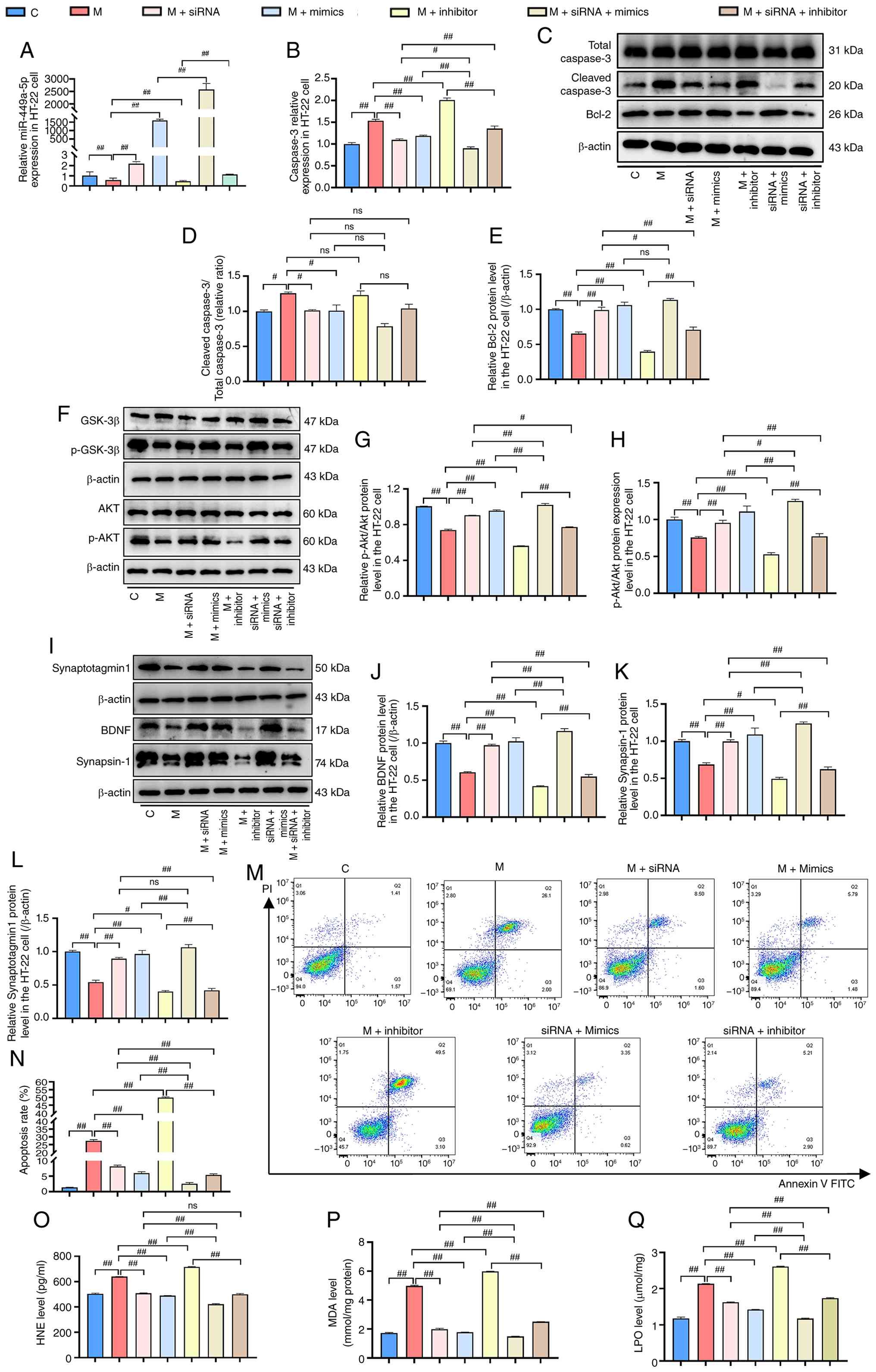 | Figure 6Effect of lncRNA
TUG1/miR-449a-5p/caspase-3 axis on neuroapoptosis in
PA-stimulated HT-22 cells. (A) Relative expression level of
miR-449a-5p in PA-stimulated HT-22 cells pretreated with si-lncRNA
TUG1 and/or miR-449a-5p mimics/inhibitor, determined by
RT-qPCR. (B) Relative expression level of caspase-3 mRNA. (C)
Representative western blot bands of Bcl-2, cleaved caspase-3 and
total caspase-3. Semi-quantification of (D) the cleaved caspase-3
to total caspase-3 ratio and (E) Bcl-2 protein levels. (F)
Representative western blot bands of Akt, p-Akt, GSK-3β and
p-GSK-3β. Semi-quantification of the (G) p-Akt/Akt and (H)
p-GSK-3β/GSK-3β ratios. (I) Representative western blot bands of
Synapsin-1, Synaptotagmin-1 and BDNF. Semi-quantification of the
(J) BDNF, (K) Synaptotagmin-1 and (L) Synapsin-1 protein levels.
(M) Flow cytometry determination of the apoptosis levels in
PA-stimulated HT-22 cells pretreated as indicated. (N) Statistical
analysis of the apoptosis rates. Determination of the (O) 4-HNE,
(P) MDA and (Q) LPO levels. The data are presented as the mean ±
SEM, with n=3 for each group. #P<0.05,
##P<0.01. 4-HNE, 4-hydroxynonenal; BDNF,
brain-derived neurotrophic factor; C, control; LDH, lactate
dehydrogenase; lncRNA, long non-coding RNA; LPO, lipid
peroxidation; M, model (PA); MDA, malondialdehyde; miR, microRNA;
ns, not significant; p, phosphorylated; PA, palmitic acid; si,
small interfering (RNA); TUG1, taurine upregulated gene 1. |
Pharmacological validation of the
Akt/GSK-3β pathway as a downstream effector of the lncRNA
TUG1/miR-449a-5p axis
To further verify the critical role of the
Akt/GSK-3β pathway in the neuroprotective effects of the lncRNA
TUG1/miR-449a-5p axis, rescue experiments were performed
using a specific pharmacological inhibitor of Akt, namely, Akt
inhibitor VIII. Two sets of rescue assays were conducted. In the
lncRNA TUG1 knockdown set, the groups included control (C),
model (M; 100 μM PA), M + NC (negative control siRNA), M +
siRNA (si-lncRNA TUG1), M + Akt inhibitor VIII, M + siRNA +
DMSO and M + siRNA + Akt inhibitor VIII. In the miR-449a-5p
overexpression set, the groups were C, M, M + NC (mimic NC), M +
mimics (miR-449a-5p mimics), M + Akt inhibitor VIII, M + mimics +
DMSO and M + mimics + Akt inhibitor VIII. As presented in Fig. 7A-J, compared with the M group, M
+ Akt inhibitor VIII did not further decrease Bcl-2 protein
expression, increase the cleaved caspase-3 to total caspase-3
ratio, significantly affect the p-GSK-3β/GSK-3β and p-Akt/Akt
ratios or further reduce the protein expression of Synapsin-1,
Synaptotagmin-1 and BDNF. However, compared with the M + siRNA +
DMSO group, the M + siRNA + Akt inhibitor VIII group reversed the
protective effects of lncRNA TUG1 knockdown, as evidenced by
decreased Bcl-2 protein expression, reduced p-GSK-3β/GSK-3β and
p-Akt/Akt ratios and decreased protein expression of Synapsin-1,
Synaptotagmin-1 and BDNF. Notably, no significant difference was
observed in the cleaved caspase-3 to total caspase-3 ratio. These
results further confirmed that the neuroprotective effects of
lncRNA TUG1 knockdown may depend on the functional
activation of the Akt/GSK-3β signaling pathway. Similarly,
inhibition of Akt reversed the protective effects of miR-449a-5p
overexpression, indicating that the protective actions of
miR-449a-5p also require the Akt/GSK-3β pathway. Specifically,
compared with the M + mimics + DMSO group, the M + mimics + Akt
inhibitor VIII group exhibited an increased cleaved caspase-3 to
total caspase-3 ratio, decreased Bcl-2 protein expression, reduced
p-GSK-3β/GSK-3β and p-Akt/Akt ratios, and decreased protein
expression of Synapsin-1, Synaptotagmin-1 and BDNF (Fig. 7K-T). No statistically significant
differences were detected in any of the measured parameters between
the M + siRNA group and its corresponding DMSO vehicle control
group (M + siRNA + DMSO) or between the M + mimics group and its
DMSO control (M + mimics + DMSO) (Fig. 7A-T), demonstrating that the
vehicle DMSO did not interfere with the observed biological
effects. Collectively, these data provide direct pharmacological
evidence that the Akt/GSK-3β signaling pathway may be indispensable
for the neuroprotective functions of the TUG1/miR-449a-5p
axis in PA-induced neuronal injury.
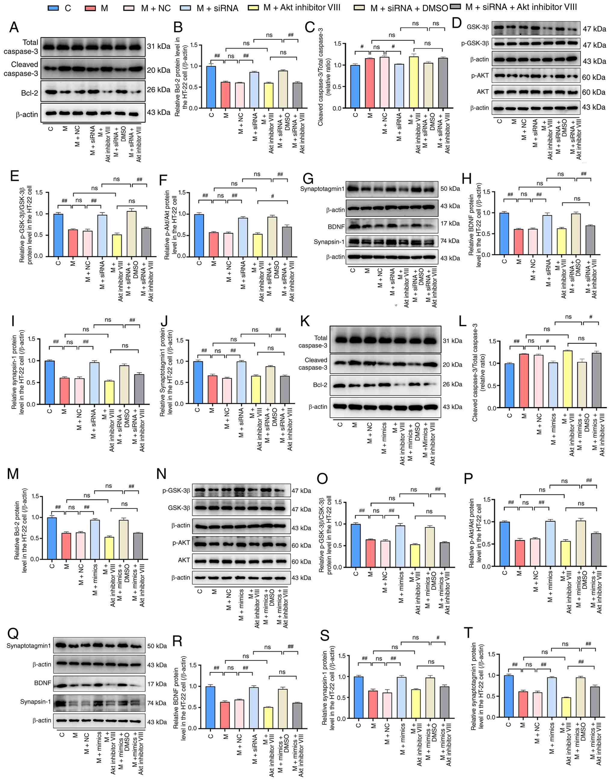 | Figure 7Pharmacological inhibition of Akt
abolishes the neuroprotective effects of lncRNA TUG1
knockdown and miR-449a-5p overexpression in PA-challenged HT-22
cells. (A) Representative western blotting bands of Bcl-2, cleaved
caspase-3 and total caspase-3. Semi-quantification of (B) Bcl-2
protein levels and (C) the cleaved caspase-3 to total caspase-3
ratio. (D) Representative western blotting bands of p-Akt, Akt,
p-GSK-3β and GSK-3β. Semi-quantification of the (E) p-GSK-3β/GSK-3β
and (F) p-Akt/Akt ratios. (G) Representative western blotting bands
of Synapsin-1, Synaptotagmin-1 and BDNF. Semi-quantification of the
(H) BDNF, (I) Synapsin-1 and (J) Synaptotagmin-1 protein levels.
(K) Representative western blotting bands of Bcl-2, cleaved
caspase-3 and total caspase-3. Semi-quantification of (L) the
cleaved caspase-3 to total caspase-3 ratio and (M) Bcl-2 protein
levels. (N) Representative western blotting bands of p-Akt, Akt,
p-GSK-3β and GSK-3β. Semi-quantification of the (O) p-GSK-3β/GSK-3β
and (P) p-Akt/Akt ratios. (Q) Representative western blotting bands
of Synapsin-1, Synaptotagmin-1 and BDNF. Semi-quantification of the
(R) BDNF, (S) Synapsin-1 and (T) Synaptotagmin-1 protein levels.
The data are presented as the mean ± SEM, with n=3 for each group.
#P<0.05, ##P<0.01. BDNF, brain-derived
neurotrophic factor; C, control; DMSO, dimethyl sulfoxide; lncRNA,
long non-coding RNA; M, model (PA); miR, microRNA; NC, negative
control; ns, not significant; p, phosphorylated; PA, palmitic acid;
siRNA, small interfering RNA; TUG1, taurine upregulated gene 1. |
Discussion
The present study identified lncRNA TUG1 as
a potential critical mediator of PA-induced neuronal lipotoxicity
via the miR-449a-5p/caspase-3 axis together with the Akt/GSK-3β
pathway. Elevated lncRNA TUG1 expression was detected in
PA-treated neuronal cells and was accompanied by increased
cytotoxicity, apoptosis and synaptic protein loss. lncRNA
TUG1 knockdown alleviated PA-induced cytotoxicity, apoptosis
and synaptic protein loss, while restoring Akt/GSK-3β
phosphorylation. Mechanistically, lncRNA TUG1 acted as a
sponge for miR-449a-5p, thus leading to the derepression of
caspase-3 expression. The pharmacological inhibition of Akt
confirmed that the Akt/GSK-3β pathway may be a required downstream
partner of this axis. These findings suggest a detrimental role for
lncRNA TUG1 in lipid-induced neurotoxicity through a
regulatory network involving both caspase-3 and Akt/GSK-3β
signaling.
A growing body of evidence implicates dysregulated
lipid metabolism in the pathogenesis of cognitive decline and
neurodegenerative disorders (1,35). Hyperlipidemia, a hallmark of
metabolic syndrome and T2DM, is independently associated with an
increased risk of all-cause dementia and AD (1,2).
Mechanistically, saturated free fatty acids such as PA, which are
elevated in the cerebrospinal fluid of patients with obesity and
cognitive impairment, can directly trigger neuronal lipotoxicity
via multiple pathways, including oxidative stress, endoplasmic
reticulum stress, mitochondrial dysfunction and autolysosomal
impairment (36). In the central
nervous system, PA disrupts autolysosomal function and
mitochondrial homeostasis, thus triggering oxidative stress and
neuroinflammatory responses, which ultimately contribute to
neurodegeneration (37).
Furthermore, PA induces dynamic alterations in histone deacetylases
and chromatin acetylation in neurons, thus suggesting that lipid
overload may have lasting effects on neuronal gene expression
beyond acute metabolic stress (2). Consistent with these findings, our
previous studies using NAFLD and T2DM animal models have
demonstrated that systemic dyslipidemia is accompanied by AD-like
neuropathological changes in the hippocampus, including impaired
synaptic plasticity, microglial activation and tau
hyperphosphorylation (9,38). The present study further
demonstrated that PA stimulation alone is sufficient to
recapitulate the core features of metabolic neuropathy in an in
vitro model, including the downregulation of synaptic proteins,
exacerbation of apoptosis and dysregulation of the Akt/GSK-3β
signaling pathway. Collectively, these observations underscore the
notion that lipid-induced neurotoxicity represents a critical yet
incompletely understood pathogenic link between peripheral
metabolic dysfunction and central nervous system impairment;
moreover, PA is not merely a metabolic byproduct but rather an
active signaling molecule that mediates the detrimental effects of
systemic hyperlipidemia on brain function.
lncRNA TUG1 has emerged as a pivotal
regulatory molecule in the nervous system, participating in a wide
array of physiological and pathological processes ranging from
neuronal differentiation to neurodegeneration. Accumulating
evidence has documented the aberrant expression of lncRNA
TUG1 in multiple neurological disorders. In ischemic stroke
models, lncRNA TUG1 expression is markedly elevated in the
ischemic penumbra, and its knockout or knockdown confers
neuroprotection by attenuating neuronal apoptosis and suppressing
microglial pyroptosis (39,40). Similarly, elevated lncRNA
TUG1 levels have been detected in the serum or cerebrospinal
fluid of patients with temporal lobe epilepsy, Parkinson's disease
and AD, and its expression has been correlated with disease
severity and poor prognosis (18,19). In the context of metabolic
dysregulation, lncRNA TUG1 has been implicated in diabetic
cardiomyopathy and NAFLD-associated hepatic injury (17,21). However, its expression profile
and functional significance in lipid-induced neuronal injury remain
unclear. In the present study, it was found that lncRNA TUG1
was robustly upregulated in both HT-22 and SH-SY5Y neuronal cells
following PA challenge, and that the magnitude of this upregulation
was significantly correlated with the severity of lipotoxic injury.
More specifically, Pearson correlation analysis revealed that
lncRNA TUG1 expression was positively associated with
cleaved caspase-3 levels and LDH release while exhibiting a
negative association with anti-apoptotic protein Bcl-2, synaptic
plasticity markers including synapsin-1, synaptotagmin-1 and BDNF
as well as the Akt and GSK-3β phosphorylation ratios. Although the
limited sample size precludes definitive causal conclusions, these
exploratory analyses provide a statistical framework for
prioritizing downstream effectors and guiding subsequent functional
validation and offer preliminary clues suggesting that lncRNA
TUG1 may functionally participate in the regulation of
neuronal apoptotic signaling networks under lipotoxic conditions.
Taken together, these findings suggest that lncRNA TUG1 is a
potential molecular sensor or amplifier of PA-induced neuronal
injury and justify its selection as the central focus of
mechanistic investigations.
Neuronal apoptosis and synaptic loss are two
inextricably linked pathological hallmarks of metabolic cognitive
impairment. Postmortem analyses of hippocampal tissue from patients
with amnestic mild cognitive impairment have revealed significantly
decreased levels of PSD95, a key postsynaptic scaffolding protein,
alongside elevated caspase-3 expression, thus suggesting that
coordinated dysregulation of synaptic integrity and apoptotic
signaling occurs early in the trajectory of cognitive decline
(41). In the context of
lipotoxicity, PA induces localized caspase-3 activation that
extends beyond its canonical role in cell death. Emerging evidence
indicates that caspase-3 can be activated at sublethal levels
within the dendritic spines, where it participates in the cleavage
of critical synaptic scaffolding proteins and contributes to the
weakening of synaptic transmission (42,43). This non-apoptotic function of
caspase-3 provides a mechanistic framework for understanding how
metabolic stressors such as PA can impair synaptic plasticity and
cognitive performance before overt neuronal loss becomes
detectable, a phenomenon that aligns with early memory deficits
frequently observed in patients with T2DM and NAFLD (37,44). The findings of the present study
extend this paradigm by demonstrating that lncRNA TUG1
functions as an upstream regulator that sustains elevated caspase-3
expression under lipotoxic conditions. By sponging miR-449a-5p and
relieving its repression of caspase-3, lncRNA TUG1 amplifies
both the apoptotic cascade and synaptic dysfunction triggered by
PA. This dual effect on neuronal survival and synaptic integrity
explains why lncRNA TUG1 expression correlated strongly with
both cleaved caspase-3 levels and the depletion of synaptic
proteins such as synapsin-1, synaptotagmin-1 and BDNF in the PA
model. Furthermore, it provides a molecular rationale for the
well-documented association between hyperlipidemia and accelerated
cognitive impairment. This caspase-3-mediated dual damage is
further exacerbated by the concurrent dysregulation of the
Akt/GSK-3β survival pathway, which is also under the control of the
lncRNA TUG1/miR-449a-5p axis.
In addition, the present study demonstrated that
lncRNA TUG1 also acts as a critical mediator in PA-induced
neuronal injury through the Akt/GSK-3β signaling pathway. This
pathway may serve as a molecular link between peripheral metabolic
dysregulation and central nervous system impairment. To explore the
functional role of the Akt/GSK-3β pathway, pharmacological
experiments using Akt inhibitor VIII were performed. The results
showed that Akt activation may be essential for the neuroprotective
effects conferred by both lncRNA TUG1 knockdown and
miR-449a-5p upregulation. In mediating PA-induced lipotoxic injury,
the Akt/GSK-3β pathway functions as an important synergistic
partner with caspase-3. The crosstalk between caspase-3 and the
Akt/GSK-3β pathway has been documented in previous studies. Akt can
phosphorylate and inactivate caspase-9, thereby suppressing
caspase-3 activation and promoting cell survival (45). Conversely, active caspase-3 can
cleave and inactivate Akt, thereby forming a feedback loop that
amplifies apoptotic signals (46). GSK-3β is a well-recognized
downstream target of Akt; Akt mediated phosphorylation at Ser9
inactivates GSK-3β, and the dysregulation of GSK-3β has been
implicated in various neurodegenerative conditions (47). In the context of lipotoxic
injury, a previous study has shown that saturated fatty acids can
simultaneously trigger caspase-3 activation and suppress Akt
signaling, ultimately leading to neuronal apoptosis (48). On the basis of the experimental
data from the present study together with these previous reports,
we propose that within the lncRNA TUG1/miR-449a-5p
regulatory axis, caspase-3 and the Akt/GSK-3β pathway do not
operate in a simple linear cascade but rather constitute an
integrated signaling network in which they mutually influence each
other to orchestrate neuronal injury.
The findings of the present study hold potential
clinical relevance in metabolic neuropathy and T2DM related
neurodegeneration. T2DM is a well-recognized risk factor for
cognitive impairment and dementia. Recent meta-analyses have
demonstrated that individuals with diabetes have a significantly
higher risk of developing dementia compared with those without
diabetes, with factors such as glycemic control and disease
duration showing significant associations with cognitive outcomes
(49,50). Among the various pathogenic
factors linking T2DM to brain dysfunction, lipotoxicity resulting
from elevated levels of saturated fatty acids, such as PA, is
considered a key contributor to neuronal injury and synaptic loss.
Emerging evidence indicates that PA disrupts autolysosomal function
and mitochondrial homeostasis, triggering oxidative stress and
neuroinflammatory responses that ultimately lead to
neurodegeneration (37). The
present study revealed that lncRNA TUG1 may act as a
critical mediator in PA-induced neuronal lipotoxicity through the
miR-449a-5p/caspase-3 axis together with the Akt/GSK-3β signaling
pathway. This pathway may serve as a molecular link between
peripheral metabolic dysregulation and central nervous system
impairment. Notably, elevated levels of lncRNA TUG1 have
been detected in both the peripheral circulation and cerebrospinal
fluid of patients with neurological disorders, and these levels are
negatively correlated with patient prognosis (51-53). This observation suggests that
lncRNA TUG1 holds promise as a potential biomarker for early
diagnosis or disease progression assessment in T2DM associated
cognitive decline. Furthermore, the neuroprotective effects
observed following lncRNA TUG1 knockdown or miR-449a-5p
overexpression in vitro in the present study indicated that
targeting this regulatory axis may offer a novel therapeutic
strategy for neuropsychiatric complications in patients with
metabolic disorders. Although further validation in animal models
and clinical samples is warranted, the present study provides a
molecular framework for developing intervention strategies that are
aimed at preventing or ameliorating T2DM related
neurodegeneration.
Although the present study elucidated the novel
role and underlying mechanisms of the lncRNA
TUG1/miR-449a-5p/caspase-3 axis in PA-induced neuronal
apoptosis, several limitations should be acknowledged. First, the
experimental findings were primarily derived from immortalized
neuronal cell lines, namely, HT-22 and SH-SY5Y cells. Although
these cell lines are widely accepted and frequently used in
neurobiological research due to their accessibility and
experimental reproducibility, they do not fully recapitulate the
complex physiological properties of primary neurons. Primary
neurons maintain intact synaptic connectivity, exhibit more
physiologically relevant electrophysiological characteristics and
may respond to metabolic stress in ways that differ substantially
from those of immortalized cell lines. Therefore, the absence of
validation in primary cultured neurons is a limitation of the
present study. Second, some of the experiments were conducted with
relatively small sample sizes. Although the results were highly
consistent across multiple independent assays, reducing the risk of
false-positive results, larger sample sizes would further improve
the robustness and reproducibility of the conclusions drawn. Third,
although bioinformatics predictions and dual-luciferase reporter
assays supported the direct targeting of miR-449a-5p and both
lncRNA TUG1 and caspase-3, additional biochemical
experiments such as RNA pull-down or argonaute 2-based RNA
immunoprecipitation assays were not performed in the present study.
Such experiments would provide further support for the direct
interactions. Therefore, the current evidence, which is consistent
with our proposed model, does not entirely exclude the possibility
of indirect regulation. Future studies employing these approaches
will help solidify this conclusion. Fourth, no loss of function or
gain of function interventions were performed in vivo, such
as stereotaxic injection of lncRNA TUG1 specific siRNA or
overexpression vectors into the hippocampus, followed by an
assessment of cognitive or behavioral changes in animals.
Consequently, the causal role of the lncRNA
TUG1/miR-449a-5p/caspase-3 signaling axis in T2DM related
neurodegeneration in vivo has not yet been established.
Furthermore, it was not validated whether targeting this pathway
could ameliorate cognitive deficits or neuropathological changes in
the animal models. These missing in vivo experiments limit
the translational relevance of the present work. Fifth, although it
was confirmed that the lncRNA TUG1/miR-449a-5p axis may
regulate caspase-3-dependent neuronal apoptosis by detecting
cleaved caspase-3 expression, Bcl-2 levels, LDH release and flow
cytometry, TUNEL or Hoechst staining to provide direct
morphological evidence of apoptosis was not performed, nor was
poly(ADP-ribose) polymerase cleavage detected or mitochondrial
membrane potential measured in the present study. These indicators
provide important supplementary evidence for the mitochondrial
apoptotic pathway and further confirm the role of caspase-3 as a
key downstream effector. This is another limitation of the present
study.
In conclusion, the present study demonstrated that
lncRNA TUG1 was upregulated in PA-induced neuronal injury
and may participate in lipotoxic neuronal damage through the
miR-449a-5p/caspase-3 axis together with the Akt/GSK-3β signaling
pathway. The findings also revealed a role for lncRNA TUG1
in PA-induced neuronal lipotoxicity, a pathological process closely
associated with hyperlipidemia-related neuronal damage. In
addition, miR-449a-5p was identified as a novel target of lncRNA
TUG1 in the regulation of lipotoxicity-associated neuronal
apoptosis and the findings confirmed that miR-449a-5p can modulate
the expression of caspase-3. These results provide experimental
evidence for understanding the pathogenesis of neuropsychiatric
dysfunction linked to lipid metabolism disorders and offer
preliminary clues for exploring lncRNA TUG1 as a potential
therapeutic target.
Supplementary Data
Availability of data and materials
The data generated in the present study may be
requested from the corresponding author.
Authors' contributions
YDW and PQC performed experiments, analyzed data
and wrote the manuscript. XZ and LQW performed experiments and
analyzed data. XYW analyzed and interpreted data. XRG analyzed and
interpreted the data. JFG conceived and designed the study, managed
the data and supervised the project. XRG and JFG confirm the
authenticity of all the raw data. All authors read and approved the
final version of the manuscript.
Ethics approval and consent to
participate
Not applicable.
Patient consent for publication
Not applicable.
Competing interests
The authors declare that they have no competing
interests.
Abbreviations:
|
4-HNE
|
4-hydroxynonenal
|
|
AD
|
Alzheimer's disease
|
|
BDNF
|
brain-derived neurotrophic factor
|
|
ceRNA
|
competing endogenous RNA
|
|
LDH
|
lactate dehydrogenase
|
|
lncRNAs
|
long non-coding RNAs
|
|
miR
|
microRNA
|
|
NAFLD
|
non-alcoholic fatty liver disease
|
|
NCBI
|
National Center for Biotechnology
Information
|
|
PA
|
palmitic acid
|
|
RT-qPCR
|
reverse transcription-quantitative
polymerase chain reaction
|
|
siRNA
|
small interfering RNA
|
|
T2DM
|
type 2 diabetes mellitus
|
|
TUG1
|
taurine upregulated gene 1
|
Acknowledgements
Not applicable.
Funding
This research was funded by the National Natural Science
Foundation of China (grant no. 82574392), the Natural Science
Foundation of Anhui Province (grant no. 2508085MH219), the Key
Project of Scientific Research in Universities of Anhui Province
(grant no. 2024AH050773) and the Scientific Research Promotion Plan
of Anhui Medical University (grant no. 2022xkjT009).
References
|
1
|
Zhao Y, Zhang H, Cheng J, Zou Y, Zhang D
and Duan X: Association between dyslipidaemia and cognitive
impairment: A meta-analysis of cohort and case-control studies. J
Integr Neurosci. 23:402024. View Article : Google Scholar : PubMed/NCBI
|
|
2
|
Iwagami M, Qizilbash N, Gregson J, Douglas
I, Johnson M, Pearce N, Evans S and Pocock S: Blood cholesterol and
risk of dementia in more than 1•8 million people over two decades:
A retrospective cohort study. Lancet Healthy Longev. 2:e498–e506.
2021. View Article : Google Scholar
|
|
3
|
Rochoy M, Rivas V, Chazard E, Decarpentry
E, Saudemont G, Hazard PA, Puisieux F, Gautier S and Bordet R:
Factors associated with Alzheimer's disease: An overview of
reviews. J Prev Alzheimers Dis. 6:121–134. 2019. View Article : Google Scholar : PubMed/NCBI
|
|
4
|
Pappolla MA, Refolo L, Sambamurti K,
Zambon D and Duff K: Hypercholesterolemia and Alzheimer's disease:
Unraveling the connection and assessing the efficacy of
lipid-lowering therapies. J Alzheimers Dis. 101(S1): S371–S393.
2024. View Article : Google Scholar : PubMed/NCBI
|
|
5
|
Libby P: The changing landscape of
atherosclerosis. Nature. 592:524–533. 2021. View Article : Google Scholar : PubMed/NCBI
|
|
6
|
Sánchez-Alegría K and Arias C: Functional
consequences of brain exposure to saturated fatty acids: From
energy metabolism and insulin resistance to neuronal damage.
Endocrinol Diabetes Metab. 6:e3862023. View Article : Google Scholar :
|
|
7
|
Moseholm KF, Jensen MK, Buzkova P, Aroner
SA, Fitzpatrick AL, Longstreth WT Jr, Lopez O, Siscovick DS, Kizer
JR, Ix JH, et al: Circulating non-esterified fatty acids, risk of
dementia and cognitive decline: The cardiovascular health study and
multi-ethnic study of atherosclerosis. Neurobiol Aging. 148:71–79.
2025. View Article : Google Scholar : PubMed/NCBI
|
|
8
|
Kalyan-Masih P, Vega-Torres JD, Miles C,
Haddad E, Rainsbury S, Baghchechi M, Obenaus A and Figueroa JD:
Western high-fat diet consumption during adolescence increases
susceptibility to traumatic stress while selectively disrupting
hippocampal and ventricular volumes. eNeuro.
3:ENEURO.0125-16.20162016. View Article : Google Scholar : PubMed/NCBI
|
|
9
|
Gao XR, Chen Z, Fang K, Xu JX and Ge JF:
Protective effect of quercetin against the metabolic dysfunction of
glucose and lipids and its associated learning and memory
impairments in NAFLD rats. Lipids Health Dis. 20:1642021.
View Article : Google Scholar : PubMed/NCBI
|
|
10
|
Chen XX, Xu YY, Wu R, Chen Z, Fang K, Han
YX, Yu Y, Huang LL, Peng L and Ge JF: Resveratrol reduces
glucolipid metabolic dysfunction and learning and memory impairment
in a NAFLD rat model: involvement in regulating the imbalance of
nesfatin-1 abundance and copine 6 expression. Front Endocrinol
(Lausanne). 10:4342019. View Article : Google Scholar : PubMed/NCBI
|
|
11
|
Gao X, Sun H, Wei Y, Niu J, Hao S, Sun H,
Tang G, Qi C and Ge J: Protective effect of melatonin against
metabolic disorders and neuropsychiatric injuries in type 2
diabetes mellitus mice. Phytomedicine. 131:1558052024. View Article : Google Scholar : PubMed/NCBI
|
|
12
|
Gao X, Sun H, Hao S, Sun H and Ge J:
Melatonin protects HT-22 cells against palmitic acid-induced
glucolipid metabolic dysfunction and cell injuries: Involved in the
regulation of synaptic plasticity and circadian rhythms. Biochem
Pharmacol. 217:1158462023. View Article : Google Scholar : PubMed/NCBI
|
|
13
|
Zhang K, Wu D and Huang C: Crosstalk
between non-coding RNA and apoptotic signaling in diabetic
nephropathy. Biochem Pharmacol. 230:1166212024. View Article : Google Scholar : PubMed/NCBI
|
|
14
|
Zhou W, Wang Z, Tao Y, Chen C, Zhang Q,
Liu Z, Li L, Xia P and Ye Z: LncRNA-MEG3 attenuates
hyperglycemia-induced damage by enhancing mitochondrial
translocation of HSP90A in the primary hippocampal neurons. Exp
Cell Res. 419:1133202022. View Article : Google Scholar : PubMed/NCBI
|
|
15
|
Che F, Han Y, Fu J, Wang N, Jia Y, Wang K
and Ge J: LncRNA MALAT1 induced by hyperglycemia promotes
microvascular endothelial cell apoptosis through activation of the
miR-7641/TPR axis to exacerbate neurologic damage caused by
cerebral small vessel disease. Ann Transl Med. 9:17622021.
View Article : Google Scholar
|
|
16
|
Young TL, Matsuda T and Cepko CL: The
noncoding RNA taurine upregulated gene 1 is required for
differentiation of the murine retina. Curr Biol. 15:501–512. 2005.
View Article : Google Scholar : PubMed/NCBI
|
|
17
|
Chen Y, Li Z, Chen X and Zhang S: Long
non-coding RNAs: From disease code to drug role. Acta Pharm Sin B.
11:340–354. 2021. View Article : Google Scholar : PubMed/NCBI
|
|
18
|
Yang B, Liang RS, Wu XY and Lin YJ: LncRNA
TUG1 inhibits neuronal apoptosis in status epilepticus rats via
targeting the miR-421/mTOR axis. Cell Signal. 76:1097872020.
View Article : Google Scholar : PubMed/NCBI
|
|
19
|
Zhai K, Liu B and Gao L: Long-noncoding
RNA TUG1 promotes parkinson's disease via modulating
MiR-152-3p/PTEN pathway. Hum Gene Ther. 31:1274–1287. 2020.
View Article : Google Scholar : PubMed/NCBI
|
|
20
|
Li X, Wang SW, Li XL, Yu FY and Cong HM:
Knockdown of long non-coding RNA TUG1 depresses apoptosis of
hippocampal neurons in Alzheimer's disease by elevating
microRNA-15a and repressing ROCK1 expression. Inflamm Res.
69:897–910. 2020. View Article : Google Scholar : PubMed/NCBI
|
|
21
|
Wang K, Lin Y, Shen H, Yu S and Xu J:
LncRNA TUG1 exacerbates myocardial fibrosis in diabetic
cardiomyopathy by modulating the microRNA-145a-5p/Cfl2 axis. J
Cardiovasc Pharmacol. 81:192–202. 2023. View Article : Google Scholar
|
|
22
|
Li C, Zheng X, Liu P and Li M: Clinical
value of lncRNA TUG1 in temporal lobe epilepsy and its role in the
proliferation of hippocampus neuron via sponging miR-199a-3p.
Bioengineered. 12:10666–10673. 2021. View Article : Google Scholar : PubMed/NCBI
|
|
23
|
He C, Li Z, Yu W, Luo R, Zhou J, He J,
Chen Q, Song Z and Cheng S: LncRNA TUG1 mediates microglial
inflammatory activation by regulating glucose metabolic
reprogramming. Sci Rep. 14:121432024. View Article : Google Scholar : PubMed/NCBI
|
|
24
|
Wang W, Miao Z, Qi X, Wang B, Liu Q, Shi X
and Xu S: LncRNA Tug1 relieves the steatosis of SelenoF-knockout
hepatocytes via sponging miR-1934-3p. Cell Biol Toxicol.
39:3175–3195. 2023. View Article : Google Scholar : PubMed/NCBI
|
|
25
|
López-Noriega L and Rutter GA: Long
non-coding RNAs as key modulators of pancreatic β-cell mass and
function. Front Endocrinol (Lausanne). 11:6102132021. View Article : Google Scholar
|
|
26
|
Gao X, Wei Y, Sun H, Hao S, Ma M, Sun H,
Zang D, Qi C and Ge J: Role of bmal1 in type 2 diabetes
mellitus-related glycolipid metabolic disorder and neuropsychiatric
injury: Involved in the regulation of synaptic plasticity and
circadian rhythms. Mol Neurobiol. 60:4595–4617. 2023. View Article : Google Scholar : PubMed/NCBI
|
|
27
|
Zhao Y and Ai Y: Overexpression of lncRNA
Gm15621 alleviates apoptosis and inflammation response resulting
from sevoflurane treatment through inhibiting miR-133a/Sox4. J Cell
Physiol. 235:957–965. 2020. View Article : Google Scholar
|
|
28
|
Feng X, Zhan F, Luo D, Hu J, Wei G, Hua F
and Xu G: LncRNA 4344 promotes NLRP3-related neuroinflammation and
cognitive impairment by targeting miR-138-5p. Brain Behav Immun.
98:283–298. 2021. View Article : Google Scholar : PubMed/NCBI
|
|
29
|
Livak KJ and Schmittgen TD: Analysis of
relative gene expression data using real-time quantitative PCR and
the 2(-Delta Delta C(T)) method. Methods. 25:402–408. 2001.
View Article : Google Scholar
|
|
30
|
Liu S, Fan M, Xu JX, Yang LJ, Qi CC, Xia
QR and Ge JF: Exosomes derived from bone-marrow mesenchymal stem
cells alleviate cognitive decline in AD-like mice by improving
BDNF-related neuropathology. J Neuroinflammation. 19:352022.
View Article : Google Scholar : PubMed/NCBI
|
|
31
|
Diez H, Garrido JJ and Wandosell F:
Specific roles of Akt iso forms in apoptosis and axon growth
regulation in neurons. PLoS One. 7:e327152012. View Article : Google Scholar : PubMed/NCBI
|
|
32
|
Jin B, Chen Y, Yang D and Wang M: Linarin
from Lycii cortex alleviates high-fat diet-induced cognitive
impairment by improving hippocampal insulin signaling and synaptic
plasticity. J Nutr Biochem. 158:1104662026. View Article : Google Scholar : PubMed/NCBI
|
|
33
|
Qiu Q, Yang Z, Zhao J, Zhang R, Zheng S,
Wang C, Xu H, Deng H, Zhao K and Liu M: Integrative analysis of
cuproptosis-related lncRNAs for prognostic risk assessment and
tumor immune microenvironment evaluation in laryngeal squamous cell
carcinoma. Int J Biol Macromol. 306:1418462025. View Article : Google Scholar : PubMed/NCBI
|
|
34
|
De Santis R, Liepelt A, Mossanen JC, Dueck
A, Simons N, Mohs A, Trautwein C, Meister G, Marx G,
Ostareck-Lederer A and Ostareck DH: miR-155 targets caspase-3 mRNA
in activated macrophages. RNA Biol. 13:43–58. 2016. View Article : Google Scholar :
|
|
35
|
Stern A, Teng P and Frishman WH: Lipid
lowering agents and late life cognitive dysfunction. Cardiol Rev.
May 16–2025.Epub ahead of print. View Article : Google Scholar : PubMed/NCBI
|
|
36
|
Horgusluoglu E, Neff R, Song WM, Wang M,
Wang Q, Arnold M, Krumsiek J, Galindo-Prieto B, Ming C, Nho K, et
al: Integrative metabolomics-genomics approach reveals key
metabolic pathways and regulators of Alzheimer's disease.
Alzheimers Dement. 18:1260–1278. 2021. View Article : Google Scholar : PubMed/NCBI
|
|
37
|
Saipuljumri EN, Zeng J and Lo CH: Palmitic
acid-induced autolysosomal dysfunction and lipotoxicity in
neuroinflammation and neurodegeneration. Neural Regen Res.
21:2806–2811. 2026. View Article : Google Scholar :
|
|
38
|
Wei YD, Chen XX, Yang LJ, Gao XR, Xia QR,
Qi CC and Ge JF: Resveratrol ameliorates learning and memory
impairments induced by bilateral hippocampal injection of
streptozotocin in mice. Neurochem Int. 159:1053852022. View Article : Google Scholar : PubMed/NCBI
|
|
39
|
Shi X, Liu S, Zou Y, Wu H, Ma J, Lin J and
Zhang X: LncRNA taurine Up-regulated 1 knockout provides
neuroprotection in ischemic stroke rats by inhibiting
nuclear-cytoplasmic shuttling of HuR. Biomedicines. 12:25202024.
View Article : Google Scholar : PubMed/NCBI
|
|
40
|
Yao M, Wang X, Lin H, Shu H, Xu Z, Tang L,
Guo W and Xu P: LncRNA Tug1 regulates post-stroke microglial
pyroptosis via PINK1/Parkin-mediated mitophagy. Inflammation.
48:2677–2691. 2025. View Article : Google Scholar : PubMed/NCBI
|
|
41
|
Sultana R, Banks WA and Butterfield DA:
Decreased levels of PSD95 and two associated proteins and increased
levels of BCl2 and caspase 3 in hippocampus from subjects with
amnestic mild cognitive impairment: Insights into their potential
roles for loss of synapses and memory, accumulation of Abeta, and
neurodegeneration in a prodromal stage of Alzheimer's disease. J
Neurosci Res. 88:469–477. 2010. View Article : Google Scholar
|
|
42
|
Li Z, Jo J, Jia JM, Lo SC, Whitcomb DJ,
Jiao S, Cho K and Sheng M: Caspase-3 activation via mitochondria is
required for long-term depression and AMPA receptor
internalization. Cell. 141:859–871. 2010. View Article : Google Scholar : PubMed/NCBI
|
|
43
|
D'Amelio M, Cavallucci V, Middei S,
Marchetti C, Pacioni S, Ferri A, Diamantini A, De Zio D, Carrara P,
Battistini L, et al: Caspase-3 triggers early synaptic dysfunction
in a mouse model of Alzheimer's disease. Nat Neurosci. 14:69–76.
2011. View Article : Google Scholar
|
|
44
|
Contreras A, Del Rio D, Martinez A, Gil C,
Morales L, Ruiz-Gayo M and Del Olmo N: Inhibition of hippocampal
long-term potentiation by high-fat diets: Is it related to an
effect of palmitic acid involving glycogen synthase kinase-3?
Neuroreport. 28:354–359. 2017. View Article : Google Scholar : PubMed/NCBI
|
|
45
|
Cardone MH, Roy N, Stennicke HR, Salvesen
GS, Franke TF, Stanbridge E, Frisch S and Reed JC: Regulation of
cell death protease caspase-9 by phosphorylation. Science.
282:1318–1321. 1998. View Article : Google Scholar : PubMed/NCBI
|
|
46
|
Widmann C, Gibson S and Johnson GL:
Caspase-dependent cleavage of signaling proteins during apoptosis.
A turn-off mechanism for anti-apoptotic signals. J Biol Chem.
273:7141–7147. 1998. View Article : Google Scholar : PubMed/NCBI
|
|
47
|
Jope RS and Johnson GVW: The glamour and
gloom of glycogen synthase kinase-3. Trends Biochem Sci. 29:95–102.
2004. View Article : Google Scholar : PubMed/NCBI
|
|
48
|
Chen K and Yu G: Tetrahydroalstonine
possesses protective potentials on palmitic acid stimulated SK-N-MC
cells by suppression of Aβ1-42 and tau through regulation of
PI3K/Akt signaling pathway. Eur J Pharmacol. 962:1762512024.
View Article : Google Scholar
|
|
49
|
Zhao Y, Wang H, Tang G, Wang L, Tian X and
Li R: Risk factors for mild cognitive impairment in type 2
diabetes: A systematic review and meta-analysis. Front Endocrinol
(Lausanne). 16:16172482025. View Article : Google Scholar : PubMed/NCBI
|
|
50
|
Tabesh M, Sacre JW, Mehta K, Chen L,
Sajjadi SF, Magliano DJ and Shaw JE: Associations of
glycaemia-related risk factors with dementia and cognitive decline
in individuals with type 2 diabetes: A systematic review and
meta-analysis. Diabet Med. 42:e701232025. View Article : Google Scholar : PubMed/NCBI
|
|
51
|
Zhang X, Chen D, Zhu C, Zhou X, Liu H and
Zhu G: Expression significance and relationship of serum miR-542-3p
and lncRNA TUG1 in STBI patients and their predictive value for
prognosis. Int J Gen Med. 18:3441–3450. 2025. View Article : Google Scholar : PubMed/NCBI
|
|
52
|
Gu X, Song Y, Liu X, Cheng Z, Min J and
Zhang Y: METTL14-mediated m6A modification of TUG1 represses
ferroptosis in Alzheimer's disease via inhibiting GDF15
ubiquitination. Front Biosci (Landmark Ed). 29:2982024. View Article : Google Scholar : PubMed/NCBI
|
|
53
|
Chen J, Su C, Ma Z, Ou X, Zhan M and Hu J:
Aerobic exercise suppresses cognitive injury in patients with
Alzheimer's disease by regulating long non-coding RNA TUG1.
Neurosci Lett. 826:1377322024. View Article : Google Scholar : PubMed/NCBI
|

















