Introduction
Gastric cancer (GC) is the fourth most common cancer
and the second leading cause of cancer-related deaths worldwide.
Its prevalence is particularly high in East Asia, including
countries such as China, Japan and Korea (1). The prognosis of GC depends on the
stage of diagnosis, as an early gastric cancer (EGC) or advanced
gastric cancer (AGC) (2). Despite
the surgical advances that have improved long-term survival of GC
patients (3,4), molecular understanding of, as well as
novel molecular biomarkers for, the condition is still urgently
required for EGC, as EGC may progress towards AGC (2).
To address this, several microarray analyses in GC
have been performed and have identified gene expression patterns
that may be useful in the prognosis and diagnosis of the cancer
(5,6); however, these approaches did not
consider the different stages or subtypes of GC. Recent studies
that did consider stage differences (2,7,8) did
not reveal the multiple phenotypes underlining EGC, because their
primary aim was to study a handful of gene sets, which
differentiate the stage differences. Accordingly, we further
explored the various hidden phenotypes, functions and pathways in
EGC by using an integrative systematic bioinformatics approach.
Here, we focus on molecular understanding of
EGC-specific expression patterns gained by employing a systematic
approach, including function and pathway, as well as
cross-experiment analyses of 27 pairs of EGC tissues and their
normal counterparts. Interestingly, the function and pathway
analyses show that the upregulated genes in EGC tissues correlate
with cell migration and metastasis, events typical of late-stage
cancer. In addition, we propose a novel association between EGC and
estrogen receptor α (ERα)-negative breast cancer that was indicated
by cross-experiment analysis, and which enables us to identify
various associated phenotypes.
Materials and methods
Patients and samples
Tissue samples were prospectively collected from
patients who underwent gastric surgery or gastroscopy at the
National Cancer Center (NCC) Hospital between 2008 and 2009. All
tissues were obtained according to the protocols approved by the
Institutional Review Board, NCC for the human subject guideline of
NCC (NCCNCS-08-127) that is in accordance with the principles of
the Declaration of Helsinki. The samples were obtained by
endoscopic biopsies from gastric cancer patients who gave informed
consent to the protocol. The samples were stored at –80°C. The
clinical and pathological features of the patients are listed in
Table I.
 | Table I.Clinical features for 27 patients with
gastric cancer. |
Table I.
Clinical features for 27 patients with
gastric cancer.
| Characteristics | No. of
patientsa |
|---|
| Total | 27 |
| Male | 20 |
| Female | 7 |
| Age at diagnosis
(years) | |
| Range | 41–78 |
| Mean ± SD | 60.3±11.2 |
| TNM stageb | |
| T
classification | |
| T1 | 17 |
| T2 | 10 |
| T3 | 0 |
| N
classification | |
| N0 | 13 |
| N1 | 10 |
| N2 | 2 |
| N3 | 2 |
| M
classification | |
| M0 | 27 |
| M1 | 0 |
| Lauren
classification | |
| Intestinal | 12 |
| Diffuse | 7 |
| Mixed | 6 |
| NAc | 2 |
RNA extraction
Total RNA was extracted from gastric cancer and
adjacent normal tissues from EGC patients using TRIzol reagent
(Invitrogen, Carlsbad, CA, USA), followed by purification of the
RNA using Qiagen RNeasy mini kit columns (Valencia, CA, USA)
according to the manufacturer’s instructions. RNA quality was
evaluated using the Agilent 2100 Bioanalyzer (Agilent Technologies,
Palo Alto, CA, USA) and concentration measured by Nanodrop 1000
(Thermo Scientific, Wilmington, DE, USA). Only RNAs showing
distinct 18S/28S ribosomal peak ratios of 1.5–2.0 in the
Bioanalyzer (Agilent Technologies) and 260/280 ratios of 1.8–2.1 in
the Nanodrop (Thermo Scientific) analyses were accepted for further
analysis.
Microarray analysis and data
processing
Genome-wide gene expression was analyzed in the
27-paired EGC tissue samples using Affymetrix GeneChip Human
Exon1.0 ST Array (Santa Clara, CA, USA). Target preparation and
microarray processing procedures were carried out as described in
the manufacturer’s instructions, and raw data were deposited in the
NCBI Gene Expression Omnibus (GSE30727). The data were preprocessed
by a default robust multi-array average (RMA) method implemented in
the Bioconductor (www.bioconductor.org) ‘oligo’ package. The
differentially expressed genes between EGC tissues and adjacent
non-cancerous gastric tissues (i.e., the up- and downregulated
genes in EGC) were filtered by a fold-change cut-off of 1.5 and a
P-value cut-off of 0.05.
Functional/pathway enrichment analysis
and cross-experimental analysis
We downloaded a Gene Ontology (GO) annotation file
(gene_association.goa_human) and an ontology file
(gene_ontology_ext.obo) from www.geneontology.org, as recommended by the BiNGO
tutorial (9). In the BiNGO
analysis, all options, except for filtering the IEA code, were set
at default values. The false discovery rate (FDR) cut-off was 0.05.
DAVID v6.7 software (http://david.abcc.ncifcrf.gov/) was used to summarize
the over-representation of the KEGG pathways (10). The gene expression signatures of
up- or downregulated genes in EGC were analyzed using the L2L
microarray analysis tool (http://depts.washington.edu/l2l/) (11).
Reverse transcription PCR
Two micrograms of total RNA were reverse transcribed
with Superscript III reverse transcriptase (Invitrogen). Reverse
transcription PCR (RT-PCR) was performed using 5 ng cDNA for 1
cycle at 94°C for 2 min, followed by 32–35 cycles of 94°C for 20
sec, 60°C for 40 sec and 72°C for 30 sec, using gene-specific
primers (Table II). Gene
expression levels were analyzed by gel electrophoresis.
| Table II.The primer sequences used in
RT-PCR. |
Table II.
The primer sequences used in
RT-PCR.
| Primer ID | Sequence (5′→3′) |
|---|
| MMP1-F |
CTGGAATTGGCCACAAAGTT |
| MMP1-R |
CCTTCTTTGGACTCACACCA |
| MMP3-F |
CCCTGGGTCTCTTTCACTCA |
| MMP3-R |
TCAAAGGACAAAGCAGGATC |
| MMP7-F |
CGGATGGTAGCAGTCTAGGG |
| MMP7-R |
TGAATGGATGTTCTGCCTGA |
| MMP9-F |
GGGAAGATGCTGCTGTTCA |
| MMP9-R |
TCAACTCACTCCGGGAACTC |
| MMP10-F |
GGCTCTTTCACTCAGCCAAC |
| MMP10-R |
TCCCGAAGGAACAGATTTTG |
| MMP12-F |
CCTTCAGCCAGAAGAACCTG |
| MMP12-R |
ACACATTTCGCCTCTCTGCT |
| MMP13-F |
TTGAGCTGGACTCATTGTCG |
| MMP13-R |
GGAGCCTCTCAGTCATGGAG |
| GAPDH-F |
TGCACCACCAACTGCTTA |
| GAPDH-R |
GGATGCAGGGATGATGTTC |
Hierarchical clustering
Independent additional cancer datasets were obtained
from NCBI GEO (www.ncbi.nlm.nih.gov/geo) and EBI ArrayExpress
(www.ebi.ac.uk/arrayexpress): GSE19536
for ERα-negative breast cancers (12), and the E-MTAB-62 dataset for
Ewing’s sarcoma, bladder cancer, small cell lung cancer and LNCaP
prostate cancer cell lines (13).
The up- and downregulated genes in the EGC tissues were compared
with these 5 cancer types. We transformed the expression of all our
EGC tissue samples, GSE19536 and E-MTAB-62, into standard scores
(z-scores), and then performed hierarchical clustering for the 6
cancers.
Results
Genome-wide expression analysis
We selected differentially expressed genes (i.e.,
up- and downregulated genes) from the 27 pairs of EGC tissue and
their adjacent normal tissue. The P-value cut-off of 0.05 in
t-tests, and the fold-change cut-off of 1.5 or 1/1.5 for up- and
downregulated genes, respectively, was used for selection. We
identified 556 upregulated genes and 417 downregulated genes. The
differentially expressed genes were then fed into function, pathway
and cross-experiment analyses to acquire a deeper understanding of
the molecular basis of EGC.
Functional enrichment analysis
The BiNGO plug-in on the Cytoscape platform
(http://www.cytoscape.org/) was used to
explore the molecular function and biological processes in GO.
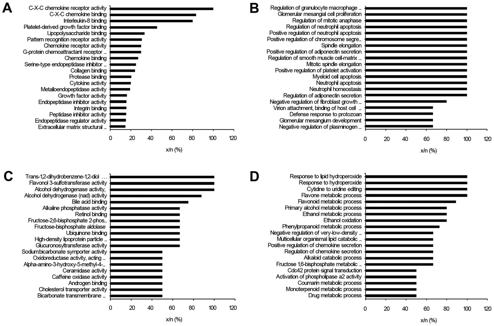
The functions of the upregulated genes of EGC
tissues were significantly associated with C-X-C and other
chemokine-related signaling, interleukin-8 binding, growth factor
binding, collagen binding, and the extracellular matrix (ECM)
(Fig. 1A). Moreover, the
biological processes involved in the upregulated genes in the EGC
tissues were strongly related to cell proliferation, mitosis,
apoptosis and cell-matrix adhesion (Fig. 1B). Additionally, wound healing
terms, cell migration terms and cell motility terms were also
listed in the upregulated genes with statistical significance
(Table III). Since cell migration,
cell motility and wound healing are typically observed in
late-stage, metastatic cancer, this may indicate that EGC tissues
could possess intrinsic aggressiveness, despite their early
detection. Conversely, the downregulated genes were strongly linked
to oxidoreductase activity (e.g., oxidoreductase activity acting on
the CH or CH2 groups, quinones) in GO molecular function (Fig. 1C). Furthermore, the downregulated
genes were enriched in various terms related to metabolic processes
(e.g., flavone and flavonoid metabolic pathways) in GO biological
processes (Fig. 1D). The GO terms
of the downregulated genes clearly indicate dysregulation of
metabolism in EGC, which is one of the emerging cancer hallmarks
(14).
| Table III.The GO biological process terms
associated with genes upregulated in gastric cancer tissues,
relating to wound healing, cell migration and cell motility. |
Table III.
The GO biological process terms
associated with genes upregulated in gastric cancer tissues,
relating to wound healing, cell migration and cell motility.
| GO-ID | P-value | Corrected
P-valuea | xb | nc | x/n (%) | Description |
|---|
| GO:0014910 | 1.54E-03 | 1.86E-02 | 4 | 14 | 29 | Regulation of
smooth muscle cell migration |
| GO:0061041 | 5.41E-07 | 3.15E-05 | 11 | 44 | 25 | Regulation of wound
healing |
| GO:0010595 | 4.41E-03 | 4.25E-02 | 5 | 29 | 17 | Positive regulation
of endothelial cell migration |
| GO:0030335 | 3.69E-07 | 2.38E-05 | 18 | 116 | 16 | Positive regulation
of cell migration |
| GO:2000147 | 3.69E-07 | 2.38E-05 | 18 | 116 | 16 | Positive regulation
of cell motility |
| GO:0030334 | 9.12E-07 | 4.44E-05 | 23 | 190 | 12 | Regulation of cell
migration |
| GO:2000145 | 1.44E-06 | 6.12E-05 | 23 | 195 | 12 | Regulation of cell
motility |
| GO:0048870 | 9.75E-10 | 1.81E-07 | 38 | 330 | 12 | Cell motility |
| GO:0042060 | 1.04E-10 | 3.09E-08 | 50 | 485 | 10 | Wound healing |
Pathway enrichment analysis
The DAVID tool (http://david.abcc.ncifcrf.gov/) was used to inspect
the KEGG biological pathways associated with the differently
expressed genes in EGC.
The upregulated genes in EGC tissues were
intrinsically associated with cytokine-cytokine receptor
interactions, ECM-receptor interactions, the cell cycle,
hematopoietic cell lineage and Toll-like receptor signaling
pathways (Table IV). In addition,
focal adhesion and cell adhesion molecule pathways were
highlighted. Thus, similar to the functional enrichment analysis,
upregulated pathways in these tumor tissues suggest a strong
potential for cell motility and metastasis, despite early
detection. In contrast, the downregulated genes in the EGC tissues
were strongly associated with xenobiotics-, drug-, retinol-,
starch- and sucrose-related metabolism, steroid hormone
biosynthesis, as well as pentose and glucuronate interconversion
pathways (Table IV).
| Table IV.Pathway enrichment analysis for up-
and downregulated genes in gastric cancer tissues. |
Table IV.
Pathway enrichment analysis for up-
and downregulated genes in gastric cancer tissues.
| Input genes | Pathways | Counta | P-value |
|---|
| Upregulated
pathways | hsa04060,
cytokine-cytokine receptor interaction | 38 | 3.61.E-11 |
| hsa04512,
ECM-receptor interaction | 17 | 3.16.E-07 |
| hsa04110, cell
cycle | 19 | 4.21.E-06 |
| hsa04640,
hematopoietic cell lineage | 13 | 2.40.E-04 |
| hsa04620, Toll-like
receptor signaling pathway | 14 | 3.02.E-04 |
| hsa04062, chemokine
signaling pathway | 20 | 3.18.E-04 |
| hsa04610,
complement and coagulation cascades | 11 | 5.85.E-04 |
| hsa04510, focal
adhesion | 19 | 2.00.E-03 |
| hsa04115, p53
signaling pathway | 10 | 2.11.E-03 |
| hsa04514, cell
adhesion molecules (CAMs) | 14 | 3.72.E-03 |
| hsa05222, small
cell lung cancer | 10 | 8.79.E-03 |
| hsa04670, leukocyte
transendothelial migration | 12 | 1.11.E-02 |
| hsa05020, prion
diseases | 6 | 1.51.E-02 |
| hsa04621, NOD-like
receptor signaling pathway | 8 | 1.53.E-02 |
| hsa05200, pathways
in cancer | 23 | 2.09.E-02 |
| hsa05332, graft vs.
host disease | 6 | 2.34.E-02 |
| hsa05322, systemic
lupus erythematosus | 10 | 2.40.E-02 |
| hsa05219, bladder
cancer | 6 | 3.13.E-02 |
| hsa04114, oocyte
meiosis | 10 | 4.32.E-02 |
| hsa04650, natural
killer cell mediated cytotoxicity | 11 | 5.53.E-02 |
| hsa04142,
lysosome | 10 | 5.97.E-02 |
| hsa04630, Jak-STAT
signaling pathway | 12 | 6.43.E-02 |
| hsa04914,
progesterone-mediated oocyte maturation | 8 | 7.17.E-02 |
| Downregulated
pathways | hsa00980,
metabolism of xenobiotics by cytochrome P450 | 22 | 3.33.E-18 |
| hsa00982, drug
metabolism | 22 | 7.29.E-18 |
| hsa00830, retinol
metabolism | 19 | 2.76.E-15 |
| hsa00140, steroid
hormone biosynthesis | 13 | 3.63.E-09 |
| hsa00500, starch
and sucrose metabolism | 11 | 2.00.E-07 |
| hsa00040, pentose
and glucuronate interconversions | 8 | 3.57.E-07 |
| hsa00590,
arachidonic acid metabolism | 12 | 3.95.E-07 |
| hsa00983, drug
metabolism | 10 | 2.69.E-06 |
| hsa00053, ascorbate
and aldarate metabolism | 7 | 5.02.E-06 |
| hsa00591, linoleic
acid metabolism | 8 | 1.04.E-05 |
| hsa00860, porphyrin
and chlorophyll metabolism | 8 | 3.32.E-05 |
| hsa00010,
glycolysis/gluconeogenesis | 10 | 4.64.E-05 |
| hsa00150, androgen
and estrogen metabolism | 8 | 7.27.E-05 |
| hsa03320, PPAR
signaling pathway | 7 | 1.41.E-02 |
| hsa00051, fructose
and mannose metabolism | 5 | 1.59.E-02 |
| hsa00330, arginine
and proline metabolism | 6 | 1.78.E-02 |
| hsa00071, fatty
acid metabolism | 5 | 2.74.E-02 |
| hsa00030, pentose
phosphate pathway | 4 | 3.42.E-02 |
| hsa00350, tyrosine
metabolism | 5 | 3.72.E-02 |
| hsa00920, sulfur
metabolism | 3 | 4.53.E-02 |
| hsa00340, histidine
metabolism | 4 | 5.00.E-02 |
| hsa00480,
glutathione metabolism | 5 | 5.54.E-02 |
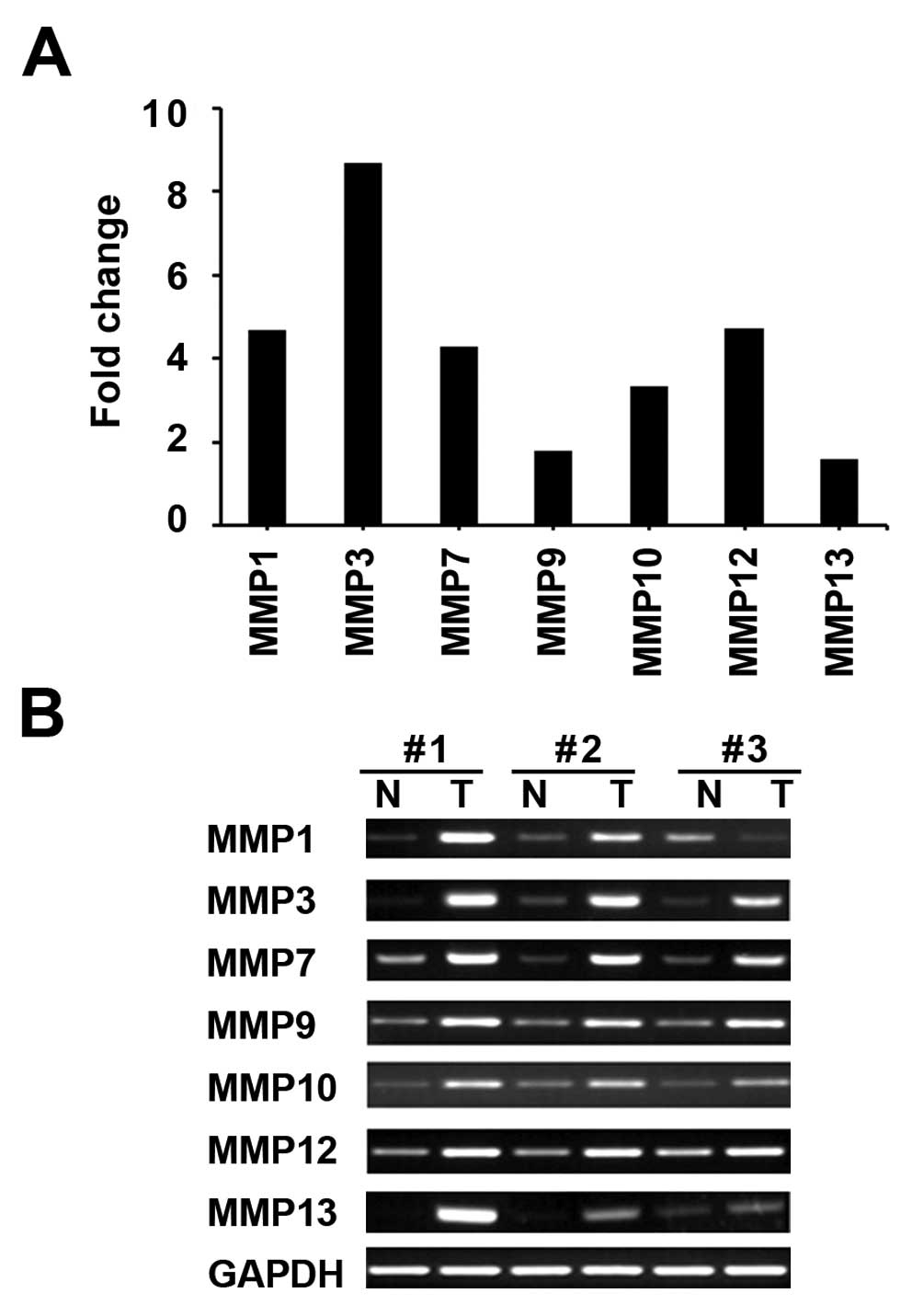
The expression of MMPs in EGC tissue
Our functional and pathway analyses demonstrated
that the significantly upregulated genes in EGC tissues are
associated with cell migration and metastasis, events typical of
late-stage cancer. To verify our findings, we further analyzed the
expression pattern of matrix metalloproteinases (MMPs), which are
well known cell migration-related genes. MMPs are also known to
play critical roles in the regulation of cell invasion by ECM
proteolysis, as well as by processing cytokine precursors in the
chemokine network.
We analyzed the expression pattern of 7 MMPs (MMP1,
−3, −7, −9, −10, −12 and −13) within the upregulated gene data in
EGC tissues. As expected, and consistent with the microarray data
where MMPs were upregulated 1.56- to 8.68-fold (Fig. 2A), RT-PCR indicated that MMP mRNA
expression was highly upregulated in the patients’ EGC tissues
(Fig. 2B).
Cross-experimental analysis
In order to investigate similar molecular signatures
between EGC and other cancer types, we compared our data of
differentially expressed genes with a public gene expression
signature warehouse, L2L. This revealed that the upregulated genes
in EGC most significantly correlated with the gene expression
signature of ERα-negative breast cancer (Table V). As summarized in Table V, the upregulated genes in EGC were
also similar to the gene expression signature related to an
undifferentiated cancer status (cancer_undifferentiated_meta_up: 69
genes commonly upregulated in undifferentiated cancer relative to
well-differentiated cancer, from a meta-analysis of the OncoMine
gene expression database), stemness (stemcell_embryonic_up:
enriched in mouse embryonic stem cells, compared to differentiated
brain and bone marrow cells) and survival (dox_resist_gastric_up:
upregulated in gastric cancer cell lines resistant to doxorubicin,
compared to parent chemosensitive lines). Together, the EGC tissues
reflect various facets of cancer-related phenotypes, viz., strong
survival, stem-like and morphology.
| Table V.The selected L2L results of genes up-
or downregulated in early gastric cancer. |
Table V.
The selected L2L results of genes up-
or downregulated in early gastric cancer.
| Input genes | L2L term (PubMed
ID) | Description | xa/nb | Binomial
P-value |
|---|
| Upregulated genes
in the gastric cancer tissues | brca_er_neg
(11823860) | Genes whose
expression is consistently negatively correlated with the gastric
cancer tissues estrogen receptor status in breast cancer - higher
expression is associated with ER-negative tumors | 145/996 | 7.50E-95 |
|
cancer_undifferentiated_meta_up
(15184677) | Sixty-nine genes
commonly upregulated in undifferentiated cancer relative to
well-differentiated cancer, from a meta-analysis of the OncoMine
gene expression database | 25/69 | 2.21E-28 |
|
dox_resist_gastric_up (14734480) | Upregulated in
gastric cancer cell lines resistant to doxorubicin, compared to
parent chemosensitive lines | 17/48 | 1.44E-19 |
|
stemcell_embryonic_up (12228720) | Enriched in mouse
embryonic stem cells, compared to differentiated brain and bone
marrow cells | 74/1350 | 1.32E-21 |
| Downregulated genes
in the gastric cancer tissues | 5azac_hepg2_up
(16854234) | Upregulated in
human hepatoma cells (HepG2) following 48 of treatment with 2.5
μM 5-aza-2-deoxycytidine (5azaC) | 60/1311 | 6.61E-20 |
| 5azac-tsa_hepg2_up
(16854234) | Upregulated in
human hepatoma cells (HepG2) following 24 h of treatment with 2.5
μM 5-aza-2-deoxycytidine (5azaC) and 24 h of treatment with
both 5azaC and 500 nM trichostatin A (TSA) | 66/1619 | 3.69E-19 |
The same L2L analysis was applied to the
downregulated genes in EGC (Table
V). Interestingly, epigenetic-related cancer gene expression
signature terms (5azac_hepg2_up and 5azac-tsa_hepg2_up in Table V) were highly ranked. This suggests
that global alterations in DNA methylation and histone modification
occur in EGCs, as it does in other cancers.
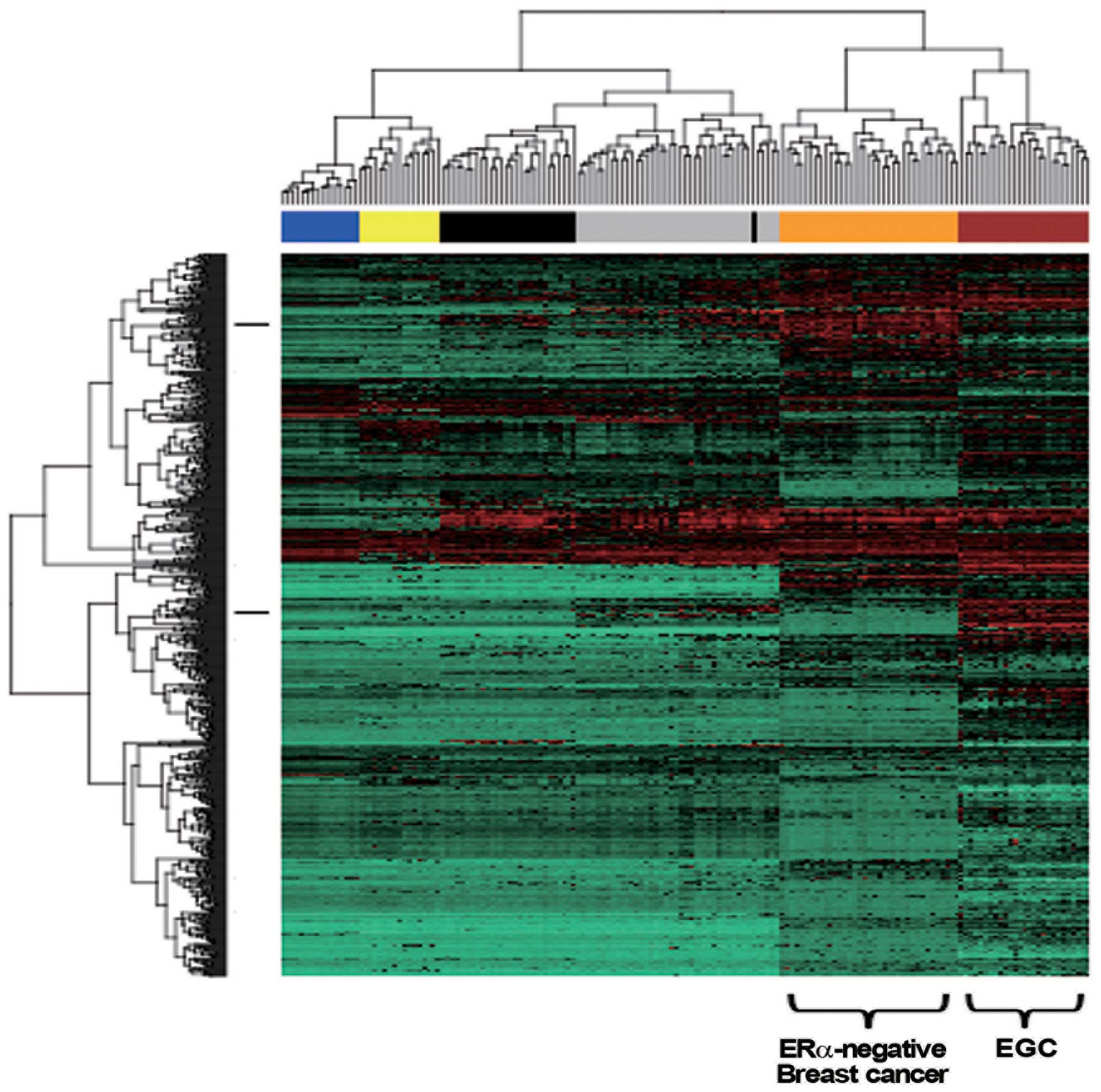
Hierarchical clustering of the EGC
tissues and other cancers
To validate the result of the L2L analysis showing a
relationship between EGC and ERα-negative breast cancer, we
performed a hierarchical clustering analysis. The expression
datasets of the differently expressed genes in EGC (556 upregulated
gene symbols and 417 downregulated gene symbols), ERα-negative
breast cancer and 4 additional cancers (small cell lung cancer,
LNCaP prostate cancer cell lines, bladder cancer and Ewing sarcoma)
were used in an unsupervised hierarchical clustering analysis. As
in the L2L analysis, the results indicated that EGC correlated most
closely with the ERα-negative cancer than with the other 4 cancers
(Fig. 3).
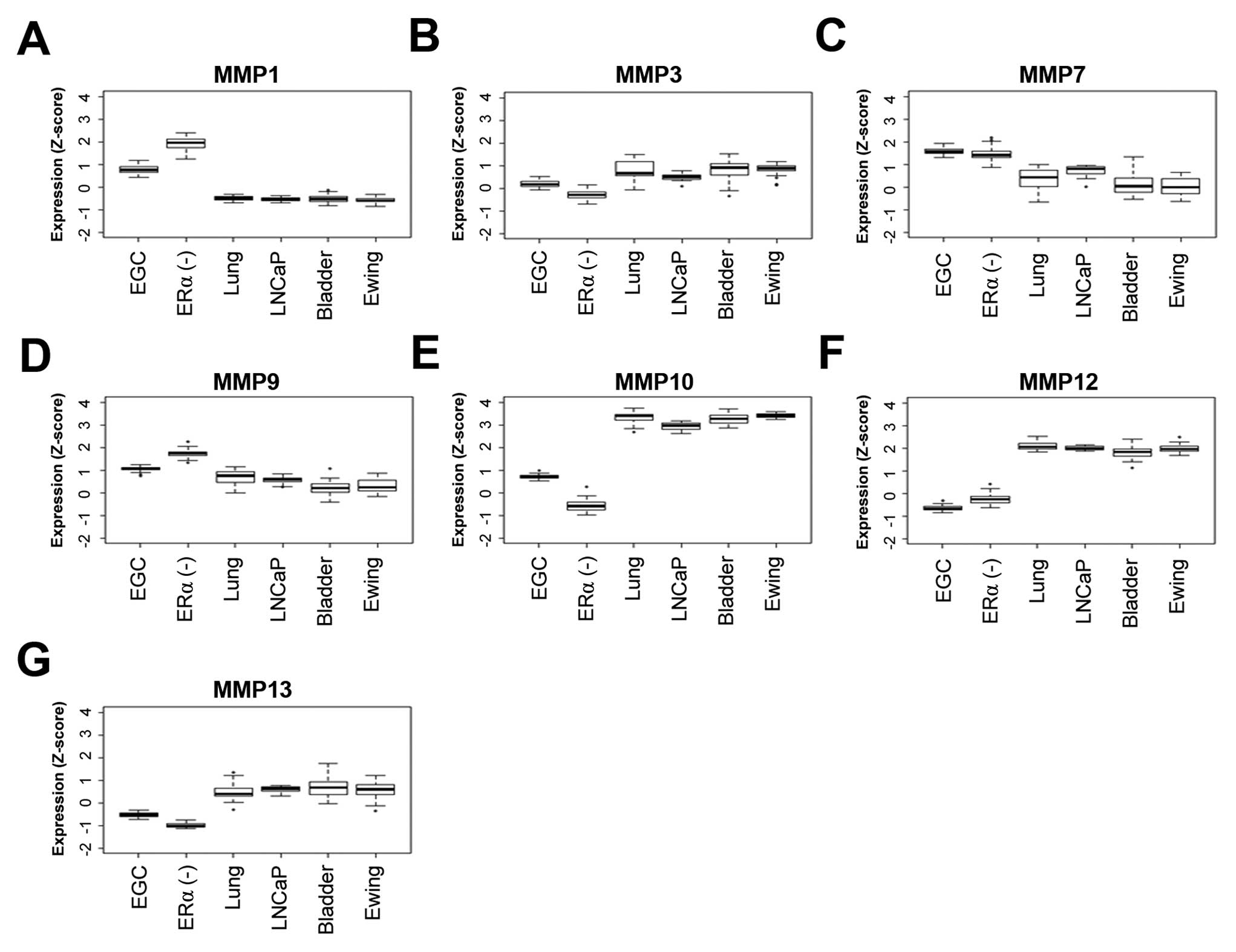
When we inspected the expression levels (z-scores)
of the 7 MMP genes (Fig. 4), the
results indicated that the ERα-negative cancer, above all other
observed cancers, showed the most similar expression patterns for
the 7 MMPs. Overall, the hierarchical clustering was consistent
with the cross-experimental analysis and strongly supported the
molecular similarity between EGC and ERα-negative breast cancer in
terms of carcinogenesis.
Discussion
We analyzed the microarray data generated from pairs
of tumor tissue and their adjacent non-cancerous tissue, obtained
from 27 EGC patients. The gene expression data were subjected to
functional and pathway analyses, as well as gene expression
signature comparison (cross-experiment analysis). This led to 2
novel findings: i) the functional and pathway analyses suggested
that metastasis-related biological processes may already be highly
expressed even in the early stage of gastric cancer, and ii) the
gene expression pattern of EGC is closely aligned to that of
ERα-negative breast cancer.
We also compared the differentially expressed genes
in our EGC tissues with other 3 previously published gene
expression studies (2,7,8). We
found that the upregulated genes in our study significantly
overlapped with the upregulated genes in the EGC groups of the 3
earlier studies (2,7,8),
under a randomization model (Table
VI). Recently, Vecchi et al suggested a carcinogenesis
model (2) in which the transition
from normal mucosa to EGC is accompanied by cell cycle
upregulation; our pathway analysis results (hsa04110, cell cycle in
Table IV) is consistent with this
model. Interestingly, AGC functions (cell migration-and ECM-related
functions), suggested by the Vecchi model were also revealed in our
EGC data, again indicating that EGC actually harbors gene
expression events that are usually observed in the later stages of
cancer, such as AGC.
| Table VI.Comparisons with other GC sources in
terms of upregulated EGC-related genes. |
Table VI.
Comparisons with other GC sources in
terms of upregulated EGC-related genes.
| Refs. | EGC
classification | na | xb | Significance
(P-value)c |
|---|
| (2) | - | 488 | 83 | <2.2E-16 |
| (7) | Well-differentiated
(WD) and moderately differentiated (MD) | 170 | 62 | <2.2E-16 |
| (8) | AJCC staging I and
II (TNM staging) | 118 | 15 | 1.601E-06 |
Based on our functional and pathway analyses, the
upregulated genes in the EGC tissues were highly enriched for genes
involved in cell proliferation, chemokine/growth factor signaling
and cell migration. The computational implication is, in fact,
closely related to MMP activity, as MMP substrates include growth
factor/chemokine precursors and E-cadherin (15,16).
We validated the upregulation of the 7 MMPs in the EGC tissues by
RT-PCR. This result suggests that the activation of multiple MMPs
may be involved in the early stage of cancer. The suggestion is
noteworthy, when considering that the roles of multiple MMPs were
mainly reported in late-stage gastric cancer (2,17).
It is also interesting to note that 6 (MMP1, −3, −7, −10, −12 and
−13) of the 7 MMPs are clustered at 11q22, implying that epigenetic
events could be involved in the upregulation of the clustered MMPs
(18).
Additionally, we found that the gene expression
pattern in EGC tissues resembles the pattern of the ERα-negative
breast cancer transcriptome. Since ERα-negative breast cancer
clusters with EGC (Fig. 3), the
similarity suggests that these two cancers may share common
molecular features. Recent breast cancer studies (19,20)
reported that high expression of cyclooygenase-2 (Cox-2), encoded
by PTGS2, is associated with poor survival in ERα-negative breast
cancer patients, when compared to ERα-positive breast cancers.
Interestingly, Cox-2 is highly involved in the
inflammation-associated carcinogenesis of the gastrointestinal
tract. In particular,
H. pylori-infected gastric epithelial cells
can experience malignant transformation via Toll-like receptor
(TLR) signaling that induces Cox-2, followed by activation of cell
proliferation (21). In fact, our
pathway analysis in EGC showed upregulation of the KEGG TLR
signaling pathway and cell cycle pathway (Table IV, Fig. 5). Our EGC also showed a markedly
increased expression of PTGS2 (5.74-fold-change). Thus, the
similarity between EGC and ERα-negative breast cancer may come from
identical subsets of immune response-related signaling between the
microenvironments of the tumors.
In conclusion, we have analyzed the differentially
expressed genes in EGC patients using an integrative systematic
approach. We found that genes highly expressed in EGC are involved
in cell migration- and metastasis-related functions typically
observed in late-stage cancer. Also, EGC may be intrinsically
similar to ERα-negative breast cancer, by sharing immune-related
signaling events, which is further dissected in both cancer types.
The functional roles of the downregulated genes in EGC
carcinogenesis remain to be elucidated in future.
Acknowledgements
This study was funded by Intramural
Research Grants of the National Cancer Center (NCC 1210460-1).
References
|
1.
|
Parkin DM, Bray F, Ferlay J and Pisani P:
Global cancer statistics, 2002. CA Cancer J Clin. 55:74–108. 2005.
View Article : Google Scholar
|
|
2.
|
Vecchi M, Nuciforo P, Romagnoli S, et al:
Gene expression analysis of early and advanced gastric cancers.
Oncogene. 26:4284–4294. 2007. View Article : Google Scholar : PubMed/NCBI
|
|
3.
|
Crew KD and Neugut AI: Epidemiology of
gastric cancer. World J Gastroenterol. 12:354–362. 2006.
|
|
4.
|
Nam SY, Choi IJ, Park KW, et al: Effect of
repeated endoscopic screening on the incidence and treatment of
gastric cancer in health screenees. Eur J Gastroenterol Hepatol.
21:855–860. 2009. View Article : Google Scholar : PubMed/NCBI
|
|
5.
|
Takeno A, Takemasa I, Doki Y, et al:
Integrative approach for differentially overexpressed genes in
gastric cancer by combining large-scale gene expression profiling
and network analysis. Br J Cancer. 99:1307–1315. 2008. View Article : Google Scholar
|
|
6.
|
Yamada Y, Arao T, Gotoda T, et al:
Identification of prognostic biomarkers in gastric cancer using
endoscopic biopsy samples. Cancer Sci. 99:2193–2199. 2008.
View Article : Google Scholar : PubMed/NCBI
|
|
7.
|
Cui J, Li F, Wang G, Fang X, Puett JD and
Xu Y: Gene-expression signatures can distinguish gastric cancer
grades and stages. PLoS One. 6:e178192011. View Article : Google Scholar : PubMed/NCBI
|
|
8.
|
Economescu MC, Necula LG, Dragu D, et al:
Identification of potential biomarkers for early and advanced
gastric adenocarcinoma detection. Hepatogastroenterology.
57:1453–1464. 2010.PubMed/NCBI
|
|
9.
|
Maere S, Heymans K and Kuiper M: BiNGO: a
cytoscape plugin to assess overrepresentation of gene ontology
categories in biological networks. Bioinformatics. 21:3448–3449.
2005. View Article : Google Scholar : PubMed/NCBI
|
|
10.
|
Kanehisa M, Goto S, Furumichi M, Tanabe M
and Hirakawa M: KEGG for representation and analysis of molecular
networks involving diseases and drugs. Nucleic Acids Res.
38:D355–D360. 2010. View Article : Google Scholar : PubMed/NCBI
|
|
11.
|
Newman JC and Weiner AM: L2L: a simple
tool for discovering the hidden significance in microarray
expression data. Genome Biol. 6:R812005. View Article : Google Scholar : PubMed/NCBI
|
|
12.
|
Enerly E, Steinfeld I, Kleivi K, et al:
miRNA-mRNA integrated analysis reveals roles for miRNAs in primary
breast tumors. PLoS One. 6:e169152011. View Article : Google Scholar : PubMed/NCBI
|
|
13.
|
Lukk M, Kapushesky M, Nikkila J, et al: A
global map of human gene expression. Nat Biotechnol. 28:322–324.
2010. View Article : Google Scholar
|
|
14.
|
Hanahan D and Weinberg RA: Hallmarks of
cancer: the next generation. Cell. 144:646–674. 2011. View Article : Google Scholar : PubMed/NCBI
|
|
15.
|
Roy R, Yang J and Moses MA: Matrix
metalloproteinases as novel biomarkers and potential therapeutic
targets in human cancer. J Clin Oncol. 27:5287–5297. 2009.
View Article : Google Scholar : PubMed/NCBI
|
|
16.
|
Foley J, Nickerson NK, Nam S, et al: EGFR
signaling in breast cancer: bad to the bone. Semin Cell Dev Biol.
21:951–960. 2010. View Article : Google Scholar : PubMed/NCBI
|
|
17.
|
Murray GI, Duncan ME, Arbuckle E, Melvin
WT and Fothergill JE: Matrix metalloproteinases and their
inhibitors in gastric cancer. Gut. 43:791–797. 1998. View Article : Google Scholar : PubMed/NCBI
|
|
18.
|
Clark IM, Swingler TE, Sampieri CL and
Edwards DR: The regulation of matrix metalloproteinases and their
inhibitors. Int J Biochem Cell Biol. 40:1362–1378. 2008. View Article : Google Scholar : PubMed/NCBI
|
|
19.
|
Glynn SA, Prueitt RL, Ridnour LA, et al:
COX-2 activation is associated with Akt phosphorylation and poor
survival in ER-negative, HER2-positive breast cancer. BMC Cancer.
10:6262010. View Article : Google Scholar : PubMed/NCBI
|
|
20.
|
Witton CJ, Hawe SJ, Cooke TG and Bartlett
JM: Cyclooxygenase 2 (COX2) expression is associated with poor
outcome in ER-negative, but not ER-positive, breast cancer.
Histopathology. 45:47–54. 2004. View Article : Google Scholar : PubMed/NCBI
|
|
21.
|
Fukata M and Abreu MT: Role of Toll-like
receptors in gastrointestinal malignancies. Oncogene. 27:234–243.
2008. View Article : Google Scholar : PubMed/NCBI
|