Introduction
Recently, the US National Toxicology Program (NTP)
released results on the toxicology and carcinogenicity of
radiofrequency (RF) radiation in rats and mice, as further
discussed below. This initiated this article for the comparison of
earlier human epidemiological studies with the NTP the findings,
including a short review of animal studies.
NTP is an interagency program established in 1978 to
coordinate toxicology research and testing across the Department of
Health and Human Services. The program was also created to
strengthen the science base in toxicology, develop and validate
improved testing methods, and provide information about potentially
toxic chemicals to health regulatory and research agencies,
scientific and medical communities, and the public. NTP is
headquartered at the National Institute of Environmental Health
Sciences (NIEHS) (https://ntp.niehs.nih.gov/about/org/index.html).
The brain is the main target of the exposure to RF
radiation during the use of handheld wireless phones; both mobile
and cordless phones (1,2). Thus, an increased risk of developing
brain tumors has long been a cause for concern.
Our study group has since the end of the 1990s
published results from case-control studies on use of wireless
phones and brain tumor risk (3). A
statistically significant increased risk for ipsilateral use of
mobile phones, the same side of the brain as the phone was used,
was published for malignant brain tumors (4) and vestibular schwannoma (5). Further scientific evidence on the
association has more recently been discussed by Carlberg and
Hardell (6).
In May, 2011 the International Agency for Research
on Cancer (IARC) concluded that radiofrequency (RF) radiation in
the frequency range 30 kHz-300 GHz is a ‘possible’ human carcinogen
Group 2B (7,8). The classification was based primarily
on evidence that long-term users of wireless phones (mobile and
cordless phones) have an increased risk for glioma and acoustic
neuroma. One major reason that the rating was not a ‘probable’ or a
‘known’ risk was the lack of clear evidence from animal studies.
IARC at the World Health Organization (WHO) is independently
financed and has its own governing and scientific councils, which
WHO staff only attend as observers (http://www.who.int/ionizing_radiation/research/iarc/en/).
Unfortunately, WHO itself has constantly refused to
acknowledge the carcinogenicity of RF radiation. In fact, WHO seems
to rely on the conclusion of the non-governmental organization
International Commission on Non-ionizing Radiation Protection
(ICNIRP) instead of the IARC evaluation. That organization is even
declared to be their in-house experts (9,10).
ICNIRP is a private non-governmental organisation (NGO) based in
Germany. New expert members can only be elected by members of the
organization. Many of the ICNIRP members have ties to the industry
that are dependent on the ICNIRP guidelines (11). This creates a conflict of interest,
since the former leader of the WHO International Electromagnetic
Field (EMF) Project is also the founder and honorary member of the
ICNIRP (11). The guidelines are
of huge economic and strategic importance to the military,
telecom/IT and power industry. These circumstances are further
discussed in a recent publication (12).
The IARC cancer classification includes all sources
of RF radiation. The exposure from mobile phone base stations, DECT
base stations, Wi-Fi access points, smart phones, laptops and
tablets can be long-term, sometimes around the clock, at home, at
the work place, at school and in the environment. For children,
this risk may be accentuated due to a cumulative effect during a
long lifetime use (13).
The exposure guidelines used by many agencies and
countries were established in 1998 by the ICNIRP and were based
only on established short-term thermal (heating) effects from RF
radiation neglecting non-thermal biological effects (14). ICNIRP provides the guideline of 2
to 10 W/m2 for RF radiation, depending on the frequency.
The ICNIRP guidelines were updated in 2009; however, they still do
not cover cancer and other long-term or non-thermal effects
(15) [see also Hardell (10)].
In contrast to the ICNIRP, the BioInitiative Reports
from 2007 and 2012 based the evaluation also on the non-thermal
health effects from RF radiation (16,17).
The scientific benchmark for possible health risks was defined to
be 30 to 60 µW/m2. In 2012, the Bioinitiative
Working Group proposed a precautionary target level of 3-6
µW/m2, using a safety factor of 10. Using the
significantly higher guideline by ICNIRP gives a ‘green card’ to
roll out the wireless digital technology, thereby not considering
non-thermal health effects from RF radiation.
The evidence of RF radiation as a carcinogen was
confirmed when NTP released preliminary results of a study of
long-term exposure of rats and mice to cell phone radiation
(18). An increased incidence of
glioma in the brain and malignant schwannoma in the heart was
found. The NTP study has now been published online for public
consultations (19,20) and is discussed below in relation to
human epidemiological studies.
Background: Evidence from previous animal
studies
There are several earlier animal studies that
demonstrate the carcinogenic potential of RF radiation. Szmigielski
et al already in 1982 published a study on the
co-carcinogenic effects of RF radiation exposure and benzopyrene in
mice (21). Cancer promotion was
found for 2,450 MHz RF radiation at either 50 or 150
W/m2. The results revealed an acceleration of
spontaneous and chemically-induced cancers.
Non-thermal 2,450 MHz continuous-wave RF radiation
has been shown to cause a biphasic effect on glioma cells (22) and lymphocytes (23). Cell proliferation was found at a
specific absorption rate (SAR) of ≤50 W/kg, whereas a higher SAR
suppressed DNA and RNA synthesis.
SAR ranged from 0.144 to 0.4 W/kg depending on the
rats' weight in a study from 1992 on 200 rats exposed to 2,450 MHz
pulsed RF radiation 21.5 h per day for 25 months (24). Compared with 200 sham-exposed rats,
a statistically significant increased incidence of primary
malignant diseases was found in exposed animals. Among the
malignancies found in the exposed rats were malignant lymphoma and
thyroid cancer. These findings are of interest since SAR values in
the study were rather low compared to the ICNIRP guideline on SAR 2
W/kg to the brain for use of mobile phones (14).
A total of 100 mice were sham-exposed and 101 were
exposed for two 30-min periods per day for up to 18 months to 900
MHz pulsed RF radiation with power densities 2.6-13 W/m2
(SAR 0.008-4.2 W/kg, averaging 0.13-1.4 W/kg). The mice carried a
lymphomagenic oncogene and their risk of developing lymphoma was
found to be statistically significantly higher in the exposed mice
than in the controls (25).
The same results were not found in the study by
Utteridge et al (26) that
has been criticized as it was not a replication study. However, the
findings on lymphoma risk by Repacholi et al (25) and Chou et al (24) are of relevance in relation to the
indications of an increased risk of non-Hodgkin lymphoma (NHL) in
human epidemiological studies on the use of wireless phones. Thus,
a statistically significant increased risk of T-Cell NHL was found
in one study (27). In another
study, NHL not otherwise specified was statistically significantly
increased among subjects with ≥6 years duration [odds ratio (OR)
=4.4 in men] for mobile phone use (28), although based on low numbers
(n=7).
The thyroid gland is among the organs with the
highest exposure to RF radiation during the use of the handheld
wireless phone, particularly smartphones (29,30).
The finding of thyroid cancer risk in the study by Chou et
al (24), and the sharp
increase in the incidence of thyroid cancer in humans during recent
years (31) are of interest in
that context.
In another study, mice were exposed to universal
mobile telecommunications system (UMTS) fields with intensities of
0 (sham), 4.8 and 48 W/m2 up to 24 months (32). The low-dose group, exposed to 4.8
W/m2, was subjected to additional prenatal
ethylnitrosourea (ENU) treatment. That group showed an increased
lung tumor rate and an increased incidence of lung carcinomas as
compared to the controls treated with ENU only. This indicated a
cocarcinogenic effect of a lifelong UMTS exposure in female mice
pretreated with ENU (32).
In a follow-up study, mice were exposed to RF
radiation: 0 (sham), 0.04, 0.4 and 2 W/kg SAR (33). The numbers of tumors of the lungs
and livers in exposed animals were statistically significantly
higher than in sham-exposed controls, and the numbers of malignant
lymphoma were also higher. A tumor-promoting effect of RF radiation
was found at low to moderate levels (0.04 and 0.4 W/kg SAR), well
below the ICNIRP exposure limits for users of mobile phones
(33).
The study by the Ramazzini Institute is the largest
long-term study ever performed on the health effects of RF
radiation, including 2,448 rats (34). Male and female Sprague-Dawley rats
were exposed from prenatal life until natural death to a 1.8 GHz
global system for mobile communication (GSM) far field of 0, 5, 25,
50 V/m with a whole-body exposure for 19 h/day. A statistically
significant increase in the incidence of malignant Schwannoma in
the heart was found in male rats at the highest dose, 50 V/m,
corresponding to 0.66 mW/cm2 and whole-body SAR of 0.1
W/Kg. An increased incidence of heart Schwann cell hyperplasia was
observed in treated male and female rats at the highest dose (50
V/m), but was not statistically significant. In treated female rats
at the highest dose (50 V/m), the incidence of malignant glial
tumors was increased, although this was not statistically
significant. The study revealed an increased incidence of tumor
types similar to those associated with the use of wireless phones,
glioma and acoustic neuroma, in human epidemiological studies.
The NTP study provides additional confirmation of
the carcinogenicity of RF radiation (19,20).
They showed an increased incidence of malignant schwannoma in the
heart and brain glioma in male rats exposed either to GSM-modulated
or code division multiple access (CDMA)-modulated cell phone RF
radiation for two years. There are also increased incidences of
some other tumor types and diseases. Below we discuss some of the
major findings.
The results on schwannoma and glioma are of
particular concern since they corroborate human epidemiological
findings. Thus, it is noteworthy that similar tumors were found in
the NTP study as in epidemiological studies on the human use of
wireless phones; mobile phones or cordless phones (DECT). Malignant
schwannoma in the heart is a similar type of tumor as vestibular
schwannoma in humans, also known as acoustic neuroma, although
acoustic neuroma is usually benign and rarely undergoes malignant
transformation.
Below, we provide an updated evaluation of the
scientific evidence of an increased risk of developing glioma and
vestibular schwannoma (acoustic neuroma) associated with the use of
wireless phones. It is pertinent to provide an updated presentation
of the NTP reports on current evidence on cancer risks associated
with the use of wireless phones.
Since the IARC evaluation in 2011, more human
epidemiological studies have been published that support a causal
association between RF radiation and brain and head tumors. A
Danish cohort study on ‘mobile phone users’ (35,36)
is not included herein due to serious methodological shortcomings
in the study design [see Söderqvist et al (37)]. The study by Benson et al
(38) is of limited value since
the use of cordless phones was not included, mobile phone use was
assessed only at baseline and no information on tumor laterality,
including ipsilateral versus contralateral use was given. In spite
of the many shortcomings, an increased risk of developing acoustic
neuroma was reported. The study will not be further discussed
below.
In the following, first, human epidemiological
studies on specific tumor types are discussed. The NTP study
findings are then presented and finally, an evaluation of the
combined evidence from human and animal studies is presented.
Glioma
Human studies
Glioma is the most common malignant brain tumor and
represents approximately 60% of all central nervous system (CNS)
tumors. Most of these are astrocytic tumors divided into low-grade
(WHO grades I-II) and high-grade (WHO grades III-IV). The most
common glioma type is glioblastoma multiforme (WHO grade IV) with a
peak incidence in the age group of 45-75 years and a median
survival less than one year (39).
No substantial increasing survival has been obtained in recent
years. Three research groups have provided results in case-control
studies on glioma, Interphone (40), Coureau et al (41) and the Hardell group in Sweden
(42-46).
The random effects model was used for a
meta-analyses of published studies, based on the test for
heterogeneity in the overall group (‘all mobile’), see also
http://www.bioinitiative.org/report/wp-content/uploads/2017/11/Hardell-2017-Sec11-Update-Use_of_Wireless_Phones.pdf.
Note that only our group also assessed the use of cordless phones.
Thus, the reference category in our studies included cases and
controls with no use of wireless phones, in contrast to the other
studies investigating only mobile phone use. Including cordless
phone use in the ‘unexposed’ group would bias the risk estimates
towards unity (45).
In Table I, results
of the highest cumulative use in hours of mobile phones are
presented. All studies reported a statistically significantly
increased risk of developing glioma and the meta-analysis yielded
OR =1.90 and 95% confidence interval (CI) =1.31-2.76. For
ipsilateral mobile phone use, the risk increased further to OR
=2.54, 95% CI =1.83-3.52 in the meta-analysis based on 247 exposed
cases and 202 exposed controls. Further support of the increased
risk of glioma associated with mobile phone use has been obtained
in additional analyses of parts of the Interphone study (47-49).
 | Table INumbers of exposed cases (Ca) and
controls (Co) and odds ratio (OR) with 95% confidence interval (CI)
for glioma in case-control studies in the highest category of
cumulative use in hours for mobile phone use. |
Table I
Numbers of exposed cases (Ca) and
controls (Co) and odds ratio (OR) with 95% confidence interval (CI)
for glioma in case-control studies in the highest category of
cumulative use in hours for mobile phone use.
| Study (ref.) | All
| Ipsilateral
|
|---|
| Ca/Co | OR | 95% CI | Ca/Co | OR | 95% CI |
|---|
| Interphone, 2010
(40) | 210/154 | 1.40 | 1.03-1.89 | 100/62 | 1.96 | 1.22-3.16 |
| Cumulative use
≥1,640 h |
| Coureau et
al, 2014 (41) | 24/22 | 2.89 | 1.41-5.93 | 9/7 | 2.11 | 0.73-6.08 |
| Cumulative use
>896 h |
| Hardell and
Carlberg, 2015 (43) | 211/301 | 2.13 | 1.61-2.82 | 138/133 | 3.11 | 2.18-4.44 |
| Cumulative use
≥1,640 h |
| Meta-analysis
(40,41,43) | 445/477 | 1.90 | 1.31-2.76 | 247/202 | 2.54 | 1.83-3.52 |
| Cumulative use
≥1,640 ha |
We previously analyzed the survival of the patients
in our studies and found a shorter survival in patients with
glioblastoma multiforme associated with the use of wireless phones
compared with patients with no use (50). Interestingly, the mutation of the
p53 gene involved in disease progression has been reported in
glioblastoma multiforme in patients using mobile phones for ≥3 h
per day. The mutation was statistically significantly associated
with a shorter overall survival time (51).
NTP study
No increased incidence of glioma was reported in the
mouse study (20).
In male rats (19),
malignant glioma and glia cell hyperplasia occurred in all groups
exposed to GSM-modulated cell phone RF radiation for two years. No
lesions were observed in the sham controls. In female rats, glial
cell hyperplasia occurred in one rat (3 W/kg), but none in the sham
controls. One malignant glioma occurred in one rat in the 6 W/kg
group but none in the sham controls.
In male rats exposed to CDMA-modulated cell phone RF
radiation for two years, there was an increased incidence of
malignant glioma with a statistically significant trend, P=0.044.
In females, three malignant glioma occurred in the 1.5 W/kg group,
but none in the other exposed groups or the sham control (P-value
for trend =0.384). Glial cell hyperplasia was observed in most
exposed groups, although this was not statistically significant
(noted in text; P-value for trend not presented in NTP table).
Evaluation
Based on human epidemiological studies supported by
the NTP animal study, there is clear evidence that RF radiation
causes glioma in humans. There is also evidence of an increased
glioma risk in occupational studies on exposure to EMF (52-54).
Meningioma
Human studies
Meningioma is an encapsulated, well-demarked and
rarely malignant tumor. It is the most common non-malignant brain
tumor that accounts for approximately 30% of intracranial
neoplasms. It develops from the pia and arachnoid membranes that
cover the CNS. It is slow-growing and presents neurological
symptoms by the compression of adjacent structures. Most common are
headaches and seizures. The incidence is greater than two-fold
higher in women than in men and meningioma develops mostly among
middle-aged and older individuals (55). The same research groups as for
glioma also included meningioma in their case-control studies with
a separate publication on meningioma by Carlberg and Hardell
(56). The results of the
meta-analyses for cumulative exposure in highest exposure category
are presented in Table II. A
statistically significant increased risk was obtained for
ipsilateral mobile phone use with OR =1.49, 95% CI =1.08-2.06.
| Table IINumbers of exposed cases (Ca) and
controls (Co) and odds ratio (OR) with 95% confidence interval (CI)
for menin-gioma in case-control studies in the highest category of
cumulative use in hours for mobile phone use. |
Table II
Numbers of exposed cases (Ca) and
controls (Co) and odds ratio (OR) with 95% confidence interval (CI)
for menin-gioma in case-control studies in the highest category of
cumulative use in hours for mobile phone use.
| Study (ref.) | All
| Ipsilateral
|
|---|
| Ca/Co | OR | 95% CI | Ca/Co | OR | 95% CI |
|---|
| Interphone, 2010
(40) | 130/107 | 1.15 | 0.81-1.62 | 46/35 | 1.45 | 0.80-2.61 |
| Cumulative use
≥1,640 h |
| Coureau et
al, 2014 (41) | 13/9 | 2.57 | 1.02-6.44 | 6/4 | 2.29 | 0.58-8.97 |
| Cumulative use
>896 h |
| Carlberg and
Hardell, 2015 (56) | 141/301 | 1.24 | 0.93-1.66 | 67/133 | 1.46 | 0.98-2.17 |
| Cumulative use
≥1,640 h |
| Meta-analysis
(40,41,56) | 284/417 | 1.27 | 0.98-1.66 | 119/172 | 1.49 | 1.08-2.06 |
| Cumulative use
≥1,640 ha |
NTP study
No increased incidence of meningioma was reported in
rats or mice (19,20).
Granular cell tumors (GCTs)
Human studies
GCTs are uncommon tumors. They are believed to be of
neuronal origin (57). They are
soft tissue tumors, which are thought to be derived from Schwann
cells (58). The immunoprofile of
granular cell tumors has revealed nerve sheath differentiation,
lending support to their neuronal origin (59). GCTs can affect any organ in the
body, although approximately 50% are found in the head and neck
region (60). In our case-control
studies on brain tumors, all diagnoses were based on a
histopathological examination; no one was diagnosed with a granular
cell tumor (42-46).
NTP study
In the rat study (19), increased incidence of malignant or
non-malignant granular cell tumors in the meninges, likely derived
from Schwann cells, occurred in the males exposed to GSM-modulated
cell phone RF radiation for two years. This was not statistically
significant (P-value for trend =0.343). In female rats, granular
cell tumors, either malignant or non-malignant were not associated
with RF radiation (P-value for trend =0.594). Since GCT is neuronal
in origin, the NTP study findings in male rats add to the evidence
that exposure to RF radiation damage nerve sheaths.
Evaluation
Based on human epidemiological studies and the NTP
animal study, there is equivocal evidence that RF radiation causes
meningeal tumors in humans (may be related to exposure).
Rate/incidence of brain tumors
The Swedish Cancer Register has not shown increasing
incidence of brain tumors in a study for the time period between
1979-2008, and has been used to dismissing epidemiological evidence
on risk associated with use of wireless phones (61). We have previously demonstrated that
descriptive studies cannot be used to dismiss results in analytical
epidemiology with individual exposure histories, such as in
case-control studies. We have also published the deficiencies in
the reporting of brain tumors to the Swedish Cancer Register
(62). The results for more recent
time periods have now been published. These articles also discuss
results from studies in other countries.
We used the Swedish National Inpatient Register
(IPR) and Causes of Death Register (CDR) to study the incidence of
brain tumors comparing with the Swedish Cancer Register data for
the time period between 1998-2013 using joinpoint regression
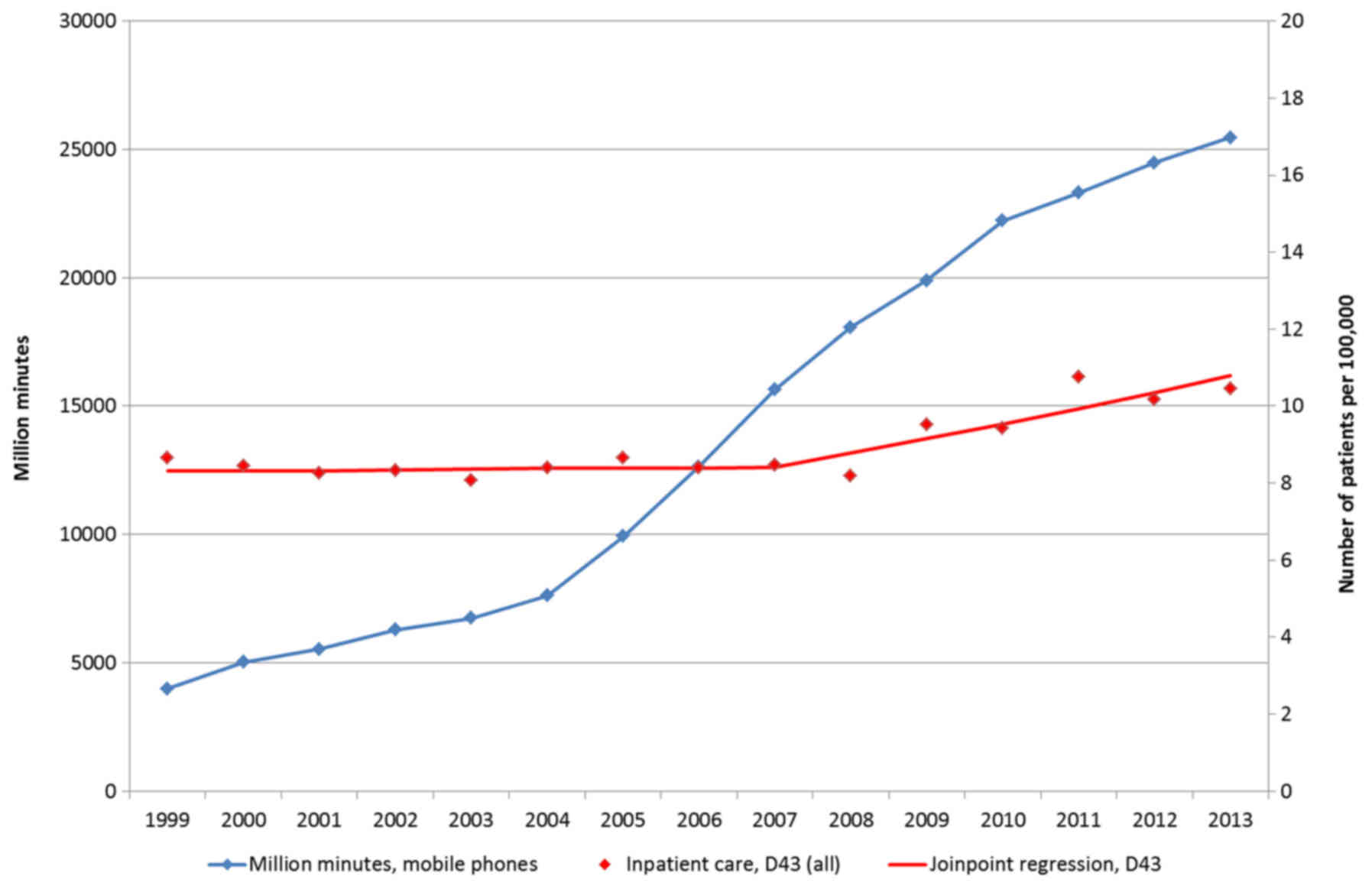
analysis (62). In the IPR, we
found a joinpoint in 2007 with Annual Percentage Change (APC)
+4.25%, 95% CI +1.98, +6.57% during the period between 2007-2013
for tumors of unknown type in the brain or CNS. Fig. 1 shows time trends in IPR for brain
tumors of unknown type (D43), red line, and mobile phone
communication; number of out-going mobile phone minutes in millions
per year (blue line). The figure shows increasing rates of brain
tumors with some latency in relation to the increasing use of
mobile phones.
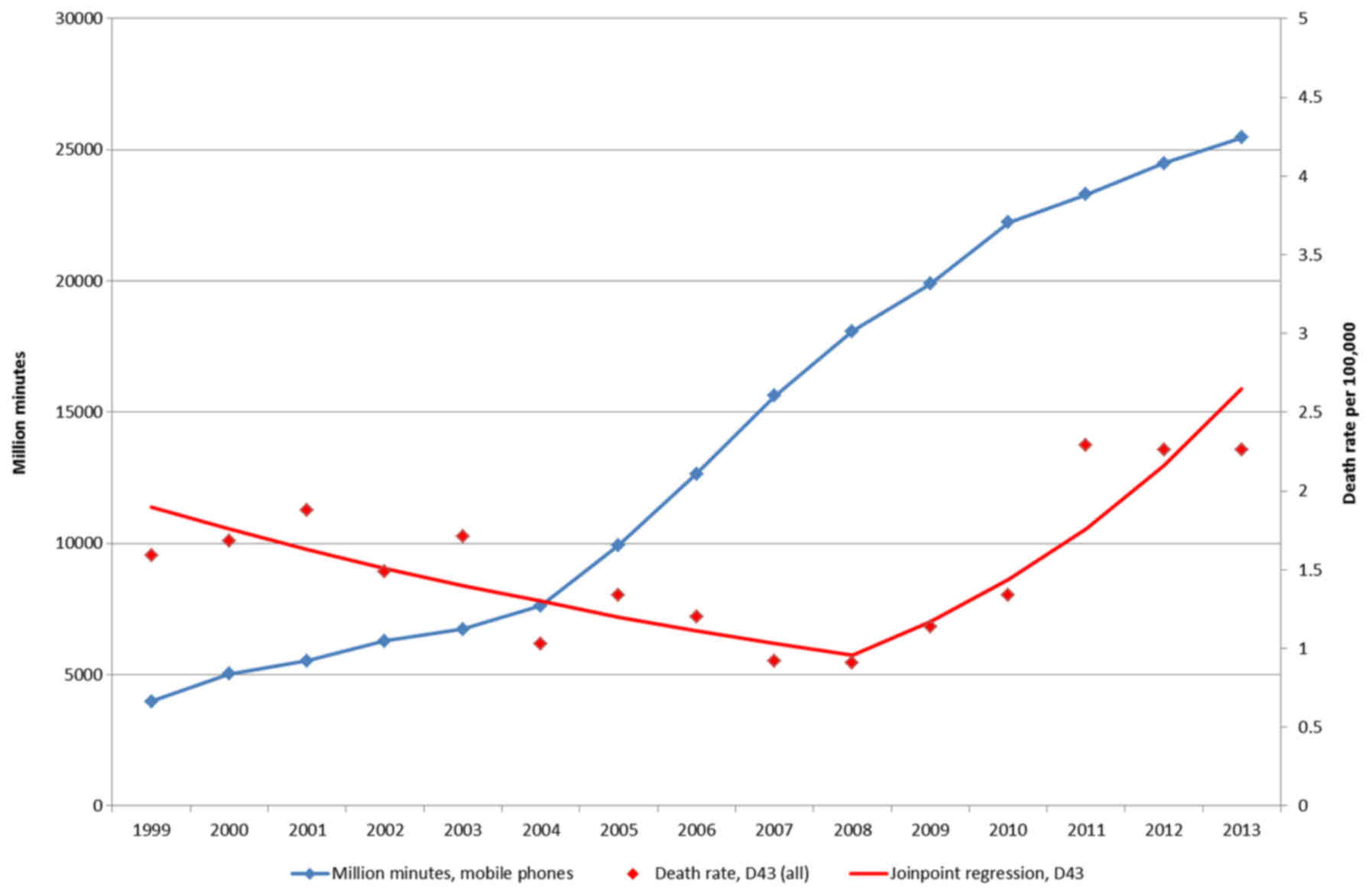
In the CDR joinpoint regression, we found one
joinpoint in 2008 with APC during the period between 2008-2013,
+22.60%, 95% CI +9.68, +37.03%. These tumor diagnoses would be
based on clinical examination, mainly CT and/or MRI, but without
histopathology or cytology. No statistically significant increasing
incidence was found in the Swedish Cancer Register during these
years. We postulated that a large part of brain tumors of unknown
type are never reported in the Cancer Register. Furthermore, the
frequency of diagnoses based on autopsy has declined substantially
due to a general decline of autopsies in Sweden, further adding to
missing cases. We concluded that the Swedish Cancer Register is not
reliable to be used to dismiss results in epidemiological studies
on the use of wireless phones and brain tumor risk.
In Fig. 2, we
present the rates per 100,000 of deaths in unknown type of brain
tumor (D43), red line, and number of out-going mobile phone minutes
in millions (blue line) during the period between 1999-2013. We
postulate that the increasing rate of patients deceased with brain
tumor may be associated with the increasing use of mobile
phones.
In an updated further analysis, we used the Swedish
IPR to analyze rates of brain tumors of unknown type (D43) during
the period between 1998-2015 in different age groups (63). The Average Annual Percentage Change
(AAPC) per 100,000 increased with +2.06%, 95% CI +1.27, +2.86% in
both sexes combined. A joinpoint was found in 2007 with APC
1998-2007 of +0.16%, 95% CI −0.94, +1.28%, and 2007-2015 of +4.24%,
95% CI +2.87, +5.63%. The highest AAPC was found in the age group
of 20-39 years.
In the Swedish Cancer Register, the age-standardized
incidence rate per 100,000 increased for brain tumors, ICD-code
193.0, during 1998-2015 with AAPC in men +0.49%, 95% CI +0.05,
+0.94%, and in women +0.33%, 95% CI −0.29, +0.45% (63). The cases with brain tumor of
unknown type lack morphological examination. Brain tumor diagnoses
in the Cancer Register were based on cytology/histopathology in 83%
for men and in 87% for women in 1980. This frequency increased to
90% in men and 88% in women in 2015. During the same time period,
CT and MRI imaging techniques were introduced and morphology is not
always necessary for diagnosis. If all brain tumors based on
clinical diagnosis with CT or MRI had been reported to the Cancer
Register the frequency of diagnoses based on cytology/histology
would have decreased in the register. The results indicate
underre-porting of brain tumor cases to the Cancer Register. The
real incidence would be higher. Thus, incidence trends based on the
Cancer Register should be used with caution. Our results support
mobile and cordless phones as risk factors for brain tumors with a
reasonable latency period.
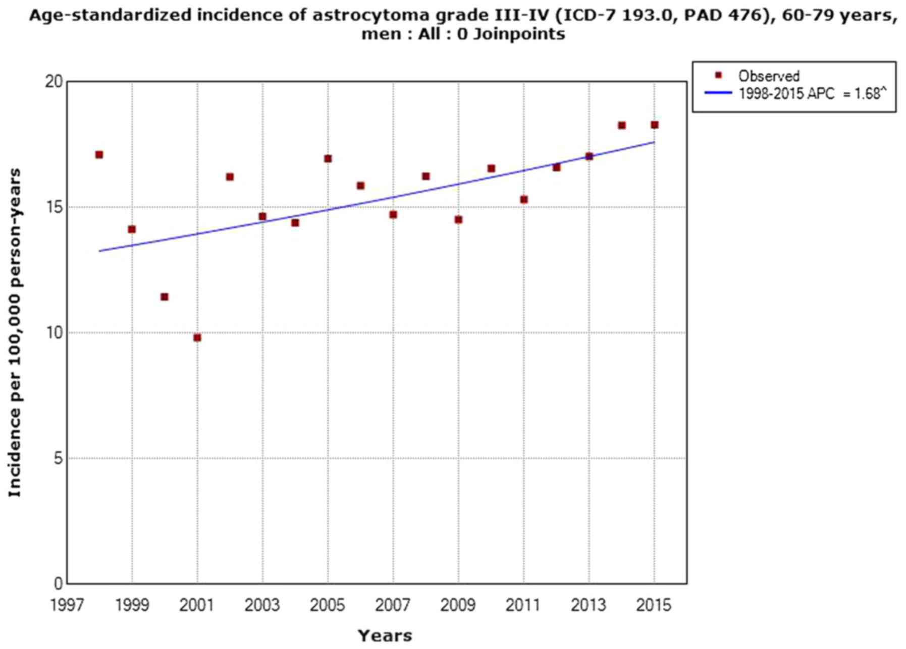
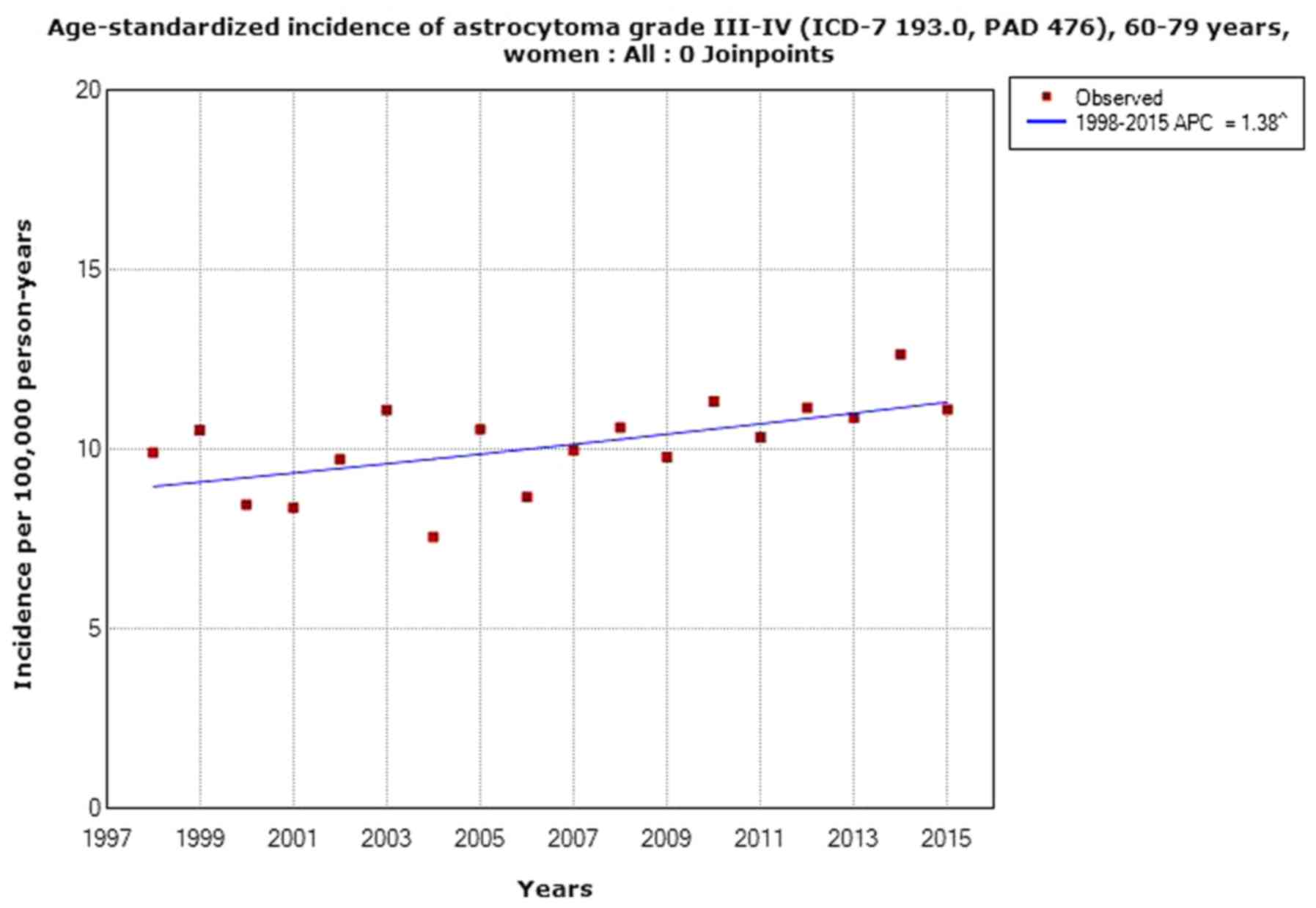
Fig. 3 shows
joinpoint regression analyses of age-standardized incidence rates
per 100,000 in men aged 60-79 years with astrocytoma grade III or
IV in the Swedish Cancer Register during the period between
1998-2015, and Fig. 4 shows
results in women (63).
Interestingly, a recent study demonstrated a similar
increase in glioblastoma multiforme in England as in Sweden
(64), ‘We report a sustained
and highly statistically significant ASR [age-standardized
incidence rates] rise in glioblastoma multiforme (GBM) across all
ages. The ASR for GBM more than doubled from 2.4 to 5.0, with
annual case numbers rising from 983 to 2531. Overall, this rise is
mostly hidden in the overall data by a reduced incidence of
lower-grade tumours.’
Evaluation
Increasing rates/incidences of brain tumors in
Sweden, a country with among the earliest use of wireless phones in
the world, have been published. Similar findings have been reported
from other countries, see above and reviewed by us (62). The results have strengthened the
evidence that RF radiation causes brain tumors in humans.
Acoustic neuroma (vestibular
schwannoma)
Human studies
Acoustic neuroma, also known as vestibular
schwannoma, is a non-malignant tumor located on the eight cranial
nerve from the inner ear to the brain. It is usually encapsulated
and grows in relation to the auditory and vestibular portions of
the nerve. It grows slowly and due to the narrow anatomical space,
may lead to the compression of vital brain stem structures. The
first symptoms of acoustic neuroma are usually tinnitus and hearing
problems. The results for the use of mobile phones in the
Interphone (65) and Hardell et
al (66) studies are presented
in Table III. A statistically
significant increased risk was found for cumulative ipsilateral use
>1,640 h yielding an OR of 2.71, 95% CI of 1.72-4.28.
| Table IIINumbers of exposed cases (Ca) and
controls (Co) and odds ratio (OR) with 95% confidence interval (CI)
for acoustic neuroma in case-control studies in the highest
category of cumulative use in hours for mobile phone use. |
Table III
Numbers of exposed cases (Ca) and
controls (Co) and odds ratio (OR) with 95% confidence interval (CI)
for acoustic neuroma in case-control studies in the highest
category of cumulative use in hours for mobile phone use.
| Study (ref.) | All
| Ipsilateral
|
|---|
| Ca/Co | OR | 95% CI | Ca/Co | OR | 95% CI |
|---|
| Interphone, 2011
(65) | 77/107 | 1.32 | 0.88-1.97 | 47/46 | 2.33 | 1.23-4.40 |
| Cumulative use
≥1,640 h |
| Hardell et
al, 2013 (66) | 27/301 | 2.40 | 1.39-4.16 | 19/133 | 3.18 | 1.65-6.12 |
| Cumulative use
≥1,640 h |
| Meta-analysis
(65,66) | 104/408 | 1.73 | 0.96-3.09 | 66/179 | 2.71 | 1.72-4.28 |
| Cumulative use
≥1,640 h |
The study by Moon et al (67) was not included in the
meta-analysis, since the data on cumulative mobile phone use with
numbers of cases and controls were not given. Support of an
increased risk was found in the case-case part of the study
(67), as also reported by Sato
et al (68) in their
case-case analysis. Pettersson et al made a case-control
study on acoustic neuroma in Sweden not overlapping our study
(69). An increased risk for the
highest category of cumulative use of both mobile phone (≥680 h OR
=1.46, 95% CI =0.98-2.17) and cordless phone (≥900 h OR =1.67, 95%
CI =1.13-2.49) was found. We did not include that study in our
meta-analysis due to the many scientific shortcomings in the study,
e.g., laterality analysis was not made for cordless phone and the
numbers in the laterality analysis for mobile phone are not
consistent in text and tables and obviously not correct, and the
‘unexposed’ reference category included subjects using either
mobile or cordless phone (70).
The Danish part of the Interphone study reported a
mean tumor volume of 1.66 cm3 among regular mobile phone
users and 1.39 cm3 for non-users (P=0.03) (71). We analyzed the percentage change in
tumor volume per year of latency and 100 h of cumulative use
(66). For all types of wireless
phones, the percentage of tumor volume increased, and was
statistically significant for analogue mobile phones per year of
latency (P=0.02) and per 100 h of cumulative use (P=0.01). Moon
et al (67) reported a
statistically significant larger mean tumor volume for heavy users
(11.32±15.43 cm3) compared with light users (4.88±5.60
cm3) based on the daily amount of mobile phone use
(P=0.026). Similar results were found for cumulative hours of use.
Taken together, these results support tumor promotion by RF
radiation.
NTP study
No malignant schwannoma was reported in the mouse
study (20).
In the rat study (19), there was a statistically
significant increased incidence of malignant schwannoma in the
heart of males exposed to GSM modulated cell phone RF radiation for
2 years; P-value for trend =0.041. The tumor was found in all
exposure categories for male rats, whereas no malignant schwannoma
was found in the sham controls. Endocardial hyperplastic Schwann
cell lesions, that are preneoplastic, were found in one 1.5 W/kg
and in two 6 W/kg males, but not in the sham control. A
statistically significant trend was found in CDMA-modulated exposed
males, P=0.011. Two female rats were diagnosed with malignant
schwannoma in the heart in the 3 W/kg group, but no malignant
schwannomas were found in the two other exposure groups or in the
sham control, P-value for trend =0.640.
Evaluation
Based on human epidemiological studies and the NTP
animal study, there is clear evidence that RF radiation causes
vestibular schwannoma (acoustic neuroma) in humans.
Pituitary tumors
Human studies
In a case-control study from Japan, no statistically
significant increased risks were found for the use of mobile phone
(72). A somewhat increased risk
was found in the highest cumulative call time in hours, OR =1.33,
95% CI =0.58-3.09. The cases were aged 30-69 years and diagnosed
during the period between 2000-2004.
In a UK case-control study with patients diagnosed
during the period between 2001-2005, overall no statistically
significant increased risks were found (73). In the group with ≥10 years of use a
somewhat increased risk was found for analog mobile phone use: OR
=1.2, 95% CI =0.6-2.4, and digital mobile phone use with OR =2.5,
95% CI =0.7-9.1.
In a case-control study from China with cases
diagnosed between 2006-2010, mobile phone use yielded an increased
risk for pituitary tumor: OR =7.6, 95% CI =2.6-21.4 and a duration
of use yielded OR =8.5, 95% CI =2.8-24.4 (74). However, no more data were
provided.
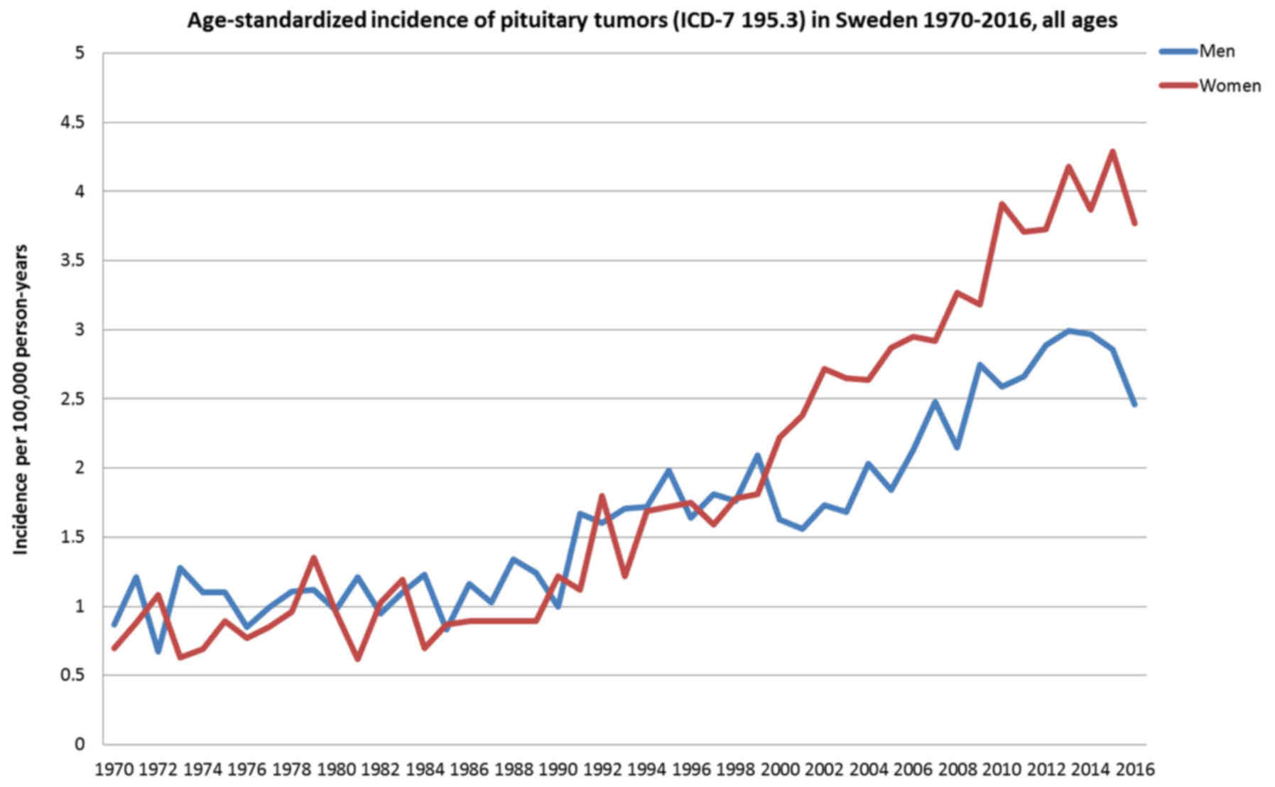
The incidence of pituitary tumors increased during
the time period between 2004-2009 in the USA (75). The incidence is increasing in
Sweden, particularly since 2000, as shown in Fig. 5. There seems to be a decrease
during the latest year, but this may be explained by a time lag in
the reporting to the Swedish Cancer Register.
NTP study
In male mice (20)
exposed to CDMA-modulated RF radiation for two years, two adenoma
and one carcinoma occurred in the pars distalis of the pituitary
gland. No carcinoma or adenoma occurred in the sham control or the
other two exposure groups. No increased incidence was found in
female mice.
In male rats exposed to GSM-modulated cell phone RF
radiation for two years (19), an
increased incidence of pituitary adenoma was found in all exposed
groups, although no statistically significance was found (P-value
for trend =0.301). In females, the incidence of adenoma in 1.5 and
6 W/kg was statistically significantly decreased (1.5 W/kg P=0.049;
6 W/kg P=0.038).
In male rats exposed to CDMA-modulated RF radiation
for two years, an increased incidence of pituitary adenoma was
found in the 1.5 W/kg (P=0.208) and 3 W/kg (P=0.030). In females
there was a statistically significantly decreased incidence of
adenoma or carcinoma in the 3 W/kg group (P=0.030).
Evaluation
Based on human epidemiological studies and the NTP
animal study, there is equivocal evidence that RF radiation causes
pituitary tumors in humans (may be related to exposure).
Thyroid cancer
Human studies
The incidence of thyroid cancer is increasing in
many countries, particularly the papillary type that is the most
radiosensitive type. We used the Swedish Cancer Register to study
the incidence of thyroid cancer during the period between 1970-2013
using joinpoint regression analysis (31). In women, the incidence increased
statistically significantly during the whole study period; AAPC
+1.19% (95% CI +0.56, +1.83%). Two joinpoints were detected, 1979
and 2001, with a high increase of the incidence during the last
period between 2001-2013 with an APC of +5.34% (95% CI +3.93,
+6.77%).
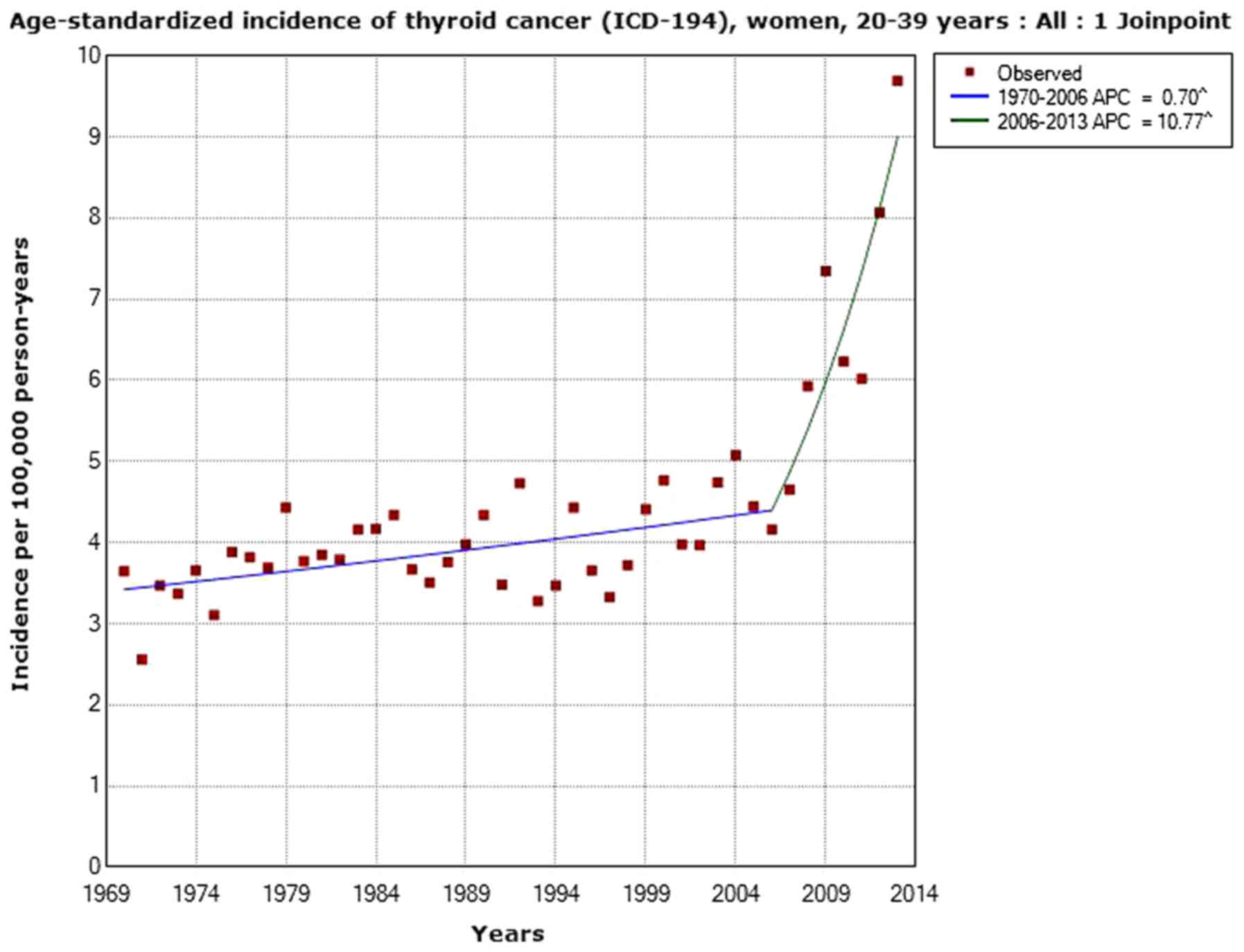
In the age group of 20-39 years, joinpoint
regression analysis of age-standardized incidence of thyroid cancer
in women, aged 20-39 years, APC increased with +10.77% (95% CI
+5.75, +16.04%) during the time period between 2006-2013 (Fig. 6).
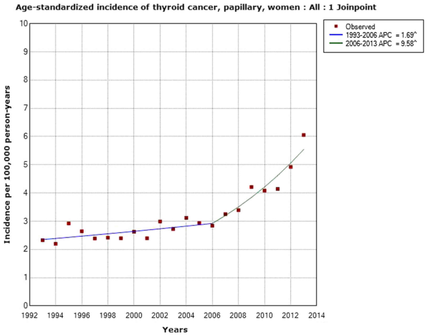
Analyses based on data from the Cancer Register
indicated that the increasing trend in Sweden was mainly caused by
thyroid cancer of the papillary type. The incidence increased
statistically significantly in women with an AAPC of +4.38% (95% CI
+2.95, +5.84%) during the period between 1993-2013 (Fig. 7). One joinpoint was detected in
2006; 1993-2006 APC +1.69% (95% CI +0.32, +3.08%), 2006-2013 APC
+9.58% (95% CI +5.85, +13.44%). The incidence of papillary cancer
increased in men during the period between 1993-2013 with an AAPC
of +3.95% (95% CI +2.20, +5.73%).
AAPC for thyroid cancer in all men during the period
between 1970-2013 was +0.77% (95% CI −0.03, +1.58%). One joinpoint
was detected in 2005 with a statistically significant increase in
incidence during the period between 2005-2013; APC +7.56% (95% CI
+3.34, +11.96%). Based on the NORDCAN data, there was a
statistically significant increase in the incidence of thyroid
cancer in the Nordic countries during the same time period. In both
women and men a joinpoint was detected in 2006. The incidence
increased during 2006-2013 in women; APC +6.16% (95% CI +3.94,
+8.42%) and in men; APC +6.84% (95% CI +3.69, +10.08%), thus
showing similar results as in the Swedish Cancer Register (31).
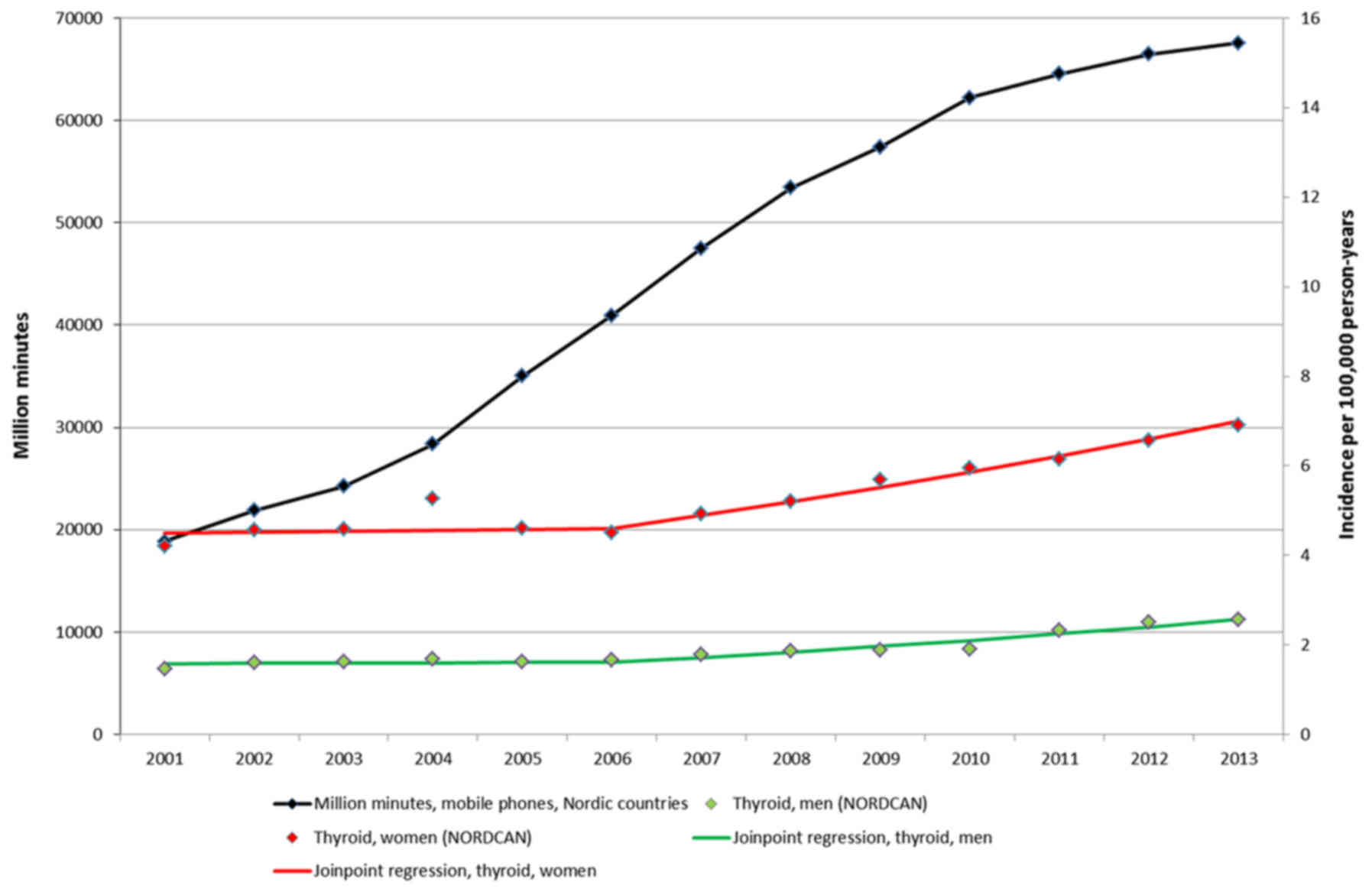
We postulate that the whole increase cannot be
attributed to better diagnostic procedures. In Fig. 8 data from the Nordic countries are
shown on number of out-going mobile phone minutes during the period
between 2001-2013 and the incidence of thyroid cancer in men (green
line) and in women (red line). Clearly, with a lag time of some
years after the increasing number of out-going calls, the thyroid
cancer incidence is increasing.
Increasing exposure to ionizing radiation, e.g.,
medical CT scans, and to RF radiation should be further studied as
causative factors to this emerging thyroid cancer health
problem.
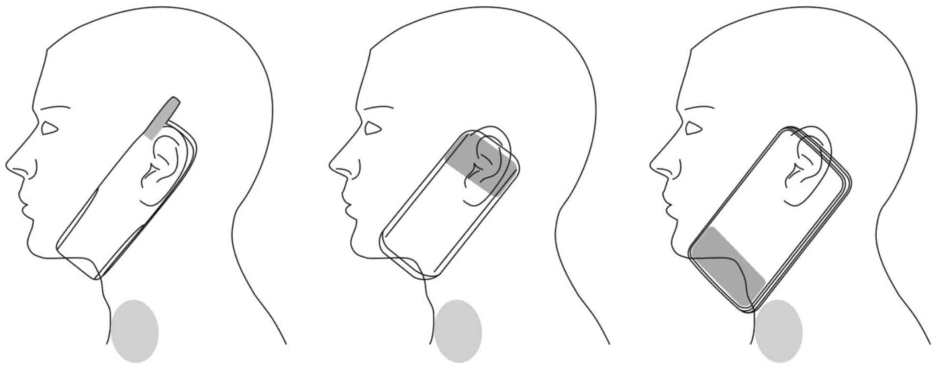
Fig. 9 presents
three developments in the antenna design in mobile phones that may
be of relevance in thyroid carcinogenesis. The second generation
(2G) mobile phones appeared in the 1990s with the external
retractable monopole or helical antennas. The 2G GSM band operated
at a 800/900 MHz frequency band, later accompanied by a 1,800 MHz
band. Around the turn of the millennium, the external antennas
began to disappear, replaced with new phone models with internal
planar or microstrip antennas. The first internal antenna was
introduced in 1998 and the first dual-band mobile phone, with the
internal antenna, was introduced on the market in 1999 (76). The internal antennas were
positioned at the top of the telephone. With the emergence of the
smartphones in the mid- and late 2000s, the internal antenna
location started to shift from the top of the phone to the bottom.
Currently, the majority of smartphone models have their antenna
positioned at the bottom of the phone, thus closer to the thyroid
gland (shown by grey color in Fig.
9). This would have a major impact on increasing radiation to
the thyroid gland from smartphones.
Some published laboratory studies are of interest,
Radiofrequency radiation at 2.45 GHz at a non-thermal level
modified the morphology of the thyroid gland in a study on rats.
The central and peripheral follicles presented increased in size
and the thickness of peripheral septa decreased. Peripheral
follicles increased in size with repeated exposure at 3 W power
(77).
In another study on rats, whole body exposure to 900
MHz pulse-modulated RF radiation that was similar to that emitted
by the global system for mobile communications (GSM) mobile phones
caused pathological changes in the thyroid gland. The gland
structure was altered and caspase-dependent pathways of apoptosis
were enhanced (78).
NTP study
In mice (20) no
increased incidence was reported.
In female rats (19) a statistically significant increased
incidence of C-cell hyperplasia was found in the two years of
GSM-exposed groups (1.5, 3 and 6 W/kg, respectively). In males, a
statistically non-significant increased incidence was observed in
the 1.5 W/kg exposure group (noted in text; P-value not given in
NTP table).
Evaluation
C-cell hyperplasia as a precursor to familial
medullary thyroid cancer in humans is well established. C-cell
hyperplasia may be a precursor to other types of thyroid cancer but
its role is not well established. Based on human cancer statistics
and the NTP animal study, there is some evidence that thyroid
cancer is caused by RF radiation in humans.
Malignant lymphoma
Human studies
Few studies exist on malignant lymphoma and exposure
to RF radiation. In a case-control study male and female subjects
aged 18-74 years living in Sweden were included during a period
from December 1, 1999 to April 30, 2002 (27). Controls were selected from the
national population registry. Exposure to different agents was
assessed by a questionnaire. In total, 910 (91%) cases and 1,016
(92%) controls participated. NHL of the B-cell type was not
associated with the use of cellular or cordless telephones. As
regards T-cell NHL and the >5 year latency period, the use of
analogue cellular phones yielded: OR =1.46, 95% CI =0.58-3.70;
digital: OR =1.92, 95% CI =0.77-4.80; and cordless phones: OR
=2.47; 95% CI =1.09-5.60. The corresponding results for certain
lymphoma, e.g., of the cutaneous and leukemia types, were for
analogue phones: OR =3.41, 95% CI =0.78-15.0; digital: OR =6.12,
95% CI =1.26-29.7; and cordless phones: OR =5.48, 95% CI
=1.26-23.9. The results indicate an association between T-cell NHL
and the use of cellular and cordless telephones; however, the study
was based on low numbers and must be interpreted with caution. As
regards B-cell NHL, no association was found.
A case-control study in USA used a questionnaire to
assess cellular telephone use in 551 NHL cases and 462
frequency-matched population controls (28). Compared to persons who had never
used cellular telephones, risks were not increased among
individuals whose lifetime use was >100 times (e.g., regular
users, OR =0.9, 95% CI =0.6-1.4). Among regular users compared to
those who had never used hand-held cellular telephones, risks of
NHL were not statistically significantly associated with minutes
per week, duration, cumulative lifetime or year of first use,
although NHL was non-significantly higher in men who used cellular
telephones for >8 years; OR =2.4, 95% CI =0.8-7.0. NHL not
otherwise specified was statistically significantly increased in
men for mobile phone use among subjects with ≥6 years duration, OR
=4.4, 95% CI =1.3-14.6. There was little evidence to link the use
of cellular telephones with total, diffuse large B-cell lymphoma or
follicular NHL. No results were presented for T-cell lymphoma.
In the USA, primary central nervous system lymphoma
(PCNSL) rates in immunocompetent men and women aged 65+ years
increased statistically significantly (1.7 and 1.6% per year,
respectively), but remained stable in other age groups during the
period between 1992-2011 (79).
Thus, the increasing rates could not be related to HIV or immune
suppression in organ transplant patients.
In Sweden, the increasing incidence of PCNSL was
reported for the time period between 2000-2013 in immunocompetent
persons (80). With 359 identified
PCNSL cases (median age, 66 years), the overall incidence was 0.26
(95% CI =0.24-0.29) per 100,000 person-years and the average annual
increase 4% (P=0.002). The increasing trend was primarily observed
among elderly individuals (70+ years). Similarly, an increase in
incidence of all brain tumors was noted only among the elderly.
No etiological factor has clearly been defined to
explain the increasing incidence of brain lymphoma. However, it has
occurred during a time period when RF radiation to the brain from
wireless phones has increased.
It should be noted that in transgenic mice, an
increased incidence of lymphoma exposed to 900 MHz GSM RF radiation
was reported; P=0.006 versus the sham group (25). No increased risk of malignant
lymphoma was found in mice exposed to GSM 900 MHz in another study
(26). However, the incidence in
the sham exposed group was higher in the study by Utteridge et
al (26) compared with the
study by Repacholi et al (25) which might have influenced the
results.
NTP study
In female mice exposed to GSM-modulated cell phone
RF radiation for two years, there were increased incidences of
malignant lymphoma in all exposed groups compared to the controls
(20). The increase was
statistically significant in the 2.5 W/kg (P=0.004) and 5 W/kg
groups (P=0.035). In the CDMA-modulated cell phone RF radiation for
two years, the incidence increased in female mice in all exposed
groups compared to the controls, and was statistically significant
in the 2.5 W/kg group (P=0.035).
No conclusive evidence of increased incidence of
malignant lymphoma was reported in female rats (19); P-value for trend =0.537 for
GSM-modulated cell phone RF radiation and P-value for trend =0.339
for CDMA-modulated cell phone RF radiation.
Evaluation
Based on human epidemiological studies and the NTP
study, there is equivocal evidence that malignant lymphoma is
caused by RF radiation in humans (may be related to exposure).
Skin (cutaneous tissue)
Human studies
Few studies exist on RF radiation and the risk of
developing skin tumors. In a Danish cohort on mobile phone
subscribers from the period between 1987-1995 followed to 2007, no
increased risks of skin cancer were observed (81). The same cohort has also been used
for studying brain tumor risk. Due to serious methodological
problems, including the misclassification of exposure the study has
been evaluated to be uninformative (8,37).
In a Swedish study on cutaneous malignant melanoma
diagnosed during the period between 2000-2003, no increased risk
was observed overall (82). In the
shortest latency period of >1-5 years and highest cumulative use
of >365 h, wireless phone use (mobile phone and/or cordless
phone) yielded OR =1.6, 95% CI =0.96-2.9. For melanoma in the most
exposed anatomical area during use of the handheld phone, temporal,
ear, cheek, the risk increased to OR =2.1, 95% CI =1.1-3.8. The
risk was overall highest for cases with first use of a wireless
phone before 20 years of age, OR =2.7, 95% CI =0.6-12, although
based on low numbers. No interaction was observed with known risk
factors for malignant melanoma, such as hair and eye color, skin
type or sunburns as a teenager.
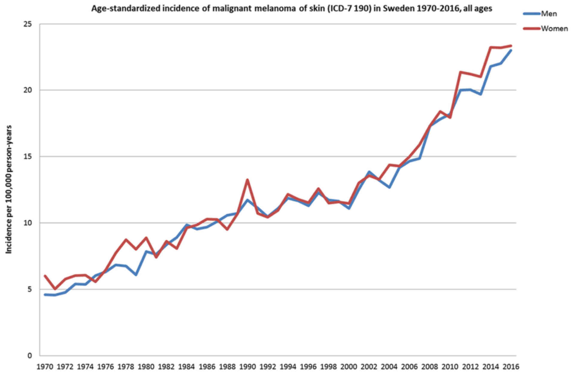
Fig. 10 displays
the rapidly increasing incidence of malignant melanoma in Sweden in
both sexes. The increase is most marked from early 2000.
NTP study
The incidences of malignant fibrous histiocytoma in
the skin were higher in 5 and 10 W/kg male mice exposed to
GSM-modulated cell phone RF radiation for two years (20). The results were not statistically
significant (5 W/kg P=0.124; 10 W/kg P=0.321). The incidences of
fibrosarcoma, sarcoma or malignant fibrous histiocytoma were higher
in exposed male mice compared with sham control, although
border-line significant, P-value for trend =0.093. No increased
incidence was observed in female mice.
Male rats exposed to GSM-modulated cell phone RF
radiation for two years (19)
exhibited higher incidences of fibroma, fibrosarcoma, myxosarcoma,
or malignant fibrous histiocytoma in the skin (subcutaneous tissue)
in all exposed groups. The increased rates were not statistically
significant (P-value for =0.428). No statistically significant
results were found in female rats (P-value for trend =0.551).
Evaluation
Based on human epidemiological studies and NTP
animal studies there is equivocal evidence that RF radiation causes
skin cancer in humans (may be related to exposure).
Concluding remarks
Based on case-control studies, as discussed above,
there is a consistent finding of an increased risk of developing
glioma and acoustic neuroma associated with the use of mobile
phones. Similar results are found for cordless phones in the
Hardell group studies. These results are supported by the results
of the NTP animal studies (19,20).
Malignant vestibular schwannoma is a similar tumor type as acoustic
neuroma, also known as vestibular schwannoma.
The findings are less consistent for meningioma
although somewhat an increased risk was observed in the
meta-analysis of ipsilateral mobile phone use. A longer follow-up
time is necessary for this type of slow-growing tumor.
The results on glioma and acoustic neuroma are
supported by results from other animal studies showing carcinogenic
and/or tumor promoting effects from RF radiation (21-25,32-34).
The NTP study showed genotoxicity of RF radiation in rats and mice
exposed to RF radiation (83).
That result supports previous findings of DNA strand breaks in rat
brain cells exposed to RF radiation (84).
One mechanism in carcinogenesis may be oxidative
stress with the production of reactive oxygen species (ROS), as
summarized by Yakymenko et al (85). This could be an indirect mechanism
for the increased brain and head tumor risk since ROS may lead to
DNA damage (86).
By now carcinogenicity has been shown in human
epidemiological studies, which has been replicated in animal
studies. Laboratory studies on RF radiation have shown increased
ROS production that can cause DNA damage. In 2013, we published the
conclusion that RF radiation should be regarded as a human
carcinogen, Group 1 according to the IARC definition, fulfilling
Bradford Hill causality criteria (87). This was further supported in our
updated article (6). That
conclusion is reinforced by the current evaluation.
The evidence that RF radiation exposure is a risk
factor for cancer is particularly worrying, taking the present
deployment of the fifth generation (5G) for wireless communication.
More than 200 scientists and medical doctors have asked for a
moratorium until studies have been performed by independent
researchers on hazards to human health and the environment
(88). These millimeter waves have
primarily effects on the skin and eye (89). Sweat ducts in the skin may act as
helical antennas and boost RF radiation exposure (90). These findings are worrying, taking
the present evaluation that present RF radiation may increase the
risk of developing skin cancer.
Discussion
The NTP report uses five categories for the
evaluation of RF radiation carcinogenicity as follows:
Clear evidence
Clear evidence of carcinogenic activity is
demonstrated by studies that are interpreted as showing a
dose-related i) increase of malignant neoplasms; ii) increase of a
combination of malignant and benign neoplasms; or iii) marked
increase of benign neoplasms if there is an indication from this or
other studies of the ability of such tumors to progress to
malignancy.
Some evidence
Some evidence of carcinogenic activity is
demonstrated by studies that are interpreted as showing a test
agent-related increased incidence of neoplasms (malignant, benign,
or combined) in which the strength of the response is less than
that required for clear evidence.
Equivocal evidence
Equivocal evidence of carcinogenic activity is
demonstrated by studies that are interpreted as showing a marginal
increase of neoplasms that may be test agent related.
No evidence
No evidence of carcinogenic activity is
demonstrated by studies that are interpreted as showing no test
agent-related increases in malignant or benign neoplasms.
Inadequate study
Inadequate evidence of carcinogenic activity is
demonstrated by studies that, due to major qualitative or
quantitative limitations, cannot be interpreted as valid for
showing either the presence or absence of carcinogenic
activity.
On March 26-28, 2018, a panel of 11 external
scientific experts met to evaluate carcinogenicity of the NTP
carcinogenicity studies (https://factor.niehs.nih.gov/2018/4/feature/feature-2-cell-phone/index.htm).
As shown in Table IV, the carcinogenicity was upgraded for seven
tumor types and/or location. Thus for glioma the vote was ‘some
evidence’ in male rats exposed to GSM or CDMA cell modulation.
Evidence for heart Schwannoma was found in male rats and was
equivocal in female rats, as shown in Table IV. Note that we have
herein discussed carcinogenesis only for tumor types with human
epidemiological data. It is of interest that animal data indicate
also increased incidence for other tumor types and/or locations
such as prostate gland, adrenal medulla, pancreas, liver and lung,
see also https://ntp.niehs.nih.gov/ntp/about_ntp/trpanel/2018/march/actions20180328_508.pdf.
In contrast to the NTP panel, ICNIRP has made its
own evaluation (https://www.icnirp.org/cms/upload/publications/ICNIRPnote2018.pdf).
They discuss mainly the Schwannoma findings and ignore glial
tumors. ICNIRP does not recognize the pattern of increased risk for
Schwannoma and glioma in both animal studies and human epidemiology
on RF radiation. They conclude that ‘ICNIRP considers that the
NTP (2018a, b) and Falcioni et al (2018) studies do not provide a
consistent, reliable and generalizable body of evidence that can be
used as a basis for revising current human exposure
guidelines.’ That conclusion is not based on scientific
evidence, but is rather an ad hoc statement.
A recent commentary discussed ‘several unfounded
criticisms about the design and results of the NTP study that have
been promoted to minimize the utility of the experimental data on
RFR for assessing human health risks. In contrast to those
criticisms, an expert peer-review panel recently concluded that the
NTP studies were well designed, and that the results demonstrated
that both GSM- and CDMA-modulated RFR were carcinogenic to the
heart (schwannomas) and brain (gliomas) of male rats.’
(91).
Our conclusion on RF radiation carcinogenicity is
the following based on human epidemiology and supported by animal
results in the NTP reports: Glioma, clear evidence; meningioma,
equivocal evidence; vestibular schwannoma (acoustic neuroma), clear
evidence; pituitary tumor (adenoma), equivocal evidence; thyroid
cancer, some evidence; malignant lymphoma, equivocal evidence; skin
(cutaneous tissue), equivocal evidence; multi-site carcinogen,
clear evidence.
There is clear evidence that RF radiation causes
cancer/tumor at multiple sites, primarily in the brain (glioma) and
head (acoustic neuroma). There is also evidence of an increased
risk of developing other tumor types. The results are similar in
both the NTP studies (19,20) and the Ramazzini Institute findings
(34). Based on the IARC preamble
to the monographs, RF radiation should be classified as Group 1:
The agent is carcinogenic to humans.
‘This category is used when there is sufficient
evidence of carcinogenicity in humans. Exceptionally, an agent may
be placed in this category when evidence of carcinogenicity in
humans is less than sufficient but there is sufficient evidence of
carcinogenicity in experimental animals and strong evidence in
exposed humans that the agent acts through a relevant mechanism of
carcinogenicity.’ (http://monographs.iarc.fr/ENG/Preamble/currentb6evalrationale0706.php)
Acknowledgments
Not applicable.
Funding
The study was supported by grants from Mr. Brian
Stein, Cancerhjälpen, Mr. Peter Sullivan, Clearlight Ventures Fund,
and Pandora-Foundation for Independent Research, Berlin,
Germany.
Availability of data and materials
The datasets generated and analyzed during the
current study are available from the corresponding author on
reasonable request.
Authors' contributions
Both LH and MC participated in the conception,
design and writing of the manuscript LH supervised the study. MC
made all statistical calculations. Both authors have read and
approved the final version.
Ethics approval and consent to
participate
Not applicable.
Patient consent for publication
Not applicable.
Competing interests
The authors declare that they have no competing
interests.
References
|
1
|
Cardis E, Deltour I, Mann S, Moissonnier
M, Taki M, Varsier N, Wake K and Wiart J: Distribution of RF energy
emitted by mobile phones in anatomical structures of the brain.
Phys Med Biol. 53:2771–2783. 2008. View Article : Google Scholar : PubMed/NCBI
|
|
2
|
Gandhi OP, Morgan LL, de Salles AA, Han
YY, Herberman RB and Davis DL: Exposure limits: The underestimation
of absorbed cell phone radiation, especially in children.
Electromagn Biol Med. 31:34–51. 2012. View Article : Google Scholar
|
|
3
|
Hardell L, Näsman A, Påhlson A, Hallquist
A and Hansson Mild K: Use of cellular telephones and the risk for
brain tumours: A case-control study. Int J Oncol. 15:113–116.
1999.PubMed/NCBI
|
|
4
|
Hardell L, Mild KH and Carlberg M:
Case-control study on the use of cellular and cordless phones and
the risk for malignant brain tumours. Int J Radiat Biol.
78:931–936. 2002. View Article : Google Scholar : PubMed/NCBI
|
|
5
|
Hardell L, Hansson Mild K, Sandström M,
Carlberg M, Hallquist A and Påhlson A: Vestibular schwannoma,
tinnitus and cellular telephones. Neuroepidemiology. 22:124–129.
2003. View Article : Google Scholar : PubMed/NCBI
|
|
6
|
Carlberg M and Hardell L: Evaluation of
mobile phone and cordless phone use and glioma risk using the
Bradford Hill viewpoints from 1965 on association or causation.
BioMed Res Int. 2017.9218486:2017.
|
|
7
|
Baan R, Grosse Y, Lauby-Secretan B, El
Ghissassi F, Bouvard V, Benbrahim-Tallaa L, Guha N, Islami F and
Galichet L: Carcinogenicity of radiofrequency electromagnetic
fields. Lancet Oncol. 12:624–626. 2011. View Article : Google Scholar : PubMed/NCBI
|
|
8
|
Non-ionizing radiation, part 2:
Radiofrequency electromagnetic fields. IARC Monographs on the
Evaluation of Carcinogenic Risks to Humans. 102:International
Agency for Research on Cancer Press; Lyon: 2013, http://monographs.iarc.fr/ENG/Monographs/vol102/mono102.pdf.
4–July, 2018
|
|
9
|
Starkey SJ: Inaccurate official assessment
of radiofrequency safety by the advisory group on non-ionising
radiation. Rev Environ Health. 31:493–503. 2016. View Article : Google Scholar : PubMed/NCBI
|
|
10
|
Hardell L: World Health Organization,
radiofrequency radiation and health - a hard nut to crack (Review).
Int J Oncol. 51:405–413. 2017. View Article : Google Scholar : PubMed/NCBI
|
|
11
|
Maisch D: Conflict of interest and bias in
health advisory committees: A case study of the WHO's
Electromagnetic Field (EMF) Task Group. J Aust Coll Nutr Environ
Med. 25:15–17. 2006.
|
|
12
|
Belpomme D, Hardell L, Belyaev I, Ernesto
Burgio E and Carpenter DO: Thermal and non-thermal health effects
of non-ionizing radiation: an international perspective. Env Poll.
242:643–658. 2018. View Article : Google Scholar
|
|
13
|
Hedendahl L, Carlberg M and Hardell L:
Electromagnetic hypersensitivity - an increasing challenge to the
medical profession. Rev Environ Health. 30:209–215. 2015.
View Article : Google Scholar
|
|
14
|
No authors listed: Guidelines for limiting
exposure to time-varying electric, magnetic, and electromagnetic
fields (up to 300 GHz). International Commission on Non-Ionizing
Radiation Protection. Health Phys. 74:494–522. 1998.
|
|
15
|
International Commission on Non-Ionizing
Radiation Protection: ICNIRP statement on the ‘Guidelines for
limiting exposure to time-varying electric, magnetic, and
electromagnetic fields (up to 300 GHz)’. Health Phys. 97:257–258.
2009. View Article : Google Scholar
|
|
16
|
BioInitiative Working Group: BioInitiative
Report: A Rationale for a Biologically-based Public Exposure
Standard for Electromagnetic Fields (ELF and RF). Sage C and
Carpenter DO: Bioinitiative. 2007, Available from: http://www.bioinitiative.org/table-of-contents/
(Accessed on 4 July, 2018).
|
|
17
|
BioInitiative Working Group: BioInitiative
Report 2012: A Rationale for a Biologically-based Exposure Standard
for Electromagnetic Fields (ELF and RF). Sage C and Carpenter DO:
Bioinitiative. 2012, Available from: http://www.bioinitiative.org/table-of-contents/
(Accessed on 4 July, 2018).
|
|
18
|
Wyde M, Cesta M, Blystone C, Bucher J,
Elmore S, Foster P, Hooth M, Kissling G, Malarkey D, Sills R, et
al: Report of Partial Findings from the National Toxicology Program
Carcinogenesis Studies of Cell Phone Radiofrequency Radiation in
Hsd: Sprague Dawley® SD rats (Whole Body Exposure).
Draft 519-2016. US National Toxicology Program (NTP). http://dx.doi.org/10.1101/055699.
Available from: http://biorxiv.org/content/biorxiv/early/2016/05/26/055699.full.pdf.
4–July. 2018
|
|
19
|
National Toxicology Program: NTP technical
report on the toxicology and carcinogenesis studies in Hsd: Sprague
Dawley sd rats exposed to whole-body radio frequency radiation at a
frequency (900 MHz) and modulations (GSM and CDMA) used by cell
phones. NTP TR. 595:March 26-28–2018.Available from: https://ntp.niehs.nih.gov/ntp/about_ntp/trpanel/2018/march/tr595peerdraft.pdf.
4–July.2018.
|
|
20
|
National Toxicology Program: NTP technical
report on the toxicology and carcinogenesis studies in B6C3F1/N
mice exposed to whole-body radio frequency radiation at a frequency
(1,900 MHz) and modulations (GSM and CDMA) used by cell phones. NTP
TR. 596:March 26-28–2018.Available from: https://ntp.niehs.nih.gov/ntp/about_ntp/trpanel/2018/march/tr596peerdraft.pdf.
4–July.2018.
|
|
21
|
Szmigielski S, Szudzinski A, Pietraszek A,
Bielec M, Janiak M and Wrembel JK: Accelerated development of
spontaneous and benzopyrene-induced skin cancer in mice exposed to
2450-MHz microwave radiation. Bioelectromagnetics. 3:179–191. 1982.
View Article : Google Scholar : PubMed/NCBI
|
|
22
|
Cleary SF, Liu LM and Merchant RE: Glioma
proliferation modulated in vitro by isothermal radiofrequency
radiation exposure. Radiat Res. 121:38–45. 1990. View Article : Google Scholar : PubMed/NCBI
|
|
23
|
Cleary SF, Liu LM and Merchant RE: In
vitro lymphocyte proliferation induced by radio-frequency
electromagnetic radiation under isothermal conditions.
Bioelectromagnetics. 11:47–56. 1990. View Article : Google Scholar : PubMed/NCBI
|
|
24
|
Chou CK, Guy AW, Kunz LL, Johnson RB,
Crowley JJ and Krupp JH: Long-term, low-level microwave irradiation
of rats. Bioelectromagnetics. 13:469–496. 1992. View Article : Google Scholar : PubMed/NCBI
|
|
25
|
Repacholi MH, Basten A, Gebski V, Noonan
D, Finnie J and Harris AW: Lymphomas in E mu-Pim1 transgenic mice
exposed to pulsed 900 MHZ electromagnetic fields. Radiat Res.
147:631–640. 1997. View Article : Google Scholar : PubMed/NCBI
|
|
26
|
Utteridge TD, Gebski V, Finnie JW,
Vernon-Roberts B and Kuchel TR: Long-term exposure of E-mu-Pim1
transgenic mice to 898.4 MHz microwaves does not increase lymphoma
incidence. Radiat Res. 158:357–364. 2002. View Article : Google Scholar : PubMed/NCBI
|
|
27
|
Hardell L, Eriksson M, Carlberg M,
Sundström C and Mild KH: Use of cellular or cordless telephones and
the risk for non-Hodgkin's lymphoma. Int Arch Occup Environ Health.
78:625–632. 2005. View Article : Google Scholar : PubMed/NCBI
|
|
28
|
Linet MS, Taggart T, Severson RK, Cerhan
JR, Cozen W, Hartge P and Colt J: Cellular telephones and
non-Hodgkin lymphoma. Int J Cancer. 119:2382–2388. 2006. View Article : Google Scholar : PubMed/NCBI
|
|
29
|
Lauer O, Frei P, Gosselin MC, Joseph W,
Röösli M and Fröhlich J: Combining near- and far-field exposure for
an organ-specific and whole-body RF-EMF proxy for epidemiological
research: A reference case. Bioelectromagnetics. 34:366–374. 2013.
View Article : Google Scholar : PubMed/NCBI
|
|
30
|
Lu M and Wu XY: Study of specific
absorption rate (SAR) induced in human endocrine glands for using
mobile phones. IEEE Asia-Pacific International Symposium on
Electromagnetic Compatibility (APEMC). 1084–1086. 2016.
|
|
31
|
Carlberg M, Hedendahl L, Ahonen M, Koppel
T and Hardell L: Increasing incidence of thyroid cancer in the
Nordic countries with main focus on Swedish data. BMC Cancer.
16:4262016. View Article : Google Scholar : PubMed/NCBI
|
|
32
|
Tillmann T, Ernst H, Streckert J, Zhou Y,
Taugner F, Hansen V and Dasenbrock C: Indication of cocarcinogenic
potential of chronic UMTS-modulated radiofrequency exposure in an
ethyl-nitrosourea mouse model. Int J Radiat Biol. 86:529–541. 2010.
View Article : Google Scholar : PubMed/NCBI
|
|
33
|
Lerchl A, Klose M, Grote K, Wilhelm AF,
Spathmann O, Fiedler T, Streckert J, Hansen V and Clemens M: Tumor
promotion by exposure to radiofrequency electromagnetic fields
below exposure limits for humans. Biochem Biophys Res Commun.
459:585–590. 2015. View Article : Google Scholar : PubMed/NCBI
|
|
34
|
Falcioni L, Bua L, Tibaldi E, Lauriola M,
De Angelis L, Gnudi F, Mandrioli D, Manservigi M, Manservisi F,
Manzoli I, et al: Report of final results regarding brain and heart
tumors in Sprague-Dawley rats exposed from prenatal life until
natural death to mobile phone radiofrequency field representative
of a 1.8 GHz GSM base station environmental emission. Environ Res.
165:496–503. 2018. View Article : Google Scholar : PubMed/NCBI
|
|
35
|
Johansen C, Boice J Jr, McLaughlin J and
Olsen J: Cellular telephones and cancer - a nationwide cohort study
in Denmark. J Natl Cancer Inst. 93:203–207. 2001. View Article : Google Scholar : PubMed/NCBI
|
|
36
|
Schüz J, Jacobsen R, Olsen JH, Boice JD
Jr, McLaughlin JK and Johansen C: Cellular telephone use and cancer
risk: Update of a nationwide Danish cohort. J Natl Cancer Inst.
98:1707–1713. 2006. View Article : Google Scholar : PubMed/NCBI
|
|
37
|
Söderqvist F, Carlberg M and Hardell L:
Review of four publications on the Danish cohort study on mobile
phone subscribers and risk of brain tumors. Rev Environ Health.
27:51–58. 2012. View Article : Google Scholar : PubMed/NCBI
|
|
38
|
Benson VS, Pirie K, Schüz J, Reeves GK,
Beral V and Green J; Million Women Study Collaborators: Mobile
phone use and risk of brain neoplasms and other cancers:
Prospective study. Int J Epidemiol. 42:792–802. 2013. View Article : Google Scholar : PubMed/NCBI
|
|
39
|
Ohgaki H and Kleihues P: Population-based
studies on incidence, survival rates, and genetic alterations in
astrocytic and oligodendroglial gliomas. J Neuropathol Exp Neurol.
64:479–489. 2005. View Article : Google Scholar : PubMed/NCBI
|
|
40
|
INTERPHONE Study Group: Brain tumour risk
in relation to mobile telephone use: Results of the INTERPHONE
international case-control study. Int J Epidemiol. 39:675–694.
2010. View Article : Google Scholar : PubMed/NCBI
|
|
41
|
Coureau G, Bouvier G, Lebailly P,
Fabbro-Peray P, Gruber A, Leffondre K, Guillamo JS, Loiseau H,
Mathoulin-Pélissier S, Salamon R, et al: Mobile phone use and brain
tumours in the CERENAT case-control study. Occup Environ Med.
71:514–522. 2014. View Article : Google Scholar : PubMed/NCBI
|
|
42
|
Hardell L and Carlberg M: Mobile phones,
cordless phones and the risk for brain tumours. Int J Oncol.
35:5–17. 2009. View Article : Google Scholar : PubMed/NCBI
|
|
43
|
Hardell L and Carlberg M: Mobile phone and
cordless phone use and the risk for glioma - Analysis of pooled
case-control studies in Sweden, 1997–2003 and 2007–2009.
Pathophysiology. 22:1–13. 2015. View Article : Google Scholar
|
|
44
|
Hardell L, Carlberg M and Hansson Mild K:
Pooled analysis of two case-control studies on use of cellular and
cordless telephones and the risk for malignant brain tumours
diagnosed in 1997–2003. Int Arch Occup Environ Health. 79:630–639.
2006. View Article : Google Scholar : PubMed/NCBI
|
|
45
|
Hardell L, Carlberg M and Hansson Mild K:
Re-analysis of risk for glioma in relation to mobile telephone use:
Comparison with the results of the Interphone international
case-control study. Int J Epidemiol. 40:1126–1128. 2011. View Article : Google Scholar
|
|
46
|
Hardell L, Carlberg M and Hansson Mild K:
Pooled analysis of case-control studies on malignant brain tumours
and the use of mobile and cordless phones including living and
deceased subjects. Int J Oncol. 38:1465–1474. 2011. View Article : Google Scholar : PubMed/NCBI
|
|
47
|
Cardis E, Armstrong BK, Bowman JD, Giles
GG, Hours M, Krewski D, McBride M, Parent ME, Sadetzki S, Woodward
A, et al: Risk of brain tumours in relation to estimated RF dose
from mobile phones: Results from five Interphone countries. Occup
Environ Med. 68:631–640. 2011. View Article : Google Scholar : PubMed/NCBI
|
|
48
|
Grell K, Frederiksen K, Schüz J, Cardis E,
Armstrong B, Siemiatycki J, Krewski DR, McBride ML, Johansen C,
Auvinen A, et al: The intracranial distribution of gliomas in
relation to exposure from mobile phones: Analyses from the
INTERPHONE study. Am J Epidemiol. 184:818–828. 2016. View Article : Google Scholar : PubMed/NCBI
|
|
49
|
Momoli F, Siemiatycki J, McBride ML,
Parent ME, Richardson L, Bedard D, Platt R, Vrijheid M, Cardis E
and Krewski D: Probabilistic multiple-bias modeling applied to the
Canadian data from the INTERPHONE study of mobile phone use and
risk of glioma, meningioma, acoustic neuroma, and parotid gland
tumors. Am J Epidemiol. 186:885–893. 2017. View Article : Google Scholar : PubMed/NCBI
|
|
50
|
Carlberg M and Hardell L: Decreased
survival of glioma patients with astrocytoma grade IV (glioblastoma
multiforme) associated with long-term use of mobile and cordless
phones. Int J Environ Res Public Health. 11:10790–10805. 2014.
View Article : Google Scholar : PubMed/NCBI
|
|
51
|
Akhavan-Sigari R, Mazloum Farsi Baf M,
Ariabod V, Rohde V and Rahighi S: Connection between cell phone
use, p53 gene expression in different zones of glioblastoma
multiforme and survival prognoses. Rare Tumors. 6:53502014.
View Article : Google Scholar : PubMed/NCBI
|
|
52
|
Turner MC, Benke G, Bowman JD, Figuerola
J, Fleming S, Hours M, Kincl L, Krewski D, McLean D, Parent ME, et
al: Occupational exposure to extremely low-frequency magnetic
fields and brain tumor risks in the INTEROCC study. Cancer
Epidemiol Biomarkers Prev. 23:1863–1872. 2014. View Article : Google Scholar : PubMed/NCBI
|
|
53
|
Carlberg M, Koppel T, Ahonen M and Hardell
L: Case-control study on occupational exposure to extremely
low-frequency electromagnetic fields and glioma risk. Am J Ind Med.
60:494–503. 2017. View Article : Google Scholar : PubMed/NCBI
|
|
54
|
Peleg M, Nativ O and Richter ED: Radio
frequency radiation-related cancer: Assessing causation in the
occupational/military setting. Environ Res. 163:123–133. 2018.
View Article : Google Scholar : PubMed/NCBI
|
|
55
|
Cea-Soriano L, Wallander MA and García
Rodríguez LA: Epidemiology of meningioma in the United Kingdom.
Neuroepidemiology. 39:27–34. 2012. View Article : Google Scholar : PubMed/NCBI
|
|
56
|
Carlberg M and Hardell L: Pooled analysis
of Swedish case-control studies during 1997–2003 and 2007–2009 on
meningioma risk associated with the use of mobile and cordless
phones. Oncol Rep. 33:3093–3098. 2015. View Article : Google Scholar : PubMed/NCBI
|
|
57
|
Shrestha B, Khalid M, Gayam V, Mukhtar O,
Thapa S, Mandal AK, Kaler J, Khalid M, Garlapati P, Iqbal S, et al:
Metachronous granular cell tumor of the descending colon.
Gastroenterol Res. 11:317–320. 2018. View Article : Google Scholar
|
|
58
|
Kim HJ and Lee MG: Granular cell tumors on
unusual anatomic locations. Yonsei Med J. 56:1731–1734. 2015.
View Article : Google Scholar : PubMed/NCBI
|
|
59
|
Rejas RA, Campos MS, Cortes AR, Pinto DD
and de Sousa SC: The neural histogenetic origin of the oral
granular cell tumor: An immunohistochemical evidence. Med Oral
Patol Oral Cir Bucal. 16:e6–e10. 2011. View Article : Google Scholar
|
|
60
|
Kamal SA and Othman EO: Granular cell
tumour of the larynx. J Laryngol Otol. 112:83–85. 1998. View Article : Google Scholar : PubMed/NCBI
|
|
61
|
Deltour I, Johansen C, Auvinen A,
Feychting M, Klaeboe L and Schüz J: Time trends in brain tumor
incidence rates in Denmark, Finland, Norway, and Sweden, 1974–2003.
J Natl Cancer Inst. 101:1721–1724. 2009. View Article : Google Scholar : PubMed/NCBI
|
|
62
|
Hardell L and Carlberg M: Increasing rates
of brain tumours in the Swedish national inpatient register and the
causes of death register. Int J Environ Res Public Health.
12:3793–3813. 2015. View Article : Google Scholar : PubMed/NCBI
|
|
63
|
Hardell L and Carlberg M: Mobile phones,
cordless phones and rates of brain tumors in different age groups
in the Swedish National Inpatient Register and the Swedish Cancer
Register during 1998–2015. PLoS One. 12:e01854612017. View Article : Google Scholar
|
|
64
|
Philips A, Henshaw DL, Lamburn G and
O'Carroll MJ: Brain tumours: Rise in glioblastoma multiforme
incidence in England 1995–2015 suggests an adverse environmental or
lifestyle factor. J Environ Public Health. 7910754:20182018.
|
|
65
|
INTERPHONE Study Group: Acoustic neuroma
risk in relation to mobile telephone use: Results of the INTERPHONE
international case-control study. Cancer Epidemiol. 35:453–464.
2011. View Article : Google Scholar : PubMed/NCBI
|
|
66
|
Hardell L, Carlberg M, Söderqvist F and
Mild KH: Pooled analysis of case-control studies on acoustic
neuroma diagnosed 1997–2003 and 2007–2009 and use of mobile and
cordless phones. Int J Oncol. 43:1036–1044. 2013. View Article : Google Scholar : PubMed/NCBI
|
|
67
|
Moon IS, Kim BG, Kim J, Lee JD and Lee WS:
Association between vestibular schwannomas and mobile phone use.
Tumour Biol. 35:581–587. 2014. View Article : Google Scholar :
|
|
68
|
Sato Y, Akiba S, Kubo O and Yamaguchi N: A
case-case study of mobile phone use and acoustic neuroma risk in
Japan. Bioelectromagnetics. 32:85–93. 2011. View Article : Google Scholar : PubMed/NCBI
|
|
69
|
Pettersson D, Mathiesen T, Prochazka M,
Bergenheim T, Florentzson R, Harder H, Nyberg G, Siesjö P and
Feychting M: Long-term mobile phone use and acoustic neuroma risk.
Epidemiology. 25:233–241. 2014. View Article : Google Scholar : PubMed/NCBI
|
|
70
|
Hardell L and Carlberg M: Long-term mobile
phone use and acoustic neuroma. Epidemiology. 25:7782014.
View Article : Google Scholar : PubMed/NCBI
|
|
71
|
Christensen HC, Schüz J, Kosteljanetz M,
Poulsen HS, Thomsen J and Johansen C: Cellular telephone use and
risk of acoustic neuroma. Am J Epidemiol. 159:277–283. 2004.
View Article : Google Scholar : PubMed/NCBI
|
|
72
|
Takebayashi T, Varsier N, Kikuchi Y, Wake
K, Taki M, Watanabe S, Akiba S and Yamaguchi N: Mobile phone use,
exposure to radiofrequency electromagnetic field, and brain tumour:
A case-control study. Br J Cancer. 98:652–659. 2008. View Article : Google Scholar : PubMed/NCBI
|
|
73
|
Schoemaker MJ and Swerdlow AJ: Risk of
pituitary tumors in cellular phone users: A case-control study.
Epidemiology. 20:348–354. 2009. View Article : Google Scholar : PubMed/NCBI
|
|
74
|
Leng L and Zhang Y: Etiology of pituitary
tumors: A case control study. Turk Neurosurg. 26:195–199.
2016.PubMed/NCBI
|
|
75
|
Gittleman H, Ostrom QT, Farah PD, Ondracek
A, Chen Y, Wolinsky Y, Kruchko C, Singer J, Kshettry VR, Laws ER,
et al: Descriptive epidemiology of pituitary tumors in the United
States, 2004–2009. J Neurosurg. 121:527–535. 2014. View Article : Google Scholar : PubMed/NCBI
|
|
76
|
Garg R, Bhartia P, Bahl I and Ittipiboon
A: Microstrip Antenna Design Handbook. Artech House; Norwood:
2001
|
|
77
|
Misa-Agustiño MJ, Jorge-Mora T,
Jorge-Barreiro FJ, Suarez-Quintanilla J, Moreno-Piquero E,
Ares-Pena FJ and López-Martín E: Exposure to non-ionizing radiation
provokes changes in rat thyroid morphology and expression of
HSP-90. Exp Biol Med (Maywood). 240:1123–1135. 2015. View Article : Google Scholar
|
|
78
|
Eşmekaya MA, Seyhan N and Ömeroğlu S:
Pulse modulated 900 MHz radiation induces hypothyroidism and
apoptosis in thyroid cells: A light, electron microscopy and
immunohisto-chemical study. Int J Radiat Biol. 86:1106–1116. 2010.
View Article : Google Scholar
|
|
79
|
Shiels MS, Pfeiffer RM, Besson C, Clarke
CA, Morton LM, Nogueira L, Pawlish K, Yanik EL, Suneja G and Engels
EA: Trends in primary central nervous system lymphoma incidence and
survival in the U.S. Br J Haematol. 174:417–424. 2016. View Article : Google Scholar : PubMed/NCBI
|
|
80
|
Eloranta S, Brånvall E, Celsing F,
Papworth K, Ljungqvist M, Enblad G and Ekström-Smedby K: Increasing
incidence of primary central nervous system lymphoma but no
improvement in survival in Sweden 2000–2013. Eur J Haematol.
100:61–68. 2018. View Article : Google Scholar
|
|
81
|
Poulsen AH, Friis S, Johansen C, Jensen A,
Frei P, Kjaear SK, Dalton SO and Schüz J: Mobile phone use and the
risk of skin cancer: A nationwide cohort study in Denmark. Am J
Epidemiol. 178:190–197. 2013. View Article : Google Scholar : PubMed/NCBI
|
|
82
|
Hardell L, Carlberg M, Hansson Mild K and
Eriksson M: Case-control study on the use of mobile and cordless
phones and the risk for malignant melanoma in the head and neck
region. Pathophysiology. 18:325–333. 2011. View Article : Google Scholar : PubMed/NCBI
|
|
83
|
Smith-Roe SL, Wyde ME, Stout MD, Winters
JW, Hobbs CA, Shepard KG, Green AS, Kissling GE, Tice RR, Bucher
JR, et al: Evaluation of the genotoxicity of cell phone
radiofrequency radiation in male and female rats and mice following
subchronic exposure. In: Environmental Mutagenesis and Genomics
Society, Annual Meeting; Raleigh, North Carolina, USA. September
9-13, 2017;
|
|
84
|
Lai H and Singh NP: Melatonin and a
spin-trap compound block radiofrequency electromagnetic
radiation-induced DNA strand breaks in rat brain cells.
Bioelectromagnetics. 18:446–454. 1997. View Article : Google Scholar : PubMed/NCBI
|
|
85
|
Yakymenko I, Tsybulin O, Sidorik E,
Henshel D, Kyrylenko O and Kyrylenko S: Oxidative mechanisms of
biological activity of low-intensity radiofrequency radiation.
Electromagn Biol Med. 35:186–202. 2016. View Article : Google Scholar
|
|
86
|
Megha K, Deshmukh PS, Banerjee BD,
Tripathi AK, Ahmed R and Abegaonkar MP: Low intensity microwave
radiation induced oxidative stress, inflammatory response and DNA
damage in rat brain. Neurotoxicology. 51:158–165. 2015. View Article : Google Scholar : PubMed/NCBI
|
|
87
|
Hardell L and Carlberg M: Using the Hill
viewpoints from 1965 for evaluating strengths of evidence of the
risk for brain tumors associated with use of mobile and cordless
phones. Rev Environ Health. 28:97–106. 2013. View Article : Google Scholar : PubMed/NCBI
|
|
88
|
The 5G Appeal: Scientists and doctors warn
of potential serious health effects of 5G. Available from:
http://www.5gappeal.eu/scientists-and-doctors-warn-of-potential-serious-health-effects-of-5g/
(Accessed on 4 July, 2018).
|
|
89
|
Russell CL: 5 G wireless
telecommunications expansion: Public health and environmental
implications. Environ Res. 165:484–495. 2018. View Article : Google Scholar : PubMed/NCBI
|
|
90
|
Betzalel N, Ben Ishai P and Feldman Y: The
human skin as a sub-THz receiver - Does 5G pose a danger to it or
not? Environ Res. 163:208–216. 2018. View Article : Google Scholar : PubMed/NCBI
|
|
91
|
Melnick RL: Commentary on the utility of
the National Toxicology Program Study on cell phone radiofrequency
radiation data for assessing human health risks despite unfounded
criticisms aimed at minimizing the findings of adverse health
effects. Environ Res. 168:1–6. 2018. View Article : Google Scholar : PubMed/NCBI
|