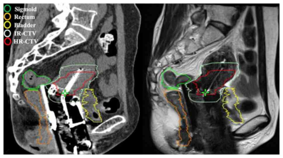
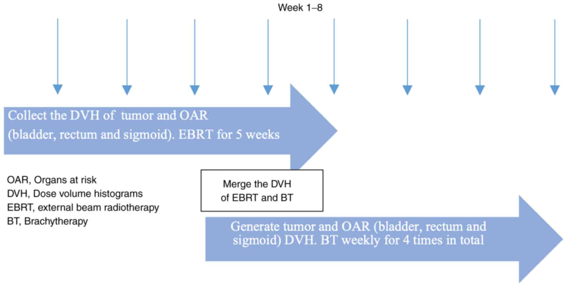
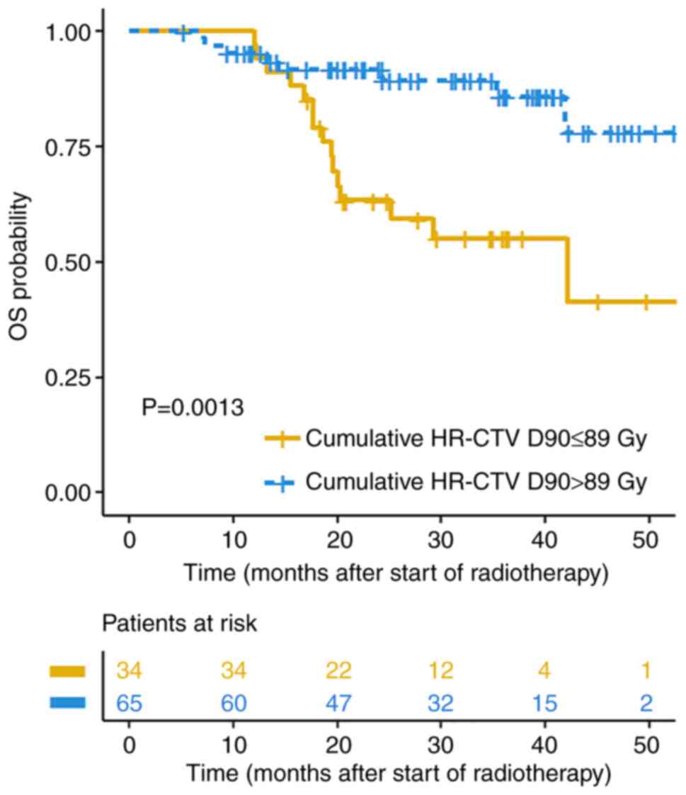
|
1
|
Siegel RL, Miller KD and Jemal A: Cancer
statistics, 2019. CA Cancer J Clin. 69:7–34. 2019.PubMed/NCBI View Article : Google Scholar
|
|
2
|
Haie-Meder C, Pötter R, Van Limbergen E,
Briot E, De Brabandere M, Dimopoulos J, Dumas I, Hellebust TP,
Kirisits C, Lang S, et al: Recommendations from gynaecological
(GYN) GEC-ESTRO working group (I): Concepts and terms in 3D image
based 3D treatment planning in cervix cancer brachytherapy with
emphasis on MRI assessment of GTV and CTV. Radiother Oncol.
74:235–245. 2005.PubMed/NCBI View Article : Google Scholar
|
|
3
|
Charra-Brunaud C, Harter V, Delannes M,
Haie-Meder C, Quetin P, Kerr C, Castelain B, Thomas L and Peiffert
D: Impact of 3D image-based PDR brachytherapy on outcome of
patients treated for cervix carcinoma in France: Results of the
French STIC prospective study. Radiother Oncol. 103:305–313.
2012.PubMed/NCBI View Article : Google Scholar
|
|
4
|
Manea E, Escande A, Bockel S, Khettab M,
Dumas I, Lazarescu I, Fumagalli I, Morice P, Deutsch E, Haie-Meder
C and Chargari C: Risk of late urinary complications following
image guided adaptive brachytherapy for locally advanced cervical
cancer: Refining bladder dose-volume parameters. Int J Radiat Oncol
Biol Phys. 101:411–420. 2018.PubMed/NCBI View Article : Google Scholar
|
|
5
|
Mazeron R, Maroun P, Castelnau-Marchand P,
Dumas I, del Campo ER, Cao K, Slocker-Escarpa A, M'Bagui R,
Martinetti F, Tailleur A, et al: Pulsed-dose rate image-guided
adaptive brachytherapy in cervical cancer: Dose-volume effect
relationships for the rectum and bladder. Radiother Oncol.
116:226–232. 2015.PubMed/NCBI View Article : Google Scholar
|
|
6
|
Zakariaee R, Hamarneh G, Brown CJ, Gaudet
M, Aquino-Parsons C and Spadinger I: Bladder accumulated dose in
image-guided high-dose-rate brachytherapy for locally advanced
cervical cancer and its relation to urinary toxicity. Phys Med
Biol. 61:8408–8424. 2016.PubMed/NCBI View Article : Google Scholar
|
|
7
|
Harmon G, Chinsky B, Surucu M, Harkenrider
M and Small W Jr: Bladder distension improves the dosimetry of
organs at risk during intracavitary cervical high-dose-rate
brachytherapy. Brachytherapy. 15:30–34. 2016.PubMed/NCBI View Article : Google Scholar
|
|
8
|
Siavashpour Z, Aghamiri MR, Jaberi R,
Manshadi HR, Ghaderi R and Kirisits C: Optimum organ volume ranges
for organs at risk dose in cervical cancer intracavitary
brachytherapy. J Contemp Brachytherapy. 8:135–142. 2016.PubMed/NCBI View Article : Google Scholar
|
|
9
|
Nesseler JP, Charra-Brunaud C, Salleron J,
Py JF, Huertas A, Meknaci E, Courrech F, Peiffert D and
Renard-Oldrini S: Effect of bladder distension on doses to organs
at risk in pulsed-dose-rate 3D image-guided adaptive brachytherapy
for locally advanced cervical cancer. Brachytherapy. 16:976–980.
2017.PubMed/NCBI View Article : Google Scholar
|
|
10
|
Siavashpour Z, Aghamiri MR, Jaberi R,
ZareAkha N, Manshadi HRD, Kirisits C and Sedaghat M: A comparison
of organs at risk doses in GYN intracavitary brachytherapy for
different tandem lengths and bladder volumes. J Appl Clin Med Phys.
17:5–13. 2016.PubMed/NCBI View Article : Google Scholar
|
|
11
|
Jamema SV, Saju S, Mahantshetty U, Pallad
S, Deshpande DD, Shrivastava SK and Dinshaw KA: Dosimetric
evaluation of rectum and bladder using image-based CT planning and
orthogonal radiographs with ICRU 38 recommendations in
intracavitary brachytherapy. J Med Phys. 33:3–8. 2008.PubMed/NCBI View Article : Google Scholar
|
|
12
|
Mahantshetty U, Shetty S, Majumder D,
Adurkar P, Swamidas J, Engineer R, Chopra S and Shrivastava S:
Optimal bladder filling during high-dose-rate intracavitary
brachytherapy for cervical cancer: a dosimetric study. J Contemp
Brachytherapy. 9:112–117. 2017.PubMed/NCBI View Article : Google Scholar
|
|
13
|
No authors listed: Prescribing, recording,
and reporting brachytherapy for cancer of the cervix. J ICRU 13:
1-2, NP, 2013.
|
|
14
|
Pötter R, Tanderup K, Kirisits C, de Leeuw
A, Kirchheiner K, Nout R, Tan LT, Haie-Meder C, Mahantshetty U,
Segedin B, et al: The EMBRACE II study: The outcome and prospect of
two decades of evolution within the GEC-ESTRO GYN working group and
the EMBRACE studies. Clin Transl Radiat Oncol. 9:48–60.
2018.PubMed/NCBI View Article : Google Scholar
|
|
15
|
Pecorelli S: Revised FIGO staging for
carcinoma of the vulva, cervix, and endometrium. Int J Gynaecol
Obstet. 105:103–104. 2009.PubMed/NCBI View Article : Google Scholar
|
|
16
|
Gay HA, Barthold HJ, O'Meara E, Bosch WR,
El Naqa I, Al-Lozi R, Rosenthal SA, Lawton C, Lee WR, Sandler H, et
al: Pelvic normal tissue contouring guidelines for radiation
therapy: A radiation therapy oncology group consensus panel atlas.
Int J Radiat Oncol Biol Phys. 83:e353–e362. 2012.PubMed/NCBI View Article : Google Scholar
|
|
17
|
Fowler JF: Review: Total doses in
fractionated radiotherapy-implications of new radiobiological data.
Int J Radiat Biol Relat Stud Phys Chem Med. 46:103–120.
1984.PubMed/NCBI View Article : Google Scholar
|
|
18
|
Bentzen SM and Joiner MC: The
linear-quadratic approach in clinical practice. In: Basic clinical
radiobiology. Joiner MC, van der Kogel A (eds). 4th edition. CRC
Press, Boca Raton, FL, pp120-134, 2009.
|
|
19
|
Chen AP, Setser A, Anadkat MJ, Cotliar J,
Olsen EA, Garden BC and Lacouture ME: Grading dermatologic adverse
events of cancer treatments: The common terminology criteria for
adverse events version 4.0. J Am Acad Dermatol. 67:1025–1039.
2012.PubMed/NCBI View Article : Google Scholar
|
|
20
|
Otter S, Coates A, Franklin A, Cunningham
M and Stewart A: Improving dose delivery by adding interstitial
catheters to fixed geometry applicators in high-dose-rate
brachytherapy for cervical cancer. Brachytherapy. 17:580–586.
2018.PubMed/NCBI View Article : Google Scholar
|
|
21
|
Sharma AD, Poddar J, Suryanarayan KU, Shah
SP, Parikh A, Mehta V and Phys M, Kumar T and Phys M: Dosimetric
analysis of the effects of the bladder volume on organs at risk
(OAR) in high-dose-rate intracavitary brachytherapy in carcinoma
cervix-an institutional study. J Contemp Brachytherapy. 10:26–31.
2018.PubMed/NCBI View Article : Google Scholar
|
|
22
|
Liao Y, Dandekar V, Chu JC, Turian J,
Bernard D and Kiel K: Reporting small bowel dose in cervix cancer
high-dose-rate brachytherapy. Med Dosim. 41:28–33. 2016.PubMed/NCBI View Article : Google Scholar
|
|
23
|
Damato AL, Buzurovic I, Bhagwat MS,
Cormack RA, Devlin PM, Friesen S, Hansen J, Lee LJ, Manuel MM, Cho
LP, et al: The value of systematic contouring of the bowel for
treatment plan optimization in image-guided cervical cancer
high-dose-rate brachytherapy. Brachytherapy. 16:579–585.
2017.PubMed/NCBI View Article : Google Scholar
|
|
24
|
Lee S, Rodney E, Traughber B, Biswas T,
Colussi V and Podder T: Evaluation of interfractional variation of
organs and displacement of catheters during high-dose-rate
interstitial brachytherapy for gynecologic malignancies.
Brachytherapy. 16:1192–1198. 2017.PubMed/NCBI View Article : Google Scholar
|
|
25
|
Tan LT, Potter R, Sturdza A, Fokdal L,
Haie-Meder C, Schmid M, Gregory D, Petric P, Jurgenliemk-Schulz I,
Gillham C, et al: Change in patterns of failure after image-guided
brachytherapy for cervical cancer: Analysis from the RetroEMBRACE
study. Int J Radiat Oncol Biol Phys. 104:895–902. 2019.PubMed/NCBI View Article : Google Scholar
|
|
26
|
Zaffino P, Pernelle G, Mastmeyer A,
Mehrtash A, Zhang H, Kikinis R, Kapur T and Francesca Spadea M:
Fully automatic catheter segmentation in MRI with 3D convolutional
neural networks: Application to MRI-guided gynecologic
brachytherapy. Phys Med Biol. 64(165008)2019.PubMed/NCBI View Article : Google Scholar
|