Introduction
Cognitive impairment is a condition characterized by
mental deficits. The most common types of cognitive deficits
include attention and language syntax disturbances, delayed recall
and executive dysfunction, which lead to difficulties with
analysis, interpretation, planning, organization, concentration and
other reductions in cognitive functions that severely affect
quality of life (1–4). Stroke is one of the most common
causes of cognitive impairment (5–8).
Approximately 25% of patients present with cognitive impairment 3
months after a stroke. Furthermore, up to 75% of stroke survivors
may be considered to have cognitive impairment when selective types
of cognitive impairment, commonly involving memory, orientation,
language and attention, are taken into account (9–11).
Although the pathogenic mechanisms of stroke and
post-stoke disabilities are complex, apoptosis has been suggested
to be one of the key elements in brain injury following ischemic
stroke (12–14). Apoptosis is triggered by intrinsic
or extrinsic stimuli. Intrinsic and extrinsic signals eventually
lead to the activation of caspases and nucleases, resulting in the
destruction of a cell (15,16).
The process of apoptosis is highly controlled by a diverse range of
intracellular pathways, including nuclear factor-κB (NF-κB)
signaling. NF-κB, one of the most important nuclear transcription
factors, is involved in the regulation of numerous critical
physiological processes. In unstimulated cells, NF-κB is
sequestered in the cytosol via interaction with inhibitory IκB
proteins. Under pathological conditions, IκB is phosphorylated by
IκB kinase (IKK), which results in the ubiquitination and
degradation of IκB proteins and leads to the release of sequestered
NF-κB. Following activation, NF-κB translocates to the nucleus,
where it regulates the expression of various critical genes
involved in apoptosis. NF-κB has been suggested to play a
bi-functional role in the death and survival of neuronal cells
(17). Although a number of
studies show that NF-κB activation prevents neuronal cells from
undergoing apoptosis (18,19), numerous other studies have
suggested that NF-κB may have a causative role in excitotoxicity
(20–23). In addition, NF-κB has been reported
to be activated in a cognitive impairment model following stroke,
where NF-κB inhibitors were shown to significantly improve
cognitive function (24).
Therefore, suppression of the NF-κB pathway may be a promising
approach for the treatment of ischemic stroke and post-stroke
disabilities.
Acupuncture, a medicinal methodology originating
from ancient China, has been used for thousands of years in several
oriental countries to treat various diseases (25). The clinical efficacy of acupuncture
in stroke and post-stroke cognitive impairment has been previously
demonstrated (26–29). In the system of traditional Chinese
medicine (TCM), Baihui (DU20) and Shenting (DU24) are acupoints
that belong to the Du Meridian and may be important in the nervous
system. Acupoint Shenting (DU24) is considered to be involved in
the improvement of human health and spirits, and Baihui (DU20) in
the adjustment of memory function. Therefore, the Baihui and
Shenting acupoints are commonly used in China to clinically treat
post-stroke cognitive impairment (30,31).
However, the precise mechanism of its effect on impaired cognition
remains to be elucidated. In the present study, we evaluated the
therapeutic efficacy of electroacupuncture against post-stroke
cognitive impairment and investigated the underlying molecular
mechanisms using a focal cerebral ischemia-reperfusion
(I/R)-injured rat model.
Materials and methods
Materials and reagents
Reverse transcriptase and a TUNEL assay kit were
provided by Promega (Madison, WI, USA). TRIzol reagent was
purchased from Invitrogen (Carlsbad, CA, USA). NF-κB p65, IκB,
phospho-IκB, Bax and β-actin antibodies and horseradish peroxidase
(HRP)-conjugated secondary antibodies were obtained from Cell
Signaling Technology, Inc. (Beverly, MA, USA). Fas antibody was
obtained from Abcam (Cambridge, UK). 2,3,5-Triphenyl tetrazolium
chloride (TTC) and all the additional chemicals used were purchased
from Sigma Chemicals (St. Louis, MO, USA), unless otherwise
stated.
Animals
Male Sprague-Dawley rats (weight, 250–280 g) were
obtained from Shanghai SLAC Laboratory Animal Co., Ltd. (Shanghai,
China) and housed under pathogen-free conditions with a 12-h
light/dark cycle. The rats had free access to food and water during
the experiment. The experiments performed in this study were
approved by the Institutional Animal Care and Use Committee of
Fujian University of Traditional Chinese Medicine (Fuzhou,
China).
Establishment of the cerebral I/R injured
rat model
The cerebral I/R injured rat model was established
by middle cerebral artery occlusion (MCAO), as previously described
by Chen et al(32).
Briefly, after the rats were anesthetized with 10% chloral hydrate
(intraperitoneal injection), the left common carotid artery (CCA),
the left external carotid artery (ECA) and the internal carotid
artery (ICA) were carefully exposed by a midline neck incision. The
left middle cerebral artery (MCA) was occluded by introducing an
embolus through the ICA. Focal cerebral ischemia started when the
tip of the catheter reached the origin of the MCA (18–22 mm).
Reperfusion was achieved by removing the thread after 2 h of
occlusion to restore blood supply to the MCA area, then the left
CCA and ECA were ligated. The rectal temperature of rats was
maintained at 37°C throughout the surgical procedure. Following
surgery, the rats were allowed to recover in pre-warmed cages.
Animal grouping and electroacupuncture
treatment
The rats were randomly divided into 3 groups
(n=15/group) as follows: i) Rats of the sham operation control (SC)
group underwent neck dissection and coagulation of the ECA without
occlusion of the MCA; ii) in rats of the ischemia control (IC)
group, blood flow to the left MCA was blocked for 2 h, followed by
reperfusion; and iii) the electroacupuncture (EA) group underwent
the same treatment of I/R as that used in the IC group. Following
recovery from surgery (2 h after I/R treatment), rats were
administered electroacupuncture for 30 min daily for 10 days in the
EA group. The acupuncture needles (diameter, 0.3 mm) were inserted
at a depth of 2–3 mm into the Baihui (DU20) and Shenting (DU24)
acupoints on the head. Stimulation was then generated using the EA
apparatus (Model G6805; SMIF, Shanghai, China) and the stimulation
parameters were set as disperse waves of 1 and 20 Hz.
Evaluation of neurological deficit
scores
The neurological deficit score was examined in a
blinded manner as previously described by Chen et
al(32) and scores were
determined as follows: Score 0, no neurological deficit; score 1
(failure to fully extend the right forepaw), mild deficits; scores
2 (circling to the right) and 3 (falling to the right), moderate
deficits; and score 4 (loss of walking), severe deficits. Rats
scoring 0 or 4 were excluded from this experiment.
Measurement of cerebral infarct
volume
Following completion of the experiment, rats were
anesthetized with 10% chloral hydrate (intraperitoneal injection).
Each rat was transcardially perfused with 0.9% NaCl and the brain
was removed. The brain of each rat was sectioned into 2-mm coronal
slices. The slices were stained with 2% TTC solution (Sigma, St.
Louis, MO, USA) at 37°C for 20 min and then fixed with 10% buffered
formalin solution. Images of the stained slices were captured using
a high-resolution digital camera (Canon SX20; Canon, Tokyo, Japan)
and the infarct volume was quantified with the Motic Med 6.0 system
as a percentage of the total brain volume.
Morris water maze
All the rats were subjected to the Morris water maze
task from the 4th day after surgery in order to investigate spatial
learning and memory ability. The water maze apparatus (Chinese
Academy of Sciences, Beijing, China) consisted of a circular pool
(diameter, 120 cm; depth, 50 cm) filled with water (depth, 30 cm;
temperature, 26±2°C). The tank was theoretically divided into four
equal quadrants and a video camera attached to a computer was
placed above the center of the tank to record and analyze the rats.
A submerged safe platform was located in the pool (2 cm below water
surface; 6 cm diameter in a fixed position). Morris water maze
tasks mainly include orientation navigation and space exploration
trials. During the first set of trials, each rat was placed in the
water at each of four equidistant locations to the platform. When
the rats arrived at the platform within the 90 sec time restriction
and remained on it for 3 sec, they were considered to have found
the platform and were scored by the time taken/length of the route.
When the rats were unable to find the platform within 90 sec, they
were placed on the platform for 10 sec and the time score was 90
sec. The time taken and the length of the route by which each rat
found the safe platform was recorded by the computer. The average
of the time taken and the length of the route for the four
quadrants as the result of each rat was assessed every day. The
duration of the first set of trials was five days, with the
experiment performed once per day. The second part of the
experiment was performed on the 9th day, to examine the time in
which rats found the location of the platform within the 90 sec
time restriction, which tested their ability to remember the
position of the platform. After the trials, the rats were dried
thoroughly with a hair drier and returned to their cages.
In situ apoptosis detection using TUNEL
staining
The rats were anesthetized and transcardially
perfused with 0.9% NaCl and 4% paraformaldehyde through the left
ventricle and the brain was removed. The samples were fixed in cold
4% paraformaldehyde and then cut into into 5-μm thick sections.
In situ apoptosis was analyzed using the TUNEL assay kit.
The nuclei of all the cells were visualized by DAPI staining and
the green fluorescence of apoptotic cells was detected using a
confocal fluorescence microscope (LSM 710; Carl Zeiss Microscopy,
Thornwood, NY, USA). Apoptotic cells were counted at four randomly
selected microscopic fields (magnification, ×200). The apoptotic
rate was calculated as the ratio of green-stained cells to the
total number of blue DAPI-stained cells.
Direct immunofluorescence analysis of
NF-κB p65 nuclear translocation
The paraffin sections of brain tissues were treated
with microwave heat-induced epitope retrieval. After the specimens
were washed three times with phosphate-buffered saline (PBS; pH
7.4), they were incubated for 1 h at 37°C in a 1:50 dilution of
rabbit anti-rat NF-κB p65 antibody (green). Following incubation,
washing was repeated. The nuclei of all the cells were
counterstained with DAPI. After three washes with PBS, the tissues
were mounted in ProLong Gold Antifade reagent. Images were captured
using a confocal fluorescence microscope (LSM 710; magnification,
×200).
Western blot analysis
Total proteins were extracted from the infarct
cortex and separated by electrophoresis on 12% SDS-PAGE gels.
Proteins were then transferred onto PVDF membranes. The membranes
were blocked for 2 h with 5% nonfat dry milk at room temperature
and detected with rabbit -NF-κB p65, -p-IκB, -Fas,
-Bax and -β-actin antibodies (dilution, 1:1,000) at 4°C
overnight, followed by incubation with the appropriate
HRP-conjugated secondary antibody for 50 min. The bands were
visualized by enhanced chemiluminescence.
RNA extraction and reverse transcription
polymerase chain reaction (RT-PCR) analysis
After total RNA was isolated with TRIzol reagent,
oligo(dT)-primed RNA (1 μg) was reverse transcribed into cDNA
according to the manufacturer's instructions. cDNA was used to
determine the levels of Fas and Bax mRNA by PCR with Taq DNA
polymerase (Fermentas Amherst, NY, USA); β-actin was used as an
internal control. The sequences of the primers used were as
follows: Fas forward, 5′-AGA AGG GAA GGA GTA CAC TAC GAC-3′ and
reverse, 5′-TGC ACT TGG TAT TCT GGG TCC-3′; Bax forward, 5′-GTT GCC
CTC TTC TAC TTT GC-3′ and reverse, 5′-ATG GTC ACT GTC TGC CAT G-3′;
β-actin forward, 5′-ACT GGC ATT GTG ATG GAC TC-3′ and reverse,
5′-CAG CAC TGT GTT GGC ATA GA-3′. The samples were analyzed by gel
electrophoresis (1.5%). The DNA bands were examined using a Gel
Documentation System (Model Gel Doc 2000; Bio-Rad, Hercules, CA,
USA).
Statistical analysis
All the values are expressed as the mean ± SE.
Statistical analysis of the data was performed using Student's
t-test and ANOVA. P<0.05 was considered to indicate a
statistically significant difference.
Results
Effect of electroacupuncture at the
Baihui (DU20) and Shenting (DU24) acupoints on neurological
deficits and infarct volumes in cerebral I/R injured rats
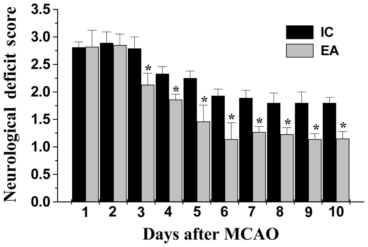
The neuroprotective effect of electroacupuncture at
the Baihui and Shenting acupoints was evaluated by determining the
neurological deficit scores. As hypothesized, rats in the SC group
did not exhibit any manifestations of neurological deficits
(Fig. 1), whereas all the rats in
the IC and EA groups had clear symptoms of cerebral injury.
However, electroacupuncture significantly improved the neurological
deficit scores (P<0.05, EA vs. IC group; Fig. 1). To further verify these results,
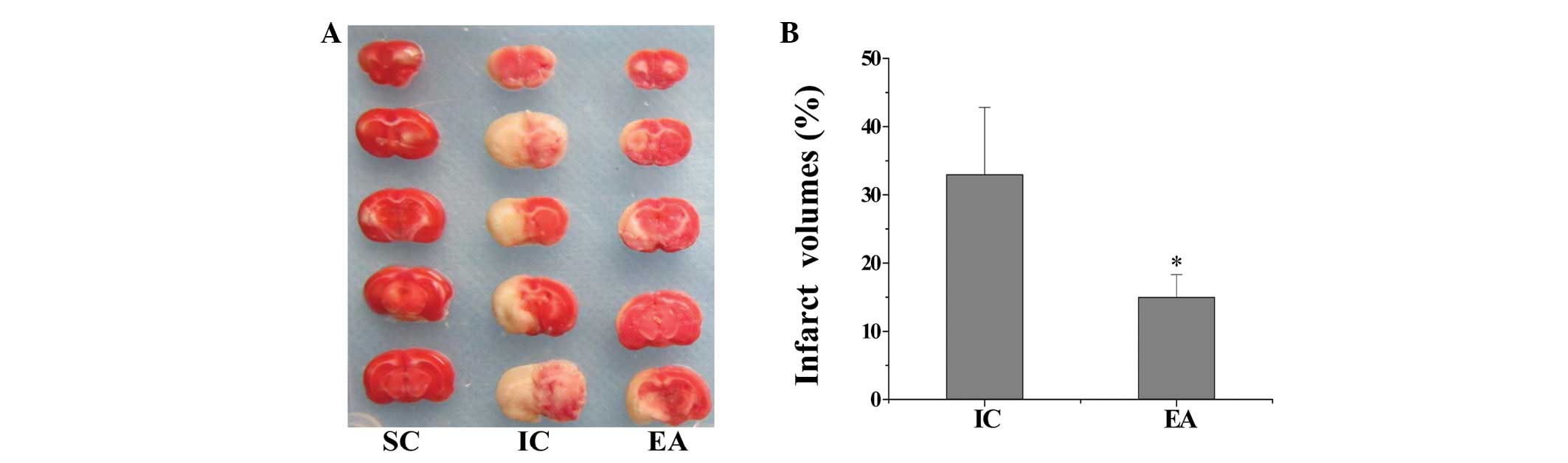
we evaluated the effect of electroacupuncture on cerebral
infarction. As shown in Fig. 2,
electroacupuncture treatment significantly reduced cerebral infarct
volumes in cerebral I/R injured rats (P<0.05, EA vs. IC group).
These results indicate that electroacupuncture at Baihui (DU20) and
Shenting (DU24) may have therapeutic efficacy against cerebral I/R
injury.
Electroacupuncture ameliorates cognitive
impairment in cerebral I/R injured rats
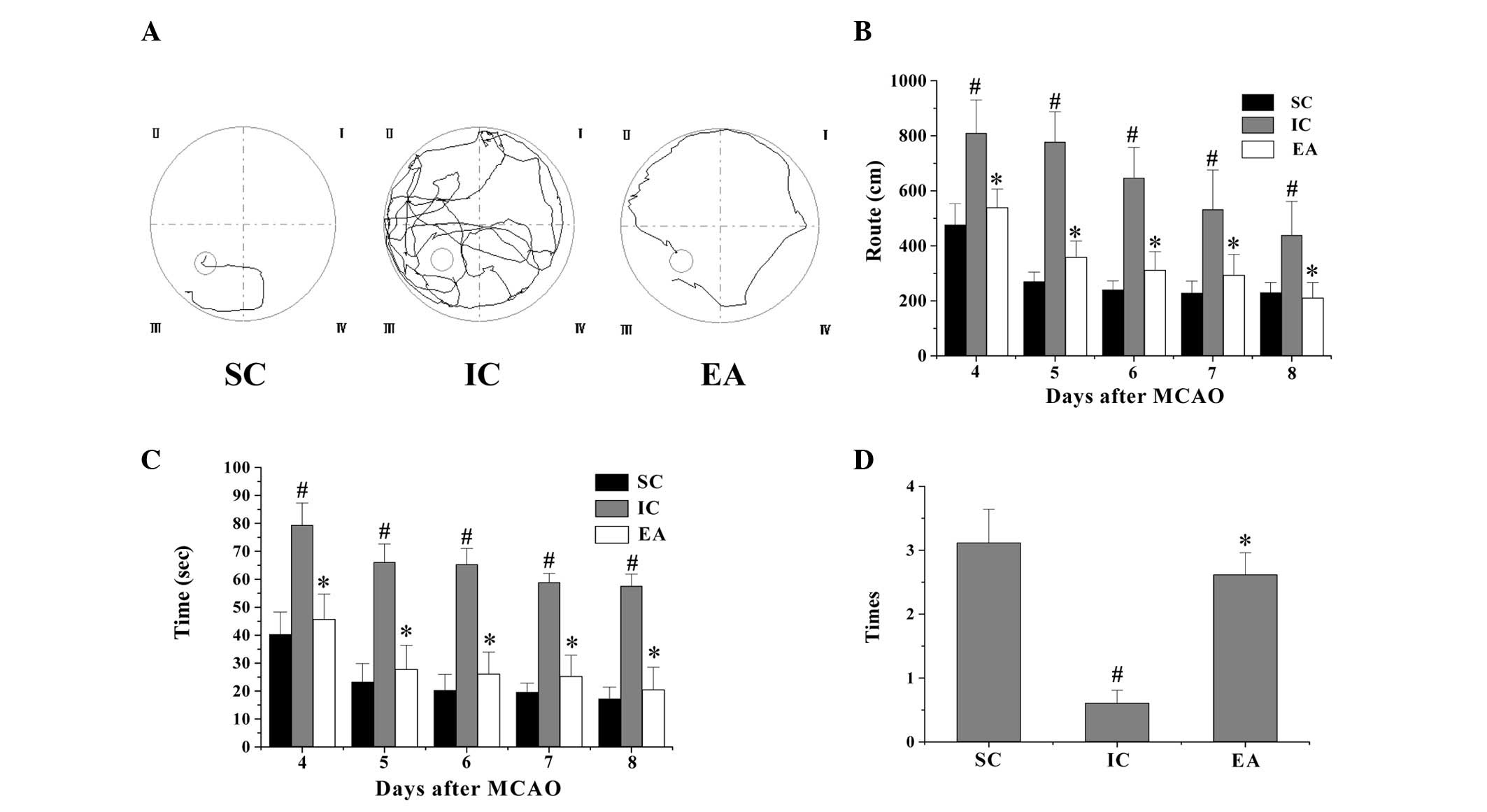
To evaluate the effect of electroacupuncture on
cognitive function, a Morris water maze test was performed on days
4–9 following MCAO surgery. As shown in Fig. 3, the latency and route for rats in
the IC group to reach the hidden platform in the Morris water maze
test markedly increased, whereas the number of times that rats
crossed the location of the platform was significantly decreased
compared with rats in the SC group (P<0.05), indicating that
cerebral I/R injury resulted in cognitive impairment. However,
electroacupuncture significantly decreased the latency and route
length, and increased the number of times the platform was crossed
in the Morris water maze test (P<0.05 vs. the IC group; Fig. 3). Collectively, these data suggest
that electroacupuncture at the Baihui and Shenting acupoints
ameliorate cognitive impairment in cerebral I/R injured rats.
Electroacupuncture inhibits cerebral cell
apoptosis in cerebral I/R injured rats
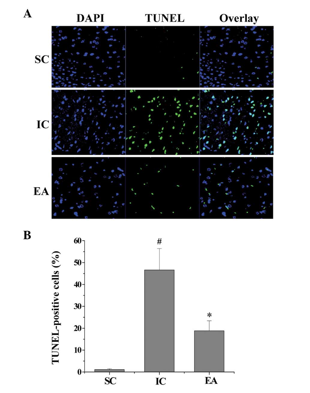
Cognitive impairment is known to be strongly
associated with neuronal cell apoptosis (33); therefore, we evaluated the effect
of electroacupuncture on cell apoptosis in ischemic cerebral
tissues using a TUNEL assay. As shown in Fig. 4, the percentage of TUNEL-positive
cells in the SC and IC groups was 1.2±0.3 and 46.7±9.7%,
respectively (P<0.05), indicating that I/R injury significantly
promoted cerebral cell apoptosis. However, the percentage of
TUNEL-positive cells in the EA group was 18.9±4.5% (P<0.05
compared with the IC group), which demonstrates that
electroacupuncture significantly inhibited the I/R-induced
apoptosis of neuronal cells. This suggests that electroacupuncture
at the Baihui and Shenting acupoints inhibits ischemia-mediated
cerebral cell apoptosis.
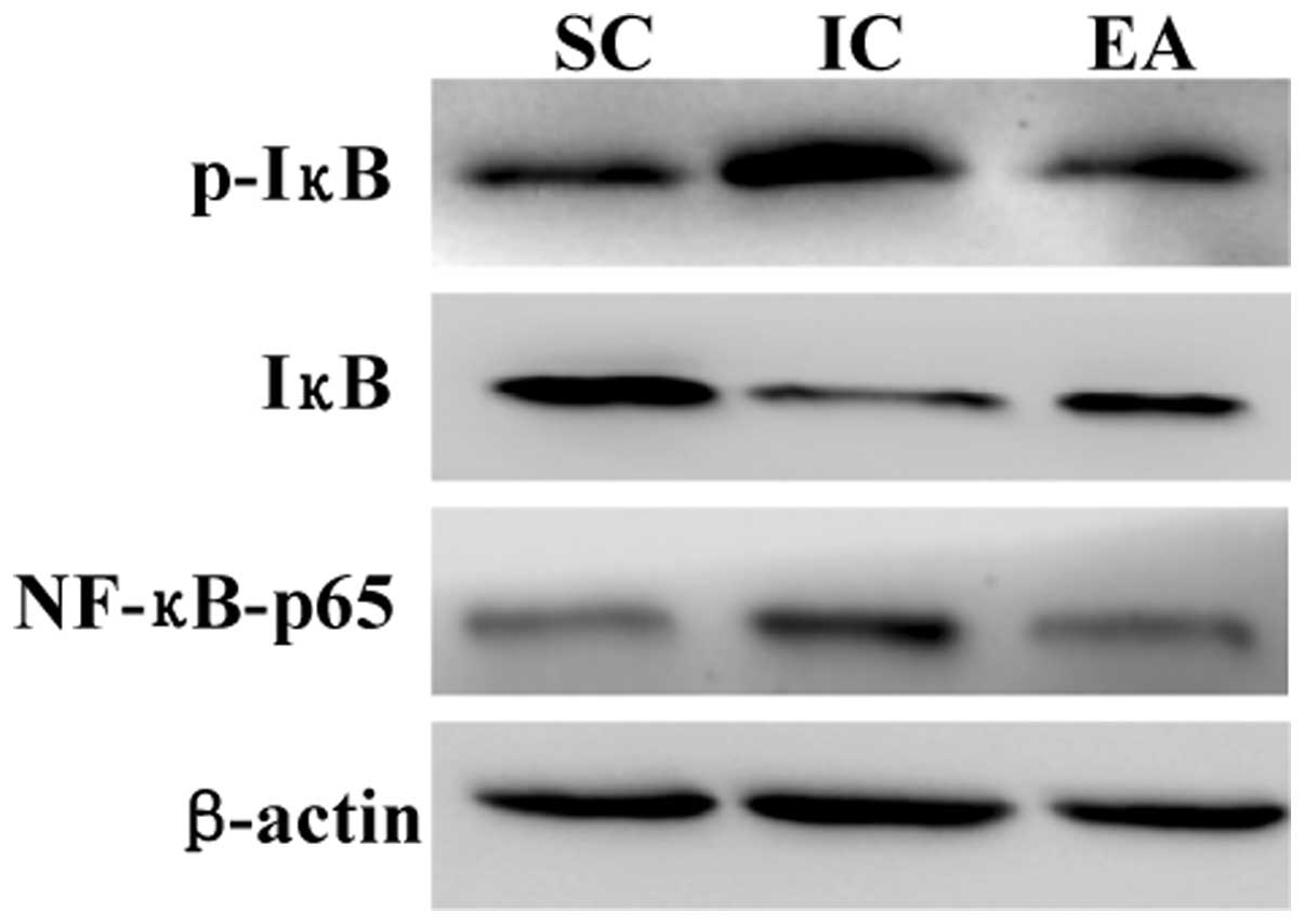
Electroacupuncture inhibits the NF-κB
signaling pathway in cerebral I/R injured rats
Since the activation of NF-κB signaling is important
in cerebral cell apoptosis in ischemic stroke, we examined the
effect of electroacupuncture on the NF-κB pathway in ischemic
cerebral tissues. As shown in Fig.
5, the protein expression of NF-κB p65 and the IκB
phosphorylation levels were significantly increased in the IC group
compared with those in the SC group, suggesting that I/R injury
significantly activates NF-κB signaling. However,
electroacupuncture neutralized the effect of model construction,
suppressing NF-κB protein expression and IκB phosphorylation in
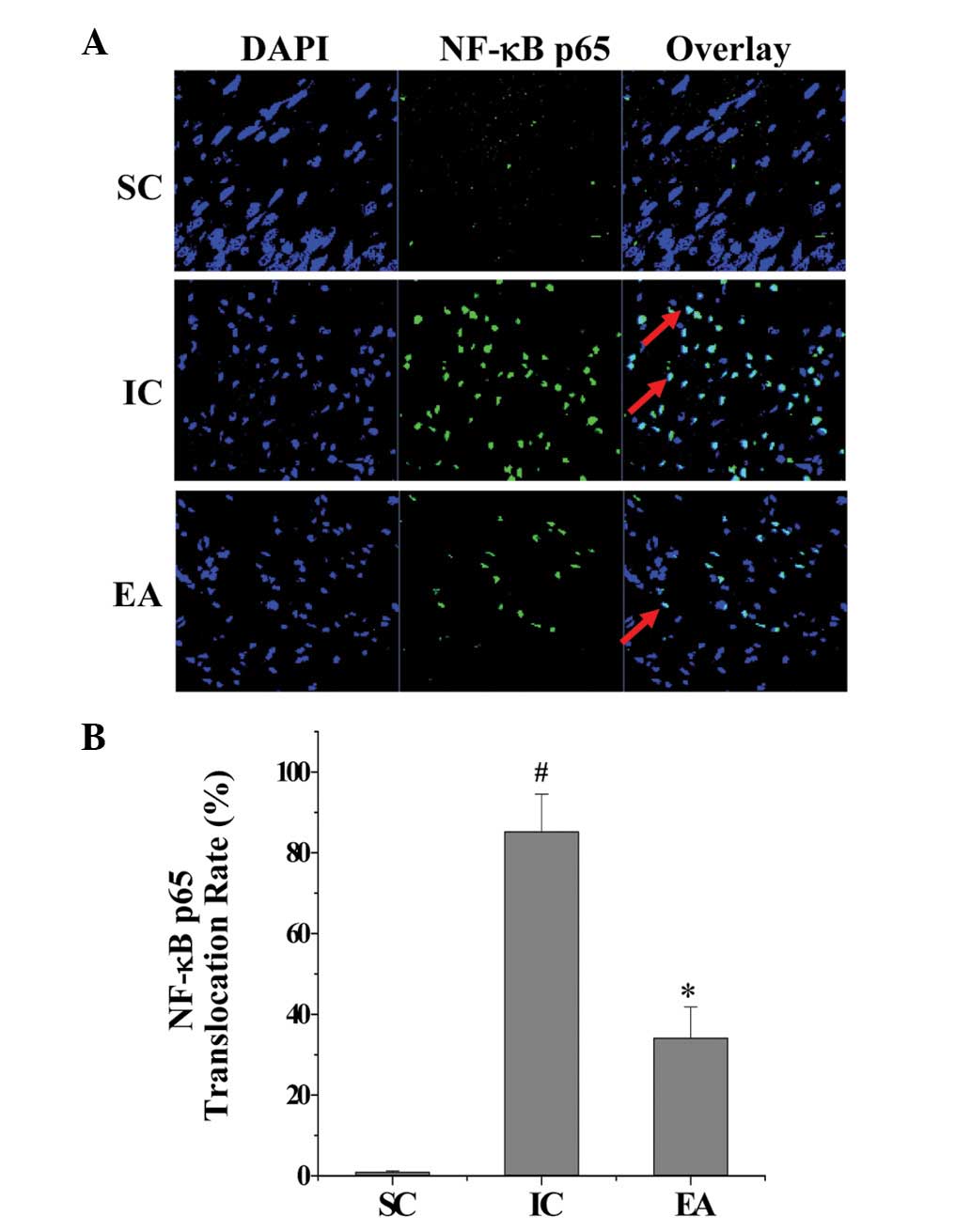
ischemic cerebral tissues. To verify these observations,
immunofluorescence staining was performed to examine the nuclear
translocation of NF-κB, a critical step for NF-κB activation. As
shown in Fig. 6, cerebral I/R
injury significantly induced the nuclear translocation of the NF-κB
p65 subunit; however, this was inhibited by electroacupuncture.
Taken together, these findings indicate that the anti-apoptotic
activity of electroacupuncture in cerebral I/R injured rats was
mediated by inhibition of the NF-κB pathway.
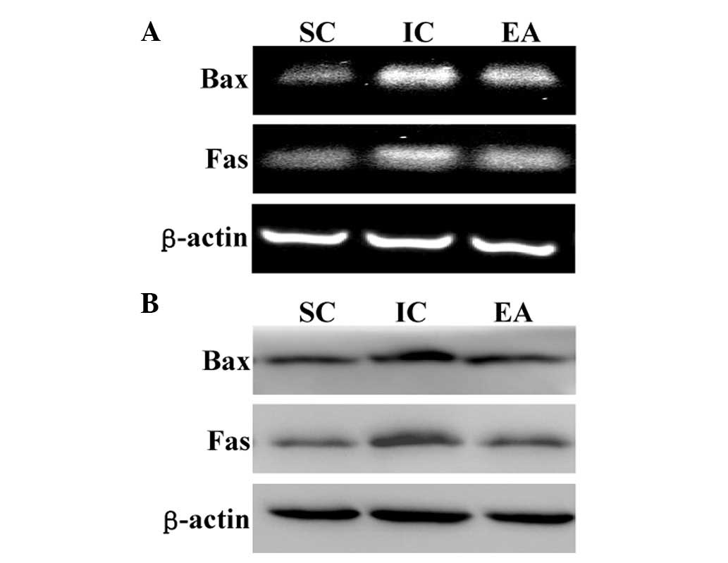
Electroacupuncture downregulates the
apoptotic Fas/Bax genes in cerebral I/R injured rats
Apoptosis is highly regulated by various factors,
including Bax and Fas. Additionally, pro-apoptotic Bax and Fas are
important downstream target genes of the NF-κB signaling pathway.
To further investigate the mechanism of the anti-apoptotic activity
of electroacupuncture, we investigated the mRNA levels and protein
expression of Fas and Bax in ischemic cerebral tissues using RT-PCR
and western blot analysis, respectively. As shown in Fig. 7, cerebral I/R injury markedly
enhanced Bax and Fas expression at transcriptional and
translational levels; however, this was neutralized by
electroacupuncture.
Discussion
Survivors of stroke frequently present with
cognitive impairment, which severely affects their quality of life.
Cognitive impairment is strongly associated with neuronal cell
apoptosis, which is tightly regulated by various intracellular
signal transduction cascades, including the NF-κB pathway (34,35).
Previous studies have demonstrated that NF-κB signaling is
activated in post-stroke cognitive impairment, suggesting that the
NF-κB pathway may be a major target for the treatment of impaired
cognition (24). In TCM,
acupuncture has been used as a complementary and alternative method
for thousands of years. Numerous studies have demonstrated the
clinical efficacy of acupuncture in stroke and cognitive impairment
(30). According to TCM, Baihui
(DU20) and Shenting (DU24) are located on the Du Meridian, which is
considered to be important in the nervous system. Consequently,
these two acupoints are commonly used in China to clinically treat
cognitive impairment (30).
However, the precise mechanism of its therapeutic effect on
impaired cognition remains unclear.
In the present study, a focal cerebral I/R rat model
was constructed and electroacupuncture at the Baihui and Shenting
acupoints was shown to have a neuroprotective effect, as it
significantly ameliorated neurological deficits and reduced
cerebral infarct volume. Additionally, a Morris water maze test
revealed that electroacupuncture improved learning and memory
ability in cerebral I/R injured rats, demonstrating its therapeutic
efficacy against post-stroke cognitive impairment. Furthermore, the
NF-κB pathway was identified to be activated after cerebral I/R
injury, which was consistent with the results of previous studies
(36). However, electroacupuncture
significantly suppressed NF-κB signaling in ischemic cerebral
tissues. The inhibitory effect of electroacupuncture on NF-κB
activation led to the inhibition of cerebral cell apoptosis.
Apoptosis is activated through two major pathways; in the intrinsic
pathway, death signals are integrated at the level of the
mitochondria, while in the extrinsic pathway, death signals are
mediated through cell surface receptors. Both pathways eventually
lead to the activation of caspases and nucleases, resulting in the
destruction of the cell. Bax and Fas, two critical downstream
target genes of the NF-κB pathway, exert their pro-apoptotic
function via the intrinsic and extrinsic pathways, respectively
(37). As hypothesized,
electroacupuncture significantly downregulated the expression of
Bax and Fas at the transcriptional and translational levels.
In conclusion, the present study showed for the
first time that electroacupuncture at the Baihui (DU20) and
Shenting (DU24) acupoints has a therapeutic function in ischemic
stroke and impaired cognition via inhibition of NF-κB-mediated
neuronal cell apoptosis. These results suggest that
electroacupuncture may be a potential therapeutic modality for the
treatment of post-stroke cognitive impairment.
Acknowledgements
This study was sponsored by the International
S&T Cooperation Program of China (ISTCP Program; No.
2011DFG33240), the key International S&T Cooperation Program of
Fujian Science and Technology Department (No. 2010I0007) and
‘Twelfth five-year’ national Technology Support Project (No.
2013BAI10B01).
Abbreviations:
|
NF-κB
|
nuclear factor κB
|
|
I/R
|
ischemia-reperfusion
|
|
MCAO
|
middle cerebral artery occlusion
|
|
TTC
|
2,3,5-triphenyl tetrazolium
chloride
|
|
TUNEL
|
terminal
deoxynucleotidyl-transferase-mediated dUTP nick end labeling
|
References
|
1
|
Jokinen H, Kalska H, Mäntylä R, et al:
Cognitive profile of subcortical ischaemic vascular disease. J
Neurol Neurosurg Psychiatry. 77:28–33. 2006. View Article : Google Scholar : PubMed/NCBI
|
|
2
|
Lindeboom J and Weinstein H:
Neuropsychology of cognitive ageing, minimal cognitive impairment,
Alzheimer's disease, and vascular cognitive impairment. Eur J
Pharmacol. 490:83–86. 2004. View Article : Google Scholar
|
|
3
|
Nyenhuis DL, Gorelick PB, Geenen EJ, et
al: The pattern of neuropsychological deficits in Vascular
Cognitive Impairment-No Dementia (Vascular CIND). Clin
Neuropsychol. 18:41–49. 2004. View Article : Google Scholar : PubMed/NCBI
|
|
4
|
Sachdev PS, Brodaty H, Valenzuela MJ, et
al: The neuropsychological profile of vascular cognitive impairment
in stroke and TIA patients. Neurology. 62:912–919. 2004. View Article : Google Scholar : PubMed/NCBI
|
|
5
|
Mok V, Chang C, Wong A, et al:
Neuroimaging determinants of cognitive performances in stroke
associated with small vessel disease. J Neuroimaging. 15:129–137.
2005. View Article : Google Scholar : PubMed/NCBI
|
|
6
|
Mok VC, Wong A, Lam WW, et al: Cognitive
impairment and functional outcome after stroke associated with
small vessel disease. J Neurol Neurosurg Psychiatry. 75:560–566.
2004. View Article : Google Scholar : PubMed/NCBI
|
|
7
|
Haring HP: Cognitive impairment after
stroke. Curr Opin Neurol. 15:79–84. 2002.
|
|
8
|
Alvarez-Sabín J and Román GC: Citicoline
in vascular cognitive impairment and vascular dementia after
stroke. Stroke. 42(Suppl 1): S40–S43. 2011.PubMed/NCBI
|
|
9
|
Hachinski V and Munoz D: Vascular factors
in cognitive impairment - where are we now? Ann NY Acad Sci.
903:1–5. 2000. View Article : Google Scholar : PubMed/NCBI
|
|
10
|
Tatemichi TK, Desmond DW, Stern Y, et al:
Cognitive impairment after stroke: frequency, patterns, and
relationship to functional abilities. J Neurol Neurosurg
Psychiatry. 57:202–207. 1994. View Article : Google Scholar : PubMed/NCBI
|
|
11
|
Desmond DW, Moroney JT, Paik MC, et al:
Frequency and clinical determinants of dementia after ischemic
stroke. Neurology. 54:1124–1131. 2000. View Article : Google Scholar : PubMed/NCBI
|
|
12
|
Mattson MP: Apoptosis in neurodegenerative
disorders. Nat Rev Mol Cell Biol. 1:120–129. 2000. View Article : Google Scholar
|
|
13
|
Nakka VP, Gusain A, Mehta SL and Raghubir
R: Molecular mechanisms of apoptosis in cerebral ischemia: multiple
neuroprotective opportunities. Mol Neurobiol. 37:7–38. 2008.
View Article : Google Scholar : PubMed/NCBI
|
|
14
|
Broughton BR, Reutens DC and Sobey CG:
Apoptotic mechanisms after cerebral ischemia. Stroke. 40:e331–e339.
2009. View Article : Google Scholar : PubMed/NCBI
|
|
15
|
Cory S and Adams JM: The Bcl2 family:
regulators of the cellular life-of-death switch. Nat Rev Cancer.
2:647–656. 2002. View
Article : Google Scholar : PubMed/NCBI
|
|
16
|
Borner C: Bcl-2 family members:
integrators of survival and death. Biochim Biophys Acta.
1644:71–72. 2004. View Article : Google Scholar : PubMed/NCBI
|
|
17
|
Baeuerle PA and Baltimore D: NF-kappa B:
ten years after. Cell. 87:13–20. 1996.PubMed/NCBI
|
|
18
|
Taglialatela G, Robinson R and Perez-Polo
JR: Inhibition of nuclear factor kappa B (NFkappaB) activity
induces nerve growth factor-resistant apoptosis in PC12 cells. J
Neurosci Res. 47:155–162. 1997. View Article : Google Scholar : PubMed/NCBI
|
|
19
|
Middleton G, Hamanoue M, Enokido Y, et al:
Cytokine-induced nuclear factor kappa B activation promotes the
survival of developing neurons. J Cell Biol. 148:325–332. 2000.
View Article : Google Scholar : PubMed/NCBI
|
|
20
|
Goodman Y and Mattson MP: Ceramide
protects hippocampal neurons against excitotoxic and oxidative
insults, and amyloid beta-peptide toxicity. J Neurochem.
66:869–872. 1996. View Article : Google Scholar : PubMed/NCBI
|
|
21
|
Mattson MP, Goodman Y, Luo H, et al:
Activation of NF-kappaB protects hippocampal neurons against
oxidative stress-induced apoptosis: evidence for induction of
manganese superoxide dismutase and suppression of peroxynitrite
production and protein tyrosine nitration. J Neurosci Res.
49:681–697. 1997. View Article : Google Scholar
|
|
22
|
Grilli M, Pizzi M, Memo M and Spano P:
Neuroprotection by aspirin and sodium salicylate through blockade
of NF-kappaB activation. Science. 274:1383–1385. 1996. View Article : Google Scholar : PubMed/NCBI
|
|
23
|
Won SJ, Ko HW, Kim EY, et al: Nuclear
factor kappa B-mediated kainite neurotoxicity in the rat and
hamster hippocampus. Neuroscience. 94:83–91. 1999. View Article : Google Scholar : PubMed/NCBI
|
|
24
|
van der Kooij MA, Nijboer CH, Ohl F, et
al: NF-kappaB inhibition after neonatal cerebral hypoxia-ischemia
improves long-term motor and cognitive outcome in rats. Neurobiol
Dis. 38:266–272. 2010.PubMed/NCBI
|
|
25
|
Wu JN: A short history of acupuncture. J
Altern Complement Med. 2:19–21. 1996. View Article : Google Scholar
|
|
26
|
Hu HH, Chung C, Liu TJ, et al: A
randomized controlled trial on the treatment for acute partial
ischemic stroke with acupuncture. Neuroepidemiology. 12:106–113.
1993. View Article : Google Scholar : PubMed/NCBI
|
|
27
|
Jansen G, Lundeberg T, Kjartansson J and
Samuelson UE: Acupuncture and sensory neuropeptides increase
cutaneous blood flow in rats. Neurosci Lett. 97:305–309. 1989.
View Article : Google Scholar : PubMed/NCBI
|
|
28
|
Johansson K, Lindgren I, Widner H, et al:
Can sensory stimulation improve the functional outcome in stroke
patients? Neurology. 43:2189–2192. 1993. View Article : Google Scholar : PubMed/NCBI
|
|
29
|
Magnusson M, Johansson K and Johansson BB:
Sensory stimulation promotes normalization of postural control
after stroke. Stroke. 25:1176–1180. 1994. View Article : Google Scholar : PubMed/NCBI
|
|
30
|
Zhao L, Zhang H, Zheng Z, et al:
Electroacupuncture on the head points for improving gnosia in
patients with vascular dementia. J Tradit Chin Med. 29:29–34. 2009.
View Article : Google Scholar : PubMed/NCBI
|
|
31
|
Chen LP, Wang FW, Zuo F, et al: Clinical
research on comprehensive treatment of senile vascular dementia. J
Tradit Chin Med. 31:178–181. 2011. View Article : Google Scholar : PubMed/NCBI
|
|
32
|
Chen AZ, Lin ZC, Lan L, et al:
Electroacupuncture at the Quchi and Zusanli acu points exerts
neuroprotective role in cerebral ischemia- reperfusion injured rats
via activation of the PI3K/Akt pathway. Int J Mol Med. 30:791–796.
2012.PubMed/NCBI
|
|
33
|
Zhang GZ, Liu AL and Zhou YB: Panax
ginseng ginsenoside-Rg2 protects memory impairment
via-anti-apoptosis in a rat model with vascular dementia. J
Ethnopharmacol. 115:440–448. 2008. View Article : Google Scholar : PubMed/NCBI
|
|
34
|
Sarnico I, Lanzillotta A and Benarese M:
NF-kappaB dimers in the regulation of neuronal survival. Int Rev
Neurobiol. 85:351–362. 2009. View Article : Google Scholar : PubMed/NCBI
|
|
35
|
Freudenthal R, Romano A and Routtenberg A:
Transcription factor NF-kappaB activation after in vivo perforant
path LTP in the mouse hippocampus. Hippocampus. 14:677–683. 2004.
View Article : Google Scholar : PubMed/NCBI
|
|
36
|
Zhang W, Potrovita I, Tarabin V, et al:
Neuronal activation of NF-kB contributes to cell death in cerebral
ischemia. J Cerebral Blood F Met. 25:30–34. 2005. View Article : Google Scholar : PubMed/NCBI
|
|
37
|
Kumar A, Takada Y, Boriek AM and Aggarwal
BB: Nuclear factor-kB: its role in health and disease. J Mol Med
(Berl). 82:434–448. 2004. View Article : Google Scholar
|