Identification of microRNAs involved in growth arrest and cell death in hydrogen peroxide-treated human dermal papilla cells

  • Authors:
    • Ok-Yeon Kim
    • Hwa Jun Cha
    • Kyu Joong Ahn
    • In-Sook An
    • Sungkwan An
    • Seunghee Bae
  • View Affiliations

  • Published online on: April 16, 2014     https://doi.org/10.3892/mmr.2014.2158
  • Pages: 145-154
Metrics: Total Views: 0 (Spandidos Publications: | PMC Statistics: )
Total PDF Downloads: 0 (Spandidos Publications: | PMC Statistics: )


Abstract

microRNAs (miRNAs) are small non‑coding RNAs that regulate various biological processes by interfering with the translation of target genes. Several studies have suggested that miRNAs are involved in cellular responses to hydrogen peroxide (H2O2). Reactive oxygen species (ROS) are involved in hair malignancies, however, the H2O2‑induced, miRNA‑dependent regulatory mechanisms of human dermal papilla (HDP) cells are not fully understood. Our previous study demonstrated that changes in miRNA expression function to regulate growth arrest and apoptosis in UVB‑irradiated HDPs. In the present study, miRNA expression was profiled in HDPs treated with H2O2. The transcriptome analysis of H2O2‑treated HDPs enabled the identification of 68 differentially expressed miRNAs (62 were upregulated and 6 were downregulated) and 14,316 putative target genes of the miRNAs. Gene ontology (GO) analysis was utilized to verify that the putative target genes of the altered miRNAs were associated with H2O2‑induced cell growth arrest and apoptosis. This bioinformatics analysis indicated that H2O2‑response pathways involved in growth arrest and apoptosis were significantly affected. The identification of miRNAs and their putative targets may offer new therapeutic strategies for H2O2‑induced hair follicle disorders.

Introduction

The dermal papilla (DP) describes the component of the hair follicle that is involved in hair growth and formation. During development of the hair follicle, the DP is generated by condensation of dermal mesenchymal cells. The DP exists at the base of hair follicles and regulates the hair cycle by providing key signals that control the timing and phase of hair follicle growth and formation (1,2). Anagen is the active growth phase of hair follicles, where the root divides rapidly and adds material to the hair follicle for rapid hair growth. DP stimulates the initiation of the anagen growth phase by secreting Fgf7/10 and TGFβ (3,4). DP-induced β-catenin/Wnt signaling sustains the anagen phase and DP-induced Notch/Wnt5a signaling induces hair follicle differentiation (5,6). The production of reactive oxygen species (ROS) in hair follicles results in hair developmental disorders, including graying and hair loss (79). One such example is alopecia, which is induced by a ROS-mediated reduction of hair growth (9,10).

Oxidative stress that results in crucial damage of DNA, proteins and lipids is implicated in several hair follicle disorders, including graying and hair loss (710). Oxidative stress is increased by the accumulation of ROS, including hydrogen peroxide (H2O2), hydroxyl radical (OH), superoxide anion (O2) and the accumulation of reactive nitrogen species (RNS), including peroxynitrite (ONOO). The generation of ROS and RNS is triggered by an imbalance between pro-oxidants and antioxidants. It has been identified that high levels of oxidative stress induces growth arrest, apoptosis and necrosis in cells (11). Mammalian cells have developed defense mechanisms to neutralize ROS, including antioxidant enzymes and non-enzymatic antioxidants (12,13). In hair follicles, low levels of ROS are generated in the mitochondria, and these act as crucial signaling molecules for hair follicle differentiation and morphogenesis (7). However, high levels of ROS directly damages cellular membranes, lipids, proteins and DNA (9). ROS levels increase with aging, which causes a decrease in the function and number of functional melanocyte cells in hair follicles (8). ROS-responsive microRNA (miRNA) expression regulates the cell cycle and apoptosis in a variety of cells (1416).

miRNAs are short oligonucleotides, consisting of ~19–24 nucleotides (17). miRNAs repress the translation of their target genes by binding to partly complementary sequences in the 3′ untranslated region of the target mRNA (18). miRNAs are involved in the control of diverse cellular processes, including cell growth, apoptosis, development, metabolism, stress adaptation, hormone signaling and differentiation (1822). In hair follicles, a deficiency of miRNAs induced by the knockout of Dicer and Drosha blocks the anagen developmental phase by repression of the catagen phase (23). It has been hypothesized that the miRNA miR-31 is involved in hair follicle growth and hair fiber formation because it targets Krt16, Krt17, Dlx3 and Fgf10 (24). A recent study compared the miRNA expression profiles in balding and non-balding dermal papilla (25).

To the best of our knowledge, no previous studies have reported the miRNA expression profile in H2O2-treated DP. Therefore, in the present study, we analyzed changes in the miRNA expression profiles in DP alone and in DP that had been treated with H2O2. The target genes of significant miRNAs (those with >2-fold changes in expression) were predicted by an in silico prediction algorithm. Based on these data, we derive a model indicating that H2O2-specific miRNAs regulate ROS-responsive cellular functions.

Materials and methods

Cell culture

Human dermal papilla (HDP) cells were purchased from Cellbio Inc. (Seoul, Korea). HDPs were maintained as a monolayer culture in Dulbecco’s modified Eagle’s medium (DMEM; Gibco-Invitrogen Life Technologies, Carlsbad, CA, USA) supplemented with 10% fetal bovine serum (FBS; Sigma-Aldrich, St. Louis, MO, USA) and 1% penicillin/streptomycin. HDPs were cultured in a humidified chamber with 5% CO2 at 37°C.

Cell viability

HDPs (5×10³ cells) were plated in 96-well culture plates and treated with H2O2 for 24 h under the growth conditions described above. The 3-(4,5-dimethylthiazol-2-yl)-2,5-diphenyltetrazolium bromide (MTT) colorimetric assay was performed by adding 0.5 μg/ml MTT (Sigma-Aldrich) to the culture medium. Following 1 h of incubation under normal cell growth conditions, the growth medium was removed and 200 μl of dimethyl sulfoxide was added to each well. The absorbance was measured at 490 nm using a microplate reader (iMark; Bio-Rad, Hercules, CA, USA).

Cell cycle analysis

The cell cycle was determined by flow cytometry via propidium iodide (PI) staining. Cell cycle analysis was performed as described previously (26).

RNA preparation and miRNA microarray

Total RNAs were extracted by RiboEX (GeneAll Biotechnology Co., Ltd., Seoul, Korea) and quantified by measuring the optical density ratio. The miRNA microarray (SurePrint G3 Human v16.0 miRNA 8×60K; Agilent Technologies, Santa Clara, CA, USA) was performed according to the manufacturer’s instructions. miRNA was stained by pCp-Cy3 (Agilent Technologies), combined with T4 ligase (Agilent Technologies) and hybridized to a probe on the microarray. The microarray was imaged using the Agilent microarray scanner and digitized by Feature extraction. The digitized data were analyzed for fold change, miRNA potential target and gene ontology (GO) using Genespring GX version 11.5 (Agilent Technologies).

Prediction of miRNA target genes and GO analysis

The putative target genes of significantly up and downregulated miRNAs were identified by the web tool TargetScan. Target gene prediction was performed on significant miRNAs with 50 context score percentile using the conserved and non-conserved database. The putative target genes were identified and sorted by GO of each gene.

Statistical analysis

Statistical significance was determined by Student’s t-test. P<0.05 was considered to indicate a statistically significant difference.

Results

H2O2 treatment decreases the viability of HDPs via cell cycle arrest or apoptosis

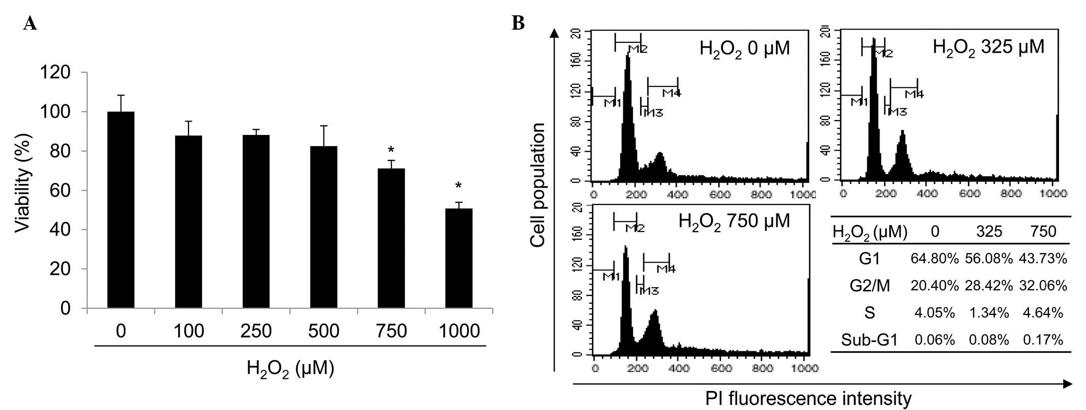
To determine the cytotoxicity of H2O2, we initially examined the viability and status of the G1 or G2/M population in HDPs. HDPs were treated with H2O2 (0–1,000 μM) and the resulting viability was measured using the MTT assay (Fig. 1A). Following 24 h of H2O2 treatment, a decrease in cell viability occurred in a dose-dependent manner. In the presence of 750 and 1,000 μM H2O2, a significant (P<0.05) decrease in cell viability to 71.23 and 50.60% of that in control cells, respectively, was observed (Fig. 1A). In the presence of 750 μM H2O2, HDPs exhibited a 21.07% reduction in G1 phase and an 11.66% increase in G2/M phase, compared with those of the control (Fig. 1B). These data suggest that H2O2 decreases cell viability of HDPs by inducing cell cycle arrest at the G2/M phase and cell death.

Identification of H2O2-induced changes in miRNA expression in HDPs

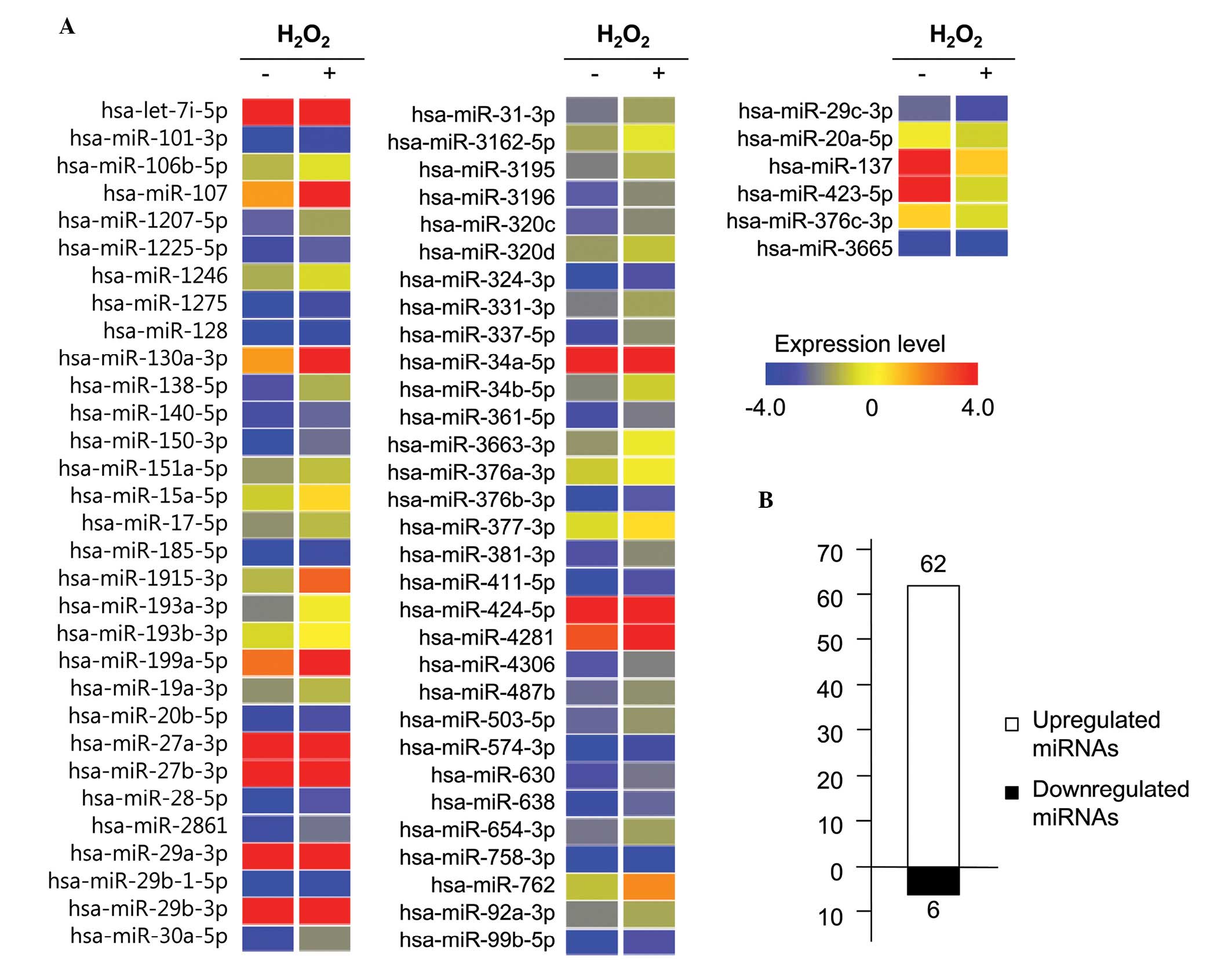
H2O2-induced changes in the miRNA expression profiles were analyzed using the Agilent miRNA microarray, which contains 1,368 probes that are able to detect 1,205 human miRNAs. The fluorescence intensity data for each sample was normalized by global normalization. To eliminate disorderly data, miRNA expression data were selected by requiring a present-flag for at least one of all samples. Using this method, 155 miRNAs were selected out of 1,205 human miRNAs detected by present-flag selection. To identify H2O2-regulated miRNAs, the refined data were compared for control HDPs grown under normal conditions and HDPs treated with 750 μM H2O2 for 24 h. The results demonstrated that the expression levels of 68 miRNAs were altered at least 1.5-fold in response to treatment with H2O2. The 68 miRNAs are presented in Fig. 2. Fold-change analysis revealed that 62 miRNAs are upregulated and 6 miRNAs are downregulated at levels of 1.5-fold or greater in control HDPs grown under normal conditions and HDPs treated with 750 μM H2O2 for 24 h (Table I).

Table I

Up and downregulated miRNAs in H2O2-treated HDPs.

Table I

Up and downregulated miRNAs in H2O2-treated HDPs.

A, Upregulated

miRNAFold changeChrmiRNAFold changeChrmiRNAFold changeChr
hsa-let-7i-5p1.52chr12hsa-miR-19a-3p1.52chr13hsa-miR-361-5p1.86chrX
hsa-miR-101-3p1.91chr1hsa-miR-20b-5p1.56chrX hsa-miR-3663-3p2.73chr10
hsa-miR-106b-5p1.59chr7hsa-miR-27a-3p2.13chr19 hsa-miR-376a-3p1.65chr14
hsa-miR-1071.55chr10hsa-miR-27b-3p1.56chr9 hsa-miR-376b-3p2.39chr14
hsa-miR-1207-5p2.08chr8hsa-miR-28-5p2.17chr3hsa-miR-42812.15chr5
hsa-miR-1225-5p1.62chr16hsa-miR-28612.29chr9hsa-miR-43061.65chr13
hsa-miR-12461.60chr2hsa-miR-29a-3p1.60chr7hsa-miR-487b1.51chr14
hsa-miR-12751.75chr6 hsa-miR-29b-1-5p2.14chr7hsa-miR-503-5p1.66chrX
hsa-miR-1281.54chr2hsa-miR-29b-3p3.26chr1hsa-miR-574-3p2.01chr4
hsa-miR-130a-3p1.63chr11hsa-miR-30a-5p3.01chr6hsa-miR-6301.66chr15
hsa-miR-138-5p2.84chr3hsa-miR-31-3p1.58chr9hsa-miR-6382.33chr19
hsa-miR-140-5p1.55chr16 hsa-miR-3162-5p2.00chr11hsa-miR-654-3p1.63chr14
hsa-miR-150-3p3.95chr19hsa-miR-31951.81chr20hsa-miR-758-3p3.00chr14
hsa-miR-151a-5p1.52chr8hsa-miR-31961.67chr20hsa-miR-7622.34chr16
hsa-miR-15a-5p1.76chr13hsa-miR-320c1.52chr18hsa-miR-92a-3p1.51chr13
hsa-miR-17-5p1.56chr13hsa-miR-320d1.52chr13hsa-miR-99b-5p2.01chr19
hsa-miR-185-5p1.64chr22hsa-miR-324-3p2.38chr17hsa-miR-377-3p1.55chr14
hsa-miR-1915-3p2.81chr10hsa-miR-331-3p1.51chr12hsa-miR-381-3p2.03chr14
hsa-miR-193a-3p3.65chr17hsa-miR-337-5p2.52chr14hsa-miR-411-5p2.47chr14
hsa-miR-193b-3p1.56chr16hsa-miR-34a-5p2.04chr1hsa-miR-424-5p1.72chrX
hsa-miR-199a-5p1.61chr1hsa-miR-34b-5p2.19chr11

B, Downregulated

miRNAFold changeChrmiRNAFold changeChrmiRNAFold changeChr

hsa-miR-29c-3p−1.51chr1hsa-miR-137−1.50chr1 hsa-miR-376c-3p−1.56chr14
hsa-miR-20a-5p−1.51chr13hsa-miR-423-5p−1.57chr17hsa-miR-3665−2.36chr13

[i] miRNAs, microRNAs; H2O2, hydrogen peroxide; HDPs, human dermal papilla cells.

Identification of H2O2-specific miRNA putative target genes and GO analysis

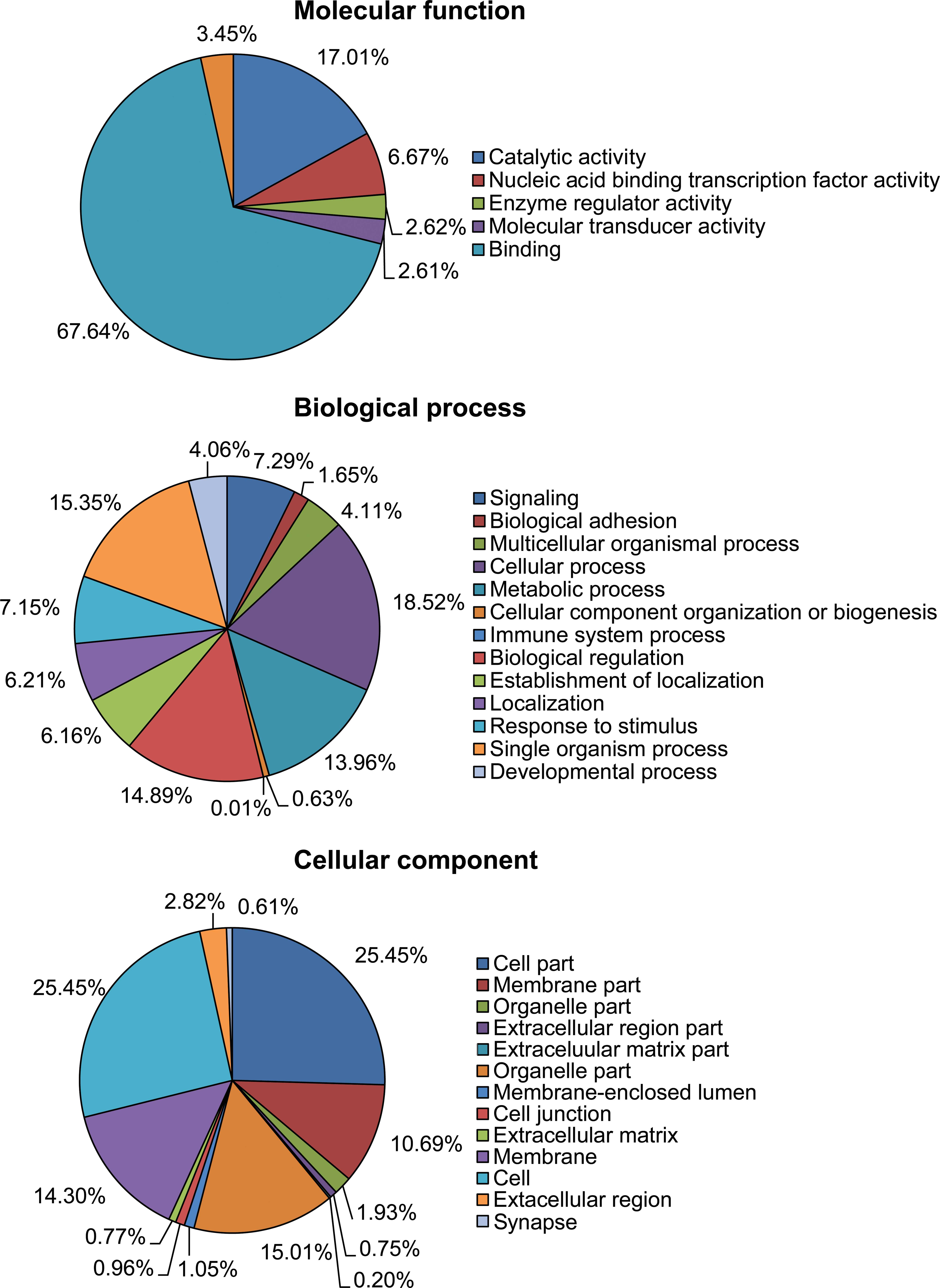
Our study identified 68 novel miRNAs that were significantly up or downregulated in response to H2O2 treatment. As miRNA functions as RNA interference during mRNA translation, we predicted that H2O2-specific miRNAs may regulate all or a number of the H2O2-response genes. Therefore, the target genes of H2O2-specific miRNAs were analyzed using the bioinformatics target gene prediction program TargetScan. For target gene analysis, default parameters were utilized, 50 context score percentile in the conserved and nonconserved database. H2O2-induced miRNAs targeted 14,046 genes; H2O2-repressed miRNAs targeted 6,019 genes. To identify the cellular functions of the putative target genes, GO analysis was performed, which is a method that categorizes the genes according to the cellular function classified for a standard for each gene. As illustrated in Fig. 3, GO analysis identified the following cellular functional activities: for molecular function, catalytic (17.01%), nucleic acid binding transcription factor (6.67%), enzyme regulator (2.62%), molecular transducer (2.61%), binding (67.63%) and transporter (3.45%); for biological process, signaling (7.29%), biological adhesion (1.65%), multicellular organismal (4.11%), cellular (18.52%), metabolic (13.96%), cellular component organization or biogenesis (0.63%), immune system (0.01%), biological regulation (14.89%), establishment of localization (6.16%), localization (6.21%), response to stimulus (7.15%), single organism (15.35%) and developmental (4.06%); for cellular component part, cell (25.45%), membrane (10.69%), organelle (1.93%), extracellular region (0.75%), extracellular matrix (0.2%), organelle (15.01%), membrane-enclosed lumen (1.05%), cell junction (0.96%), extracellular matrix (0.77%), membrane (14.3%), cell (25.45%), extracellular region (2.82%) and synapse (0.61%). All of these were implicated in UVB-mediated responses in HDPs. These GO annotations provided comprehensive information on the function of H2O2-regulated transcripts in HDPs. TargetScan was used to predict the gene targets of the top five miRNAs that demonstrated the greatest increase or decrease in expression levels. The putative miRNA target genes were sorted into cell cycle, apoptosis and cell growth, and proliferation-related GO (Tables II and III). Cell cycle-related GO included cell cycle (GO:0007049), cell cycle arrest (GO:0007049), negative regulation of cell cycle (GO:0045786) and regulation of cell cycle (GO:0051726). Apoptosis-related GO included apoptotic process (GO:0006915), apoptotic signaling pathway (GO:0097190), cell death (GO:0008219), death (GO:0016265), programmed cell death (GO:0012501), regulation of apoptotic process (GO:0042981), regulation of cell death (GO:0010941), regulation of execution phase of apoptosis (GO:1900117) and regulation of programmed cell death (GO:0043067). Cell growth-related and cell proliferation-related GO included positive regulation of cell proliferation (GO:0008284), regulation of cell growth (GO:0001558), regulation of cell proliferation (GO:0042127) and regulation of growth (GO:0040008).

Table II

Predicted target genes of the top five most upregulated miRNAs in H2O2-treated HDPs.

Table II

Predicted target genes of the top five most upregulated miRNAs in H2O2-treated HDPs.

Target genes and functions

miRNACell cycleApoptosisCell growth and proliferation
hsa-miR-150-3p---
hsa-miR-193a-3pPSRC1, JUB, CCNT2, CCPG1, DLGAP5, PAPD5, SIAH1, TSC1, CABLES2, SMC1A, ERBB2IP, SEP3, KLK10, PTEN, CCND1, NEDD9, NF1, NF2, SUFU, CTF8, SASS6, E2F6, SENP5, VASH1, SPECC1L, CADM1, APPL1, GAS1, ING1, IRF1, FOXO4, ATMBCL2L10, DIDO1, WDR92, RFFL, ZNF346, CADM1, RNF144B, GAS1, TNFRSF21, IL1A, JAK2, MCL1, MLL, OSM, SH3GLB1, PDCD2, DRAM, PSEN1, BAG1, BCL2L2, RTKN, ATXN1, ELMO2, SIAH1, TIAL1, TNFAIP3, TNFRSF1B, TRAF1, PHLDA2, DYRK2, AIFM2, TNFSF9, TNFRSF10B, SQSTM1, EBAG9, TP53INP1, ARHGEF6, PPM1FBMPR2, TDGF1, KAZALD1, PPP1R9B, WISP1, EBAG9, SOCS6, CIAO1, NET1, ENOX2, TNFSF13B, FLT1, FLT3, HOXD13, IGFBP5, KIT, KRAS, NEDD9, NODAL, OSM, CCDC88A, PRKCQ
hsa-miR-29b-3pPPM1D, CDC23, CCND2, CCNF, UBA3, CCNT2, AURKB, HDAC4, DCLRE1A, SUPT5H, TACC1, PTP4A1, CDC7, GSG2, SMPD3, RCC2, PTEN, NEDD9, ZAK, XRN1, CDK2, CDK6, HTATIP2, CGREF1, STAG2, SEP9, DBF4, MAPRE2, E2F7, SLC5A8, AHR, VASH1, MAPRE1, CLASP2, SEP6FEM1B, HTATIP2, IL24, SLC5A8, AHR, FOXO3, PPP1R13B, SIRT1, ZNF346, CECR2, BIRC2, FAS, IL2RA,, MCL1, SH3GLB1, ZAK, CYCS, DIABLO, DUSP22, BIRC6, BAK1, ATXN1, CIDEC, SGK1, ISG20L1, TNFAIP3, TNFRSF1A, TRAF5, FAM130A1, CASP7, AIFM2, BMF, RABEP1, TP53INP1, TRAF4, SLKBIRC6, PURA, TRAF5, VEGFA, EPC1, CDC7, CREG1, WISP1, CCND2, MORF4L2, CDK2, MORF4L1, CHRNA7, IFNG, IGF1, LIF, LIFR, NDN, NEDD9, PDGFRB, ING3, PPP2CA
hsa-miR-30a-5pTBRG1, CCNK, CCNA1, BCL10, LMLN, UBA3, CCNT2, CCNE2, SEP7, NEK4, TFDP1, TSC1, UBE2I, PTP4A1, EVI5, MIS12, SUV39H2, CHAF1B, CDC7, RECK, RGS2, RCBTB1, NEK1, NF1, PNN, RBM5, STAG2, DBF4, ESCO1, ESCO2, DDX11, E2F3, EPHB2, CLASP2, SEP6, CD2AP, NSL1, MTBP, INCENP, JAG2, KIF11, RHOBBCL2L11, EDAR, NLRP3, RFFL, TICAM1, PRUNE2, TRIM35, SIRT1, RNF144B, TCTN3, GJA1, CECR2, SH3KBP1, HTT, HIP1, IL1A, IL2RA, IL17A, MAP3K5, MLL, MNT, BCL2L15, NAIP, DDIT4,. C8orf4, AVEN, PTGER3, BIRC6, TRIB3, ATXN1, SOX9, ACTC1, TFDP1, TIA1, CASP3, UNC5C, TNFSF9, TNFRSF10D, TNFRSF10B, BCL10, ATG12, EBAG9, DLG5, ARHGEF6, ATG5C19orf10, BIRC6, BNC1, SOX9, VIPR1, CAMK2D, CDC7, CDCA7, CUL3, SOCS1, CREG1, OCS3, EBAG9, CFDP1, ENOX2, ADRA1D, ADRA2A, ADRB2, DDX11, TLX1, IL7, JAG2, KRAS, LIFR, LYN, MAFG, NOV, PDGFRB, PPP2CA
hsa-miR-758-3pPARD6G, CCNG1, AURKB, MPHOSPH1, DLGAP5, DMTF1, BMP7, NEK4, TACC1, TP53BP2, VHL, WEE1, PPAPDC1B, RBL1, PPP1CB, NUSAP1, WWOX, ZAK, CDKN1B, KHDRBS1, STAG2, FOXN3, SGOL1, E2F6, CLASP2, PDS5A, CD2AP, CADM1, EID1, APPL1, LIN9, STRN3, ING1, LIG4, MCM3, MCM6MALT1, UNC5D, CTSB, RYBP, CADM1, APAF1, PAK1, ZAK, KRT20, APTX, IFT57, TNFRSF19, BCL2L1, ATXN1, PERP, UBE2Z, BNIP2, TIA1, TIAL1, TP53BP2, C10orf97, ACTN4, RABEP1, EBAG9, BAG2, BRESLAMF1, STAT5B, KLF5, BLZF1, CDC2L5, EBAG9, SOCS6, NAMPT, CDKN1B, CFDP1, ENOX2, TCFL5, FGF7, ID4, IGF1, ILK, LIFR, NDN, CRIM1, POU3F2

[i] miRNAs, microRNAs; H2O2, hydrogen peroxide; HDPs, human dermal papilla cells.

Table III

Predicted target genes of the top five most downregulated miRNAs in H2O2-treated HDPs.

Table III

Predicted target genes of the top five most downregulated miRNAs in H2O2-treated HDPs.

Target genes and functions

miRNACell cycleApoptosisCell growth and proliferation
hsa-miR-376c-3pJUB, CCNB3, CCNT2, NOLC1, MTSS1, SIAH2, AURKA, VHL, HMGA2, CHAF1B, ERBB2IP, PTEN, NPAT, PAFAH1B1, GMNN, XRN1, MPHOSPH8, SPIN1, POLS, LZTS1, CHEK2, SASS6, GADD45A, MAPRE1, KANK1, SASH1, APPL1, ANXA1, HPGD, SEP14, MN1EGLN3, RFFL, ACVR1C, PARP4, CNTN4, GADD45A, DAPK2, TNFAIP8, SGMS1, ARF6, MCL1, APIP, GULP1, RHOT1, DUSP22, ROCK1, ATXN1, SHB, SIAH2, ACTC1, PHLDA2, FASTKD3, CASP3, CASP7, AIFM1, ATG12, CD5LMARK4, CXCL5, BMPR2, SSR1, STAT5A, TGFBR1, TNFSF4, HMGA2, SOCS2, SOCS6, CD86, CD47, MORF4L2, DNAJA2, CFDP1, TNFSF13B, TBC1D8, SOCS4, FGF7, FLT1, FLT3, RBM9, ANXA1, IL2, CXCL10, KRAS, NOV, IL17RB
hsa-miR-423-5pJUB, CCNF, PKMYT1, MPHOSPH1, DMTF1, SUV39H1, TACC1, DBF4B, BIN3, MAPK3, MAPK4, PTCH1, RAP1A, UPF1, TSPYL2, NF2, PAFAH1B1, CINP, SUFU, PFDN1, RBM5, CDKN1A, HMG20B, TXNIP, SPIN1, RCC1, FOXN3, LZTS1, PMF1, E2F7, KCTD11, SNF1LK, DAB2IP, CYLD, DDX11, E2F2, EP300, PDS5A, NSL1, SGSM3, BIRC5, INCENP, IRF1, MCM7, MN1, NBL1, NBN, SEP3, SEP8BCL2L11, IL24, DIDO1, RFFL, CLU, ACVR1C, DAP, DEDD2, DOCK1, E2F2, EIF5A, EP300, ERN1, PTK2B, FOSL2, CYFIP2, CIDEB, HIPK2, SAP30BP, CARD10, HTT, KCNIP3, HIP1, BIRC5, IAPP, IL17A, LTBR, MDM4, MLL, NGFR, NDUFA13, PDCD1, RNF216, PPARD, PRKCG, BAG1, BCL2L1, RTKN, NOD2, BNIP1, BOK, PRDX2, CACNA1A, BCL2L14, BIRC7, CASP2, PLA2G6, TRAF7, AIFM2, LGALS12, RIPK1, TNFSF12, TNFRSF14, TNFRSF10D, SQSTM1, NOL3, BMF, DNAJA3, TAOK2, NTN1, BREC19orf10, RAC2, SHC1, STAT5A, STAT5B, TGFBR2, VEGFB, PROK1, CUL3, CDC2L5, FGF18, NRP1, SOCS3, SCGB3A1, S1PR2, TAOK2, CIAO1, HTRA3, NTN1, CLU, ADM, CSF1, ADRA1D, KCTD11, CTF1, ADRA2A, DDX11, DHPS, S1PR3, PTK2B, FOXO1, FLT3LG, RBM9, HOXC10, VSX2, IGF1, IGFBP6, KRT6A, MAFG, ODC1, PDGFRB, C9orf127, IL17RB, CCDC88A, PRKCQ
hsa-miR-3665---
hsa-miR-20a-5pPARD6B, JUB, RUNX3, CDC23, PCAF, BCL10, CCND2, CCNG2, RNF8, CCNT2, PKMYT1, ZNF830, NEK9, KIF23, RASSF2, RB1CC1, SEP7, CDC25A, DLEC1, DMTF1, PAPD5, BMP2, STK11, TACC1, BUB1, VHL, WEE1, PTP4A1, SUV39H2, MAP9, PPP6C, ERBB2IP, MAPK1, MAPK4, PCNP, PTEN, RAD17, RAP1A, RB1, RBL1, RBL2, CCND1, TIPIN, SEP6, NPAT, PAFAH1B1, FZR1, ZAK, XRN1, CHAF1A, CDKN1A, TXNIP, CETN2, SEP2, CIT, TUSC2, SNF1LK, CYLD, E2F1, E2F3, FANCD2, C11orf82, HEPACAM, MAPRE1, MAPRE3, SEP9, PDS5A, SASH1, CLASP1, RABGAP1, GAK, NSL1, APPL1, FBXO5, CKAP2, PDCD4, C13orf15, GRLF1, RACGAP1, BIRC5, TMPRSS11A, IRF1, MCC, MCM3, NBL1NOD1, FASTK, EGLN3, NLRP3, E2F1, EIF4G2, C11orf82, CARD8, ACIN1, FAIM2, SULF1, RYBP, SGMS1, MAGI3, CKAP2, GJA1, TNFRSF21, PDCD4, BIRC5, APP, MCL1, MDM4, MAP3K5, BCL2L15, OSM, SH3GLB1, PDE1B, ZAK, RHOT1, BCAP29, PTGER3, BCL2, PERP, ISG20L1, BNIP2, TNFAIP3, SLTM, C10orf97, RNF34, FXR1, CASP2, PLA2G6, CASP7, CASP8, CUL1, AIFM2, TNFRSF10D, TNFRSF10B, IER3, SQSTM1, SGPL1, TAX1BP1, BCL10, RABEP1, ATG12, DEDD, CD5L, STK17B, TP53INP1, ATG5, LITAF, TRAF4, SLKPURA, BCL2, CXCL5, BMPR2, SSR1, TBX3, TGFBR2, TSG101, CAMK2D, ITCH, CDCA7, BRMS1L, CUL3, CCND2, S1PR2, SOCS6, ARHGEF11, DNAJA2, CFDP1, HOXB13, MORF4L1, TBC1D8, CHRNA7, S1PR3, ERBB3, HEPACAM, FGF4, FGF7, LRP12, IGFBP7, LIF, LIFR, OSM, DERL2, CRIM1, ING3, TIPIN, PPP2CA
hsa-miR-29c-3pPPM1D, CDC23, CCND2, CCNF, UBA3, CCNT2, AURKB, HDAC4, DCLRE1A, SUPT5H, TACC1, PTP4A1, CDC7, SMPD3, RCC2, PTEN, NEDD9, ZAK, XRN1, CDK2, CDK6, HTATIP2, GREF1, STAG2, SEP9, DBF4, MAPRE2, FOXN3, E2F7, SLC5A8, AHR, VASH1, MAPRE1, CLASP2, SEP6, PPP1R13B, MCC, MLF1, NASPFEM1B, HTATIP2, IL24, SLC5A8, AHR, FOXO3, PPP1R13B, SIRT1, ZNF346, CECR2, BIRC2, FAS, IL2RA, MCL1, SH3GLB1, ZAK, CYCS, DIABLO, DUSP22, BIRC6, BAK1, ATXN1, CIDEC, SGK1, ISG20L1, TNFAIP3, TNFRSF1A, TRAF5, FAM130A1, CASP7, AIFM2, BMF, RABEP1, TP53INP1, TRAF4, SLKBIRC6, PURA, TRAF5, VEGFA, EPC1, CDC7, CREG1, CCND2, MORF4L2, CDK2, MORF4L1, CHRNA7, RBM9, IFNG, IGF1, LIF, LIFR, NDN, NEDD9, PDGFRB, ING3, PPP2CA

[i] miRNAs, microRNAs; H2O2, hydrogen peroxide; HDPs, human dermal papilla cells.

Discussion

ROS, such as H2O2, are generated as reactive byproducts of cellular metabolism in the mitochondria. The intracellular level of ROS is distinctly regulated by the cellular antioxidant system, including non-enzymatic and enzymatic antioxidants (27). The ROS level is increased in response to environmental stresses, including UV irradiation, toxic chemicals, heat and even high glucose concentrations (27,28). High levels of ROS induce cell cycle arrest, senescence and apoptosis due to ROS damage of cellular membranes, lipids, proteins and DNA (710). In the present study, and in others previously, it has been identified that H2O2 induces growth arrest in HDPs (Fig. 1) (2931). The H2O2-induced growth arrest occurs later within the cascade of events activated in response to H2O2 treatment. Previous investigations identified H2O2-responsive miRNAs, including miR-34 and miR-145, which also are implicated in the ROS-responsive pathway (32,33). Therefore, miRNA appears to be required for H2O2-dependent growth arrest.

The present study identified 68 miRNAs that were regulated by H2O2 in HDPs (Fig. 2). miR-193-3p and miR-29b increased in H2O2-treated HDPs and induced apoptosis by targeting MCL-1 (34,35). MCL-1 is a BCL-2 family member that is involved in mitochondria-dependent intrinsic apoptosis (36). MCL-1 represses apoptosis by preventing the formation of mitochondrial membrane potential (36). Our data, together with those of Lin et al (15), demonstrated that miR-193a-3p increased in response to ROS and subsequently induced cell death. A previous study revealed that miR-30a-5p was upregulated by H2O2, it repressed autophagy by targeting beclin-1 and eventually induced apoptosis (37). In the present study, it was demonstrated that miR-20a-5p and miR-423-5p were repressed by H2O2 in HDPs (Table I). miR-20a-5p had multiple target genes, including BNIP2, APP, ASK1 and TNKS2. Therefore, miR-20a-5p regulated proliferation, migration, invasion and inflammation by the regulation of its target genes (3841). miR-424-5p targeted p21Cip1/Waf1, which functions in proliferation and G1 phase transition (42).

Numerous studies have demonstrated that ROS, including H2O2, induces intrinsic apoptosis (4345). H2O2-induced apoptosis is regulated by mitochondrial membrane permeability, which is regulated by the BCL-2 family (36). The anti-apoptotic BCL-2 family, including BCL2L10, BCL2L11, BCL2L2 and BCL10 were predicted as targets of miRNAs that were upregulated by H2O2 (Table II). The pre-apoptotic BCL-2 family members BOK and BAK1 were predicted as targets of miRNAs that were downregulated by H2O2 (Table III). Cell cycle regulating proteins, such as cyclins and CDKs, function during each phase of the cell cycle (G1, S, G2 and M) (46). CCNA1 and CDK2 are required for the regulation of the G2/M phase (47,48). The results of the present study predict that hsa-miR-30a-5p and hsa-miR-29b-3p target CCNA1 and CDK2. These data suggest that H2O2-mediated growth arrest and cell death in HDPs is associated with the changes in expression of specific miRNAs.

Bioinformatics analysis of miRNA expression profiles, miRNA target genes and the GO of target genes provided a more holistic view of the underlying cellular mechanisms that occur in response to H2O2-induced growth arrest and apoptosis. The identification of miRNAs and their putative targets may offer new therapeutic strategies for H2O2-induced hair follicle disorders, such as hair loss.

Acknowledgements

The authors are grateful to all the members of our research group for their support and advice regarding this study. This study was supported by the KU Research Professor Program of Konkuk University.

References

1 

Oliver RF and Jahoda CA: Dermal-epidermal interactions. Clin Dermatol. 6:74–82. 1988. View Article : Google Scholar : PubMed/NCBI

2 

Driskell RR, Clavel C, Rendl M and Watt FM: Hair follicle dermal papilla cells at a glance. J Cell Sci. 124:1179–1182. 2011. View Article : Google Scholar : PubMed/NCBI

3 

Greco V, Chen T, Rendl M, Schober M, Pasolli HA, Stokes N, Dela Cruz-Racelis J and Fuchs E: A two-step mechanism for stem cell activation during hair regeneration. Cell Stem Cell. 4:155–169. 2009. View Article : Google Scholar : PubMed/NCBI

4 

Oshimori N and Fuchs E: Paracrine TGF-β signaling counterbalances BMP-mediated repression in hair follicle stem cell activation. Cell Stem Cell. 10:63–75. 2012.

5 

Enshell-Seijffers D, Lindon C, Kashiwagi M and Morgan BA: β-Catenin activity in the dermal papilla regulates morphogenesis and regeneration of hair. Dev Cell. 18:633–642. 2010.

6 

Enshell-Seijffers D, Lindon C, Wu E, Taketo MM and Morgan BA: β-Catenin activity in the dermal papilla of the hair follicle regulates pigment-type switching. Proc Natl Acad Sci USA. 107:21564–21569. 2010.

7 

Hamanaka RB, Glasauer A, Hoover P, Yang S, Blatt H, Mullen AR, Getsios S, Gottardi CJ, DeBerardinis RJ, Lavker RM and Chandel NS: Mitochondrial reactive oxygen species promote epidermal differentiation and hair follicle development. Sci Signal. 6:ra82013. View Article : Google Scholar : PubMed/NCBI

8 

Wood JM, Decker H, Hartmann H, Chavan B, Rokos H, Spencer JD, Hasse S, Thornton MJ, Shalbaf M, Paus R and Schallreuter KU: Senile hair graying: H2O2-mediated oxidative stress affects human hair color by blunting methionine sulfoxide repair. FASEB J. 23:2065–2075. 2009.PubMed/NCBI

9 

Trüeb RM: Oxidative stress in ageing of hair. Int J Trichology. 1:6–14. 2009.PubMed/NCBI

10 

Luanpitpong S, Nimmannit U, Chanvorachote P, Leonard SS, Pongrakhananon V, Wang L and Rojanasakul Y: Hydroxyl radical mediates cisplatin-induced apoptosis in human hair follicle dermal papilla cells and keratinocytes through Bcl-2-dependent mechanism. Apoptosis. 16:769–782. 2011. View Article : Google Scholar

11 

Chekulayeva LV, Shevchuk IN, Chekulayev VA and Ilmarinen K: Hydrogen peroxide, superoxide, and hydroxyl radicals are involved in the phototoxic action of hematoporphyrin derivative against tumor cells. J Environ Pathol Toxicol Oncol. 25:51–77. 2006. View Article : Google Scholar : PubMed/NCBI

12 

Koruk M, Taysi S, Savas MC, Yilmaz O, Akcay F and Karakok M: Oxidative stress and enzymatic antioxidant status in patients with nonalcoholic steatohepatitis. Ann Clin Lab Sci. 34:57–62. 2004.PubMed/NCBI

13 

Bartosz G: Non-enzymatic antioxidant capacity assays: limitations of use in biomedicine. Free Radic Res. 44:711–720. 2010. View Article : Google Scholar : PubMed/NCBI

14 

Lukiw WJ and Pogue AI: Induction of specific micro RNA (miRNA) species by ROS-generating metal sulfates in primary human brain cells. J Inorg Biochem. 101:1265–1269. 2007. View Article : Google Scholar : PubMed/NCBI

15 

Lin Y, Liu X, Cheng Y, Yang J, Huo Y and Zhang C: Involvement of MicroRNAs in hydrogen peroxide-mediated gene regulation and cellular injury response in vascular smooth muscle cells. J Biol Chem. 284:7903–7913. 2009. View Article : Google Scholar : PubMed/NCBI

16 

Wang Z, Liu Y, Han N, Chen X, Yu W, Zhang W and Zou F: Profiles of oxidative stress-related microRNA and mRNA expression in auditory cells. Brain Res. 1346:14–25. 2010. View Article : Google Scholar : PubMed/NCBI

17 

He L and Hannon GJ: MicroRNAs: small RNAs with a big role in gene regulation. Nat Rev Genet. 5:522–531. 2004. View Article : Google Scholar : PubMed/NCBI

18 

Winter J and Diederichs S: MicroRNA biogenesis and cancer. Methods Mol Biol. 676:3–22. 2011. View Article : Google Scholar

19 

Papagiannakopoulos T and Kosik KS: MicroRNAs: regulators of oncogenesis and stemness. BMC Med. 6:152008. View Article : Google Scholar : PubMed/NCBI

20 

Lin SL, Chiang A, Chang D and Ying SY: Loss of mir-146a function in hormone-refractory prostate cancer. RNA. 14:417–424. 2008. View Article : Google Scholar : PubMed/NCBI

21 

Kogo R, Mimori K, Tanaka F, Komune S and Mori M: Clinical significance of miR-146a in gastric cancer cases. Clin Cancer Res. 17:4277–4284. 2011. View Article : Google Scholar : PubMed/NCBI

22 

Hou Z, Xie L, Yu L, Qian X and Liu B: MicroRNA-146a is down-regulated in gastric cancer and regulates cell proliferation and apoptosis. Med Oncol. 29:886–892. 2012. View Article : Google Scholar : PubMed/NCBI

23 

Teta M, Choi YS, Okegbe T, Wong G, Tam OH, Chong MM, Seykora JT, Nagy A, Littman DR, Andl T and Millar SE: Inducible deletion of epidermal Dicer and Drosha reveals multiple functions for miRNAs in postnatal skin. Development. 139:1405–1416. 2012. View Article : Google Scholar : PubMed/NCBI

24 

Mardaryev AN, Ahmed MI, Vlahov NV, Fessing MY, Gill JH, Sharov AA and Botchkareva NV: Micro-RNA-31 controls hair cycle-associated changes in gene expression programs of the skin andhair follicle. FASEB J. 24:3869–3881. 2010. View Article : Google Scholar : PubMed/NCBI

25 

Goodarzi HR, Abbasi A, Saffari M, Fazelzadeh Haghighi M, Tabei MB and Noori Daloii MR: Differential expression analysis of balding and nonbalding dermal papilla microRNAs in male pattern baldness with a microRNA amplification profiling method. Br J Dermatol. 166:1010–1016. 2012. View Article : Google Scholar

26 

Lee JP, Cha HJ, Lee KS, Lee KK, Son JH, Kim KN, Lee DK and An S: Phytosphingosine-1-phosphate represses the hydrogen peroxide-induced activation of c-Jun N-terminal kinase in human dermal fibroblasts through the phosphatidylinositol 3-kinase/Akt pathway. Arch Dermatol Res. 304:673–678. 2012. View Article : Google Scholar : PubMed/NCBI

27 

Cheng Z and Ristow M: Mitochondria and metabolic homeostasis. Antioxid Redox Signal. 19:240–242. 2013. View Article : Google Scholar : PubMed/NCBI

28 

Caputo F, Vegliante R and Ghibelli L: Redox modulation of the DNA damage response. Biochem Pharmacol. 84:1292–1306. 2012. View Article : Google Scholar : PubMed/NCBI

29 

Li M, Zhao L, Liu J, Liu AL, Zeng WS, Luo SQ and Bai XC: Hydrogen peroxide induces G2 cell cycle arrest and inhibits cell proliferation in osteoblasts. Anat Rec (Hoboken). 292:1107–1113. 2009. View Article : Google Scholar : PubMed/NCBI

30 

Cho SD, Li G, Hu H, Jiang C, Kang KS, Lee YS, Kim SH and Lu J: Involvement of c-Jun N-terminal kinase in G2/M arrest and caspase-mediated apoptosis induced by sulforaphane in DU145 prostate cancer cells. Nutr Cancer. 52:213–224. 2005. View Article : Google Scholar : PubMed/NCBI

31 

He L, Nan MH, Oh HC, Kim YH, Jang JH, Erikson RL, Ahn JS and Kim BY: Asperlin induces G2/M arrest through ROS generation and ATM pathway in human cervical carcinoma cells. Biochem Biophys Res Commun. 409:489–493. 2011.

32 

Iekushi K, Seeger F, Assmus B, Zeiher AM and Dimmeler S: Regulation of cardiac microRNAs by bone marrow mononuclear cell therapy in myocardial infarction. Circulation. 125:1765–1773. S1–S7. 2012. View Article : Google Scholar : PubMed/NCBI

33 

Li R, Yan G, Li Q, Sun H, Hu Y, Sun J and Xu B: MicroRNA-145 protects cardiomyocytes against hydrogen peroxide (H2O2)-induced apoptosis through targeting the mitochondria apoptotic pathway. PLoS One. 7:e449072012. View Article : Google Scholar : PubMed/NCBI

34 

Chen J, Zhang X, Lentz C, Abi-Daoud M, Paré GC, Yang X, Feilotter HE and Tron VA: miR-193b regulates Mcl-1 in melanoma. Am J Pathol. 179:2162–2168. 2011. View Article : Google Scholar : PubMed/NCBI

35 

Mott JL, Kobayashi S, Bronk SF and Gores GJ: mir-29 regulates Mcl-1 protein expression and apoptosis. Oncogene. 26:6133–6140. 2007. View Article : Google Scholar : PubMed/NCBI

36 

Yang-Yen HF: Mcl-1: a highly regulated cell death and survival controller. J Biomed Sci. 13:201–204. 2006. View Article : Google Scholar : PubMed/NCBI

37 

Zhu H, Wu H, Liu X, Li B, Chen Y, Ren X, Liu CG and Yang JM: Regulation of autophagy by a beclin 1-targeted microRNA, miR-30a, in cancer cells. Autophagy. 5:816–823. 2009. View Article : Google Scholar : PubMed/NCBI

38 

Fan X, Liu Y, Jiang J, Ma Z, Wu H, Liu T, Liu M, Li X and Tang H: miR-20a promotes proliferation and invasion by targeting APP in human ovarian cancer cells. Acta Biochim Biophys Sin (Shanghai). 42:318–324. 2010. View Article : Google Scholar : PubMed/NCBI

39 

Chai H, Liu M, Tian R, Li X and Tang H: miR-20a targets BNIP2 and contributes chemotherapeutic resistance in colorectal adenocarcinoma SW480 and SW620 cell lines. Acta Biochim Biophys Sin (Shanghai). 43:217–225. 2011. View Article : Google Scholar : PubMed/NCBI

40 

Philippe L, Alsaleh G, Pichot A, Ostermann E, Zuber G, Frisch B, Sibilia J, Pfeffer S, Bahram S, Wachsmann D and Georgel P: MiR-20a regulates ASK1 expression and TLR4-dependent cytokine release in rheumatoid fibroblast-like synoviocytes. Ann Rheum Dis. 72:1071–1079. 2013. View Article : Google Scholar : PubMed/NCBI

41 

Kang HW, Wang F, Wei Q, Zhao YF, Liu M, Li X and Tang H: miR-20a promotes migration and invasion by regulating TNKS2 in human cervical cancer cells. FEBS Lett. 586:897–904. 2012. View Article : Google Scholar : PubMed/NCBI

42 

Lin J, Huang S, Wu S, Ding J, Zhao Y, Liang L, Tian Q, Zha R, Zhan R and He X: MicroRNA-423 promotes cell growth and regulates G(1)/S transition by targeting p21Cip1/Waf1 in hepatocellular carcinoma. Carcinogenesis. 32:1641–1647. 2011. View Article : Google Scholar : PubMed/NCBI

43 

Lin HJ, Wang X, Shaffer KM, Sasaki CY and Ma W: Characterization of H2O2-induced acute apoptosis in cultured neural stem/progenitor cells. FEBS Lett. 570:102–106. 2004.PubMed/NCBI

44 

Herrera B, Alvarez AM, Sánchez A, Fernández M, Roncero C, Benito M and Fabregat I: Reactive oxygen species (ROS) mediates the mitochondrial-dependent apoptosis induced by transforming growth factor (beta) in fetal hepatocytes. FASEB J. 15:741–751. 2001. View Article : Google Scholar : PubMed/NCBI

45 

Cai J and Jones DP: Superoxide in apoptosis. Mitochondrial generation triggered by cytochrome c loss. J Biol Chem. 273:11401–11404. 1998. View Article : Google Scholar : PubMed/NCBI

46 

Arellano M and Moreno S: Regulation of CDK/cyclin complexes during the cell cycle. Int J Biochem Cell Biol. 29:559–573. 1997. View Article : Google Scholar : PubMed/NCBI

47 

Rivera A, Mavila A, Bayless KJ, Davis GE and Maxwell SA: Cyclin A1 is a p53-induced gene that mediates apoptosis, G2/M arrest, and mitotic catastrophe in renal, ovarian, and lung carcinoma cells. Cell Mol Life Sci. 63:1425–1439. 2006. View Article : Google Scholar : PubMed/NCBI

48 

Chung JH and Bunz F: Cdk2 is required for p53-independent G2/M checkpoint control. PLoS Genet. 6:e10008632010. View Article : Google Scholar : PubMed/NCBI

Related Articles

Journal Cover

July-2014
Volume 10 Issue 1

Print ISSN: 1791-2997
Online ISSN:1791-3004

Sign up for eToc alerts

Recommend to Library

Copy and paste a formatted citation
x
Spandidos Publications style
Kim O, Cha HJ, Ahn KJ, An I, An S and Bae S: Identification of microRNAs involved in growth arrest and cell death in hydrogen peroxide-treated human dermal papilla cells. Mol Med Rep 10: 145-154, 2014
APA
Kim, O., Cha, H.J., Ahn, K.J., An, I., An, S., & Bae, S. (2014). Identification of microRNAs involved in growth arrest and cell death in hydrogen peroxide-treated human dermal papilla cells. Molecular Medicine Reports, 10, 145-154. https://doi.org/10.3892/mmr.2014.2158
MLA
Kim, O., Cha, H. J., Ahn, K. J., An, I., An, S., Bae, S."Identification of microRNAs involved in growth arrest and cell death in hydrogen peroxide-treated human dermal papilla cells". Molecular Medicine Reports 10.1 (2014): 145-154.
Chicago
Kim, O., Cha, H. J., Ahn, K. J., An, I., An, S., Bae, S."Identification of microRNAs involved in growth arrest and cell death in hydrogen peroxide-treated human dermal papilla cells". Molecular Medicine Reports 10, no. 1 (2014): 145-154. https://doi.org/10.3892/mmr.2014.2158