Introduction
Breast cancer is the most commonly diagnosed type of
cancer in female individuals, and 99% of the cases of breast cancer
originate in the ductal and lobular epithelia (1,2). The
breast cancer screening tools available at present, including
mammography and careful palpation of the breasts do not detect up
to 40% of early cases of breast cancer and are the least effective
in detecting cancer in young females, whose tumors are often more
aggressive (2). In addition,
further invasive diagnostic methods, including needle aspiration or
surgical biopsy, are required to determine whether the breast
lesion is cancerous (3). Despite
the use of mammography, breast cancer is often undetected at an
early stage in Brazil (4). Thus,
the development of a noninvasive and effective method to diagnose
early-stage breast cancer is necessary to improve patient
prognosis.
Nipple aspirate fluid (NAF) is continuously secreted
and reabsorbed in non-pregnant and non-lactating women from the
ductal and lobular system of the breast (5). NAF is a potential source of
biomarkers for the early diagnosis or risk assessment of breast
cancer, it is produced at all ages between puberty and the
menopause, and can arise from malignant breast tumors and benign
diseases, including inflammation, fibrocystic diseases and ductal
ectasia (6).
As currently used techniques for the collection of
NAF are either invasive or are pump-based, the application of
Guthrie cards for collecting small quantities of NAF for subsequent
protein extraction was investigated in the present study. Guthrie
cards are routinely used for the collection of blood spots from
heel prick assessements in newborns, to screen for metabolic
disorders, including phenylketonuria. In addition, Guthrie cards
are appropriate for protein conservation (7).
The present study aimed to investigate whether
proteins can be extracted from dried NAF spots on Guthrie cards. In
order to verify the feasibility of this method, a qualitative
proteomics analysis was performed using liquid chromatography
quadrupole time of flight (LC-Q-TOF) for certain proteins obtained
by this method, which were separated in high-resolution gradient
gels.
Materials and methods
Subjects
The Ethical Committees from the Gaffrée e Guinle
University Hospital (Rio de Janeiro, Brazil) approved subject
participation for the present study; written informed consent was
obtained from all patients. Between May 2007 and December 2010, 88
eligible female individuals, ≥18 years old, were recruited with
spontaneous nipple discharge at HUGG. Reasons for exclusion
included pregnancy, lactation within the last 12 months, previous
subareolar or other surgery, which may have disrupted the ductal
systems and immunological deficiency by virus. Certain individuals
had specimens analyzed from one breast only, whereas others had
specimens analyzed from two breasts. In 50 females, NAF was
secreted from one breast, whereas NAF was secreted from both
breasts in 28 females. As for the origin of the discharge, certain
females exhibited effusion from one orifice in the nipple, while
others exhibited multiple orifices in the nipple, providing
spontaneous NAF. Specimens were only collected when the NAF was
easily obtainable. The NAFs were classified by their macroscopic
characteristics, including whether they were watery, citrine,
serous, bloody or mixed (seropurulent). All participants were
subject to mammography and breast ultrasonography examinations and
clinical evaluation.
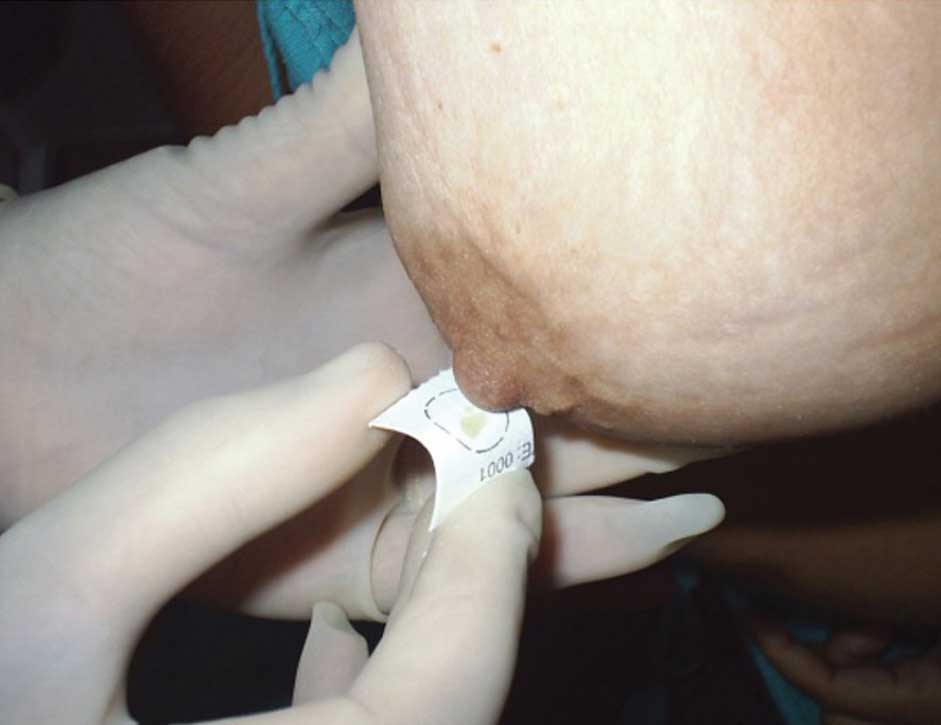
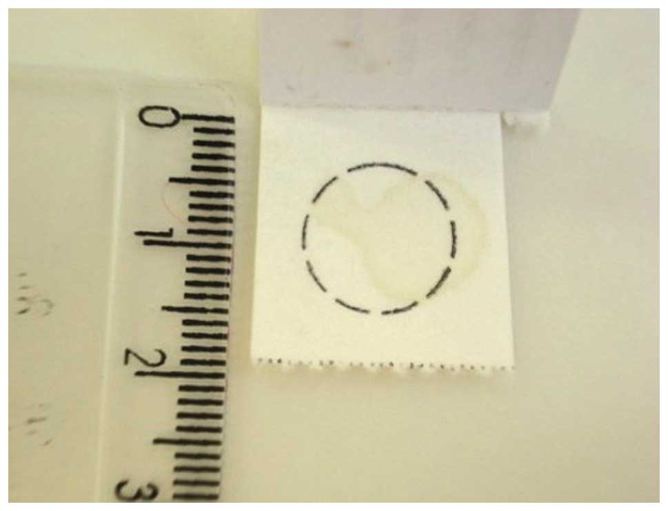
NAF collection
NAF was collected using a modification of a
previously described technique (8,9). The
participants had their breasts warmed for 10–30 min using bilateral
hot compress pads wrapped in towels. The nipple was cleaned with
alcohol and the breast was gently massaged from the chest wall
toward the nipple for 1 min. Subsequently, the participant gently
compressed the breast with two hands and the fluid, which appeared
in the form of droplets, was collected onto Guthrie cards (GE
Healthcare Bio-Sciences, Pittsburgh, PA, USA), as shown in Fig. 1 and were stored at room temperature
(Fig. 2). Up to three attempts
were made to obtain fluid on each breast. If no fluid appeared
following the third attempt, the participant was considered a
non-provider. The NAF-Guthrie cards used in the present study had
been stored for a period of 2–4 years.
Protein extraction and gels
The Guthrie spots were cut into sections of ~6
mm2 and each was incubated in 100 μl
double-distilled water for 30 min at 56°C. The soluble NAF proteins
were mixed with 1 μl phenylmethanesulfonyl fluoride (0.2
mg/ml; Sigma-Aldrich, St. Louis, MO, USA) and measured using the
bicinchoninic acid or Smith reagent methods (Pierce BCA Protein
Assay kit; Pierce Biotechnology, Inc., Rockford, IL, USA) (10).
The NAF proteins were separated by polyacrylamide
gel electrophoresis using SDS-PAGE and β-mercaptoethanol
(Sigma-Aldrich). The stacking gel was prepared at room temperature
using 4% acrylamide (Sigma-Aldrich) Tris-HCl (0.5 M pH 6.8;
Sigma-Aldrich), containing 0.4% SDS, and the separating gel was
prepared at room temperature using 12% acrylamide Tris-HCl buffer
(1.5 M, pH 8.8), containing 0.4% SDS (Sigma-Aldrich). The electrode
buffer used was Tris-glycine (0.025 M Tris base and 0.192 M
glycine; pH 8.3; All from Sigma-Aldrich), containing 0.1% SDS. Each
sample (20 μg of each) was mixed with the sample buffer to a
final concentration of 0.06 M Tris-HCl pH 6.8, 2% SDS, 5%
β-mercaptoethanol, 10% glycerol and 0.025% bromophenol blue. The
samples were heated to 95°C for 3 min and loaded onto the gel in
the mini-Protean II system (Bio-Rad Laboratories, Inc., Hercules,
CA, USA), running at 39 mA/120V for 90 min at room temperature. The
molecular weight standard was BenchMark Protein Ladder (Invitrogen
Life Technologies, Carlsbad, CA, USA). Following electrophoresis,
the gels were fixed for 1 h in a solution of 40% (v/v) aqueous
ethanol (99.8%; Sigma-Aldrich) and 10% (v/v) acetic acid (Merck
Millipore, Darmstadt, Germany) at room temperature. The gels were
then washed for 30 min in fresh fixing solution and incubated with
Coomassie Blue R-250 0.2% diluted in fixative solution for 2 h at
room temperature (Sigma-Aldrich). The gels were destained using
fixative solution for 2 h, followed by incubation in water at room
temperature until complete destaining.
The NAF proteins were also separated using Amersham
ECL high resolution gradient gels (GE Healthcare Life Sciences,
Chalfont, UK), with concentrations of 4–12, 8–16 and 4–20%. Total
protein (5 μg) was added onto the gel with sample buffer 1:1
(50 mM Tris-HCl, pH 6.8, 2% SDS, 0.1% bromophenol blue and 10%
glycerol). This system has a horizontal electrophoresis field and
the gels comprise buffers, which improve the resolution of complex
samples. The molecular weight standard used was Benchmark Protein
Ladder (Invitrogen Life Technologies). Following electrophoresis,
the gels were stained with Coomassie Blue R-250, according to the
manufacturer’s instructions, and were analyzed for protein
integrity and the to determine the profile of the revealed
bands.
Enzymatic digestion for
nano(n)LC-Q-TOF
Selected bands, as described by Manello et al
(11), were excised for destaining
and were subjected to enzymatic digestion, according to Shevchenko
et al (12) with
modification of the destaining phase, where the bands were
destained in a solution of 25 mM ammonium bicarbonate (pH 8.8/50%;
Sigma-Aldrich) and acetonitrile (ACN) overnight in a shaker, at
room temperature. All the samples were concentrated in a Speed-Vac
Centrifuge (Thermo Fisher Scientific, Waltham, MA, USA) at 3,000 ×
g for 5 min to produce a 20 μl final volume of digested
ultrafiltrate sample (DIUs).
Analysis of DIUs by nLC-Q-TOF
Prior to the nLC-Q-TOF analysis of the DIUs, they
underwent manual desalination Zip Tip (Eppendorf, Hamburg,
Germany). Each Zip Tip was activated with 10 μl ACN (100%;
Merck Millipore), was washed three times with 10 μl
ultrapure sterile water, and 10 μl sample was loaded by
pipetting up and down 10 times within the tube. Each Zip Tip was
then washed three times using sterile ultrapure water, and ACN
elution was performed. Subsequently, the samples were reduced to a
final volume of 20 μl and were stored at −20°C until
analysis using mass spectrometry (Q-TOF Ultima Global; Waters,
Manchester, UK).
The extracted peptides from the SDS-PAGE gel slice
were loaded into an electrospray ionization quadrupole
time-of-flight (ESI-Q-TOF) mass spectrometer (Waters Corporation,
Wilmslow, UK). The DIU samples were loaded onto the Waters
nanoACQUITY UPLC® System (Waters Corporation, Milford,
MA, USA), with a Waters Opti-Pak C18 trap column coupled to Q-Tof
Ultima® (Waters Corporation, Milford, MA, USA).
Subsequently, 3.0 μl sample was injected into a nanoEase C18
150 mM × 75 μm column (Waters Corporation) at a flow rate of
0.6 μl/min, and eluted with ACN containing 0.1% formic acid.
The instrument control and data acquisition were performed using a
MassLynx data system (Version 4.0, Waters Corporation). The
experiments were performed by scanning from a mass-to-charge ratio
(m/z) of between 200 and 2,000. The exact mass was automatically
determined using the Q-Tof’s LockSpray™ (Waters Corporation,
Milford, MA, USA).
Database searching
The data were processed using ProteinLynx Global
Server (version 2.0, Waters Corporation) for ESI-Q-TOF analysis.
The proteins were identified by the correlation of tandem mass
spectra to the NCBInr proteins and MSDB database, using MASCOT
online software (www.matrixscience.com). The first analysis considered
all taxonomies, while the second analysis was restricted to Homo
sapiens to remove redundant protein identification.
Breast imaging
Conventional mammography was performed in a Mediman
HFG/B unit with Kodak min-R-2000 film and a Kodak RP-X-Omat
processor (Kodak, Rochester, NY, USA). Ultrasonography was used as
a complementary examination to the conventional mammography, and
was performed using an Image Point Hx unit (HP Labs, Palo Alto, CA,
USA), with two transducers (7.5 and 10 MHz) that measured the
diameters of the breast ducts. The images were then classified
using a breast imaging reporting and data system (BI-RADS).
Results
From the 88 female individuals enrolled in the
present study, NAF was obtained from 80 (91%) on the first visit,
which was collected and absorbed onto Guthrie cards, using the
gentle massage and warming procedure. Of these 80 individuals, two
were excluded due to subsequently identified immunological
deficiency, and the remaining group was composed of 78 individuals,
with a mean age of 50.24 years (range: 23–77 years) and menarche at
a mean age of 13.24 years (range: 9–18 years). A total of 43 (55%)
were postmenopausal. The mean age at menopause was 48.58 years
(range: 36–54 years). A total of 64 became pregnant and of these,
11 did not breastfeed. Of the total group, 18 individuals (23%) had
either an abortion (8; 44%) or a miscarriage (10; 66%). A total of
52 women (67%) reported a family history of cancer, however, only
four reported a family history of breast cancer, of which there was
only one confirmed case of hereditary breast cancer. The results of
the mammography examinations assigned 73% of the individuals to the
BI-RADS 0 category (inconclusive diagnosis), which required
additional assessments, 46% of which had ultrasonography and 77%
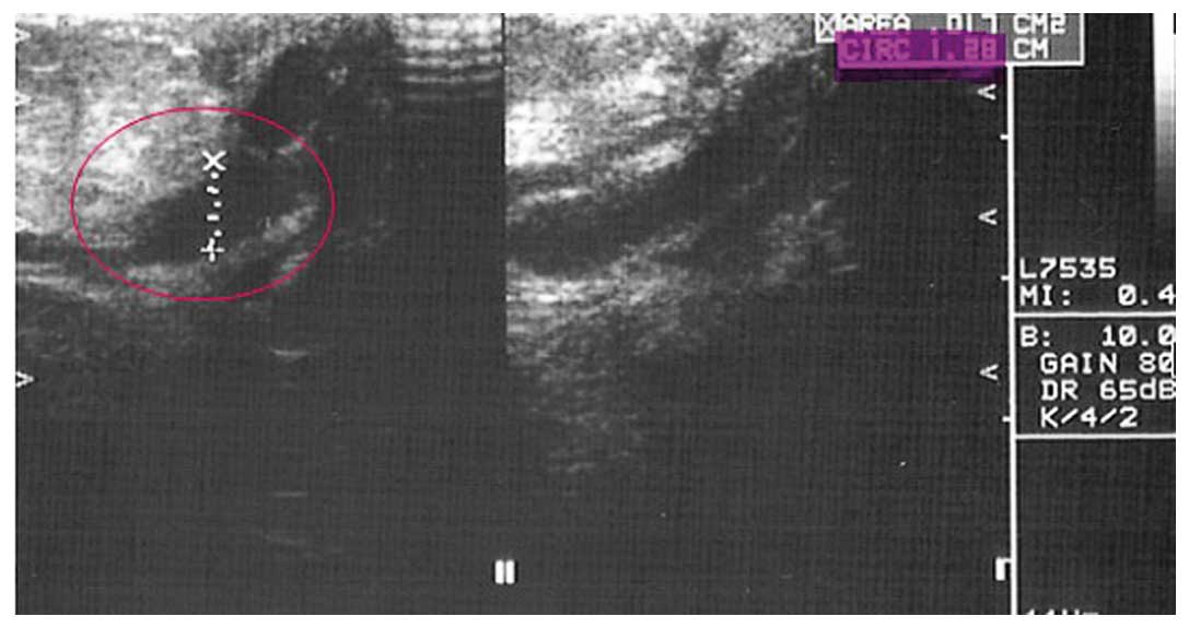
were identified as BI-RADS 3 (benign lesions). Ultrasound was
performed, which revealed the predominance of ductal ectasias
(Fig. 3). In addition, other
injuries were observed, including the presence of nodules, axillary
lymph nodes and microcalcification.
A total of 106 NAF spots were obtained on the
Guthrie cards, which were characteristically classified into the
following five types: Watery, citrine, serous, bloody and mixed
(Table I). NAF was obtained from
both breasts in 28 of the females, explaining why the total number
of NAF spots was higher than the number of individuals enrolled.
The NAF classification from both breasts were the same, with the
exception of three cases.
 | Table IClassification of the breast fluid
collected. |
Table I
Classification of the breast fluid
collected.
| Type | Number | % |
|---|
| Watery | 18 | 17 |
| Citrin | 5 | 5 |
| Serous | 35 | 33 |
| Blood | 12 | 11 |
| Mixed
(seropurulent) | 36 | 34 |
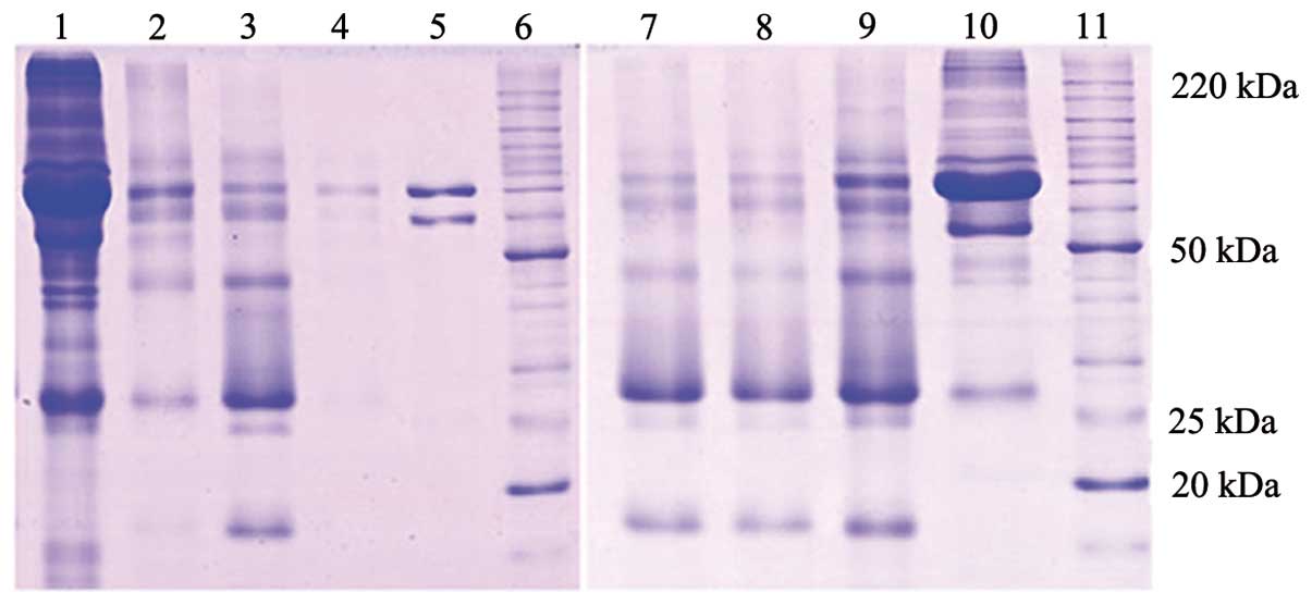
The protein concentration of NAF ranged between 6.8
and 11.2 μg/μl, with a mean value of 9.2
μg/μl. Analysis of the NAF proteins was performed
using one-dimensional SDS-PAGE 12% gel electrophoresis, which
revealed five major bands, with each sample containing similar
quantities of protein. Using the Guthrie card collection method,
the proteins were found to have a similar band pattern as those
described by Mannello et al and Varnum et al using an
aspiration system (11,13). Differences from the default bands
in the protein gel were classified according to band presence,
absence and intensity variation (Fig.
4). No differences were observed in the bands in the watery and
mixed NAF groups, compared to the higher molecular weight bands of
the citrine group, between the gradient gels and the SDS-PAGE 12%
gels. The 4–12% gel exhibited the highest resolution (Fig. 5) and was selected for use in the
gradient gels. The greatest difference was confirmed in the bands
<20 kDa. This difference requires further investigation, but
were considered to be associated with cystic breast disease and
benign breast lesions (13,14).
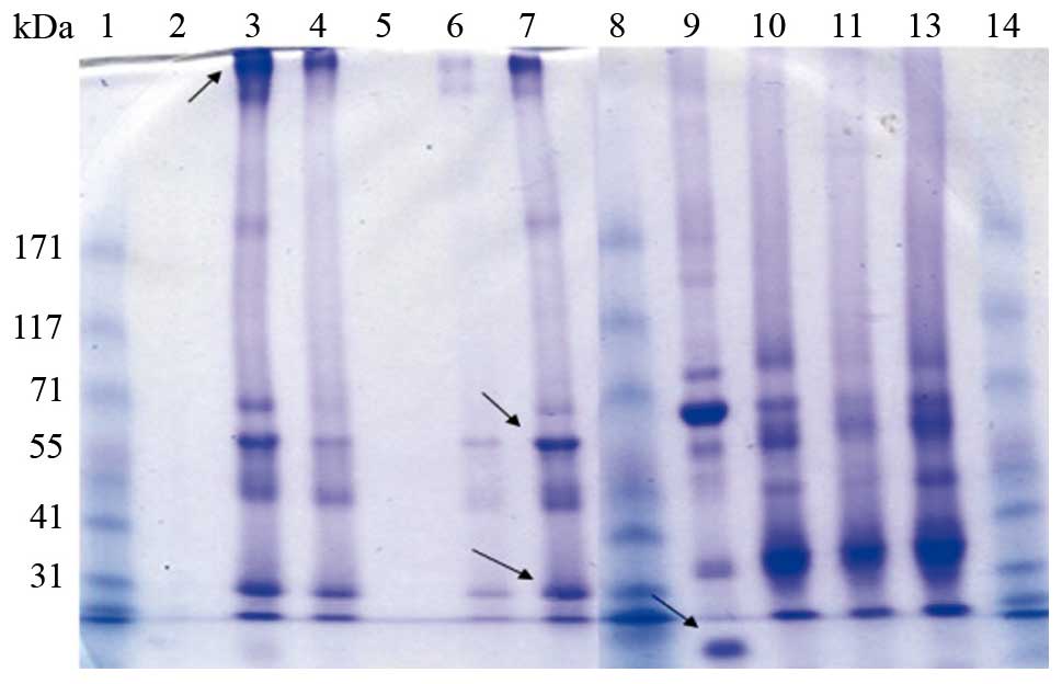 | Figure 5High resolution gradient gel 4-12%
electrophoresis of the NAF proteins. Lanes 1, 8 and 13, molecular
weight; lanes 2 and 5, blanks; lanes 3 and 7, bloody NAF; lanes 4
and 10, mixed NAF, lane 6, watery NAF; lane 9, citrine NAF; lanes
11 and 12, serous NAF. Arrows indicate the bands that were excised
for subsequent proteomic analysis. NAF, nipple aspirate fluid. |
Of the bands excised, the spectra predominates were
identified and the peptide score was calculated as −10Log(P), where
P (0.05) is the probability that the observed match was a random
event. Table II shows the major
proteins that were identified with a score >50 in the nLC-Q-TOF.
Immunoglobulins, Zn-α2-Glicoprotein, apoliprotein D and prolactin
inducible protein were among the bands assessed. The NAF-Guthrie
card collection has not been applied previously, however, NAF
proteins have been identified using other collecting methods
(11,15), confirming the feasibility of the
NAF-Guthrie card collecting method.
| Table IIList of the major proteins identified
by liquid chromatography quadrupole time of flight. |
Table II
List of the major proteins identified
by liquid chromatography quadrupole time of flight.
| Band position | Protein | Score (Ion
score>50) | Mass (m/z) | Match (n) |
|---|
| 15 (kDa) | Hemoglobin subunit
β | 1937 |
16102 | 59 |
| Hemoglobin subunit
δ | 760 |
16159 | 36 |
|
Prolactin-inducible protein | 523 |
16847 | 25 |
| Apoliprotein D
(fragment) | 219 |
15305 | 27 |
| Ig α-1 chain C
region | 214 |
38486 | 11 |
| Ig γ-1 chain C
region |
81 |
36596 |
3 |
| Putative
zinc-α-2-glycoprotein-like 1 |
79 |
23080 |
4 |
| Immunoglobulin J
chain (Fragment) |
78 |
18509 |
2 |
| 20 (kDa) | Serum albumin | 4942 |
71317 | 163 |
| Apoliprotein
D | 880 |
24541 | 40 |
| Clusterin
(fragment) | 147 |
33794 |
9 |
|
Prolactin-inducible protein | 132 |
16847 | 12 |
| α-1-Antitrypsin | 54 |
46878 |
2 |
| 50 (kDa) | Ig α-1 chain C
region | 1483 |
38486 | 59 |
| Ig α-2 chain C
region | 1483 |
37301 | 62 |
| Apoliprotein
D | 777 |
24541 | 36 |
| Serum albumin | 625 |
71317 | 23 |
| Ig heavy chain V-III
region BRO | 291 |
13332 |
9 |
| Ig heavy chain
V-III region CAM |
102 |
13773 |
6 |
|
Prolactin-inducible protein |
227 |
16847 | 15 |
| Complement C4 β
chain |
172 | 194351 |
3 |
|
α-1-Antitrypsin |
154 |
46878 |
9 |
| Ig γ-1 chain C
region |
140 |
36596 | 13 |
| Secretoglobin
family 1D member 2 |
117 |
10260 |
6 |
|
Zinc-α-2-glycoprotein |
101 |
34465 |
7 |
| Polymeric
immunoglobulin receptor |
88 |
84429 |
7 |
| Vasorin |
79 |
72751 |
2 |
| Ig κ chain C
region |
75 |
11773 |
6 |
| Ig γ-2 chain C
region |
68 |
36505 |
9 |
|
Chromosome-associated kinesin KIF4A |
54 | 141390 |
2 |
| 200 (kDa) | Serum albumin | 1290 |
71317 | 60 |
| Apoliprotein D
(fragment) |
102 |
24541 |
8 |
| Cdc42 effector
protein 4 |
53 |
30253 |
2 |
Discussion
Examining the breast epithelium directly using core
needle biopsy or ductal lavage is uncomfortable and invasive. By
contrast, the NAF-Guthrie card collecting method is inexpensive,
non-invasive, reliable and painless. This method may broaden the
applicability of NAF sample collection and may have an advantage
over the method described by Sauter et al (16), which used an aspiration device. In
addition, Guthrie cards occupy little space and can be stored at
room temperature with dried NAF. These characteristics enable the
cards to be sent to a laboratory for analysis.
Identifying associations in the results of imaging
techniques is hindered by their own limitations in the public
health system in Brazil (4),
however, a significant association was observed between ductal
ectasia and the secretion of NAF. In addition, the majority of the
mammogram results were BI-RADS 0, which are flagged as abnormal due
to the ability of non-palpable lesions to disturb results, leading
to a false-negative diagnosis (17).
A low percentage of the female individuals selected
for the present study were receiving hormone therapy or oral
contraceptives (data not shown), which did not enable the
investigation of associations between the secretion of NAF and
these variables. The small 20 kDa protein, in particular the Gross
cystic disease fluid protein, as described by Mannello et al
is markedly associated with alterations in the breast (15,16).
Human epidermal growth factor receptor-2 (HER2) is a breast cancer
subtype biomarker, the amplification/overexpression of which is
associated with aggressive disease and a poorer prognosis (18). In ductal carcinoma in situ,
overexpression of HER has been observed by immunohistochemistry and
is correlated with higher proliferative activity (19). HER2 has previously been detected in
NAF, in addition to breast related hormones, metabolites and growth
factors (18–24). Thus, it was suggested that breast
cancer biomarkers can be detected using dried NAF spots in Guthrie
cards, followed by mass spectrometric analysis. In the present
study, gels were produced from the NAF proteins in order to
characterize them; however, for clinical use, the shotgun mass
spectrometry approach, with no in-between gel, is considered more
appropriate.
In conclusion, the NAF-Guthrie card collecting
method used in the present study, was confirmed as being suitable
for modern mass spectrometric analysis. This method has potential
application for early breast cancer screening and subtype
classification.
Acknowledgements
The authors would like to acknowledge the Mass
Spectrometry Facility at the Brazilian Biosciences National
Laboratory, CNPEM (Campinas, Brazil) for their support in mass
spectrometric analysis and Professors Márcia Regina Soares da Silva
and Rosane Nunes from the Instituto de Química of Universidade
Federal do Rio de Janeiro (Rio de Janeiro, Brazil) for their
technical assistance.
This study was supported by the Fundação de Amparo a
Pesquisa do Rio de Janeiro (noa. Bolsa 102.558/2012, APQ1
E-26/110.319/2008 and APQ1 E-26/110.803/2009) and the Programa de
Oncobiologia, Rio de Janeiro, Brazil.
References
|
1
|
West KE, Wojcik EM, Dougherty TA, et al:
Correlation of nipple aspiration and ductal lavage cytology with
histopathologic findings for patients before scheduled breast
biopsy examination. Am J Surg. 191:57–60. 2006. View Article : Google Scholar : PubMed/NCBI
|
|
2
|
Alexander H, Stegner AL, Wagner-Mann C, et
al: Proteomic analysis to identify breast cancer biomarkers in
nipple aspirate fluid. Clin Cancer Res. 10:7500–7510. 2004.
View Article : Google Scholar : PubMed/NCBI
|
|
3
|
Balci FL and Feldman SM: Exploring breast
with therapeutic ductoscopy. Gland Surg. 3:136–141. 2014.PubMed/NCBI
|
|
4
|
Liedke PE, Finkelstein DM, Szymonifka J,
et al: Outcomes of breast cancer in Brazil related to health care
coverage: a retrospective cohort study. Cancer Epidemiol Biomarkers
Prev. 23:126–133. 2014. View Article : Google Scholar
|
|
5
|
Lang JE and Kuerer HM: Breast ductal
secretions: clinical features, potential uses, and possible
applications. Cancer Control. 14:350–359. 2007.PubMed/NCBI
|
|
6
|
Hirose M, Nobusawa H and Gokan T: MR
ductography: comparison with conventional ductography as a
diagnostic method in patients with nipple discharge. Radiographics.
27:S183–S196. 2007. View Article : Google Scholar
|
|
7
|
Guthrie R and Susi A: A simple
phenylalanine method for detecting phenylketonuria in large
populations of newborn infants. Pediatrics. 32:338–343.
1963.PubMed/NCBI
|
|
8
|
Borte S, Janzi M, Pan-Hammarström Q, et
al: Placental transfer of maternally-derived IgA precludes the use
of guthrie card eluates as a screening tool for primary
immunodeficiency diseases. PLoS One. 7:e434192012. View Article : Google Scholar : PubMed/NCBI
|
|
9
|
van Ommen CC, Elvers LH, Notermans DW, et
al: Antibody levels against B. pertussis in neonates measured in
dried blood spots. Vaccine. 30:2697–2700. 2012. View Article : Google Scholar : PubMed/NCBI
|
|
10
|
Smith PK, Krohn RI, Hermanson GT, Mallia
AK, Gartner FH, Provenzano MD, et al: Measurement of protein using
bicinchoninic acid. Anal Biochem. 150:76–85. 1985. View Article : Google Scholar : PubMed/NCBI
|
|
11
|
Manello F, Medda V and Tonti GA: Protein
profile analysis of the breast microenvironment to differentiate
healthy women from breast cancer patients. Expert Rev Proteomics.
43:43–60. 2009. View Article : Google Scholar
|
|
12
|
Shevchenko A, Wilm M, Vorm O, et al: Mass
spectrometric sequencing of proteins silver-stained polyacrylamide
gels. Anal Chem. 68:850–858. 1996. View Article : Google Scholar : PubMed/NCBI
|
|
13
|
Varnum SM, Covington CC, Woodbury RL, et
al: Proteomic characterization of nipple aspirate fluid:
identification of potential biomarkers of breast cancer. Breast
Cancer Res Treat. 80:87–97. 2003. View Article : Google Scholar : PubMed/NCBI
|
|
14
|
Sartorius OW, Smith HS, Morris P, et al:
Cytologic evaluation of breast fluid in the detection of breast
disease. J Natl Cancer Inst. 59:1073–1080. 1977.PubMed/NCBI
|
|
15
|
Manello F, Tonti GA and Papa S: Human
gross cyst breast disease and cystic fluid: bio-molecular,
morphological, and clinical studies. Breast Cancer Res Treat.
97:115–129. 2006. View Article : Google Scholar
|
|
16
|
Sauter ER, Ross E, Daly M, et al: Nipple
aspirate fluid: a promising non-invasive method to identify
cellular markers of breast cancer risk. Br J Cancer. 76:494–501.
1997. View Article : Google Scholar : PubMed/NCBI
|
|
17
|
Hamy AS, Giacchetti S, Albiter M, et al:
BI-RADS categorisation of 2,708 consecutive nonpalpable breast
lesions in patients referred to a dedicated breast care unit. Eur
Radiol. 22:9–17. 2012. View Article : Google Scholar
|
|
18
|
Borg A, Tandon AK, Sigurdsson H, et al:
HER-2/neu amplification predicts poor survival in node-positive
breast cancer. Cancer Res. 50:4332–4337. 1990.PubMed/NCBI
|
|
19
|
Allred DC, Clark GM, Molina R, et al:
Overexpression of HER-2/neu and its relationship with other
prognostic factors change during the progression of in situ to
invasive breast cancer. Hum Pathol. 23:974–979. 1992. View Article : Google Scholar : PubMed/NCBI
|
|
20
|
Kuerer HM, Thompson PA, Krishnamurthy S,
et al: High and differential expression of HER-2/neu extracellular
domain in bilateral ductal fluids from women with unilateral
invasive breast cancer. Clin Cancer Res. 9:601–605. 2003.PubMed/NCBI
|
|
21
|
Gann PH, Geiger AS, Helenowski IB, et al:
Estrogen and progesterone levels in nipple aspirate fluid of
healthy premenopausal women: relationship to steroid precursors and
response proteins. Cancer Epidemiol Biomarkers Prev. 15:39–44.
2006. View Article : Google Scholar : PubMed/NCBI
|
|
22
|
Loud JT, Gierach GL, Veenstra TD, et al:
Circulating estrogens and estrogens within the breast among
postmenopausal BRCA1/2 mutation carriers. Breast Cancer Res Treat.
143:517–529. 2014. View Article : Google Scholar : PubMed/NCBI
|
|
23
|
Fought AJ, McGathey C, Scholtens DM, et
al: Hormonal determinants of nipple aspirate fluid yield among
breast cancer cases and screening controls. Cancer Epidemiol
Biomarkers Prev. 22:2277–2284. 2013. View Article : Google Scholar : PubMed/NCBI
|
|
24
|
Tredwell GD, Miller JA, Chow HH, et al:
Metabolomic characterization of nipple aspirate fluid by (1)H NMR
spectroscopy and GC-MS. J Proteome Res. 13:883–889. 2014.
View Article : Google Scholar
|