Open Access

Implication of microRNA regulation in para-phenylenediamine-induced cell death and senescence in normal human hair dermal papilla cells

  • Authors:
    • Ok‑Kyu Lee
    • Hwa Jun Cha
    • Myung Joo Lee
    • Kyung Mi Lim
    • Jae Wook Jung
    • Kyu Joong Ahn
    • In‑Sook An
    • Sungkwan An
    • Seunghee Bae
  • View Affiliations

  • Published online on: March 13, 2015     https://doi.org/10.3892/mmr.2015.3487
  • Pages: 921-936
  • Copyright: © Lee et al. This is an open access article distributed under the terms of Creative Commons Attribution License [CC BY_NC 3.0].

Metrics: Total Views: 0 (Spandidos Publications: | PMC Statistics: )
Total PDF Downloads: 0 (Spandidos Publications: | PMC Statistics: )


Abstract

Para-phenylenediamine (PPD) is a major component of hair coloring and black henna products. Although it has been largely demonstrated that PPD induces allergic reactions and increases the risk of tumors in the kidney, liver, thyroid gland and urinary bladder, the effect on dermal papilla cells remains to be elucidated. Therefore, the current study evaluated the effects of PPD on growth, cell death and senescence using cell‑based assays and microRNA (miRNA) microarray in normal human hair dermal papilla cells (nHHDPCs). Cell viability and cell cycle analyses demonstrated that PPD exhibited a significant cytotoxic effect on nHHDPCs through inducing cell death and G2 phase cell cycle arrest in a dose‑dependent manner. It was additionally observed that treatment of nHHDPCs with PPD induced cellular senescence by promoting cellular oxidative stress. In addition, the results of the current study indicated that these PPD‑mediated effects were involved in the alteration of miRNA expression profiles. Treatment of nHHDPCs with PPD altered the expression levels of 74 miRNAs by ≥2‑fold (16 upregulated and 58 downregulated miRNAs). Further bioinformatics analysis determined that these identified miRNA target genes were likely to be involved in cell growth, cell cycle arrest, cell death, senescence and the induction of oxidative stress. In conclusion, the observations of the current study suggested that PPD was able to induce several cytotoxic effects through alteration of miRNA expression levels in nHHDPCs.

Introduction

Para-phenylenediamine (PPD), also known as 1,4-diaminobenzene, is a key primary precursor of the oxidative dyes used in hair coloring and tattoos (1,2). However, accumulating evidence has suggested that this compound sensitizes skin to allergic reactions (3,4). Further studies have identified that these allergic reactions predominantly occur due to PPD-mediated activation of dendritic cells (5). Additionally, PPD is a potential carcinogen, which was reported to increase the risk of tumorigenesis in the kidney, liver, thyroid gland and urinary bladder in mice and rats (6,7). In addition, in vitro studies have demonstrated that PPD induced reactive oxygen species (ROS)-mediated DNA damage in uroepithelial cells and activated p38 mitogen-activated protein kinase (MAPK) and c-Jun N-terminal kinase in Chang liver cells (8,9). A case report demonstrated that PPD induced severe acute hair loss in females within six days of application (10). However, it remains to be elucidated whether PPD contributes to hair loss by inducing damage to normal human hair dermal papilla cells (nHHDPCs).

MicroRNAs (miRNAs) are small (18–24 nucleotides) noncoding RNAs that repress the translation of target genes through imperfect base pairing to the 3′-untranslated region of their target mRNAs (11,12). miRNAs have been reported to be key regulators of apoptosis, proliferation and differentiation (13). Regarding the role of miRNA in hair, it has been reported that the expression levels of miRNA-31 (miR-31) were upregulated in the anagen phase of the hair growth cycle and controlled the expression levels of Krt16, Krt17, Dlx3 and Fgf10 (14). In addition, miR-24 was reported to regulate the development of hair follicles by targeting the hair keratinocyte stemness regulator Tcf-3 (15). Furthermore, a previous study demonstrated that Dicer, an miRNA-processing enzyme, was essential for the morphogenesis of hair follicles (16).

The aim of the current study was to investigate the effects of PPD on cell growth, death and senescence in nHHDPCs. In addition, the role of PPD in the regulation of the expression profile and the mechanisms of specific miRNAs was evaluated using bioinformatics analysis.

Materials and methods

Cells and culture conditions

nHHDPCs (Innoprot, Biscay, Spain) were cultured in Dulbecco’s modified Eagle’s medium (Gibco Life Technologies, Grand Island, NY, USA) supplemented with 10% fetal bovine serum (Gibco Life Technologies) and 1% penicillin-streptomycin (Gibco Life Technologies) at 37°C in an atmosphere of 5% CO2. PPD was purchased from Sigma-Aldrich (St. Louis, MO, USA).

Cell viability assay

Cell viability was monitored using the water-soluble tetrazolium salt (WST-1) assay (EZ-Cytox Cell Viability Assay kit; ITSbio, Seoul, Korea). A total of 5×10³ nHHDPCs were seeded into 96-well plates and treated with various concentrations of PPD (0, 100, 200, 300, 400, 500 and 600 μM) for 24 h. Following treatment, nHHDPCs were mixed with 10 μl WST-1 solution and incubated at 37°C for 0.5 h. Cell viability was then determined by measuring absorbance at 450 nm using an iMark plate reader (Bio-Rad Laboratories, Inc., Hercules, CA, USA).

Propidium iodide (PI)-based cell cycle analysis

The cell cycle was analyzed using PI (Sigma-Aldrich) staining of DNA. nHHDPCs were plated and treated with various concentrations of PPD (0, 200, 400 and 600 μM) for 24 h. Cells were then trypsinized (using 0.25% trypsin-EDTA; Gibco Life Technologies), centrifuged (3,500 × g, 2 min), washed with phosphate-buffered saline (PBS; Gibco Life Technologies) and fixed in 70% ethanol (Merck Millipore, Darmstadt, Germany) at 4°C for 3 h. The fixed cells were incubated with staining solution [50 μg/ml PI, 0.1 μg/ml RNase (Life Technologies, Grand Island, NY, USA) and 0.05% Triton X-100 (Sigma-Aldrich) in PBS] at 37°C for 1 h and then analyzed using a FACSCalibur flow cytometer (BD Biosciences, San Jose, CA, USA). The mean PI fluorescence intensity was determined based on analysis of 10,000 cells using the FLH-2 detection channel (585±42 nm).

Detection of cellular senescence

For the assessment of cellular senescence, nHHDPCs (2×10⁶) were seeded into 60-mm cell culture dishes and treated with 0 or 400 μM PPD. Following 48 h of treatment, cells were fixed using Fixative solution (included in Senescence Detection kit; BioVision, Inc., Milpitas, CA, USA) and senescence-associated-β-galactosidase (SA-β-gal) activity was measured using the Staining Solution Mix, including Staining Solution, Staining Supplements and X-gal substrate for (SA-β-gal) within the Senescence Detection kit, according to the manufacturer’s instructions. Cells stained for SA-β-gal were counted under a light microscope (CKX41; Olympus Corporation, Tokyo, USA) (magnification, ×200) and the percentage of SA-β-gal positive cells were calculated.

Detection of intracellular ROS

Staining for ROS in cultured cells was conducted using a 2′,7′-dichlorodihydrofluorescein diacetate (DCF-DA; Sigma-Aldrich) assay. Briefly, 2×10⁶ nHHDPCs were plated in 60-mm culture dishes and treated with PPD. Following treatment for 24 h, the cells were stained by adding DCF-DA to the culture medium to a final concentration of 20 μM and then incubating for 1 h. Distribution of the stained cell population was determined using a FACSCalibur flow cytometer.

miRNA expression profiling

In order to analyze the miRNA expression profile, nHHDPCs (2×10⁶) were seeded into 60-mm culture dishes and treated with 400 μM PPD. Following 24 h of treatment, total RNA was purified using TRIzol reagent (Life Technologies) according to the manufacturer’s instructions. Total RNA was dephosphorylated and labeled with pCp-Cy3 using an Agilent miRNA Labeling kit (Agilent Technologies, Inc., Santa Clara, CA, USA). Labeled RNAs were hybridized using a SurePrint G3 Human v16 miRNA 8×60K microarray (Agilent Technologies, Inc.) at 65°C for 20 h. The miRNA expression profile was digitized using Feature Extraction version 10.7 software (Agilent Technologies). Fold changes in miRNA expression levels were determined using GeneSpring GX software, version 11.5 (Agilent Technologies).

Prediction of potential target genes of PPD-regulated miRNAs and gene ontology (GO) analysis

Potential target genes of PPD-regulated miRNAs were predicted using the DNA Intelligent Analysis (DIANA) microT-CDS version 5.0 bioinformatics tool (http://diana.cslab.ece.ntua.gr/). GO of each putative target gene was identified using the Database for Annotation, Visualization and Integrated Discovery (DAVID) bioinformatics resource, version 6.7 (http://david.abcc.ncifcrf.gov). Target genes were categorized into four GO terms: Aging, skin development, apoptosis and cell proliferation. Furthermore, target gene-associated signaling pathways were determined using the Kyoto Encyclopedia of Genes and Genomes (KEGG) pathway algorithm (http://david.abcc.ncifcrf.gov/summary.jsp) within the DAVID resource.

Statistical analyses

Values are expressed as the mean ± standard error of the mean of three independent experiments. Statistical significance was determined by Student’s t-test and P<0.05 was considered to indicate a statistically significant difference between values.

Results

PPD treatment reduces the proliferation rate of nHHDPCs

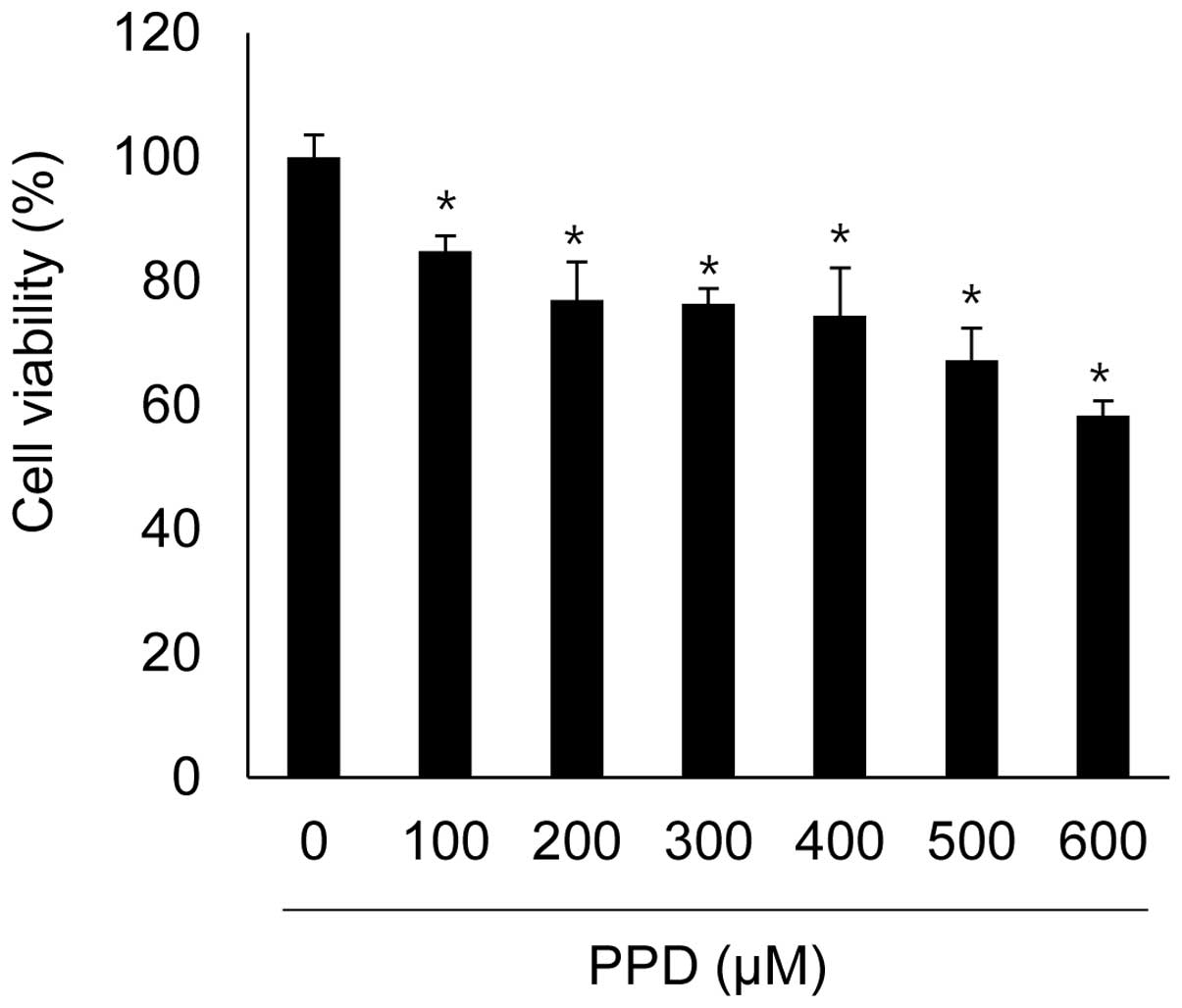
Previous studies reported that exposure to >250 μM and >60 μM PPD resulted in considerable cytotoxicity in dendritic cells (17) and keratinocytes (18), respectively. Therefore, the current study aimed to determine the cytotoxic effects of PPD on nHHDPCs at concentrations of 0, 100, 200, 300, 400, 500 and 600 μM using a WST-1-based cell viability assay. As shown in Fig. 1, the viability of nHHDPCs was significantly reduced following exposure to PPD for 24 h. Maximal toxicity was observed at 600 μM, at which concentration cell viability was reduced to 58.33±2.39% of the control value (n=3; P<0.05). The IC25 (a 25% reduction in viability) of PPD was 400 μM, at which concentration cell viability was reduced to 74.42±6.08% of control value (P<0.05) (Fig. 1).

PPD treatment increases cell death and cell cycle arrest in nHHDPCs

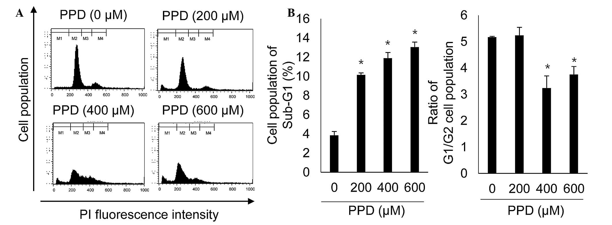
The present study investigated whether PPD-induced loss of cell viability occurred due to cell cycle arrest and cell death. Cells were treated with PPD (0, 200, 400 and 600 μM) for 24 h and the distribution of cells in the different cell cycle phases was analyzed using flow cytometry. As shown in Fig. 2, treatment with 200, 400 and 600 μM PPD led to significant accumulation in the sub-G1 phase, compared with that of control DMSO-treated cells (P<0.05). In addition, the proportion of cells in G1/G2 was significantly reduced by PPD (P<0.05), indicating that PPD increased the G2 population. These data therefore demonstrated that PPD induced cell death and G2 arrest in nHHDPCs.

PPD treatment leads to the accumulation of intracellular ROS and senescence-like growth

Chye et al (8) observed that PPD increased intracellular ROS and induced apoptosis in Chang normal human liver cells. Therefore, the current study investigated the effects of PPD on the regulation of intracellular ROS production in nHHDPCs. Intracellular ROS levels were determined using an DCF-DA probe, which is oxidized to fluorescent DCF in the presence of ROS. As presented in Fig. 3A, exposure to PPD resulted in a marked increase in fluorescent DCF-positive cells compared with that of the control cells, indicating that PPD increased intracellular ROS production in nHHDPCs. As ROS have been implicated in cellular senescence (19), the present study investigated whether the PPD-mediated increase in ROS was associated with increased senescence by analyzing the activity of SA-β-gal, a marker of cellular senescence. Consistent with the observed ROS increase, PPD was identified to promote an increase in SA-β-gal activity (Fig. 3B), indicating that PPD induces cellular senescence in nHHDPCs.

Identification of differentially expressed miRNAs in PPD-treated nHHDPCs

Fig. 2 and 3 demonstrated that cell cycle arrest in G2 phase and PPD-mediated cell death were characterized by increased ROS production. Therefore, in order to determine whether ROS-mediated cell cycle arrest and cell death are associated with miRNA expression, miRNA microarray analysis was conducted using the SurePrint G3 Human v16 miRNA 8×60K microarray, which contained 2,006 human miRNA probes. Significant miRNAs exhibiting a ≥2.0-fold increase or reduction in expression were selected using GeneSpring GX software. As presented in Table I, PPD differentially regulated the expression levels of 74 miRNAs. Notably, 16 of 74 miRNAs were significantly upregulated and 58 miRNAs were significantly downregulated. In particular, miR-425-3p exhibited the greatest increase in expression (230.60-fold) and miR-3656 the greatest reduction (112.15-fold), compared with the corresponding miRNAs in control cells. These results suggested that the PPD-mediated cellular effects were associated with alterations in expression of specific miRNAs.

Table I

MicroRNAs exhibiting a ≥2-fold alteration in expression following treatment of normal human hair dermal papilla cells with para-phenylenediamine.

Table I

MicroRNAs exhibiting a ≥2-fold alteration in expression following treatment of normal human hair dermal papilla cells with para-phenylenediamine.

MicroRNAFold changeDirection of regulation
miR-100-3p57.90Up
miR-1225-3p3.43Up
miR-1228-3p2.14Up
miR-12382.34Up
miR-18252.08Up
miR-18b-3p106.08Up
miR-191-3p2.06Up
miR-3180-5p100.21Up
miR-33b-3p50.55Up
miR-425-3p230.60Up
miR-42862.03Up
miR-43132.17Up
miR-43232.02Up
miR-6342.26Up
miR-766-3p3.78Up
miR-93351.08Up
miR-4102.48Down
miR-513a-5p2.54Down
miR-500a2.76Down
miR-36513.07Down
miR-1207-5p3.14Down
miR-7624.29Down
miR-150-3p15.14Down
let-7a-3p23.13Down
miR-1181110.44Down
miR-1226-5p32.72Down
miR-125a-3p81.56Down
miR-12845.79Down
miR-13438.97Down
miR-138-2-3p45.91Down
miR-146b-5p30.17Down
miR-148b-3p83.28Down
miR-17-3p58.57Down
miR-181d34.46Down
miR-185-5p106.57Down
miR-195-5p43.11Down
miR-197-3p73.04Down
miR-202-3p64.16Down
miR-214-5p30.65Down
miR-23a-5p17.14Down
miR-28-5p50.28Down
miR-301a-3p45.29Down
miR-30a-3p64.02Down
miR-30e-3p37.05Down
miR-324-5p93.16Down
miR-342-3p38.57Down
miR-365331.43Down
miR-3656112.15Down
miR-3663-3p89.50Down
miR-369-3p45.40Down
miR-369-5p42.07Down
miR-37043.81Down
miR-371a-5p15.21Down
miR-37836.78Down
miR-392630.72Down
miR-409-5p76.96Down
miR-423-5p58.63Down
miR-427184.01Down
miR-429196.44Down
miR-431-5p50.91Down
miR-431-3p93.55Down
miR-431753.97Down
miR-487a35.23Down
miR-501-5p38.33Down
miR-505-3p30.72Down
miR-513b46.33Down
miR-539-5p42.35Down
miR-548c-3p19.22Down
miR-57229.95Down
miR-642b-3p68.28Down
miR-65022.21Down
miR-660-5p38.17Down
miR-770-5p55.39Down
miR-87453.92Down

[i] miR, microRNA.

Bioinformatic analysis of PPD-modulated miRNAs

As miRNAs perform their biological functions through regulation of target mRNA translation (11), the present study aimed to predict the target genes of the miRNAs deregulated in response to PPD. The biological functions of the upregulated and downregulated genes were then determined following categorization into the four groups: Aging, apoptosis, cell proliferation and skin development, using DAVID (Tables II and III, respectively). In addition, in order to identify the specific signaling pathways of the deregulated miRNAs, the correlation between KEGG pathway-associated genes and the target genes of each miRNA were analyzed. The meaningful KEGG pathways with a value >1% (percentage of target genes/total genes involved in each pathway) were selected. The analysis identified a wide distribution of cellular functions, which are presented in Tables IV and V. The results indicated that the upregulated miRNAs were implicated in signaling pathways in cancer, ubiquitin-mediated proteolysis, melanogenesis, cell cycle, Wnt signaling, MAPK signaling, neurotrophin signaling, cell adhesion molecules (CAMs), long-term potentiation, natural killer cell-mediated cytotoxicity, calcium signaling, neuroactive ligand-receptor interactions, glycosphingolipid biosynthesis, arrhythmogenic right ventricular cardiomyopathy, axon guidance, ErbB signaling, gonadotropin-releasing hormone signaling, tight junctions and viral myocarditis (Table IV). In addition, PPD-induced downregulated miRNAs were implicated in signaling pathways in cancer, regulation of actin cytoskeleton, Wnt signaling, oocyte meiosis, glycerolipid metabolism, MAPK signaling, insulin signaling, chemokine signaling, cytokine-cytokine receptor interaction, Janus kinase-signal transducer and activator of transcription signaling, calcium signaling, mammalian target of rapamycin signaling, axon guidance, cell cycle, ubiquitin mediated proteolysis, regulation of actin cytoskeleton, ErbB signaling, melanogenesis, TGF-β signaling, vascular smooth muscle contraction, tight junction, neuroactive ligand-receptor interactions, CAMs, glycerophospholipid metabolism, adipocytokine signaling and neurotrophin signaling (Table V).

Table II

Predicted targets of microRNAs upregulated in response to para-phenylenediamine treatment in normal human hair dermal papilla cells.

Table II

Predicted targets of microRNAs upregulated in response to para-phenylenediamine treatment in normal human hair dermal papilla cells.

MicroRNABiological processes and target genes
AgingSkin developmentApoptosisCell proliferation
has-miR-100-3pRPS6KB1, SLC1A2, RTN4LEF1, GNASNEUROD1, HIPK1, MDM2, RTN4, LEF1, SIX1, RILPL1, SKP2, RPS6KB1, GSDMA, PHF17, RTN3, MAP3K7, SON, MAPK31, ESPL1LEF1, SIX1, MDM2, SAV1, BHLH41, AKIRN2, PDSSB, ODC1, SKP2, HIPK1, E2F3, RPOX1, NFIB, IRF2, TOB1
has-miR-1225-3pHMGA1, SREBF2, P2RY2EGLN3, LRP5, ESR1, BIRC5, CASP5, MEF2D, MAX, BMF, IL2, CLN3, AR, MAP3K1, IL2RBKRT4, TGFBI, CD274, HDGF, EGLN3, LRP5, ESR1, BIRC5, IL2, IGFBP6, AR, RAPGEF3, CD34, GDF2, GFER
hsa-miR-1228-3pSLC32A1, AMFREYA1, UBE2B, TJP1, MOAP1, AGAP2, PRDX2, WT1, BMP7, RNF41, CEBPG, CADM1, USP7AGAP2, PRDX2, WT1, BMP7, THAP1, TSC1, PDCD1LG2, EYA1, WNT9A, MAPRE2, TNS3, LHX9, STAT6
hsa-miR-1238MSH6APCSIX1, THOC1, ING4, CCAR1, MSH6, APC, CTSH, BRCA1, SLIT3, ERCC3, API5APC, ING4, CD160, SIX1, SP6, BLM, EPS8, BRCA1, TACC3, NASP, DPP4, CTSH, BCAT1, CNBP, PBX1, PTN, IGFBP5
hsa-miR-1825SERPINE1, RELA, ILK, LMNA, TERF2DDR1, COL5A3TM2D1, ROCK1, FXR1, MAX, SOS2, SERPINE1, RELA, ILK, HELLS, ITCH, SULF1, PKN2, LMNA, TRIM2, PDCD5, CECR2SERPINE1, RELA, ILK, OSR1, DDR1, JAG1, KIT, SMARCA2, NRP1, PDPN, FANCA, PMP22, ITCH, PPARG, OSR1, TGFB2, ETS1, MAP3K11, PPARG, TIAM1, SULF1, ETS1, TGFB2, HELLS, MAP3K11, ACHE, FZD6, STAT6
hsa-miR-18b-3pHMGA2, SHC1,HMGA2, ITGB1, VHL, IP6K2, RAD21, DUSP6, RRP8, BCL2L2HMGA2, SHC1, ITGB1, VHL, ACVRL1, CIAO1, RPRD1B, EMP2 NAMPT
hsa-miR-191-3pHOXA5, GLI2HOXA5, GLI2
hsa-miR-3180-5pCTNNA1, CTSC, CASP7, PTGS1, DCN, MGEA5APCCTNNA1, CTSC, CASP7, APC, GPAM, ERBB4, DLC1, NAE1, HOXA5, NAIP, DDX5, GAS2, BRCA1, HIPK2, SRPK2, NF1, ERBB2, MED1, CD38, STK4, MAP3K9, RBM25, TP53INP1, ROCK1, USP47, WNK3, BAXHOXA5, NDRG2, KIF14, IRS1, ESRRB, BRCA1, NF1, SRPK2, FER, CTBP1, ST8SIA1, FKTN, PDPN, CD274, NFIB, ERBB2, STK4, GPAM, ERBB4, DLC1, MED1, PTGS1, HIPK2, APC, SESN1, CNBP, DOCK2, CDKN2B, FZD9, CDK13
hsa-miR-33b-3pRTN4BCL11BRTN4, DUSP22, DCUN1D3, BCL11B, PDE5A, FGFR1, UBE2Z, RPS3, URI1BCL11B, PDE5A, DUSP22, FGFR1. NAMPT, FOXF1, CD274, FEZF1, RPS15A, CHRM3
hsa-miR-425-3p
hsa-miR-4286RELACOL5A2RELA, MED1, PTK2, PIM1, RRN3, MAP2K7, AGTR2, PSMD11, DYNLL2RELA, MED1, PTK2, PIM1, RRN3, DCNT2, FOXO4, ACVR2A
hsa-miR-4313TNFSF9, FZD5, JAK3TNFSF9, FZD5, JAK3, WNT3
hsa-miR-4323VDR, CTSC, MAP2K1VDR, CTSC, JAK3, PIM1, ID1, TAOK2, GSK3BVDR, MAP2K1, JAK3, PIM1, TGFB1, GABBR1, MMP14
has-miR-634EDNRA, TGFBR1, RXRA, FURINEDNRA, RXRA, TRAF5, HRK, HIPK1, TGFBR1, NF1, RNF41, CXCR4, PDCD6IP, MAP2K4FURIN, TRAF5, HIPK1, PIM1, EDNRA, CASK, ROGDI, NF1, TGFBR1, RXRA, ABI1
hsa-miR-766-3pCHEK2, TH, SCAPBDNF, NKX2 5, CUL5, SKP2, ESPL1, SLIT3, CARD8, DPF1, CHEK2, ESR2, MKL1, YARS, XAF1, KDM2B, PLECESR2, KDM2B, BDNF, NKX2 5, CUL5, SKP2, CAS8, STAT6, DOCK2, ABCB1, ERF
hsa-miR-933ING4, BDNF, MEF2AING4, BDNF

[i] miR, microRNA; hsa, homo sapiens.

Table III

Predicted targets of microRNAs downregulated in response to para-phenylenediamine treatment in normal human hair dermal papilla cells.

Table III

Predicted targets of microRNAs downregulated in response to para-phenylenediamine treatment in normal human hair dermal papilla cells.

MicroRNABiological processes and target genes
AgingSkin developmentApoptosisCell proliferation
has-let-7a-3pCNR1, TFRC, TGFBR1, F3, LRP2, ID2, VCAM1, SLC1A2TCF7L2, JUP, ITGA2FOXO1, ITCH, TLR4, PDE5A, TGFBR1, F3, CNR1, TCF7L2, MALT1, SGK3, SOX2, CUL1, RHOA, HIPK2, CUL5, JAK2, IGF1R, MEF2C, LRP6, SMO, SKIL, ECT2, ROCK1, OPA1, ID1, PKN2, RNF34, CREB1, APP, HIP1, PAK2, RAD21, UBE2B, DSG2, SOS2TLR4, PDE5A, MALT1, SGK3, MEF2C, SOX2, CUL1, IGF1R, CDC42, PLAG1, HHIP, NRP3, ID4, CDC73, NR5A2, PROX1, TOB1, RAX6, SNAI2, NR2F2, SALL1, NRP2, UTP20, EMP2, LRP6, SMO, PDSSB, MTSS1, TGFBR1, F3, VCAM1, LRP2, ID2, TCF7L2, RHOA, HIPK2, CUL5, JAK2, FOXO1, ITCH, TCF3, JAG1, FZD3
hsa-miR-150-3pTFCP2L1INHBA, IL1A, RHOA, ATG7, BCL3, MECOM, CRH, MPO, MAP3K5, NF1, ARHGEF7RHOA, NF1, PHOX2B, CD164, INHBA, IL1A, MECOM, EVI5, TIMELESS, FER, NDN, FYN, NRP1, BTRC
hsa-miR-1181
hsa-miR-1207-5pRXRA, TERF1, WNT16, TMEM115, GNAO1DHCR24, TFAP2A, ATP7AEDAR, ACAA2, GDNF, ALX4 DHCR24, TFAP2A, MAP2K5, PDPK1, ATG7, PACS2, TRIO, RXRA, PIM1, MSX1, IGF1R, ERBB2, PDE1B, BIRC6, CDH1, TERF1, APBB1MAFG, FOXO4, PBX1, RARA, ERBB2, RAC2, CSF1, MXD4, RXRA, DHCR24, TMEM115, IGF1R, PIM1, MSX1, BIRC6, TFAP2A, WNT16, MAP2K5, TSC2, TENC1, STAT6, MGFEB, SIX5
hsa-miR-1226-5pTBX3, EDNRADDR1GLI2, DICER1, NF1, MLLT11, TBX3, EDNRA, PTK2, ING4, SKP2, RTN3, SORT1, PEG10, BCL2L2, EIF2AK3GLI2, DICER1, NF1, NACC2, IL9R, EMX2, HOXC10, SIX2, TBX3, EDNRA, PTK2, ING4, SKP2, SIX1, DDR1, TENC1, MLL2, FKTN
hsa-miR-125a-3pULK3RHOA, TAOK1RHOA, TAOK1, TIRAP
hsa-miR-128EDNRA, MET, CNR1, SIRT1, F3, MAPK14, MNT, MME, TGFBR1, GRB2APC, NGFR, COL5A1NGFR, PIK3R1, HIPK2, BMI1, CITED2, ERCC1, PHB, SIRT1, TGFBR1, CYLD, SLIT1, APC, DAPK1, SOS1, BTG2, CASP8, MAP2K4, MAGI3, PDPK1, MNT, CNR1, MAPK14, FOXO1, NTRK2, TRIM32, MCF2L, WNK3, FOXQ1, PLK2, GSK3B, BCL3EIF2S2, FOXO4, NRP2, GAB1, SIRT1, TGFBR1, EDNRA, F3, ERCC1, PHB, CITED2, BTG2, RUNX1, SOX11, IRS1, TSC1, NRG1, ASPH, FOXP2, ZEB1, PRKX, JAG1, RAP1B, FZD3, HGF, HIPK2, FOXO1, BMI1, NTRK2, TRIM32, ADAM10, MNT, APC, NGFR, PIK3R1, FBXW7, VEFGC, EBI3
hsa-miR-134EDN1, CISD2COL5A2CXCR2, PAWR, TCHP, BDNF, EDN1, YAP1, STAT5B, PAX8, HIPK2, PDCD7, BARD1, SMAD6, CREB1, ITSN1EDN1, YAP1, CXCR2, PAWR, CDK13, PKD2, OSMR, KRAS, HIPK2, BDNF, ASCC3, XCL1, STAT5B, MAGI2
has-miR-138-2-3pRPS6KB1, HMGA2, CHEK2, PNPT1CTNNB1, APC, SHHCHEK2, APC, NF1, TP53INP1, FOXO3, MEF2C, SIX1, POLB, HIPK2, SHH, BCL2L1, SLIT3, HMGA2, HMGA2, RPS6KB1, BECN1, PEG3, RNF152, GSK3BNF1, HIPK2, BCL2L1, BECN1, HMGA2, HMGA2, APC, SHH, FOXO3, MEF2C, SIX1, TOB1, OTP, FER, FRK, PGR, WNT2, LHX9, CD34, E2F3, POU3F2, FGF5, PAX6, KLF5, PLAG1, STAT3, MBD2, CDK13
hsa-miR-146b-5pNOX4, SMC5, CCL5, LRP2, ATRAPC, ERRFI1, COL4A3CCL5, APC, ADAM17, CCK, COL4A3, ROBO1, SORT1, SIAH2, TRAF6CCK, ERBB4, ROBO1, FZD3, CCL5, LRP2, NOX4, APC, ADAM17, CD80, HHIP, NRAS, SMAD4, NUMB
hsa-miR-148b-3pPTEN, RTN4, SCAP, UCP3, SERPINE1, MNT, IL15, LRP2, CANXAPC, ATP7A, COL2A1PPARG, MDM4, MITF, USP7, SULF1, IGF1, ROBO1, SOS1, ETS1, ROCK1, HIPK3, MAX, PTEN, SERPINE1, CDKN1B, MNT, RTN4, APC, COL2A1, SIK1PTEN, SERPINE1, MNT, IL15, MITF, CDKN1B, E2F7, NRAS, APC, PPARG, SULF1, MDM4, LRP2, ETS1, IGF1, ROBO1, FLT1, MXD1, CDK13
hsa-miR-17-3pPTEN, SERP1, TGFBR1, HMGA2, ICAM1, DLDBCL11B,PTEN, TGFBR1, BCL6, BDNF, HMGA2, BCL11B, MAPK1, PPARG, PIK3CA, TRIM2, MEF2C, DIDER, TOPOS, WNK3, TIA1PTEN, TGFBR1, BCL6, BDNF, HMGA2, BCL11B, MAPK1, MEF2C, DIDER, TOPOS, PPARG, TSC1, NF1B
hsa-miR-181dPTEN, ATM, RPS6KB1, VCAM, TIMP3, MMERPS6KB1, PTEN, ATM, CUL5, NOTCH2, GATA6, BCL6, BIRC6, CREB1RPS6KB1, PTEN, ATM, CUL5, NOTCH2, GATA6, BCL6, BIRC6, RUNX1, VIP
hsa-miR-185-5pRELA, CDK6, MAPK14, ACANCDSN, EDARELA, MAPK14, BRCA1, AR, MAP2K6, MED1, ERCC3, APP, PAK6, BCL2L2RELA, CDK6, MED1, BRCA1, AR, CDK2, BAI1, INSIG1
hsa-miR-195-5pBCL2, PDCD4, MAP2K1, MNT, DLDWNT7A, ATP7A, EDAMNT, BCL2, PDCD4, WNT7A, NOTCH2, IGF1R, VEGFA, FOXO1, RAF1, BCL2L2, BFAR, SIAH1RAF1, CCND1, FBXW7, E2F3, MNT, BCL2, MAP2K1, IGF1R, HMGA1, WNT7A, NOTCH2, VEGFA, FOXO1, ABCB1, FKBP1B
hsa-miR-197-3pTERTDDR1TERT, BRCA1, MAPK7, ING3, PSMD1, MEF2ABRCA1, DDR1, FTO, LHX9, ASCC3, PDGFRA, INSL4
hsa-miR-202-3pNOX4TFAP2B, ERRFI1TRAF5, YAP1, MEF2C, RAG1, DICER1, DUSP1, TRAF6NOX4, TFAP2B, DICER1, TRAF5, YAP1, MEF2C, GPC3
hsa-miR-214-5pCDK6, KL, SLC1A2, TERF2BCL11B, ERRFI1BCL11B, COL18A1, OSR1, RERP, E2F2, TP53I3CDK6, BCL11B, COL18A1, OSR1, DISC1, ABCB1, SMAD4, KLF5
hsa-miR-23a-5pP2RY2, DCNTFAP2C, WNT10AHSPA9, CLIP3, CARD8, NMT1TLR4, FOXC1, ADAM10, CSF1, MAPRE1, NCK2
hsa-miR-28-5pAPC, CDSNAPC, NF1, BAG1, WNK3APC, NF1, SESN1, TRIM27
hsa-miR-301a-3pPTEN, EDN1, CDKN1A, LRP2, MET CANX, UCP3PTGES3, EDAIRF1, PRNP, RUNX3, ROBO1, SIK1, RAG1, ZMAT3, TRIM2, SULF1, USP28, SOS2, PAK6, EDN1, TGFA, MDM4, E2F2, PTEN, CDK5R1, CDKN1A, USP47, ROCK1, ROBO2ROBO1, SULF1, USP28, TSC1, MDM4, PRNP, RUNX3, LIPA, MET, IRF1, TGFA, CDK5R1, WNT2B, HOXA3, RBFOX2, CDKN1A, PTEN, EDN1, E2F7
hsa-miR-30a-3pCACYBP, HMGA2,PTEN, HMGA2, CDKN1B, MEF2C, BIRC6, TRIM2,CDKN1B, MEF2C, CREBBP, BLM, BIRC6, SUZ12,
hsa-miR-30e-3pPTENCREB1, AKAP13, SOS1PTEN, MXD1, ODZ1, HMGA2
hsa-miR-324-5pMSH6MSH6, VDAC1, PSME3CTLA4, FYN, PBX1
hsa-miR-342-3pADRB1, EDRNA, BCL2, CASP2SOX21, EDA, COL5A2, COL1A2, TCF15BRCA1, BCL6, BCL2L1, E2F1, BCL2, EDNRA, DAPK1, TIA1, NOTCH2, CASP2, ERBB4, PAK2, DUSP6, DRAM1BCL6, BCL2L1, E2F1, ERBB4, BCL2, NOTCH2, IRAK4, ID4, EDNRA, BRCA1, TACC1, CSF1, PGR, KAT2B
hsa-miR-3651MSH6MSH6, KLHL20, HIPK1HIPK1, PRMT5
hsa-miR-3653HMGA2, PTEN, SOCS2TFAP2CPTEN, HMGA2, SOCS2, MEF2C, SORT1, CREBPTEN, HMGA2, ADAM10, MEF2C, TFAP2C, RUNX1
hsa-miR-3656
hsa-miR-3663-3pFAS, CASP2, CDKN1A, SREBF1BCL11B, APC, ADAMTS2, COL1A1, COL3A1 CASP2, CDKN1A, DUSP2, COMPBCL11B, APC, USP28, DSG2, MEF2D, FAS, ARAF, PTH1R, APC, TGFB2, FABP1, CDKN1A, FAS, LIPG, CD80, BCL11B, USP28, DBN1, VSIG4, IL20RB
hsa-miR-369-3pSIRT1, WRN, MAP2K1, NUAK1, BRCA2, ADH5BCL11B, ATP7A,HDAC2, WRN, WNT5A, AHR, CARD11, JAK2, BMP2, XIAP, SATB1, PAWR, SOX2, FGF2, SOS1, OPA1, NDNF, MEF2D, SOX4, HGF, BIRC3, SLIT3, BRCA2, SIRT1, BCL11B, RASSF6, NEUROD1USP8, CEBPA, ODZ1, PROX1, CARD11, JAK2, BMP2, XIAP, MEGF10, FGF5, ZEB1, PAX6, SATB1, PAWR, SOX2, FGF2, SIRT1, MAP2K1, ADAM10, WNT5A, SOX4, AHR, HGF, NUAK1, BRCA2, HDAC2, CD47, ZEB2, VEGFC, WNT3
hsa-miR-369-5p
hsa-miR-370PNPT1APCAPC, SMO, RAF1, CCL21, BFAR, PIK3CA, AKAP13APC, SMO, RAF1, PRTFDC1, FGF7, SMARCD3, CASK, SBDS, RXRB
hsa-miR-371a-5pLEF1, ATP7ALEF1, STK4, CITED2, BARD1LEF1, STK4, SOX2, COL8A1
hsa-miR-378a-3pMNAT1, IGF1R, SKP2, NAE1MNAT1, IGF1R, SKP2, TOB2
hsa-miR-3926INO80CTMX1, BIRC6, SATB1, IGF1R, CKAP2,TMX1, BIRC6, SATB1, IGF1R, CDK7, WDR6, ABCB1, ARNT
hsa-miR-409-5pCREB1, NAIPUBE2L3, FGFR1OP
hsa-miR-410HMGA2, DCN, TOP2A, EDN1, SOCS3, CDK1, PTEN, ADM, LRP2, SMC5BAG1, ELMO2, AHI1, MAGI3, HMGA2, EDN1 PTEN, SAV1, CD38, TIAL1, MDM2, BMP2, XIAP1, TBX5, FGF2, ARAF1, TOP2A, ETS1, BDNF, PTK2, PEG3, SNAI1HMGA2, CDK1, EDN1, PTEN, BDNF, PTK2, TIAL1, MDM2, COL4A3, NBN, XIAP, YAP2, ADM, LRP2, WNT16, EST1, ISL1, NUMB, TOB1, PEX2, E2F7, CBLB, FGF9, KLF5, IRS1, STAT3, LIFR, FST
hsa-miR-423-5pMSH6, GSN, PML, ILK, CASP2CASP2, PML, ILK, APBB1, MSH6, GSN, LRP5, BAG1PML, ILK, PTH1R, LRP5, BAP1, KDM4C, ELF4
hsa-miR-4271HMGA1, SLC6A3, AMFRFOXO3, EI24, MEF2D, MAPT, SPN, CBX4, WNT7B, DAPL1, MAPK1, HMOX1, THRAWNT7B, FOXO3, SPN, MBD2, HMGA1, MAPK1, HMOX1, MXD11, MLL2, PDGFB, FOXO4, COL4A3BP
hsa-miR-4291VDRVDR, PIM1, DDX5, CREB1VDR, PIM1, PGF, IRS1, MCC
hsa-miR-431-5pCANXTCF7L1TNFSF8, SOX9, IRS1, HIPK3TNFSF8, SOX9, IRS2, NKX6 1
hsa-miR-431-3p
hsa-miR-4317CTNNA1APCCTNNA1, APC, IRS2CTNNA1, APC, IRS2, ESR1
hsa-miR-487aSIRT1, CNR1, MARCH5SIRT1, CNR1, TMX1, SGK3 BMI1, SGK3SIRT1, TMX1,
hsa-miR-500aMORC3AHR, BTG1, SULF1, HDAC2, ERBB3, SRGN, GRID2MORC3, AHR, BTG1, HDAC1, ERBB3, EMP1, SOX11, PRG4, FGF9, PEX2, AKIRIN2, BLM
hsa-miR-501-5pNR3C1, MAP2K1, TFRCNR3C1, CUL1, MEF2C, BMI1, ING3, TMBIM4, TRIM39NR3C1, CUL1, MEF2C, BMI1, MAP2K1, BCAT1, NOX1
hsa-miR-505-3pCHEK1TBX5, HMGB1TBX5, ATP8A2, IL11
hsa-miR-513a-5pPRKCD, NEK6, MORC3, MAP2K1, CDK6, ACAN, SERP1APC, SFN, WNT7A, TFAP2BPRKCD, NEK6, APC, WNT7A, GATA3, XIAP, STAT1, MITF, BCL6, PPARG, ISL1, GZMB, MED1, HDAC2, SFN, EYA1, TRIO, SOS1, ECE1, VAV2, USP47, HGF, BCL2L11, WNK3, OCLN, MALPRKCD, MORC3, CDK6, APC, SFN, WNT7A, TFAP2B, BCL6, PCM1, RXRB, PDXK, FOXP2, GATA3, STAT1, MITF, BTG1, HDAC2, XIAP, ISL1, MAGI2, MAP2K1, XRCC5, PPARG, E2F7, NFIB, VIP, ATF3, PURA, CSF1, KRAS
has-miR-513bTGFBR1, ID2, SIRT1, RNT4, ADH5BCL11B, TFAP2A, ATP7ABCL2L1, PPARG, BMF, PAK6, TGFBR1, SIRT1, RNT4, PAK1, SNAI2, BCL11B, MOAP1, TCF7L2, ERBB4, XIAP, CREB1, API5, BCL2L11TGFBR1, SIRT1, MARCKSL1, SNAI2, ERBB4, PAK1, XIAP, LIFR, FER, BCL11B, AGTR1, GATA2, VIP, BCL2L1, IRF2, ID2, FOXP2, WDR6, VTI1B
hsa-miR-539-5pSMC5, SLC1A2TCF7L2, TFAP2ACUL2, ERBB4, ELMO2, SET, TCF7L2, TFAP2A, HDAC2, TRIM2, PPARGC1A, SIX4ERBB4, FRS2, HDAC2, PBX1, TCF7L2, CUL2, CD47, DAB2, TFAP2A, VAX1, FKTN
hsa-miR-548c-3pNR3C1, PTEN, MORC3, SMC5SHH, BCL11B, TCF7L2MITF, ERBB4, CITED2, XIAP, SGK3, TAOK1, RAG1, HIPK3, NR3C1, PTEN, SHH, TCF7L2, BTG1, LRP6, GATA6, REST, TIA1GATA6, REST, ERBB4, SGK3, NR3C1, PTEN, MORC3, SHH, ADAM10, FER, HHIP, KRAS, TCF7L2, BTG1, LRP6, MITF, CITED2, XIAP, SSR1, EREG, SOX11, RUNX1, PURA, E2F3
hsa-miR-572
hsa-miR-642b-3pHMGA2, MET, CDKN1A, PTENBCL11BBCL11B, PDCD10, WT1, RB1, HMGA2, PTEN, CDKN1A, EIF2AK2, IFNG, MAPK8BCL11B, PDCD10, WT1, RB1, CDKN1A, NR2F2, PROX1, PTEN, EIF2AK2, HMGA2, PAX6, AMBN, PHOX2B
hsa-miR-650MADCAM1, SHC1LRP6, MITF, PIM1, CUL2, JAK3, MUL1, CSTB, AXLSHC1, LRP6, MITF, PIM1, CUL2, JAK3, ERF, FTO
hsa-miR-660-5pTFAP2BTFAP2B, BRCA1, HIPK1TFAP2B, BRCA1, HIPK1, SF1
hsa-miR-762RELA, PMLRELA, PML, MAPK1, MEN1, PAK4, ITCH, BCL6, PPARD, ADD1, PAX7, ITGB2, MYO18ARELA, PML, MAPK1, MEN1, PAK4, ITCH, BCL6, PPARD, MMP14, BAP1, FTO, WAR5, FSCN1, XIRP1. TENC1
hsa-miR-770-5pHMGA2, CNR1RYR1HMGA2, CNR1, MED1, SGK3, XAF1, HELLS, MAP3K1HMGA2, BTG1, LRP6, SGK3, HELLS, CCND2, MED1, PBX1, TSG101, NFIB
hsa-miR-874DDCESR1, HIPK2, PAK7, THRA, SORT1RXRB, COMT, BMRP2, NPR1, ESR1, HIPK2, PAK7, CD276, MXI1

[i] miR, microRNA; hsa, homo sapiens.

Table IV

Main functions of upregulated microRNAs predicted using bioinformatics analysis.

Table IV

Main functions of upregulated microRNAs predicted using bioinformatics analysis.

MicroRNAPutative target genesKEGG pathwayGenes involved in the term% involved genesP-value
has-miR-100-3p167Pathways in cancer74.21.10E-02
Ubiquitin mediated proteolysis53.08.00E-03
Melanogenesis42.42.00E-02
Cell cycle42.43.60E-02
Wnt signaling pathway42.45.80E-02
hsa-miR-1225-3p183MAPK signaling pathway73.86.20E-02
Neurotrophin signaling pathway52.74.20E-02
Cell adhesion molecules52.75.10E-02
hsa-miR-1228-3p198Wnt signaling pathway42.01.30E-01
MAPK signaling pathway42.03.80E-01
hsa miR-1238130
hsa miR-1825321Pathways in cancer92.86.90E-02
MAPK signaling pathway82.56.40E-02
hsa-miR-18b-3p108Long term potentiation43.73.80E-03
Natural killer cell mediated cytotoxicity43.72.40E-02
Calcium signaling pathway43.74.80E-02
hsa-miR-191-3p11
hsa-miR-3180-5p489
hsa-miR-33b-3p121Neuroactive ligand receptor interaction54.16.10E-02
Calcium signaling pathway43.38.40E-02
hsa-miR-425-3p8
hsa-miR-428687Glycosphingolipid biosynthesis22.36.30E-02
hsa-miR-431357Arrhythmogenic right ventricular cardiomyopathy35.32.60E-02
Melanogenesis35.34.20E-02
Wnt signaling pathway35.38.90E-02
hsa-miR-4323153Axon guidance53.32.10E-02
ErbB signaling pathway42.63.40E-02
GnRH signaling pathway42.64.60E-02
hsa-miR-634207GnRH signaling pathway52.44.10E-02
hsa-miR-766-3p357Tight junction102.84.60E-04
Viral myocarditis82.22.00E-04
hsa-miR-9339

[i] miR, microRNA; hsa, homo sapiens; KEGG, Kyoto Encyclopedia of Genes and Genomes; MAPK, mitogen-activated protein kinase; GnRH, gonadotropin-releasing hormone.

Table V

Main functions of downregulated microRNAs predicted using bioinformatics analysis.

Table V

Main functions of downregulated microRNAs predicted using bioinformatics analysis.

MicroRNAPutative target genesKEGG pathwayGenes involved in the term% involved genesP-value
has-let-7a-3p626Pathways in cancer243.84.60E-04
Regulation of actin cytoskeleton152.41.20E-02
Wnt signaling pathway132.14.20E-03
has-miR-150-3p184Wnt signaling pathway52.76.00E-02
Oocyte meiosis42.29.30E-02
has-miR-11812
has-miR-1207-5p503Regulation of actin cytoskeleton,112.22.50E-02
MAPK signaling pathway112.28.60E-02
has-miR-1226-5p219
has-miR-125a-3p42Glycerolipid metabolism24.87.70E-02
has-miR-128642MAPK signaling pathway223.41.50E-04
Insulin signaling pathway132.01.50E-03
has-miR-134245Chemokine signaling pathway72.91.70E-02
Cytokine-cytokine receptor interaction72.97.10E-02
Jak-STAT signaling pathway62.42.90E-02
Calcium signaling pathway62.44.60E-02
has-miR-138-2-3p345Pathways in cancer144.13.90E-03
MAPK signaling pathway113.21.60E-02
has-miR-146b-5p314
has-miR-148b-3p454Pathways in cancer204.45.30E-04
has-miR-17-3p307MAPK signaling pathway103.31.30E-02
Pathways in cancer92.99.60E-02
Insulin signaling pathway72.31.20E-02
mTOR signaling pathway62.08.20E-04
has-miR-181d286Insulin signaling pathway62.12.10E-02
has-miR-185-5p423Axon guidance102.45.70E-04
has-miR-195-5p506Pathways in cancer214.21.30E-03
MAPK signaling pathway142.84.80E-02
Wnt signaling pathway132.61.40E-03
Insulin signaling pathway122.41.90E-03
Cell cycle102.01.10E-02
Ubiquitin mediated proteolysis102.01.90E-02
has-miR-197-3p216Ubiquitin mediated proteolysis62.81.10E-02
Calcium signaling pathway52.39.90E-02
has-miR-202-3p223Axon guidance52.27.80E-02
has-miR-214-5p196Regulation of actin cytoskeleton63.16.70E-02
has-miR-23a-5p99
has-miR-28-5p157MAPK signaling pathway74.51.20E-02
has-miR-301a-3p470Regulation of actin cytoskeleton132.81.30E-02
has-miR-30a-3p221Pathways in cancer94.13.70E-02
ErbB signaling pathway52.31.90E-02
has-miR-30e-3p185Pathways in cancer84.35.70E-02
Jak-STAT signaling pathway52.78.10E-02
ErbB signaling pathway42.26.40E-02
Melanogenesis42.28.70E-02
has-miR-324-5p39
has-miR-342-3p386Calcium signaling pathway92.32.60E-02
TGF-β signaling pathway71.88.00E-03
has has-miR-365157
has-miR-365387
has-miR-365610
has-miR-3663-3p305MAPK signaling pathway123.95.90E-03
has-miR-369-3p743MAPK signaling pathway152.09.90E-02
has-miR-369-5p2
has-miR-370260Pathways in cancer83.16.10E-02
Chemokine signaling pathway62.35.00E-02
has-miR-371a-5p216Wnt signaling pathway52.36.70E-02
has-miR-378a-3p101Pathways in cancer76.92.00E-02
has-miR-3926274
has-miR-409-5p16
has-miR-410852Pathways in cancer293.44.80E-04
MAPK signaling pathway202.32.50E-02
Wnt signaling pathway162.02.50E-03
has-miR-423-5p218Calcium signaling pathway62.82.20E-02
MAPK signaling pathway62.89.80E-02
Insulin signaling pathway52.33.50E-02
has-miR-4271247Chemokine signaling pathway62.49.40E-02
has-miR-429188
has-miR-431-5p172Wnt signaling pathway52.92.70E-02
has-miR-431-3p2
has-miR-431767Vascular smooth muscle contraction34.59.00E-02
has-miR-487a184MAPK signaling pathway73.84.90E-02
Tight junction63.31.00E-02
has-miR-500a260Ubiquitin mediated proteolysis72.08.50E-02
has-miR-501-5p179Ubiquitin mediated proteolysis63.48.20E-03
has-miR-505-3p30
has-miR-513a-5p773Pathways in cancer243.12.00E-02
MAPK signaling pathway202.62.80E-02
Focal adhension182.37.90E-03
Regulation of actin cytoskeleton172.23.00E-02
has-miR-513b557Neuroactive ligand-receptor interaction122.28.60E-02
has-miR-539-5p340
has-miR-548c-3p438Pathways in cancer173.92.20E-03
Wnt signaling pathway122.74.70E-04
Insulin signaling pathway92.19.90E-03
has-miR-5724
has-miR-642b-3p180Pathways in cancer73.94.60E-02
Cell adhesion molecules52.82.10E-02
has-miR-650151Glycerophospholipid metabolism32.09.90E-02
has-miR-660-5p104Adipocytokine signaling pathway32.97.70E-02
has-miR-762342Axon guidance102.93.80E-04
MAPK signaling pathway92.69.40E-02
Cell adhesion molecules (CAMs)72.02.70E-02
has-miR-770-5p171Neurotrophin signaling pathway63.54.00E-03
MAPK signaling pathway63.57.90E-02
has-miR-87499

[i] miR, microRNA; hsa, homo sapiens; KEGG, Kyoto Encyclopedia of Genes and Genomes; MAPK, mitogen-activated protein kinase; Jak-STAT, Janus kinase-signal transducer and activator of transcription; mTOR, mammalian target of rapamycin; TGF-β, transforming growth factor-β.

Discussion

Although PPD has been widely used in hair dyes and tattoos, previous studies have demonstrated that PPD may be an important etiological factor for allergic contact dermatitis (20,21). However, the side effects of PPD in hair follicle cells remain to be fully elucidated. The results of the current study provided evidence for senescence and cell death as key responses of nHHDPCs to PPD. The senescence-associated alterations observed included G2 phase arrest and increases in ROS production as well as SA-β-gal activity. To the best of our knowledge, the present study was to first to report these responses in dermal papilla cells, although PPD-mediated hair loss has been described in a clinical report (10). PPD was reported to promote apoptosis through oxidative stress-induced DNA damage in kidney and liver cells (22,23). The data of the current study confirmed the apoptotic effect of PPD in dermal papilla cells; however, maximal toxicity was obtained at 600 μM, which only increased the proportion of apoptotic cells 9.21%. The most notable observation in the present study was the implication of PPD in the induction of G2 phase arrest, cellular ROS production and senescence in dermal papilla cells. The proportion of cells in G1/G2 phase was significantly reduced at 400 μM, at which concentration ROS production was increased by 50.61% compared with the control group. Consistent with the data described, 400 μM PPD increased the number of senescent cells by 15.90%. It has been previously demonstrated that PPD increased intracellular ROS levels and induced apoptosis in Chang normal human liver cells (8). In the present study, marked alterations in the levels of cell death and senescence were observed in dermal papilla cells following PPD treatment. The data collected indicated that PPD induced G2 arrest and ROS production, which in turn triggered cellular senescence leading to cell death in HHDPCs.

Under identical experimental conditions, the present study identified 74 miRNAs that were differentially expressed by ≥2-fold following PPD treatment in nHHDPCs. Among these, 16 miRNAs were significantly upregulated and 58 miRNAs were significantly downregulated in PPD-treated nHHDPCs. Of note, the expression levels of miR-146b-5p were significantly downregulated by 30.17-fold following PPD treatment of the cells. miR-146b-5p has been previously reported to negatively regulate cellular senescence via targeting inter-leukin-1 receptor-associated kinase 1 in fibroblasts (24). In addition, miR-378, which was downregulated by 36.78-fold in the present study, has been previously reported to promote cell survival, tumor growth and angiogenesis through targeting suppressor of fused and fused in sarcoma-1 (25). Together, miR-146b-5p and miR-378 are known to be critical miRNAs involved in cell survival and anti-senescence; thus, regulation of their expression is a promising strategy for the treatment of PPD-mediated cellular senescence in dermal papilla cells. The biological functions of potential target genes of the altered miRNAs were further demonstrated using GO analysis and the web-based program DAVID. The target genes were categorized into four GO terms: Aging, skin development, apoptosis and cell proliferation. Additionally, KEGG pathway analysis identified that the target genes of the miRNAs upregulated by PPD treatment were predominantly implicated in the Wnt and MAPK signaling pathways. The Wnt signaling pathway has been demonstrated to maintain the balance between cell proliferation and differentiation (26). Notably, the Wnt signaling pathway has an important involvement in hair follicle morphogenesis via activation of β-catenin (27). In addition, in bald patients, activation of Wnt signaling was reported to induce reactivation of hair growth (28). Therefore, the results of the KEGG pathway analysis in the present study indicated that PPD regulated hair growth, morphogenesis and proliferation of dermal papilla cells via miRNA-mediated regulation of the Wnt signaling pathway. MAPKs are important intracellular signaling molecules which have pivotal roles in proliferation, differentiation, development, transformation and apoptosis (29). Therefore, the results of the present study suggested that PPD regulated MAPK signaling pathways through altering the expression of specific miRNAs, which in turn altered cell proliferation in dermal papilla cells.

In conclusion, to the best of our knowledge the present study was the first to use cell-based assays and miRNA microarray analysis to demonstrate that PPD significantly induced dermal papilla cell death and senescence through alteration of the expression levels of specific miRNAs. The results of the current study also suggested that the identified miRNAs may be potential candidates for the development of novel treatment strategies for PPD-induced cell dysfunction.

Acknowledgments

The current study was supported by a grant from the Korean Health Technology R&D Project, Ministry of Health & Welfare, Republic of Korea (grant no. HN13C0075). Dr Seunghee Bae was additionally supported by the KU Research Professor Program of Konkuk University.

References

1 

Anderson JS: The chemistry of hair colorants. Colorant Tech. 116:193–196. 2000.

2 

Corbett JF: An historical review of the use of dye precursors in the formulation of commercial oxidation hair dyes. Dyes Pigments. 41:127–136. 1999. View Article : Google Scholar

3 

Tukenmez Demirci G, Kivanc Altunay I, Atis G and Kucukunal A: Allergic contact dermatitis mimicking angioedema due to para-phenylendiamine hypersensitivity: a case report. Cutan Ocul Toxicol. 31:250–252. 2012. View Article : Google Scholar

4 

Marcoux D, Couture-Trudel PM, Riboulet-Delmas G and Sasseville D: Sensitization to para-phenylenediamine from a streetside temporary tattoo. Pediatr Dermatol. 19:498–502. 2002. View Article : Google Scholar : PubMed/NCBI

5 

Coulter EM, Farrell J, Mathews KL, Maggs JL, Pease CK, Lockley DJ, Basketter DA, Park BK and Naisbitt DJ: Activation of human dendritic cells by p-phenylenediamine. J Pharmacol Exp Ther. 320:885–892. 2007. View Article : Google Scholar

6 

Rojanapo W, Kupradinun P, Tepsuwan A, Chutimataewin S and Tanyakaset M: Carcinogenicity of an oxidation product of p-phenylenediamine. Carcinogenesis. 7:1997–2002. 1986. View Article : Google Scholar : PubMed/NCBI

7 

Sontag JM: Carcinogenicity of substituted-benzenediamines (phenylenediamines) in rats and mice. J Natl Cancer Inst. 66:591–602. 1981.PubMed/NCBI

8 

Chye SM, Tiong YL, Yip WK, Koh RY, Len YW, Seow HF, Ng KY, Ranjit DA and Chen SC: Apoptosis induced by para-phenylenediamine involves formation of ROS and activation of p38 and JNK in chang liver cells. Environ Toxicol. 29:981–990. Nov 22–2014.Epub ahead of print. View Article : Google Scholar

9 

Huang YC, Hung WC, Chye SM, Chen WT and Chai CY: para-Phenylenediamine-induced autophagy in human uroepithelial cell line mediated mutant p53 and activation of ERK signaling pathway. Toxicol In Vitro. 25:1630–1637. 2011. View Article : Google Scholar : PubMed/NCBI

10 

Ishida W, Makino T and Shimizu T: Severe hair loss of the scalp due to a hair dye containing para phenylenediamine. ISRN Dermatol. 2011:9472842011. View Article : Google Scholar

11 

Bartel DP: MicroRNAs: genomics, biogenesis, mechanism and function. Cell. 116:281–297. 2004. View Article : Google Scholar : PubMed/NCBI

12 

Carrington JC and Ambros V: Role of microRNAs in plant and animal development. Science. 301:336–338. 2003. View Article : Google Scholar : PubMed/NCBI

13 

Wan G, Mathur R, Hu X, Zhang X and Lu X: miRNA response to DNA damage. Trends Biochem Sci. 36:478–484. 2011. View Article : Google Scholar : PubMed/NCBI

14 

Mardaryev AN, Ahmed MI, Vlahov NV, Fessing MY, Gill JH, Sharov AA and Botchkareva NV: Micro-RNA-31 controls hair cycle-associated changes in gene expression programs of the skin and hair follicle. FASEB J. 24:3869–3881. 2010. View Article : Google Scholar : PubMed/NCBI

15 

Amelio I, Lena AM, Bonanno E, Melino G and Candi E: miR-24 affects hair follicle morphogenesis targeting Tcf-3. Cell Death Dis. 4:e9222013. View Article : Google Scholar : PubMed/NCBI

16 

Andl T, Murchison EP, Liu F, Zhang Y, Yunta-Gonzalez M, Tobias JW, Andl CD, Seykora JT, Hannon GJ and Millar SE: The miRNA-processing enzyme dicer is essential for the morphogenesis and maintenance of hair follicles. Curr Biol. 16:1041–1049. 2006. View Article : Google Scholar : PubMed/NCBI

17 

Coulter EM, Farrell J, Mathews KL, Maggs JL, Pease CK, Lockley DJ, Basketter DA, Park BK and Naisbitt DJ: Activation of human dendritic cells by p-phenylenediamine. J Pharmacol Exp Ther. 320:885–892. 2007. View Article : Google Scholar

18 

So Y, Shin C, Song M, Rha Y and Ryu J: Gene expression profiling of hair-dying agent, para-phenylenediamine, in human keratinocytes (HaCaT) cells. Mol Cell Toxicol. 7:339–346. 2011. View Article : Google Scholar

19 

Colavitti R and Finkel T: Reactive oxygen species as mediators of cellular senescence. IUBMB Life. 57:277–281. 2005. View Article : Google Scholar : PubMed/NCBI

20 

Akelma AZ, Cizmeci MN, Sarifakioglu E and Mete E: A child with allergic contact dermatitis due to para-phenylenediamine. J Allergy Clin Immunol Pract. 1:399–400. 2013. View Article : Google Scholar

21 

Goebel C, Troutman J, Hennen J, Rothe H, Schlatter H, Gerberick GF and Blömeke B: Introduction of a methoxymethyl side chain into p-phenylenediamine attenuates its sensitizing potency and reduces the risk of allergy induction. Toxicol Appl Pharmacol. 274:480–487. 2014. View Article : Google Scholar

22 

Chen SC, Chen CH, Chern CL, Hsu LS, Huang YC, Chung KT and Chye SM: p-Phenylenediamine induces p53-mediated apoptosis in Mardin-Darby canine kidney cells. Toxicol In Vitro. 20:801–807. 2006. View Article : Google Scholar : PubMed/NCBI

23 

Chen SC, Chen CH, Tioh YL, Zhong PY, Lin YS and Chye SM: Para-phenylenediamine induced DNA damage and apoptosis through oxidative stress and enhanced caspase-8 and -9 activities in Mardin-Darby canine kidney cells. Toxicol In Vitro. 24:1197–1202. 2010. View Article : Google Scholar : PubMed/NCBI

24 

Bhaumik D, Scott GK, Schokrpur S, Patil CK, Orjalo AV, Rodier F, Lithgow GJ and Campisi J: MicroRNAs miR-146a/b negatively modulate the senescence-associated inflammatory mediators IL-6 and IL-8. Aging (Albany NY). 1:402–411. 2009.

25 

Lee DY, Deng Z, Wang CH and Yang BB: MicroRNA-378 promotes cell survival, tumor growth and angiogenesis by targeting SuFu and Fus-1 expression. Proc Natl Acad Sci USA. 104:20350–20355. 2007. View Article : Google Scholar

26 

Teo JL and Kahn M: The Wnt signaling pathway in cellular proliferation and differentiation: A tale of two coactivators. Adv Drug Deliv Rev. 62:1149–1155. 2010. View Article : Google Scholar : PubMed/NCBI

27 

Huelsken J, Vogel R, Erdmann B, Cotsarelis G and Birchmeier W: beta-Catenin controls hair follicle morphogenesis and stem cell differentiation in the skin. Cell. 105:533–545. 2001. View Article : Google Scholar : PubMed/NCBI

28 

Beaudoin GM III, Sisk JM, Coulombe PA and Thompson CC: Hairless triggers reactivation of hair growth by promoting Wnt signaling. Proc Natl Acad Sci USA. 102:14653–14658. 2005. View Article : Google Scholar : PubMed/NCBI

29 

Zhang W and Liu HT: MAPK signal pathways in the regulation of cell proliferation in mammalian cells. Cell Res. 12:9–18. 2002. View Article : Google Scholar : PubMed/NCBI

Related Articles

Journal Cover

July-2015
Volume 12 Issue 1

Print ISSN: 1791-2997
Online ISSN:1791-3004

Sign up for eToc alerts

Recommend to Library

Copy and paste a formatted citation
x
Spandidos Publications style
Lee OK, Cha HJ, Lee MJ, Lim KM, Jung JW, Ahn KJ, An IS, An S and Bae S: Implication of microRNA regulation in para-phenylenediamine-induced cell death and senescence in normal human hair dermal papilla cells. Mol Med Rep 12: 921-936, 2015
APA
Lee, O., Cha, H.J., Lee, M.J., Lim, K.M., Jung, J.W., Ahn, K.J. ... Bae, S. (2015). Implication of microRNA regulation in para-phenylenediamine-induced cell death and senescence in normal human hair dermal papilla cells. Molecular Medicine Reports, 12, 921-936. https://doi.org/10.3892/mmr.2015.3487
MLA
Lee, O., Cha, H. J., Lee, M. J., Lim, K. M., Jung, J. W., Ahn, K. J., An, I., An, S., Bae, S."Implication of microRNA regulation in para-phenylenediamine-induced cell death and senescence in normal human hair dermal papilla cells". Molecular Medicine Reports 12.1 (2015): 921-936.
Chicago
Lee, O., Cha, H. J., Lee, M. J., Lim, K. M., Jung, J. W., Ahn, K. J., An, I., An, S., Bae, S."Implication of microRNA regulation in para-phenylenediamine-induced cell death and senescence in normal human hair dermal papilla cells". Molecular Medicine Reports 12, no. 1 (2015): 921-936. https://doi.org/10.3892/mmr.2015.3487