Introduction
Major depressive disorder (MDD) is a severe
psychiatric disease, characterized by anorexia, pessimism and a
series of depressive-like behaviors. The World Health Organization
reports that depression may become the 2nd most prevalent disease
worldwide by 2020 (1,2). However, the exact mechanism
underlying depression remains largely unknown. The majority of
current antidepressants are based on monoamine neurochemical
alterations, including tricyclic antidepressants, and selective
serotonin and noradrenalin reuptake inhibitors (3). Numerous patients suffering from MDD
are resistant to currently available antidepressants, experiencing
a long delay in activity and adverse effects (4). Consequently, the development of novel
antidepressant drugs is a primary research concern.
Numerous studies have demonstrated that the
pathophysiology of depression involves complex signaling networks,
including abnormal cytokine secretion, monoamine deficiency, and
disturbed neurogenesis and neuroplasticity (5). In addition, it has been proposed that
dysfunctional synaptic plasticity is a basis of the etiology of
depression (6,7). Furthermore, postmortem brain tissues
from patients with MDD display increased levels of
apoptosis-associated factors (8).
Recent studies have indicated that neuronal autophagy signaling
pathways are also involved in the pathophysiology of MDD (9,10).
Dysregulation of autophagy may result in a cellular ‘traffic jam’
during neuronal development and synaptic plasticity, leading to
neurodevelopmental disorders (9).
Dysregulation of the autophagy pathway in neurons may result in
depression (10). Certain
antidepressants or molecules alleviate the symptoms of depression
by regulating apoptosis or autophagy (10).
Bafilomycin A1 (Baf A1), a macrolide antibiotic
isolated from Streptomyces species, is an inhibitor of
vacuolar H+-ATPase (11). It has been reported that Baf A1 has
potent bioactivity, including inhibition of cell growth,
differentiation and apoptosis, and anti-inflammatory and anti-tumor
properties (12,13). At high doses (0.1–1 mM) Baf A1 has
frequently been used as an inhibitor to block the fusion between
autophagosomes and lysosomes, or to inhibit lysosomal activity, a
critical step in late-stage autophagy (12,14).
Previous research has reported that Baf A1 may improve cell
survival in PC12 cells and cerebral ischemia-reperfusion injury in
rats by regulating autophagy and the apoptosis pathway (15), suggesting it may serve an important
role in diseases of the nervous system. However, the antidepressant
effects of Baf A1 in chronic unpredictable mild stress (CUMS)
depression in rats have not been elucidated.
In the present study, the antidepressant effects of
Baf A1 were investigated in a CUMS-induced depressive rat model.
The present results revealed that Baf A1 significantly alleviated
depressive-like behaviors, which may be mediated by
anti-inflammatory effects, the regulation of autophagy and the
apoptosis pathway.
Materials and methods
Animals and drug treatments
A total of 105 Male Sprague Dawley rats (15 weeks
old, 180–200 g) were purchased from the Experimental Animal Center
of Zhejiang province (Zhejiang, China). Animals were single-housed
under controlled temperature and relative humidity (25±2°C and
55±5%, respectively) and a 12-h light/dark cycle (lights on from 7
am to 7 pm). Prior to the experiment, the animals were allowed 1
week to adapt to the new environment. Rats were randomly divided
into the following groups following three sessions of sucrose
training: Control, CUMS, CUMS + fluoxetine (10 mg/kg fluoxetine;
Sigma-Aldrich; Merck KGaA, Darmstadt, Germany), and CUMS + Baf A1
(10, 25, 50 or 100 mg/kg; Sigma-Aldrich) (n=10 rats/group). All
drugs were dissolved in 0.9% saline containing 0.5% carboxymethyl
cellulose. After 6 weeks of CUMS exposure, all drugs were orally
administered once daily for a further 4 weeks in each group.
Following this, behavioral tests were performed and animals were
sacrificed for further biochemical determinations. All animal
experiments were approved by the Ethics Committee of the Medical
College of Jiaxing University (Jiaxing, China). All procedures
adhered to the National Institutes of Health Guide for the Care and
Use of Laboratory Animals (16).
CUMS procedure
The stress procedure was performed as stipulated in
a previous protocol (1). The
stressors included swimming in ice water (4°C; 5 min), a reversed
light/dark cycle (24 h), damp bedding (200 ml sterile water added
to the cage; 12 h), noise (100 dB; 5 min), oscillation (2 min),
tail pinch (1 cm from the top of the tail; 2 min), water and food
deprivation (24 h), cage tilt (45°; 12 h) and foot shock (36 V;
duration 10 sec, intershock interval 30 sec, 15 shocks). Each rat
received two or three random stressors daily.
Body weight measurement and sucrose
preference test (SPT)
Body weight was measured at 9:00 a.m. on the same
day each week. during the experimental period. Rats were allowed to
adapt to 1% sucrose solution for 24 h, then deprived of food and
water for 24 h prior to the SPT. Each animal was allowed to imbibe
two drinking bottles of 1% sucrose water or tap water for 12 h. In
order to avoid position preference, the positions of the drinking
bottles were switched halfway through the test. Finally, the
weights of sucrose solution and water consumed were recorded
accordingly. The preference of sucrose consumption was defined as
the percentage of consumed sucrose water out of the total amount of
liquid drunk during the 12-h test. The sucrose preference value was
calculated using the following formula: Sucrose preference
(%)=sucrose solution intake (g)/[sucrose solution intake (g) +
water intake (g)] ×100. During the experimental period, the SPT was
carried out at the end of each week in the same way.
Open-field test (OFT)
Rats were placed in an empty square box (40 cm long
×60 cm wide ×50 cm high) made of compressed wood to evaluate
locomotor activity and spontaneous exploration in a novel
environment. The apparatus was divided into 12 equal squares and
the number of crossings were recorded. OFT was performed as
previously described (17). Rats
were placed individually into the center of the apparatus and
tracked for 5 min with a video camera. The total distance within
the last 4 min was recorded. The apparatus was cleaned using a
detergent and subsequently dried between each rat test. The data
were collected and analyzed using the Open Field Scan system
(TopScan Realtime Option Version 2.0; CSI-OF; Clever Sys Inc.,
Reston, VA, USA).
Forced swim test (FST)
The FST was performed as previously described
(18). Rats were placed in a glass
cylinder (46 cm height; 30 cm diameter; filled with 25±1°C water to
a depth of 30 cm) for 6 min. During the test, the immobility time
was defined as the time for which the rat floated in the water
without struggling and only made movements necessary to keep its
head above the water. The duration of immobility during the last 5
min of the test was recorded by a video camera. The data were
recorded and analyzed using Tail Suspension Scan (Tail Suspension
Scan™ Version 2.0; CSI-FSW, Clever Sys Inc.).
Western blotting
At the end of the behavioral tests, the rats were
sacrificed and the brain region of the whole hippocampus was
dissected on a cold plate and immediately frozen in liquid
nitrogen. The tissue samples were stored at −80°C until assay. A
Cytoplasmic Protein Extraction kit (Beyotime Institute of
Biotechnology) was used to extract proteins from hippocampal
tissues. The protein concentration was quantified using a
bicinchoninic acid protein assay kit (Pierce; Thermo Fisher
Scientific, Inc.). The samples (30 µg) were separated by 10 or 15%
SDS-PAGE, and electro-transferred to polyvinylidene difluoride
membranes (EMD Millipore, Billerica, MA, USA). Membranes were
blocked in 5% skimmed milk at room temperature for 2 h, and
incubated with primary antibodies against: Beclin 1 (1:1,000; cat.
no. 3738; Cell Signaling Technology, Inc., Danvers, MA, USA),
apoptosis regulator Bcl-2 (Bcl-2; 1:1,000; cat. no. 15071; Cell
Signaling Technology, Inc.), cleaved caspase-3 (C-Cas-3; 1:1,000;
cat. no. 9661; Cell Signaling Technology, Inc.), synaptophysin
(SYP; 1:200; cat. no. sc-136271; Santa Cruz Biotechnology, Inc.,
Dallas, TX, USA), postsynaptic density protein 95 (PSD 95; 1:200;
cat. no. sc-71936; Santa Cruz Biotechnology, Inc.),
microtubule-associated proteins 1A/1B light chain 3 (LC3; 1:2,000;
cat. no. ab48394; Abcam, Cambridge, MA, USA) and β-actin (1:2,000;
cat. no. ab8226; Abcam) overnight at 4°C. Membranes were incubated
with horseradish peroxidase-conjugated anti-rabbit or anti-mouse
immunoglobulin (Ig)G secondary antibodies (1:2,000; cat. nos.
BA1054 or BA1050; Boster Biological Technology, Pleasanton, CA,
USA) at room temperature for 1 h and enhanced chemiluminescence
reagents (Immobilon Western, EMD Millipore) as recommended by the
manufacturer. The quantitation analysis was performed using Image J
software (version 4.0; National Institutes of Health, Bethesda, MD,
USA), and immunoreactivity was normalized to the β-actin loading
control for each protein.
Reverse transcription-quantitative
polymerase chain reaction (RT-qPCR)
Total RNA was isolated using TRIzol®
reagent (Thermo Fisher Scientific, Inc.) from hippocampal tissues
and 2–3 µg total RNA was used to synthesize first-strand cDNA using
SuperScript II reverse transcriptase kit (Invitrogen; Thermo Fisher
Scientific, Inc.), following the manufacturer's protocol. qPCR
reactions were performed using SYBR® Green Realtime
Master Mix and ABI PRISM® 7700 (Applied Biosystems;
Thermo Fisher Scientific, Inc.). The cycling conditions were:
Denaturation at 95°C for 30 sec, followed by 40 cycles of DNA
synthesis at 95°C for 5 sec and 60°C for 34 sec. The primers used
were: SYP sense, 5′-CATCTTCGCCTTTGCTACG-3′; SYP antisense,
5′-CACTGAGGTGTTGAGTCCTGA-3′; PSD95 sense, 5′-ACAACCAAGAAATACCGC-3′;
PSD95 antisense, 5′-ATACTCCATCTCCCCCTC-3′; β-actin sense,
5′-CCAGATCATGTTTGAGACC-3′; β-actin antisense,
5′-ATGTCACGCACGATTTCCC-3′. β-actin was used as an internal
standard. All results were calculated using the 2™ΔΔCq
method (19).
Immunofluorescence
At the end of the behavioral tests, the rats were
sacrificed, and the brains were removed and fixed in 4%
paraformaldehyde for 24 h at 4°C. For immunofluorescence, all
procedures were performed as previously described (20). Sections of the brain at a thickness
of 30 µm were prepared using standard protocols and stained with
primary anti-PSD95 antibody (1:50; cat. no. sc-71936; Santa Cruz
Biotechnology, Inc.) overnight at 4°C, followed by Alexa Fluor 488
goat anti-mouse IgG (1:300; cat. no. B40912; Invitrogen; Thermo
Fisher Scientific, Inc.) for 1 h at 37°C and then stained with
propidium iodide (PI, 50 µg/ml) in the dark for 30 min and
observed. Images were captured using a confocal microscope system
at ×200 magnification (LSM510; Carl Zeiss AG, Oberkochen,
Germany).
ELISA
Following the behavioral tests, the rats were
sacrificed, and the brains were immediately removed. The hippocampi
were dissected and homogenized, then centrifuged at 1,000 × g for
20 min at 4°C. The levels of interleukin (IL)-1β (cat. no. RLB00)
and tumor necrosis factor (TNF)-α (cat. no. RTA00) were measured
using ELISA kits (R&D Systems, Minneapolis, MN, USA), according
to the manufacturer's protocols.
Statistical analysis
Statistical analyses were performed using GraphPad
Prism 5.0 Software (version 2.0; GraphPad Software, Inc., La Jolla,
CA, USA). Significant differences were analyzed by one-way analysis
of variance, followed by Tukey's post hoc test. The data are
presented as the mean ± standard error of the mean of three
independent experiments. P<0.05 was considered to indicate a
statistically significant difference.
Results
Baf A1 improves depressive-like
behaviors in CUMS rats
As presented in Fig.
1A, CUMS rats exhibited decreased body weights compared with
control group rats. In addition, CUMS rats exhibited lower sucrose
preference, longer durations of forced swim immobility and a
reduced crossing time in the OFT when compared with the control
group (Fig. 1B-D). A 4-week
treatment with Baf A1 (25, 50 and 100 mg/kg) and fluoxetine (10
mg/kg) significantly increased the body weights and percentages of
sucrose preference, decreased the immobility time in the FST and
increased the crossing time in the OFT compared with the CUMS group
(Fig. 1A-D). However, there were
no statistically significant differences between the CUMS group and
the Baf A1 (10 mg/kg) group. The results suggested that Baf A1
exerted antidepressant effects in CUMS rats. However, there was
almost no difference between the Baf A1 25, 50 and 100 mg/kg
groups, thus the dose of Baf A1 used in the subsequent experiments
was 25 mg/kg.
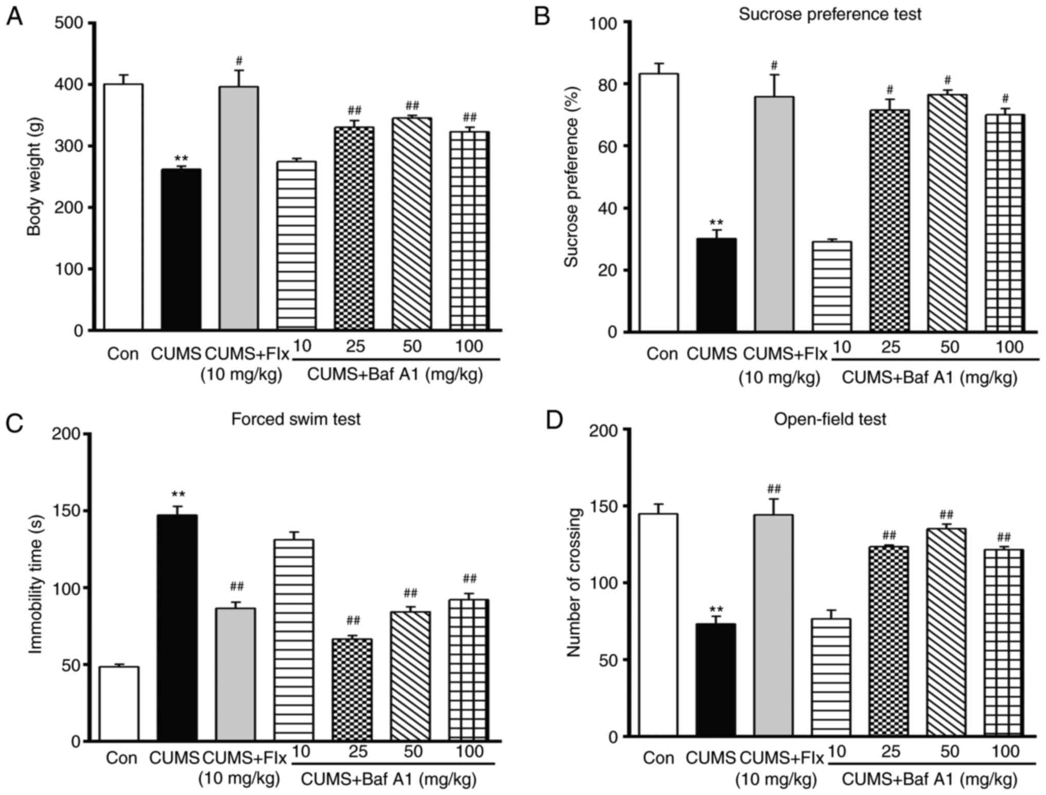 | Figure 1.Effects of Baf A1 on CUMS-induced
depressive-like behaviors. Effects of Baf A1 (10, 25, 50 and 100
mg/kg) or Flx (10 mg/kg) on (A) CUMS-induced rat body weight, (B)
sucrose preference, (C) immobility time in the forced swim test,
and (D) the total distance in the open field test. Data are
expressed as the mean ± standard error of the mean; n=10 per group.
**P<0.01 vs. Con; #P<0.05, ##P<0.01
vs. CUMS. CUMS, chronic unpredictable mild stress; Con, control;
Flx, fluoxetine; Baf A1, Bafilomycin A1. |
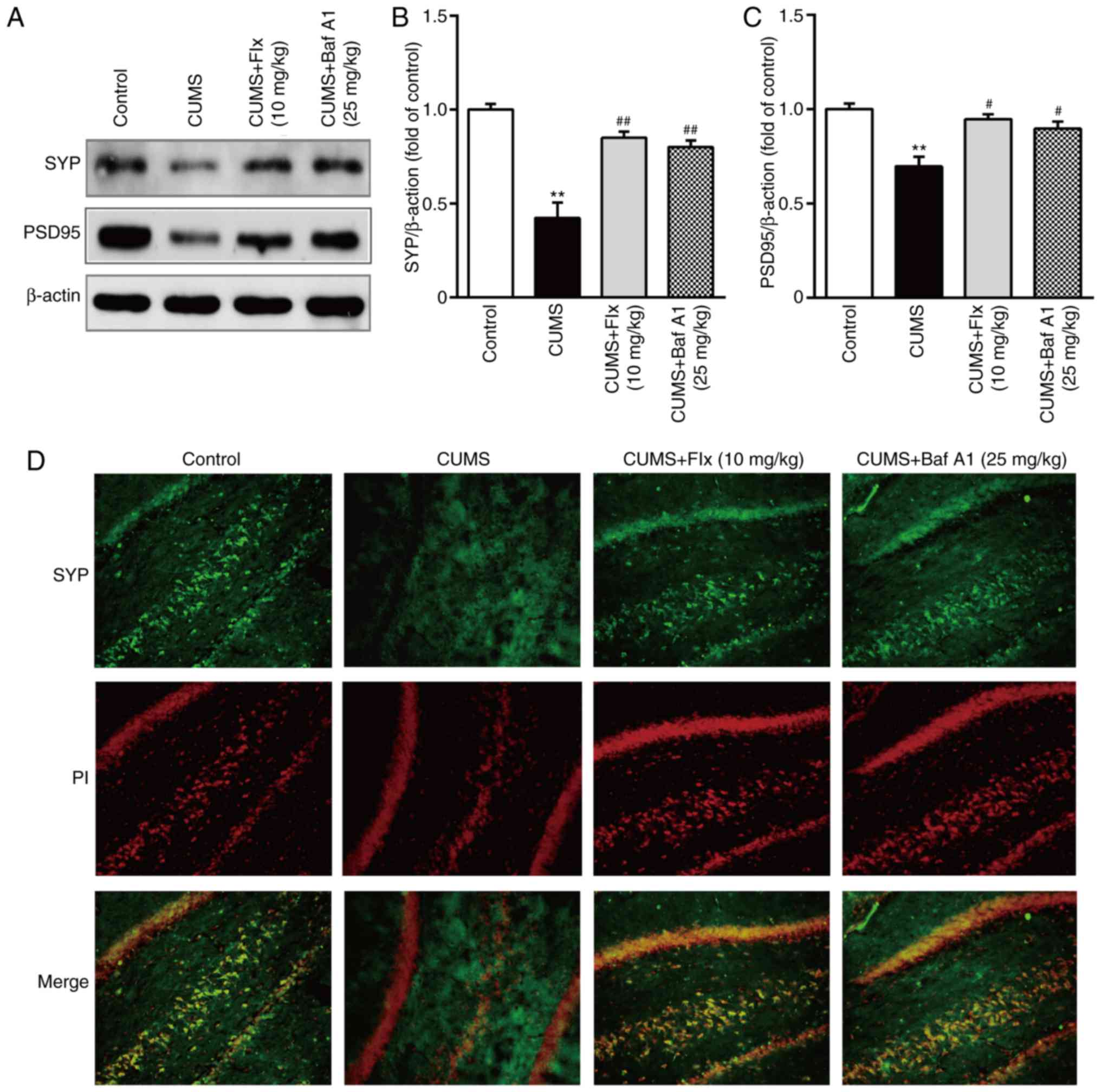
Baf A1 increases synaptic protein
expression in CUMS rats
As presented in Fig.
2A-C, the expression levels of SYP and PSD 95 were
significantly decreased in the hippocampi of CUMS rats compared
with the control group. The administration of Baf A1 (25 mg/kg) and
fluoxetine (10 mg/kg) for 4 weeks significantly increased the
expression levels of SYP and PSD 95 in the hippocampus compared
with the CUMS group. Subsequently, the present study also detected
the immunoreactivity of SYP in the hippocampi of rats. The
immunofluorescence results demonstrated that treatment with Baf A1
(25 mg/kg) and fluoxetine (10 mg/kg) markedly increased the
fluorescence intensity of SYP compared with the CUMS group
(Fig. 2D).
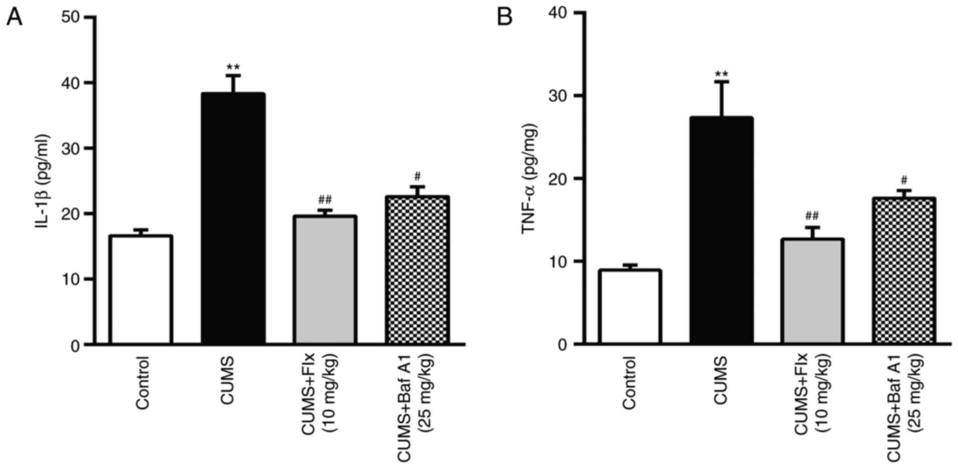
Baf A1 ameliorates the inflammatory
response in the hippocampi of CUMS rats
Inflammation is closely associated with the
development of depressive symptoms, and the anti-inflammatory
effects of Baf A1 in CUMS rats were further examined. As
demonstrated in Fig. 3, the levels
of pro-inflammatory cytokines IL-1β and TNF-α were significantly
increased in the hippocampi of CUMS rats compared with the control
group. However, this increase was significantly suppressed by
treatment with Baf A1 (25 mg/kg) and fluoxetine (10 mg/kg) for 4
weeks, suggesting that Baf A1 may have the ability to ameliorate
CUMS-induced increases in the levels of IL-1β and TNF-α in the
hippocampus.
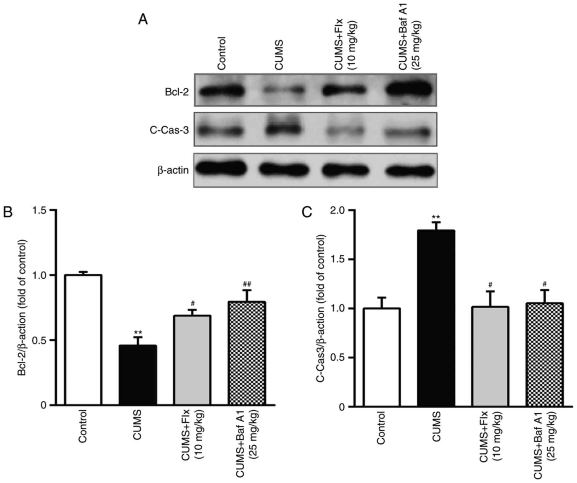
Baf A1 inhibits hippocampal cell
apoptosis in CUMS rats
The expression levels of the apoptosis-associated
proteins Bcl-2 and C-Cas-3 in the hippocampi of CUMS rats were
assessed by western blotting. As presented in Fig. 4, the expression of Bcl-2 was
significantly decreased in the hippocampi of the CUMS group
compared with the control group. The expression levels of C-Cas-3
in the hippocampi were significantly increased in the CUMS group
compared with the control group. Administration of Baf A1 (25
mg/kg) and fluoxetine (10 mg/kg) for 4 weeks significantly
increased the expression of Bcl-2 and decreased the expression of
C-Cas-3 compared with the CUMS group. These results indicated that
Baf A1 inhibited hippocampal cell apoptosis in CUMS rats.
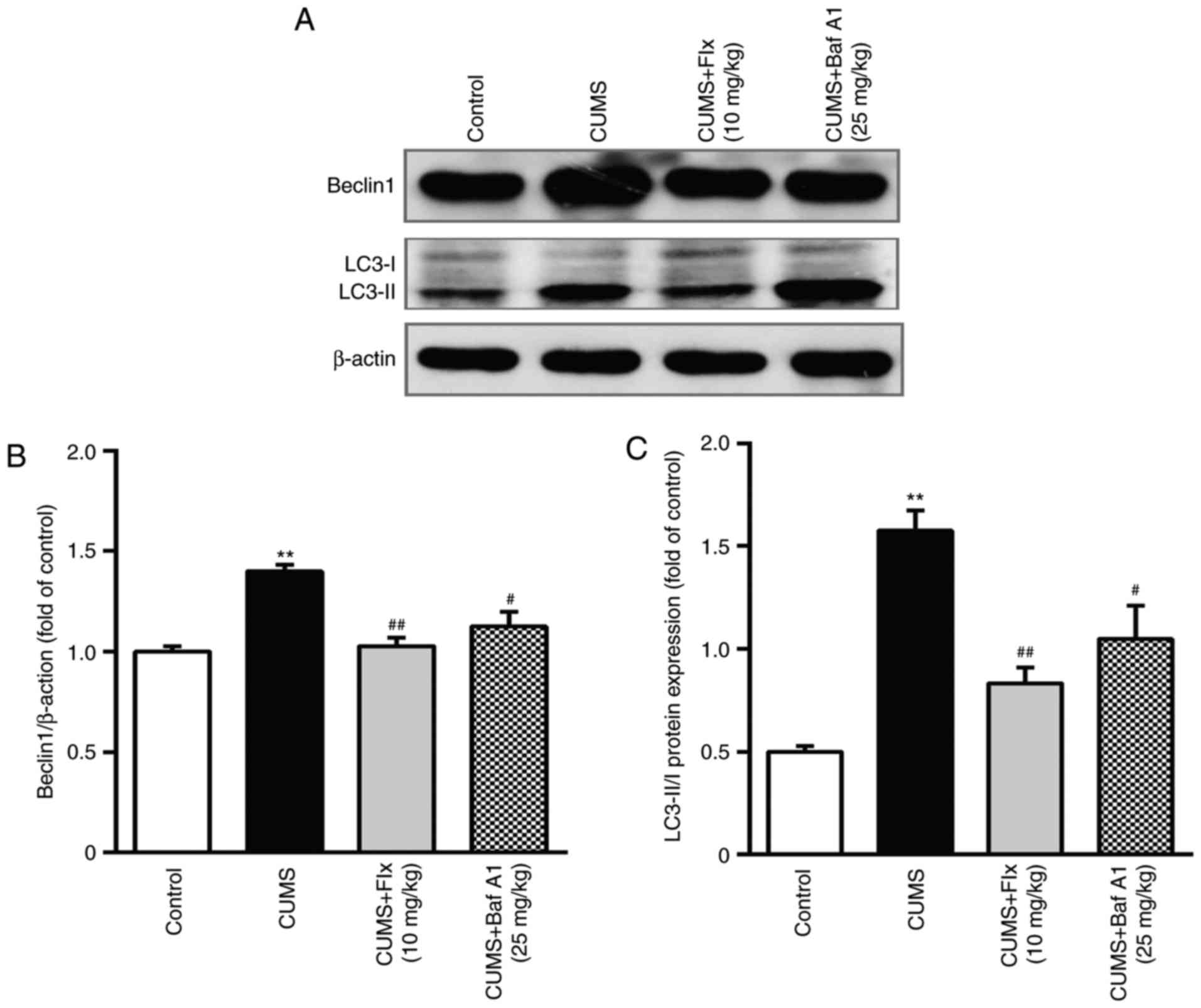
Bafilomycin A1 inhibits autophagy in
CUMS rats
The expression of two critical markers in the
process of autophagy, Beclin 1 and LC3 II/LC3 I were determined by
western blotting (Fig. 5). The
results demonstrated that the expression levels of Beclin 1 and LC3
II/LC3 I were significantly increased in the CUMS group. These
alterations suggested that CUMS induced autophagic stress. However,
the CUMS-exposed rats treated with Baf A1 (25 mg/kg) and fluoxetine
(10 mg/kg) for 4 weeks exhibited significantly decreased Beclin1
expression and LC3 II/LC3 I ratios. These results suggested that
Baf A1 inhibited autophagy in CUMS rats.
Discussion
In the present study, the results indicated that Baf
A1 may possess antidepressant-like effects in the CUMS rat model.
CUMS is considered to be one of the best animal models for
mimicking the symptoms of depression and is a widely used model of
depression (21). According to the
observations from the present study, rats exposed to CUMS exhibited
a significantly decreased body weight and percentage of sucrose
preference, increased immobility time in the FST and decreased
crossing time in the OFT. Administration of Baf A1 for 4 weeks
significantly ameliorated the depressive-like behaviors in CUMS
rats.
SYP and PSD 95 are associated with the survival and
growth of neurons by regulating synaptic transmission and promoting
synapse stability (22). These two
synapse-associated proteins have been confirmed to serve important
roles in neurogenesis and synaptogenesis (23). Clinical studies have indicated that
the levels of PSD 95 and SYP are significantly decreased in
patients with major depressive disorder (24,25).
A previous study reported that restraint stress tended to decrease
the expression of SYP in the rat hippocampus, although this was
restored to pre-stress levels after 2 weeks of treatment with
venlafaxine (2). The present
results revealed that CUMS caused a significant reduction in PSD 95
and SYP expression in the hippocampus, which was consistent with a
previous study (26).
Administration of Baf A1 for 4 weeks significantly increased the
expression levels of SYP and PSD 95 in the CUMS group. The
expression levels of synaptic plasticity proteins in the
hippocampus were almost parallel to the antidepressant-like
behavioral responses, as demonstrated by results obtained from
western blotting and immunofluorescence analysis.
As mentioned, the activation of the inflammatory
response system may serve an important role in the pathophysiology
of depressive disorders (27). A
previous study indicated that pro-inflammatory cytokines
significantly increase in depressed patients and CUMS mice
(28). In addition,
antidepressants, including ketamine and fluoxetine, result in a
decrease in pro-inflammatory cytokines, including IL-6 and TNF-α in
the hippocampi of CUMS rats (29).
The present results demonstrated that treatment with Baf A1 for 4
weeks significantly decreased the levels of IL-1β and TNF-α
compared with the CUMS group, suggesting that Baf A1 inhibited
inflammatory responses. In addition, high levels of
pro-inflammatory cytokines may lead to a widespread increase in
damaged proteins and autophagy (29).
Autophagy is a ubiquitous phenomenon in eukaryotic
cells that serves a key role in maintaining cellular homeostasis
(30). Dysregulation of autophagy
may cause a cellular ‘traffic jam’ during neuronal development and
synaptic plasticity, leading to neurodevelopmental disorders
(31). Previous research has
suggested that autophagy is widely involved in the neural mechanism
of depression, which may be a novel target for antidepressants
(32). Rapamycin, a
serine/threonine-protein kinase mTOR inhibitor, has also been
reported to have antidepressant activity in animal models (33). In addition, autophagy markers,
including Beclin 1, are increased in the mouse brain following
treatment with antidepressants (34). Autophagy may have favorable and
unfavorable consequences in MDD, which may be the reason why
certain patients with MDD remain resistant to certain
antidepressants (31). The present
study elucidated that Baf A1 reduced the expression of Beclin 1 and
the ratio of LC3II/I in CUMS rats, which was consistent with
results from previous studies (12,15).
These results indicated that Baf A1 alleviates depression-like
behaviors, and this may be mediated by inhibition of autophagy.
In conclusion, the present study demonstrated that
Baf A1 alleviates depression-like behaviors induced by CUMS, which
may be mediated by inhibition of hippocampal inflammation,
apoptosis and autophagy. It may therefore be proposed that Baf A1
may be a novel antidepressant candidate for the treatment of
depression.
Acknowledgements
Not applicable.
Funding
The present study was supported by the Experimental
Animal Science and Technology Plan Projects of Zhejiang Province
(grant nos. 2018C37092, 2018 and 2014C37019, 2014]; Scientific and
Technological Projects of Jiaxing (grant no. 2015AY23066, 2015);
and College Students' Science and Technology Innovation Activity
Plan and Talent Plan Project of Zhejiang Province (grant no.
2015R417004, 2015).
Availability of data and materials
The analyzed data sets generated during the present
study are available from the corresponding author on reasonable
request.
Authors' contributions
ZW wrote the manuscript and interpreted the data. SL
and WP analyzed the data and revised the manuscript, YG searched
the literature and collected the data. ZS designed the study.
Ethics approval and consent to
participate
All animal experiments were approved by the Ethics
Committee of the Medical College of Jiaxing University (Jiaxing,
China; approval number JUMC2018-012) and all procedures adhered to
the National Institutes of Health Guide for the Care and Use of
Laboratory Animals.
Patient consent for publication
Not applicable.
Competing interests
The authors declare that they have no competing
interests.
References
|
1
|
Barcelos-Ferreira R, Nakano EY, Steffens
DC and Bottino CM: Quality of life and physical activity associated
to lower prevalence of depression in community-dwelling elderly
subjects from Sao Paulo. J Affect Disord. 150:616–622. 2013.
View Article : Google Scholar : PubMed/NCBI
|
|
2
|
O'Neil A, Fisher AJ, Kibbey KJ, Jacka FN,
Kotowicz MA, Williams LJ, Stuart AL, Berk M, Lewandowski PA, Taylor
CB and Pasco JA: Depression is a risk factor for incident coronary
heart disease in women: An 18-year longitudinal study. J Affect
Disord. 196:117–124. 2016. View Article : Google Scholar : PubMed/NCBI
|
|
3
|
Chandrasekhar Y, Ramya EM, Navya K, Phani
Kumar G and Anilakumar KR: Antidepressant like effects of
hydrolysable tannins of Terminalia catappa leaf extract via
modulation of hippocampal plasticity and regulation of monoamine
neurotransmitters subjected to chronic mild stress (CMS). Biomed
Pharmacother. 86:414–425. 2017. View Article : Google Scholar : PubMed/NCBI
|
|
4
|
Murphy JA and Byrne GJ: Prevalence and
correlates of the proposed DSM-5 diagnosis of Chronic Depressive
Disorder. J Affect Disord. 139:172–180. 2012. View Article : Google Scholar : PubMed/NCBI
|
|
5
|
Gaspersz R, Nawijn L, Lamers F and Penninx
BWJH: Patients with anxious depression: Overview of prevalence,
pathophysiology and impact on course and treatment outcome. Curr
Opin Psychiatry. 31:17–25. 2018. View Article : Google Scholar : PubMed/NCBI
|
|
6
|
Kraus C, Castren E, Kasper S and
Lanzenberger R: Serotonin and neuroplasticity-Links between
molecular, functional and structural pathophysiology in depression.
Neurosci Biobehav Rev. 77:317–326. 2017. View Article : Google Scholar : PubMed/NCBI
|
|
7
|
Kwon SE and Chapman ER: Synaptophysin
regulates the kinetics of synaptic vesicle endocytosis in central
neurons. Neuron. 70:847–854. 2011. View Article : Google Scholar : PubMed/NCBI
|
|
8
|
Politi P, Brondino N and Emanuele E:
Increased proapoptotic serum activity in patients with chronic mood
disorders. Arch Med Res. 39:242–245. 2008. View Article : Google Scholar : PubMed/NCBI
|
|
9
|
Komatsu M, Waguri S, Chiba T, Murata S,
Iwata J, Tanida I, Ueno T, Koike M, Uchiyama Y, Kominami E and
Tanaka K: Loss of autophagy in the central nervous system causes
neurodegeneration in mice. Nature. 441:880–884. 2006. View Article : Google Scholar : PubMed/NCBI
|
|
10
|
Zschocke J, Zimmermann N, Berning B, Ganal
V, Holsboer F and Rein T: Antidepressant drugs diversely affect
autophagy pathways in astrocytes and neurons-dissociation from
cholesterol homeostasis. Neuropsychopharmacology. 36:1754–1768.
2011. View Article : Google Scholar : PubMed/NCBI
|
|
11
|
Williamson LC and Neale EA: Bafilomycin A1
inhibits the action of tetanus toxin in spinal cord neurons in cell
culture. J Neurochem. 63:2342–2345. 1994. View Article : Google Scholar : PubMed/NCBI
|
|
12
|
Pivtoraiko VN, Harrington AJ, Mader BJ,
Luker AM, Caldwell GA, Caldwell KA, Roth KA and Shacka JJ: Low-dose
bafilomycin attenuates neuronal cell death associated with
autophagy-lysosome pathway dysfunction. J Neurochem. 114:1193–1204.
2010.PubMed/NCBI
|
|
13
|
Yuan N, Song L, Zhang S, Lin W, Cao Y, Xu
F, Fang Y, Wang Z, Zhang H, Li X, et al: Bafilomycin A1 targets
both autophagy and apoptosis pathways in pediatric B-cell acute
lymphoblastic leukemia. Haematologica. 100:345–356. 2015.
View Article : Google Scholar : PubMed/NCBI
|
|
14
|
Redmann M, Benavides GA, Berryhill TF,
Wani WY, Ouyang X, Johnson MS, Ravi S, Barnes S, Darley-Usmar VM
and Zhang J: Inhibition of autophagy with bafilomycin and
chloroquine decreases mitochondrial quality and bioenergetic
function in primary neurons. Redox Biol. 11:73–81. 2017. View Article : Google Scholar : PubMed/NCBI
|
|
15
|
Cui D, Wang L, Qi A, Zhou Q, Zhang X and
Jiang W: Propofol prevents autophagic cell death following oxygen
and glucose deprivation in PC12 cells and cerebral
ischemia-reperfusion injury in rats. PLoS One. 7:e353242012.
View Article : Google Scholar : PubMed/NCBI
|
|
16
|
Qiu ZK, Zhong DS, He JL, Liu X, Chen JS
and Nie H: The anxiolytic-like effects of puerarin are associated
with the changes of monoaminergic neurotransmitters and
biosynthesis of allopregnanolone in the brain. Metab Brain Dis.
33:167–175. 2018. View Article : Google Scholar : PubMed/NCBI
|
|
17
|
Li H, Lin S, Qin T, Li H, Ma Z and Ma S:
Senegenin exerts anti-depression effect in mice induced by chronic
un-predictable mild stress via inhibition of NF-κB regulating NLRP3
signal pathway. Int Immunopharmacol. 53:24–32. 2017. View Article : Google Scholar : PubMed/NCBI
|
|
18
|
Porsolt RD, Bertin A and Jalfre M:
Behavioral despair in mice: A primary screening test for
antidepressants. Arch Int Pharmacodyn Ther. 229:327–336.
1977.PubMed/NCBI
|
|
19
|
Livak KJ and Schmittgen TD: Analysis of
relative gene expression data using real-time quantitative PCR and
the 2(-Delta Delta C(T)) method. Methods. 25:402–408. 2001.
View Article : Google Scholar : PubMed/NCBI
|
|
20
|
Zhao Z, Zhang L, Guo XD, Cao LL, Xue TF,
Zhao XJ, Yang DD, Yang J, Ji J, Huang JY and Sun XL: Rosiglitazone
exerts an anti-depressive effect in unpredictable chronic
mild-stress-induced depressive mice by maintaining essential neuron
autophagy and inhibiting excessive astrocytic apoptosis. Front Mol
Neurosci. 10:2932017. View Article : Google Scholar : PubMed/NCBI
|
|
21
|
Yang JL, Liu X, Jiang H, Pan F, Ho CS and
Ho RC: The effects of high-fat-diet combined with chronic
unpredictable mild stress on depression-like behavior and
Leptin/LepRb in male rats. Sci Rep. 6:352392016. View Article : Google Scholar : PubMed/NCBI
|
|
22
|
Aguilar-Arredondo A, López-Hernández F,
García-Velázquez L, Arias C and Zepeda A: Behavior-associated
neuronal activation after kainic acid-induced hippocampal
neurotoxicity is modulated in time. Anat Rec (Hoboken).
300:425–432. 2017. View
Article : Google Scholar : PubMed/NCBI
|
|
23
|
Duman CH and Duman RS: Spine synapse
remodeling in the pathophysiology and treatment of depression.
Neurosci Lett. 601:20–29. 2015. View Article : Google Scholar : PubMed/NCBI
|
|
24
|
Zhao J, Bao AM, Qi XR, Kamphuis W,
Luchetti S, Lou JS and Swaab DF: Gene expression of GABA and
glutamate pathway markers in the prefrontal cortex of non-suicidal
elderly depressed patients. J Affect Disord. 138:494–502. 2012.
View Article : Google Scholar : PubMed/NCBI
|
|
25
|
Gilabert-Juan J, Varea E, Guirado R,
Blasco-Ibáñez JM, Crespo C and Nácher J: Alterations in the
expression of PSA-NCAM and synaptic proteins in the dorsolateral
prefrontal cortex of psychiatric disorder patients. Neurosci Lett.
530:97–102. 2012. View Article : Google Scholar : PubMed/NCBI
|
|
26
|
Liu XL, Luo L, Mu RH, Liu BB, Geng D, Liu
Q and Yi LT: Fluoxetine regulates mTOR signalling in a
region-dependent manner in depression-like mice. Sci Rep.
5:160242015. View Article : Google Scholar : PubMed/NCBI
|
|
27
|
Leighton SP, Nerurkar L, Krishnadas R,
Johnman C, Graham GJ and Cavanagh J: Chemokines in depression in
health and in inflammatory illness: A systematic review and
meta-analysis. Mol Psychiatry. 23:48–58. 2018. View Article : Google Scholar : PubMed/NCBI
|
|
28
|
Kochar B, Barnes EL, Long MD, Cushing KC,
Galanko J, Martin CF, Raffals LE and Sandler RS: Depression is
associated with more aggressive inflammatory bowel disease. Am J
Gastroenterol. 113:80–85. 2018. View Article : Google Scholar : PubMed/NCBI
|
|
29
|
Wang N, Yu HY, Shen XF, Gao ZQ, Yang C,
Yang JJ and Zhang GF: The rapid antidepressant effect of ketamine
in rats is associated with down-regulation of pro-inflammatory
cytokines in the hippocampus. Ups J Med Sci. 120:241–248. 2015.
View Article : Google Scholar : PubMed/NCBI
|
|
30
|
Du D, Hu L, Wu J, Wu Q, Cheng W, Guo Y,
Guan R, Wang Y, Chen X, Yan X, et al: Neuroinflammation contributes
to autophagy flux blockage in the neurons of rostral ventrolateral
medulla in stress-induced hypertension rats. J Neuroinflammation.
14:1692017. View Article : Google Scholar : PubMed/NCBI
|
|
31
|
Jia J and Le W: Molecular network of
neuronal autophagy in the pathophysiology and treatment of
depression. Neurosci Bull. 31:427–434. 2015. View Article : Google Scholar : PubMed/NCBI
|
|
32
|
Ma J, Hou LN, Rong ZX, Liang P, Fang C, Li
HF, Qi H and Chen HZ: Antidepressant desipramine leads to C6 glioma
cell autophagy: Implication for the adjuvant therapy of cancer.
Anticancer Agents Med Chem. 13:254–260. 2013. View Article : Google Scholar : PubMed/NCBI
|
|
33
|
Cleary C, Linde JA, Hiscock KM, Hadas I,
Belmaker RH, Agam G, Flaisher-Grinberg S and Einat H:
Antidepressive-like effects of rapamycin in animal models:
Implications for mTOR inhibition as a new target for treatment of
affective disorders. Brain Res Bull. 76:469–473. 2008. View Article : Google Scholar : PubMed/NCBI
|
|
34
|
Gassen NC, Hartmann J, Zschocke J, Stepan
J, Hafner K, Zellner A, Kirmeier T, Kollmannsberger L, Wagner KV,
Dedic N, et al: Association of FKBP51 with priming of autophagy
pathways and mediation of antidepressant treatment response:
Evidence in cells, mice, and humans. PLoS Med. 11:e10017552014.
View Article : Google Scholar : PubMed/NCBI
|