Stem cell-based therapy has been demonstrated as a
good option for cardiac repair following myocardial infarction
(MI). Previous clinical trials have demonstrated efficacy using a
traumatic method of bone marrow aspiration, culture of the bone
marrow stem cells and intracoronary infusion of this end-product
(1). Another less invasive option
is to use granulocyte colony-stimulating factor (G-CSF) to mobilize
bone marrow-derived progenitor cells to undertake tissue repair
(2). The advantage of this
technique is that it is less traumatic, requiring only subcutaneous
injections. However, the results on its therapeutic efficacy have
thus far been conflicting. In the FIRSTLINE-AMI study, subcutaneous
injection of G-CSF shortly after successful primary percutaneous
coronary intervention (PCI) in patients suffering from acute ST
elevation MI was demonstrated to mobilize mononuclear blood stem
cells (3). This resulted in
enhanced resting wall thickening of the infarcted myocardium,
improvement of wall motions and systolic function without apparent
re-stenosis. Further studies have demonstrated a good safety
profile without reports of adverse effects (2,4,5).
However, the REVIVAL-2 study demonstrated that delayed application
of G-CSF 5 days after successful PCI did not significantly reduce
infarct size or improve left ventricular function (6). Furthermore, usage of G-CSF in acute
MI may be a double-edged sword, due to the fact that it may
paradoxically reduce the migratory capacity of bone marrow-derived
progenitor cells into the ischaemic myocardium and thus there is a
need to optimize the cytokine profile with other agents to ensure
their successful migration (7).
Animal studies have proven useful in the study of
the properties of G-CSF. A previous study used a porcine model of
ischaemic heart disease to demonstrate improvement in cardiac
contractile function in chronic myocardial ischaemia after G-CSF
administration (8). Whilst its
efficacy in reversing mechanical dysfunction in the heart has been
observed, the potential benefits in chronic obliterative arterial
disease remain to be fully investigated. Thus, the present study
aimed to investigate the effects of G-CSF in a novel rabbit model
of chronic obliterative arterial disease, which was generated by
clamping the carotid artery using a surgical ameroid constrictor.
These results were compared with those of an established porcine
model of ischamic heart disease generated by clamping of the left
coronary artery also using this constrictor.
A novel rabbit model of chronic obliterative
arterial disease was produced by clamping the right carotid artery
using an ameroid constrictor. The current study was approved by the
Animal Welfare Ethics Committee of the Tongji Hospital of Tongji
University. A total of 36 New Zealand rabbits were used (male, six
months old male weight 2–3 kg; SPF grade Ivl; Experimental Animal
Centre, Tongji Hospital). The animals were housed at 22±1°C and
50–60% relative humidity with a 12-h light/dark cycle. They were
divided into 2 groups, with one group being fed a high fat diet for
two months to induce atherosclerosis (AS), whereas the other group
was fed a normal fat diet, acting as the control group (CON). Each
group was further subdivided into three groups: Sham group (carotid
arteries surgically exposed but no ameroid constrictors were
applied; SHAM), ameroid constrictor group receiving hypodermic
saline (0.5 ml daily for 5 days; NS) and ameroid constrictor group
receiving G-CSF (15 µg.kg−1 daily; Shanghai 3D
Biotechnology Co., Ltd.). The 6 groups were therefore: AS-SHAM,
AS-NS, AS-G-CSF; CON-SHAM, CON-NS and CON-G-CSF.
The following parameters were measured using Doppler
ultrasound Vevo770 Imaging system (VisualSonics, Inc., Toronto, ON,
Canada) at four different time points (prior to surgery and 3, 5 or
7 weeks after surgery): Peak systolic flow velocity, resistance
index and the end-diastolic velocity. Stenosis rate was calculated
by (difference in diameters between left and right carotid
arteries)/(left carotid artery).
RNA was extracted and purified from the proximal
portion of the right carotid artery using TRIzol (Invitrogen;
Thermo Fisher Scientific, Inc., Waltham, MA, USA). Quantification
was achieved using a spectrophotometer. A total of 2 µg RNA was
used as a template for a RT-qPCR with random hexamers (Toyobo Life
Science, Osaka, Japan) as primers. Then, 10 µl cDNA was purified
with an additional ethanol washing step. Levels of mRNA were
quantified by RT-qPCR using a Rotor-Gene 2000 machine (Corbett
Research, Mortlake, Australia). The results were presented as gene
copy numbers relative to GAPDH. The primers were used as follows:
Endothelin 1 (ET-1), upstream, 5′-CTCTCTGCTGTTGGTGGCTTT-3′ and
downstream 5′-TGGGTTTTCCGCTCCTGT-3′; endothelial nitric oxide
synthase (eNOS), upstream 5′-AGGCCTCCTGTGAGACTTTC-3′ and downstream
5′-AAGGAGTCGAGGACTGGATG-3′; GAPDH, upstream
5′-CCACTTTGTGAAGCTCATTTCCT-3′ and downstream
5′-TCGTCCTCCTCTGGTGCTCT-3′.
The appearance of the endothelium of the rabbit
carotid arteries close to the site of ameroid constrictor
application was studied using a scanning electron microscope
(HITACHI-S520; Hitachi, Tokyo, Japan).
A well-established porcine model of chronic ischemic
heart disease was also used in the present study, which involved
clamping the left coronary artery using an ameroid constrictor. A
total of 24 pigs (male, 1 year old, weight ~40 kg; Beijing
University of Agriculture, China) The animals were housed in a cage
with straw at 22±1°C and 50–60% relative humidity with a 12-h
light/dark cycle and water and food ad libitum. They were equally
divided into four groups, receiving normal saline 0.5 ml (control),
G-CSF 2.5 µg.kg−1.day−1 (low dose), 5
µg.kg−1.day−1 (medium dose) and 10
µg.kg−1.day−1 (high dose) for 5 days. G-CSF
was diluted using normal saline.
In the porcine model, the proximal portion of
occluded coronary arteries were obtained four weeks after the
operation, which allowed effects of G-CSF on histology to be
determined. Hematoxylin and eosin (H&E) staining was used to
examine angiogenesis and quantified using a Leica Qwin V3 imaging
system (Leica Microsystems GmbH, Wetzlar, Germany). Mallory stain
was used to study collagen content. Immunohistochemical staining
were used to examine vascular endothelial growth factor (VEGF) and
tumor necrosis factor (TNF)-α, which reflected angiogenesis and
inflammation, respectively.
All data were presented as mean ± standard error.
Fisher's exact test was used as appropriate using SPSS (version
11.5; SPSS, Inc., Chicago, IL, USA). P<0.05 was considered to
indicate a statistically significant difference.
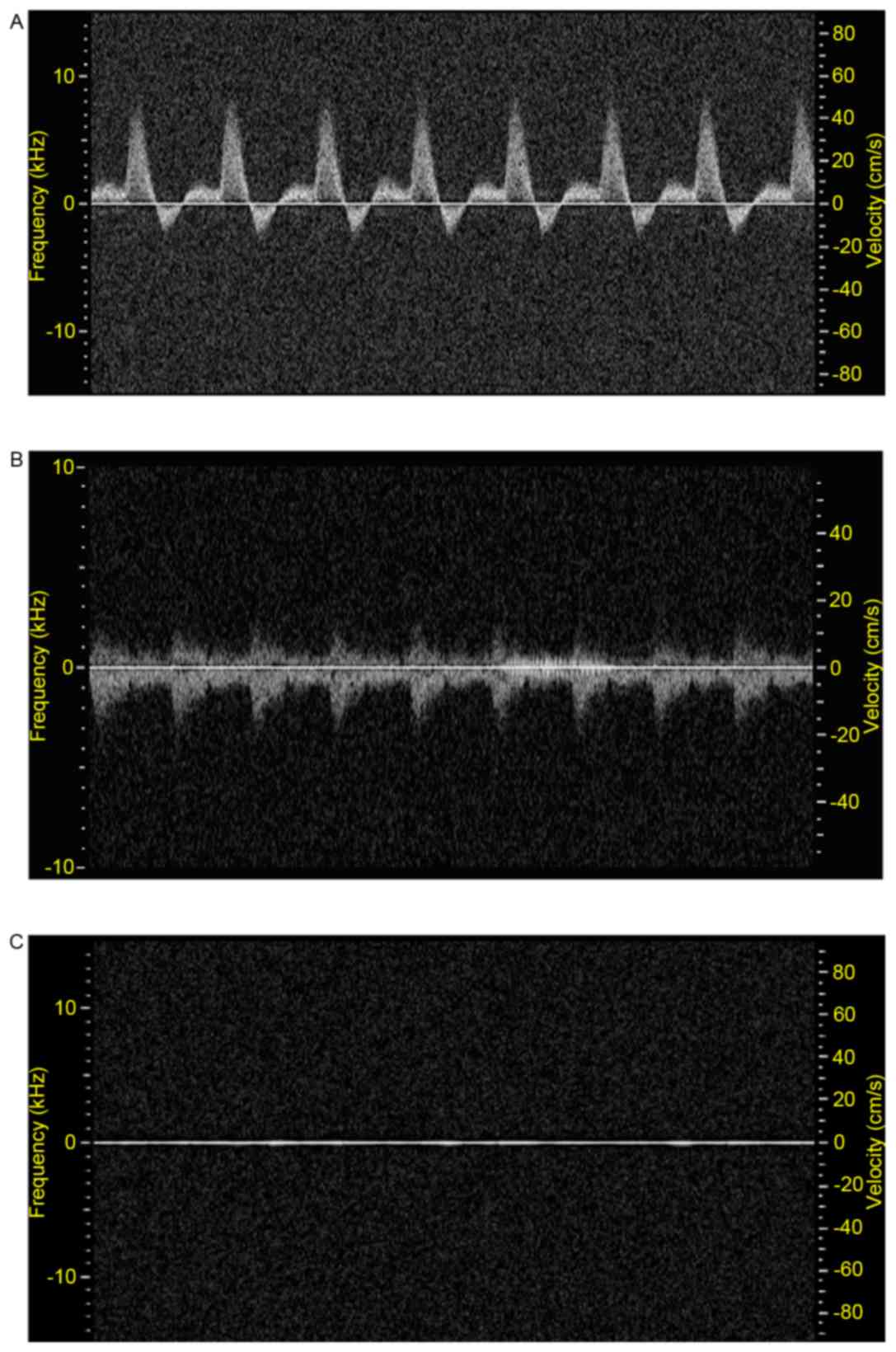
Ultrasound was used for assessing the functional
status of the right carotid artery of rabbit hearts. Firstly,
B-mode ultrasonography was used to determine real-time information
on the lumen and vessel wall (9,10).
Secondly, Doppler imaging was used to determine flow parameters
(Fig. 1). Three weeks after the
operation, the right carotid artery developed stenosis (Table I). Five weeks later, total
occlusion was observed in all groups at different rates (Table II).
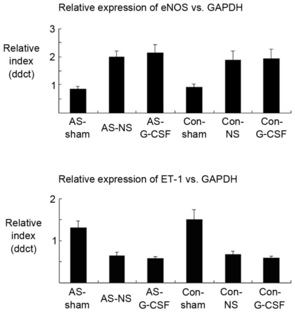
RNA was extracted from the proximal portion of the
right carotid arteries in the different groups (Fig. 2). Expression indices of endothelial
nitric oxide synthase (eNOS) in the AS-SHAM and CON-SHAM groups
were 0.85±0.11 and 0.91±0.12, respectively. They were increased to
2±0.206 and 1.89±0.33 for the AS-NS and CON-NS groups. However,
these values were not significantly different from the values in
the presence of G-CSF (2.14±0.30 and 1.94±0.32 for AS-G-CSF and
CON-G-CSF). The expression levels of ET-1 were 1.3±0.17 and
1.5±0.25 (AS-SHAM and CON-SHAM), 0.65±0.08 and 0.67±0.09 (AS-NS and
CON-NS) and 0.57±0.05 and 0.59±0.05 (AS-G-CSF and CON-G-CSF). There
were no significant difference between the groups treated with
normal saline compared with those treated with G-CSF.
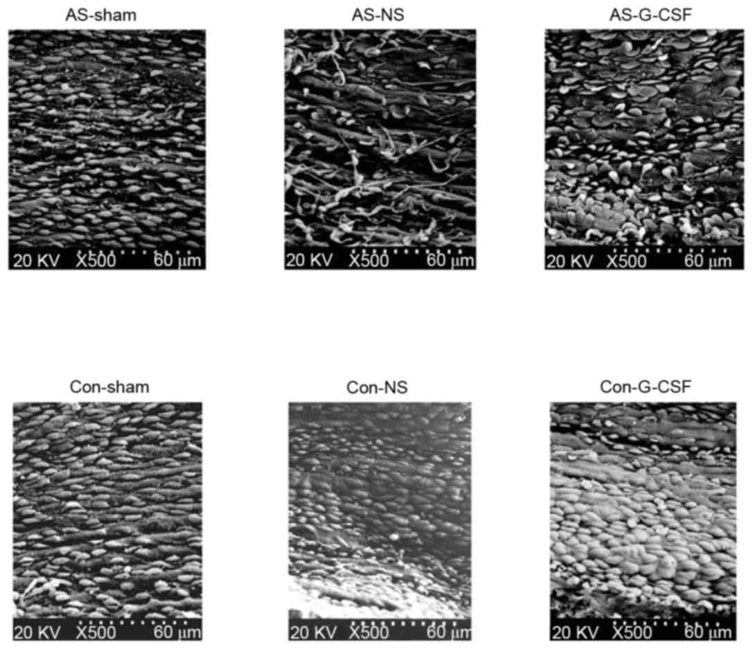
The morphology of endothelial cells of the proximal
carotid artery were assessed using scanning electron microscopy
(Fig. 3). In the AS-SHAM and
CON-SHAM groups, endothelial cells took on an oval shape, which was
due to linear blood flow within the artery. When compared with the
CON-SHAM group, the AS-SHAM group, the endarterium developed a
rougher morphology with a smaller number of endothelial cells, and
had a more shrunken image with tiny lipid droplets. With
application of the ameroid constrictor, this morphology was changed
to one of roundness due to the turbulent flow distal to the site of
occlusion. Such a change was more prominent in the AS-SHAM than
CON-SHAM group. The number of endothelial cells increased after
G-CSF injections.
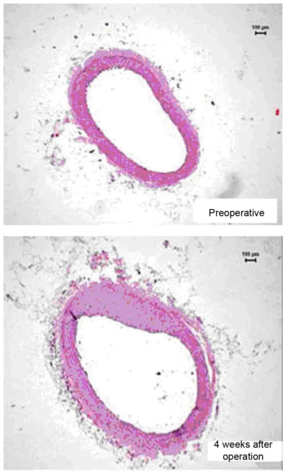
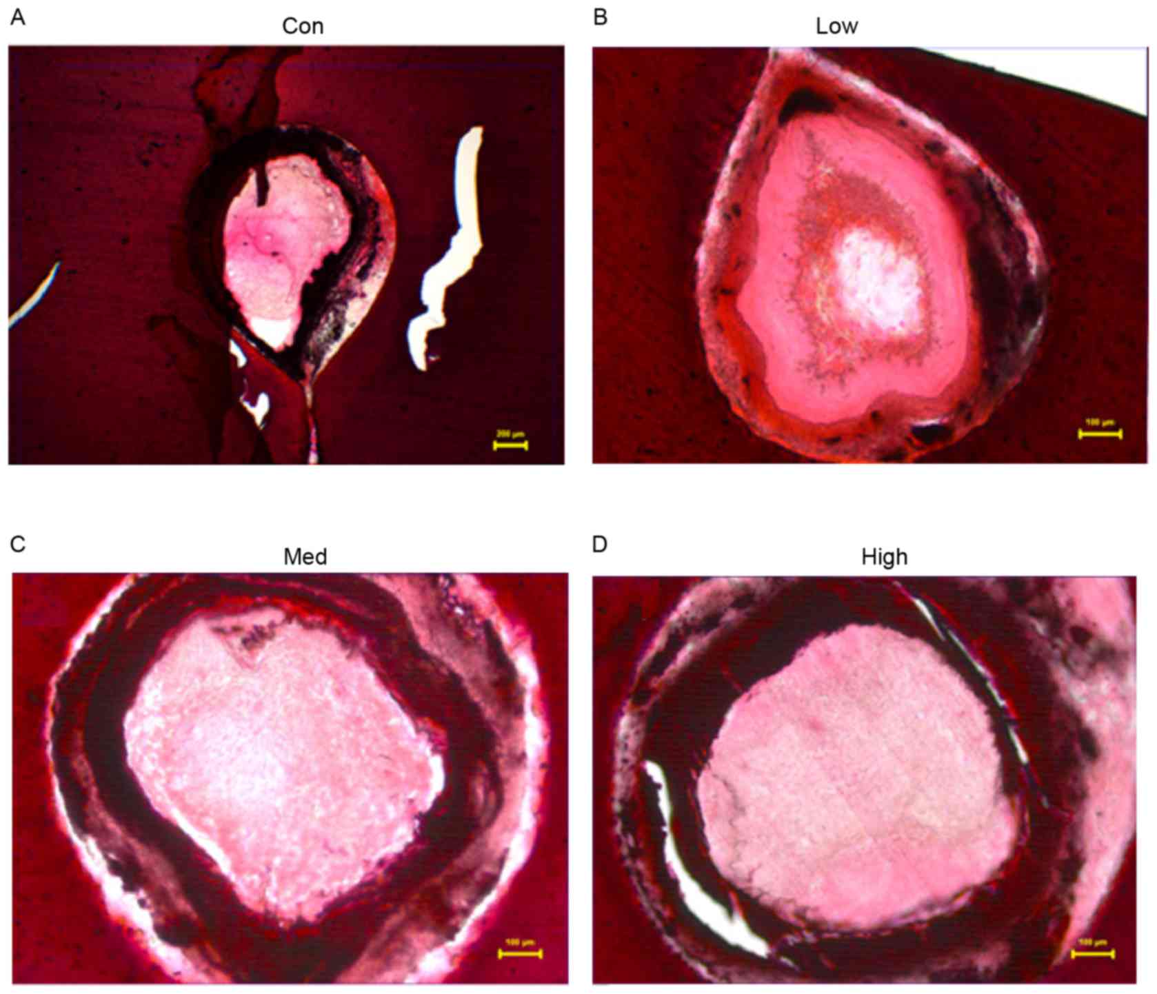
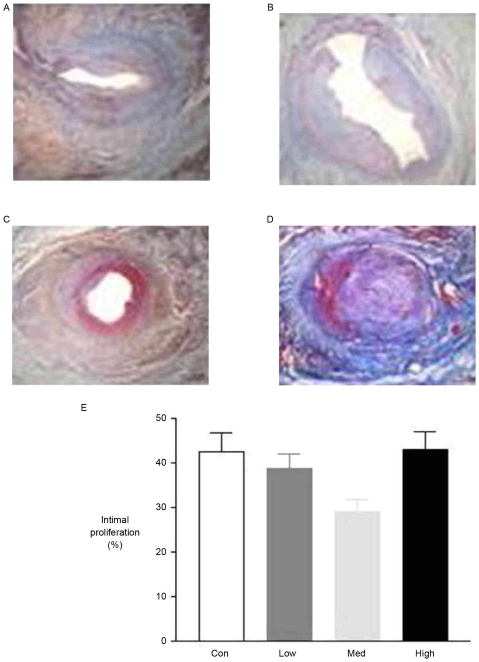
Four weeks after the operation, histological studies
exhibited intimal proliferation together with eccentric narrowing
in the left coronary arteries (Fig.
4). H&E staining indicated varying degrees of intimal
hyperplasia in the different groups. Intimal hyperplasia was more
severe in the low and high dose G-CSF groups compared with the
control group, whereas it was less severe in the medium dose group
compared with the control (Fig.
5). The areas of tunica intima and vessel lumen were analyzed
using Leica Qwin Plus Graphic Processing System, which allowed
proliferation area to be calculated (Fig. 6).
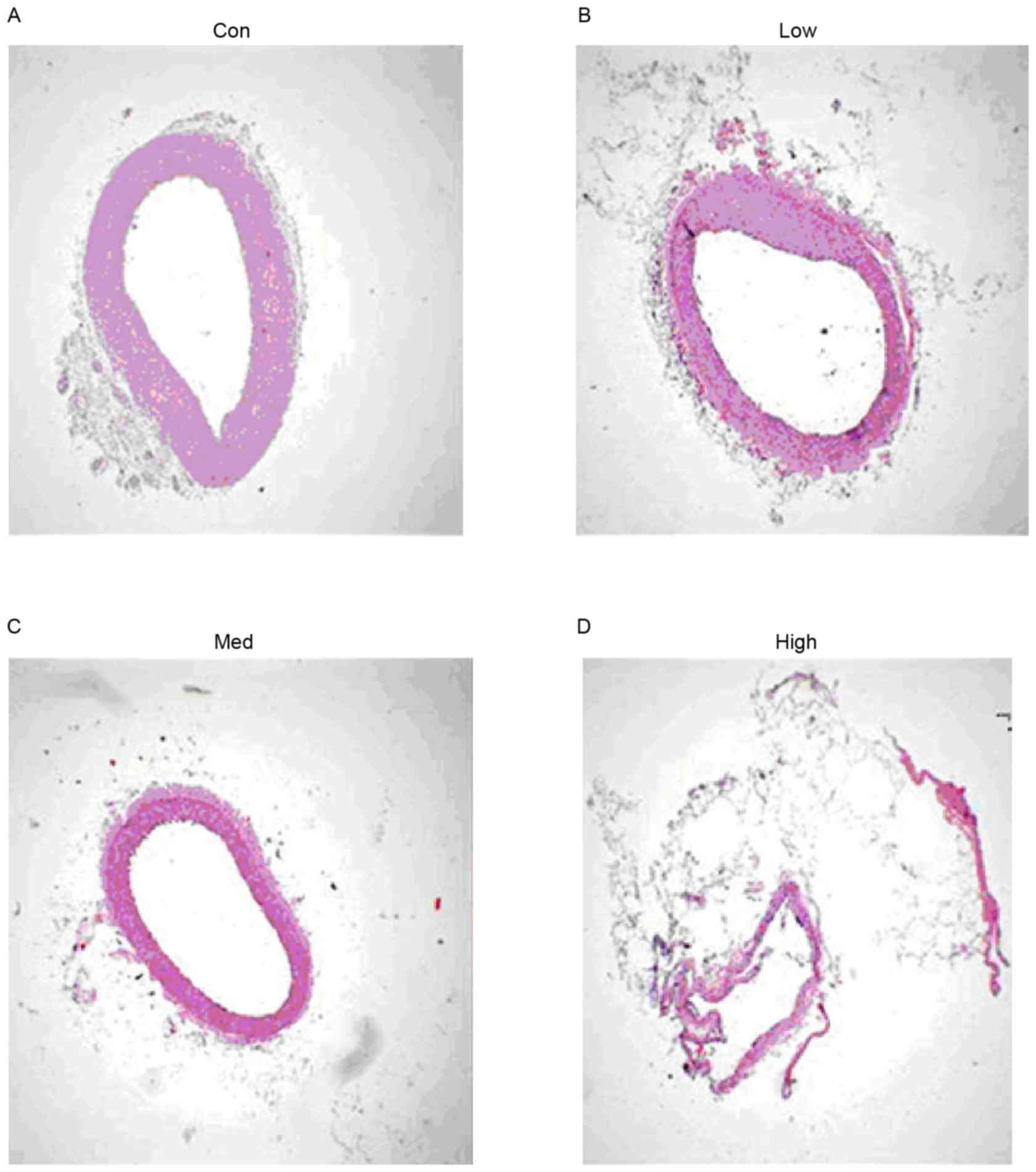
Mallory's trichrome staining of the proximal
coronary artery indicated significantly smaller amounts of collagen
in the medium and high dose groups compared with the control group.
However, there was no significant difference between the low dose
and control group (Fig. 7).
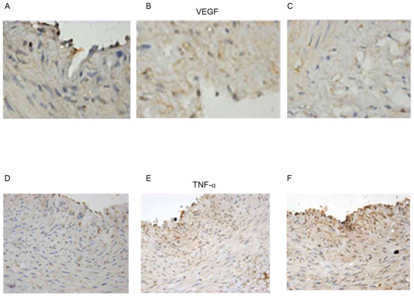
In order to examine the role of angiogenesis and
inflammation, immunohistochemistry was used to determine the
expression of VEGF and TNF-α. G-CSF exerted a dose-dependent effect
on VEGF and TNF- α, increasing them both at increasing
concentrations (Fig. 8).
G-CSF has exhibited some efficacy in reversing
mechanical dysfunction in chronic ischaemic heart disease and
following MI. A previous studies demonstrated its safety in humans,
however results regarding its therapeutic effects remain
conflicting (10). A previous
study failed to demonstrate improvement in ventricular function
post MI using G-CSF when compared with the control group (11), in contrast to other studies in
which improved left ventricular systolic function was observed
(4,5). However, G-CSF was demonstrated to
reduce the migratory capacity of bone marrow-derived progenitor
cells into the ischaemic myocardium (7). In addition, patients suffering from
coronary artery disease responded to G-CSF by exhibiting increased
numbers of endothelial progenitor cells (EPCs) and higher
expression of the chemokine receptor CXCR4, which directed EPCs to
ischaemic tissue (12). However,
increased mobilization did not equate to reversal of damage, no
significant improvement in wall motion, perfusion or exercise
duration after G-CSF use was observed (13). However, intra-coronary infusion of
G-CSF did mobilize peripheral blood stem cells and improved
ejection fraction in patients with acute MI although not in those
with chronic (≥6±1.2 months) MI (14).
The mechanism of action of G-CSF remains to be fully
elucidated. Different animal models have been useful for modelling
the molecular mechanisms of human diseases ADDIN EN.CITE (15–35),
resulting in translational insights (36–41).
Mice and rats have been used because of their amenability to
genetic modification, as demonstrated by the study of ion channel
mutations on cardiovascular physiology (42–44).
The effects of increased oxidative stress on endothelial function
have been studied in diabetic mice and spontaneously hypertensive
rats, modelling human cardio-metabolic disorders (45–57).
Larger animals such as guinea pigs and pigs have haemodynamic
parameters more similar to humans, and are therefore useful for
studying flow properties, however it remains a challenge to study
using methods involving large machines, such as cardiac magnetic
resonance imaging (34,35). In a rat model, inhibition of
neointimal formation and increased re-endothelialisation of injured
arteries has been observed, potentially via a direct effect on the
heart and the arteries (58). In
the current study, two animal models were employed by applying
ameroid surgical constrictors to the right carotid artery of
rabbits or to the left coronary artery of pigs.
In the rabbit model, a high fat diet led to the
development of atherosclerosis, a chronic inflammatory disease
(59). The number of endothelial
cells increased after G-CSF injection at a dose of 15 µg/kg/day as
demonstrated using scanning electron microscopy. However, there was
no change in endothelial function assessed using RT-qPCR for the
endothelial markers endothelial nitric oxide synthase and ET-1.
Whether or not G-CSF is protective in atherosclerosis remains
controversial. At a dose of 10 µg/kg/day, G-CSF has been
demonstrated to worsen atherosclerosis in apolipoprotein
E-deficient mice (60), however by
contrast, at a high dose of 100 µg/kg/day, it prevented progression
of atherosclerosis in heritable hyperlipidemic rabbits and
following vascular injury induced by angioplasty balloon in rabbits
(61). In an iliac artery injury
model of rabbits, G-CSF at a dose of 70 µg/day mobilized vascular
progenitor cells, which induced neointimal overgrowth at the
stented vessels and enhanced endothelial healing was observed when
drug-eluting stent was used when compared with a bare metal stent
(62). In a rat model, G-CSF
increased the number of mononuclear cells in the circulation,
increased endothelial adhesion markers and re-endothelialization of
the denuded vessels, which resulted in parallel reductions in
inflammation in the vessel wall (63).
The present study in porcine hearts observed
increasing intimal hyperplasia after low (2.5 µg/kg/day) or high
(10 µg/kg/day) doses of G-CSF. Furthermore, the mobilization of
bone marrow-derived progenitor cells using G-CSF resulted in
increased endothelial expressions of VEGF and TNF-α, markers of
angiogenesis and inflammation, respectively, as assessed using
immunohistochemistry and western blot analysis. G-CSF has
pleotropic effects, and is capable of inducing hyperplasia,
angiogenesis and inflammation (62). Whether it is beneficial or harmful
depends on the relative contributions to the above processes. In
the present study, intimal hyperplasia was increased in chronic
obliterative arterial disease, in contrast to observations in
humans where G-CSF was demonstrated to be effective in acute as
opposed to chronic (≥6±1.2 months ± 1.2) MI (14). Other beneficial effects of G-CSF
have been reported in rats, where a neuroprotective role was
demonstrated in transient middle cerebral artery occlusion
(64). However, G-CSF can be
ineffective or potentially harmful. For example, experiments in a
swine model of ischaemia-reperfusion injury indicated that G-CSF
accelerated angiogenesis, reduced fibrosis however did not improve
either ejection fraction or end-diastolic volume (65). Furthermore, intramyocardial VEGF
gene transfer followed by bone marrow stem cell mobilization using
G-CSF was safe however did not significantly improve myocardial
perfusion as assessed by single photon emission computerized
tomography (66). G-CSF aggravated
in-stent re-stensosis, which was partly dependent on VEGF and
STAT-3, although this may be reduced by using a sirolimus
drug-eluting stent (67).
In conclusion, the present study identified that: i)
G-CSF did not alter endothelial function in the carotid artery of a
rabbit model of atherosclerosis; ii) its effects on chronic
ischaemic heart disease in pigs are dose-dependent, worsening
intimal hyperplasia when used at a low or high dose, however
improving it at a medium dose. Therefore, its therapeutic role
warrants further attention.
The authors would like to thank Dr Dingzhiwen of the
Department of Cardiology, Shanghai Institute of Cardiovascular
Diseases, Zhongshan Hospital (Shanghai, China), for providing the
facilities for operations on animals and analysis on tissue
pathology.
Professor Gary Tse was supported by the
Biotechnology and Biological Sciences Research Council Doctoral
Training Award at the University of Cambridge. In addition, the
current study was supported by grants from the National 973 Program
of China (grant no. 2007CB512003), National Natural Science
Foundation of China (grant no. 81300150) and Science and Technology
Commission of Shanghai (grant no. 074107020).
The datasets used and/or analyzed during the current
study are available from the corresponding author on reasonable
request.
The manuscript was written with contributions from
all authors. All authors have given approval to the final version
of the manuscript. ZH performed the research and experiments. ZC,
YW, JJ, GT, WX, JG and BS contributed to data analysis. ZH and ZC
were responsible for the overall project design and manuscript
organization.
The present study was approved by the Animal Welfare
Ethics Committee of the Tongji Hospital of Tongji University and
the approval number was 2017-DW-006.
Not applicable.
The authors declare no conflict of interest.
|
1
|
Traverse JH, McKenna DH, Harvey K,
Jorgenso BC, Olson RE, Bostrom N, Kadidlo D, Lesser JR, Jagadeesan
V, Garberich R and Henry TD: Results of a phase 1, randomized,
double-blind, placebo-controlled trial of bone marrow mononuclear
stem cell administration in patients following ST-elevation
myocardial infarction. Am Heart J. 160:428–434. 2010. View Article : Google Scholar : PubMed/NCBI
|
|
2
|
Ince H, Petzsch M, Kleine HD, Eckard H,
Rehders T, Burska D, Kische S, Freund M and Nienaber CA: Prevention
of left ventricular remodeling with granulocyte colony-stimulating
factor after acute myocardial infarction: Final 1-year results of
the front-integrated revascularization and stem cell liberation in
evolving acute myocardial infarction by granulocyte
colony-stimulating factor (FIRSTLINE-AMI) trial. Circulation 112 (9
Suppl). I73–I80. 2005.
|
|
3
|
Ince H, Petzsch M, Kleine HD, Schmidt H,
Rehders T, Körber T, Schümichen C, Freund M and Nienaber CA:
Preservation from left ventricular remodeling by front-integrated
revascularization and stem cell liberation in evolving acute
myocardial infarction by use of granulocyte-colony-stimulating
factor (FIRSTLINE-AMI). Circulation. 112:3097–3106. 2005.
View Article : Google Scholar : PubMed/NCBI
|
|
4
|
Takano H, Hasegawa H, Kuwabara Y, Nakayama
T, Matsuno K, Miyazaki Y, Yamamoto M, Fujimoto Y, Okada H, Okubo S,
et al: Feasibility and safety of granulocyte colony-stimulating
factor treatment in patients with acute myocardial infarction. Int
J Cardiol. 122:41–47. 2007. View Article : Google Scholar : PubMed/NCBI
|
|
5
|
Valgimigli M, Rigolin GM, Cittanti C,
Malagutti P, Curello S, Percoco G, Bugli AM, Della Porta M,
Bragotti LZ, Ansani L, et al: Use of granulocyte-colony stimulating
factor during acute myocardial infarction to enhance bone marrow
stem cell mobilization in humans: Clinical and angiographic safety
profile. Eur Heart J. 26:1838–1845. 2005. View Article : Google Scholar : PubMed/NCBI
|
|
6
|
Zohlnhöfer D, Ott I, Mehilli J, Schömig K,
Michalk F, Ibrahim T, Meisetschläger G, von Wedel J, Bollwein H,
Seyfarth M, et al: Stem cell mobilization by granulocyte
colony-stimulating factor in patients with acute myocardial
infarction: A randomized controlled trial. JAMA. 295:1003–1010.
2006. View Article : Google Scholar : PubMed/NCBI
|
|
7
|
Brunner S, Huber BC, Fischer R, Groebner
M, Hacker M, David R, Zaruba MM, Vallaster M, Rischpler C, Wilke A,
et al: G-CSF treatment after myocardial infarction: Impact on bone
marrow-derived vs cardiac progenitor cells. Exp Hematol.
36:695–702. 2008. View Article : Google Scholar : PubMed/NCBI
|
|
8
|
Hasegawa H, Takano H, Iwanaga K, Ohtsuka
M, Qin Y, Niitsuma Y, Ueda K, Toyoda T, Tadokoro H and Komuro I:
Cardioprotective effects of granulocyte colony-stimulating factor
in swine with chronic myocardial ischemia. J Am Coll Cardiol.
47:842–849. 2006. View Article : Google Scholar : PubMed/NCBI
|
|
9
|
Kagawa R, Moritake K, Shima T and Okada Y:
Validity of B-mode ultrasonographic findings in patients undergoing
carotid endarterectomy in comparison with angiographic and
clinicopathologic features. Stroke. 27:700–705. 1996. View Article : Google Scholar : PubMed/NCBI
|
|
10
|
Handa N, Matsumoto M, Maeda H, Hougaku H,
Ogawa S, Fukunaga R, Yoneda S, Kimura K and Kamada T: Ultrasonic
evaluation of early carotid atherosclerosis. Stroke. 21:1567–1572.
1990. View Article : Google Scholar : PubMed/NCBI
|
|
11
|
Ripa RS, Jørgensen E, Wang Y, Thune JJ,
Nilsson JC, Søndergaard L, Johnsen HE, Køber L, Grande P and
Kastrup J: Stem cell mobilization induced by subcutaneous
granulocyte-colony stimulating factor to improve cardiac
regeneration after acute ST-elevation myocardial infarction: Result
of the double-blind, randomized, placebo-controlled stem cells in
myocardial infarction (STEMMI) trial. Circulation. 113:1983–1992.
2006. View Article : Google Scholar : PubMed/NCBI
|
|
12
|
Powell TM, Paul JD, Hill JM, Thompson M,
Benjamin M, Rodrigo M, McCoy JP, Read EJ, Khuu HM, Leitman SF, et
al: Granulocyte colony-stimulating factor mobilizes functional
endothelial progenitor cells in patients with coronary artery
disease. Arterioscler Thromb Vasc Biol. 25:296–301. 2005.
View Article : Google Scholar : PubMed/NCBI
|
|
13
|
Hill JM, Syed MA, Arai AE, Powell TM, Paul
JD, Zalos G, Read EJ, Khuu HM, Leitman SF, Horne M, et al: Outcomes
and risks of granulocyte colony-stimulating factor in patients with
coronary artery disease. J Am Coll Cardiol. 46:1643–1648. 2005.
View Article : Google Scholar : PubMed/NCBI
|
|
14
|
Kang HJ, Lee HY, Na SH, Chang SA, Park KW,
Kim HK, Kim SY, Chang HJ, Lee W, Kang WJ, et al: Differential
effect of intracoronary infusion of mobilized peripheral blood stem
cells by granulocyte colony-stimulating factor on left ventricular
function and remodeling in patients with acute myocardial
infarction versus old myocardial infarction: The MAGIC Cell-3-DES
randomized, controlled trial. Circulation 114 (1 Suppl). I145–I151.
2006.
|
|
15
|
Tse G, Hothi SS, Grace AA and Huang CL:
Ventricular arrhythmogenesis following slowed conduction in
heptanol-treated, Langendorff-perfused mouse hearts. J Physiol Sci.
62:79–92. 2012. View Article : Google Scholar : PubMed/NCBI
|
|
16
|
Tse G, Tse V, Yeo JM and Sun B: Atrial
anti-arrhythmic effects of heptanol in Langendorff-perfused mouse
hearts. PLoS One. 11:e01488582016. View Article : Google Scholar : PubMed/NCBI
|
|
17
|
Tse G, Yeo JM, Tse V and Sun B: Gap
junction inhibition by heptanol increases ventricular
arrhythmogenicity by decreasing conduction velocity without
affecting repolarization properties or myocardial refractoriness in
Langendorff-perfused mouse hearts. Mol Med Rep. 14:4069–4074. 2016.
View Article : Google Scholar : PubMed/NCBI
|
|
18
|
Tse G, Wong ST, Tse V and Yeo JM:
Restitution analysis of alternans using dynamic pacing and its
comparison with S1S2 restitution in heptanol-treated, hypokalaemic
Langendorff-perfused mouse hearts. Biomed Rep. 4:673–680. 2016.
View Article : Google Scholar : PubMed/NCBI
|
|
19
|
Tse G, Tse V and Yeo JM: Ventricular
anti-arrhythmic effects of heptanol in hypokalaemic,
Langendorff-perfused mouse hearts. Biomed Rep. 4:313–324. 2016.
View Article : Google Scholar : PubMed/NCBI
|
|
20
|
Tse G, Sun B, Wong ST, Tse V and Yeo JM:
Anti-arrhythmic effects of hypercalcemia in hyperkalemic,
Langendorff-perfused mouse hearts. Biomed Rep. 5:301–310. 2016.
View Article : Google Scholar : PubMed/NCBI
|
|
21
|
Choy L, Yeo JM, Tse V, Chan SP and Tse G:
Cardiac disease and arrhythmogenesis: Mechanistic insights from
mouse models. Int J Cardiol Heart Vasc. 12:1–10. 2016.PubMed/NCBI
|
|
22
|
Tse G, Wong ST, Tse V, Lee YT, Lin HY and
Yeo JM: Cardiac dynamics: Alternans and arrhythmogenesis. J
Arrhythm. 32:411–417. 2016. View Article : Google Scholar : PubMed/NCBI
|
|
23
|
Tse G, Lai ET, Yeo JM and Yan BP:
Electrophysiological mechanisms of Bayés syndrome: Insights from
clinical and mouse studies. Front Physiol. 7:1882016. View Article : Google Scholar : PubMed/NCBI
|
|
24
|
Tse G, Lai ET, Lee AP, Yan BP and Wong SH:
Electrophysiological mechanisms of gastrointestinal
arrhythmogenesis: Lessons from the heart. Front Physiol. 7:2302016.
View Article : Google Scholar : PubMed/NCBI
|
|
25
|
Tse G, Fiona Chan YW, Keung W and Yan BP:
Electrophysiological mechanisms of long and short QT syndromes:
Insights from mouse models. Int J Cardiol Heart Vasc. 14:8–13.
2017.PubMed/NCBI
|
|
26
|
Tse G, Lai TH, Yeo JM, Tse V and Wong SH:
Mechanisms of electrical activation and conduction in the
gastrointestinal system: Lessons from cardiac electrophysiology.
Front Physiol. 7:1822016. View Article : Google Scholar : PubMed/NCBI
|
|
27
|
Tse G, Lai ET, Tse V and Yeo JM: Molecular
and electrophysiological mechanisms underlying cardiac
arrhythmogenesis in diabetes mellitus. J Diabetes Res.
2016:28487592016. View Article : Google Scholar : PubMed/NCBI
|
|
28
|
Tse G, Yan BP, Chan YW, Tian XY and Huang
Y: Reactive oxygen species, endoplasmic reticulum stress and
mitochondrial dysfunction: The link with cardiac arrhythmogenesis.
Front Physiol. 7:3132016. View Article : Google Scholar : PubMed/NCBI
|
|
29
|
Chen Z, Sun B, Tse G, Jiang J and Xu W:
Reversibility of both sinus node dysfunction and reduced HCN4 mRNA
expression level in an atrial tachycardia pacing model of
tachycardia-bradycardia syndrome in rabbit hearts. Int J Clin Exp
Pathol. 9:8526–8531. 2016.
|
|
30
|
Tse G, Yeo JM, Chan YW, Lai ET and Yan BP:
What is the arrhythmic substrate in viral myocarditis? Insights
from clinical and animal studies. Front Physiol. 7:3082016.
View Article : Google Scholar : PubMed/NCBI
|
|
31
|
Tse G and Yeo JM: Conduction abnormalities
and ventricular arrhythmogenesis: The roles of sodium channels and
gap junctions. Int J Cardiol Heart Vasc. 9:75–82. 2015.PubMed/NCBI
|
|
32
|
Tse G: Mechanisms of cardiac arrhythmias.
J Arrhythm. 32:75–81. 2016. View Article : Google Scholar : PubMed/NCBI
|
|
33
|
Vassiliou V, Chin C, Perperoglou A, Tse G,
Ali A, Raphael C, Jabbour A, Newby D, Pennell D, Dweck M and Prasad
S: 93 ejection fraction by cardiovascular magnetic resonance
predicts adverse outcomes post aortic valve replacement. Heart. 100
(Suppl 3):A53–A54. 2014. View Article : Google Scholar
|
|
34
|
Tse G, Ali A, Prasad SK, Vassiliou V and
Raphael CE: Atypical case of post-partum cardiomyopathy: An overlap
syndrome with arrhythmogenic right ventricular cardiomyopathy? BJR
Case Rep. 1:201501822015.PubMed/NCBI
|
|
35
|
Tse G, Ali A, Alpendurada F, Prasad S,
Raphael CE and Vassiliou V: Tuberculous constrictive pericarditis.
Res Cardiovasc Med. 4:e296142015.PubMed/NCBI
|
|
36
|
Tse G: Both transmural dispersion of
repolarization and of refractoriness are poor predictors of
arrhythmogenicity: A role for iCEB (QT/QRS)? J Geriatr Cardiol.
13:813–814. 2016.PubMed/NCBI
|
|
37
|
Tse G, Wong ST, Tse V and Yeo JM:
Monophasic action potential recordings: Which is the recording
electrode? J Basic Clin Physiol Pharmacol. 27:457–462. 2016.
View Article : Google Scholar : PubMed/NCBI
|
|
38
|
Tse G and Yan BP: Novel arrhythmic risk
markers incorporating QRS dispersion: QRSd ×
(Tpeak-Tend)/QRS and QRSd ×
(Tpeak-Tend)/(QT × QRS). Ann Noninvasive
Electrocardiol. 22:Nov;2016.(doi: 10.1111/anec.12397). PubMed/NCBI
|
|
39
|
Tse G: Novel conduction-repolarization
indices for the stratification of arrhythmic risk. J Geriatr
Cardiol. 13:811–812. 2016.PubMed/NCBI
|
|
40
|
Tse G: (Tpeak-Tend)/QRS and
(Tpeak-Tend)/(QT × QRS): Novel markers for predicting arrhythmic
risk in Brugada syndrome. Europace. 19:6962017. View Article : Google Scholar : PubMed/NCBI
|
|
41
|
Tse G and Yan BP: Traditional and novel
electrocardiographic markers for predicting arrhythmic risk and
sudden cardiac death. Europace. 19:712–721. 2016. View Article : Google Scholar
|
|
42
|
Tse G, Wong ST, Tse V and Yeo JM:
Depolarization vs. repolarization: What is the mechanism of
ventricular arrhythmogenesis underlying sodium channel
haploinsufficiency in mouse hearts? Acta Physiol (Oxf).
218:234–235. 2016. View Article : Google Scholar : PubMed/NCBI
|
|
43
|
Tse G, Wong ST, Tse V and Yeo JM:
Variability in local action potential durations, dispersion of
repolarization and wavelength restitution in aged wild-type and
Scn5a+/− mouse hearts modelling human Brugada syndrome.
J Geriatr Cardiol. 13:930–931. 2016.PubMed/NCBI
|
|
44
|
Tse G, Wong ST, Tse V and Yeo JM:
Determination of action potential wavelength restitution in
Scn5a+/− mouse hearts modelling human Brugada syndrome.
J Physiol. 14:595–596. 2017.
|
|
45
|
Murugan D, Lau YS, Lau WC, Mustafa MR and
Huang Y: Angiotensin 1–7 protects against angiotensin II-induced
endoplasmic reticulum stress and endothelial dysfunction via mas
receptor. PLoS One. 10:e01454132015. View Article : Google Scholar : PubMed/NCBI
|
|
46
|
Wei LH, Huang XR, Zhang Y, Li YQ, Chen HY,
Heuchel R, Yan BP, Yu CM and Lan HY: Deficiency of Smad7 enhances
cardiac remodeling induced by angiotensin II infusion in a mouse
model of hypertension. PLoS One. 8:e701952013. View Article : Google Scholar : PubMed/NCBI
|
|
47
|
Wong WT, Tian XY and Huang Y: Endothelial
dysfunction in diabetes and hypertension: Cross talk in RAS, BMP4,
and ROS-dependent COX-2-derived prostanoids. J Cardiovasc
Pharmacol. 61:204–214. 2013. View Article : Google Scholar : PubMed/NCBI
|
|
48
|
Ma S, Tian XY, Zhang Y, Mu C, Shen H,
Bismuth J, Pownall HJ, Huang Y and Wong WT: E-selectin-targeting
delivery of microRNAs by microparticles ameliorates endothelial
inflammation and atherosclerosis. Sci Rep. 6:229102016. View Article : Google Scholar : PubMed/NCBI
|
|
49
|
Lin Z, Pan X, Wu F, Ye D, Zhang Y, Wang Y,
Jin L, Lian Q, Huang Y, Ding H, et al: Fibroblast growth factor 21
prevents atherosclerosis by suppression of hepatic sterol
regulatory element-binding protein-2 and induction of adiponectin
in mice. Circulation. 131:1861–1871. 2015. View Article : Google Scholar : PubMed/NCBI
|
|
50
|
Yuen CY, Wong SL, Lau CW, Tsang SY, Xu A,
Zhu Z, Ng CF, Yao X, Kong SK, Lee HK and Huang Y: From skeleton to
cytoskeleton: Osteocalcin transforms vascular fibroblasts to
myofibroblasts via angiotensin II and toll-like receptor 4. Circ
Res. 111:e55–e66. 2012. View Article : Google Scholar : PubMed/NCBI
|
|
51
|
Zhang H, Liu J, Qu D, Wang L, Luo JY, Lau
CW, Liu P, Gao Z, Tipoe GL, Lee HK, et al: Inhibition of miR-200c
restores endothelial function in diabetic mice through suppression
of COX-2. Diabetes. 65:1196–1207. 2016. View Article : Google Scholar : PubMed/NCBI
|
|
52
|
Cheang WS, Tian XY, Wong WT, Lau CW, Lee
SS, Chen ZY, Yao X, Wang N and Huang Y: Metformin protects
endothelial function in diet-induced obese mice by inhibition of
endoplasmic reticulum stress through 5′adenosine
monophosphate-activated protein kinase-peroxisome
proliferator-activated receptor δ pathway. Arterioscler Thromb Vasc
Biol. 34:830–836. 2014. View Article : Google Scholar : PubMed/NCBI
|
|
53
|
Morrow JP, Katchman A, Son NH, Trent CM,
Khan R, Shiomi T, Huang H, Amin V, Lader JM, Vasquez C, et al: Mice
with cardiac overexpression of peroxisome proliferator-activated
receptor γ have impaired repolarization and spontaneous fatal
ventricular arrhythmias. Circulation. 124:2812–2821. 2011.
View Article : Google Scholar : PubMed/NCBI
|
|
54
|
Xie L, Feng H, Li S, Meng G, Liu S, Tang
X, Ma Y, Han Y, Xiao Y, Gu Y, et al: SIRT3 mediates the antioxidant
effect of hydrogen sulfide in endothelial cells. Antioxid Redox
Signal. 24:329–343. 2016. View Article : Google Scholar : PubMed/NCBI
|
|
55
|
Xu A and Huang Y: A tireless giant in
vascular research. J Cardiovasc Pharmacol. 67:359–360. 2016.
View Article : Google Scholar : PubMed/NCBI
|
|
56
|
Chen Y, Liu J, Zheng Y, Wang J, Wang Z, Gu
S, Tan J, Jing Q and Yang H: Uncoupling protein 3 mediates
H2O2 preconditioning-afforded
cardioprotection through the inhibition of MPTP opening. Cardiovasc
Res. 105:192–202. 2015. View Article : Google Scholar : PubMed/NCBI
|
|
57
|
Zhang Y, Liu J, Luo JY, Tian XY, Cheang
WS, Xu J, Lau CW, Wang L, Wong WT, Wong CM, et al: Upregulation of
angiotensin (1–7)-mediated signaling preserves endothelial function
through reducing oxidative stress in diabetes. Antioxid Redox
Signal. 23:880–892. 2015. View Article : Google Scholar : PubMed/NCBI
|
|
58
|
Li Y, Fukuda N, Yokoyama S, Kusumi Y,
Hagikura K, Kawano T, Takayama T, Matsumoto T, Satomi A, Honye J,
et al: Effects of G-CSF on cardiac remodeling and arterial
hyperplasia in rats. Eur J Pharmacol. 549:98–106. 2006. View Article : Google Scholar : PubMed/NCBI
|
|
59
|
Hansson GK: Immune mechanisms in
atherosclerosis. Arterioscler Thromb Vasc Biol. 21:1876–1890. 2001.
View Article : Google Scholar : PubMed/NCBI
|
|
60
|
Haghighat A, Weiss D, Whalin MK, Cowan DP
and Taylor WR: Granulocyte colony-stimulating factor and
granulocyte macrophage colony-stimulating factor exacerbate
atherosclerosis in apolipoprotein E-deficient mice. Circulation.
115:2049–2054. 2007. View Article : Google Scholar : PubMed/NCBI
|
|
61
|
Hasegawa H, Takano H, Ohtsuka M, Ueda K,
Niitsuma Y, Qin Y, Tadokoro H, Shiomi M and Komuro I: G-CSF
prevents the progression of atherosclerosis and neointimal
formation in rabbits. Biochem Biophys Res Commun. 344:370–376.
2006. View Article : Google Scholar : PubMed/NCBI
|
|
62
|
Cho HJ, Kim TY, Cho HJ, Park KW, Zhang SY,
Kim JH, Kim SH, Hahn JY, Kang HJ, Park YB and Kim HS: The effect of
stem cell mobilization by granulocyte-colony stimulating factor on
neointimal hyperplasia and endothelial healing after vascular
injury with bare-metal versus paclitaxel-eluting stents. J Am Coll
Cardiol. 48:366–374. 2006. View Article : Google Scholar : PubMed/NCBI
|
|
63
|
Kong D, Melo LG, Gnecchi M, Zhang L,
Mostoslavsky G, Liew CC, Pratt RE and Dzau VJ: Cytokine-induced
mobilization of circulating endothelial progenitor cells enhances
repair of injured arteries. Circulation. 110:2039–2046. 2004.
View Article : Google Scholar : PubMed/NCBI
|
|
64
|
Sehara Y, Hayashi T, Deguchi K, Zhang H,
Tsuchiya A, Yamashita T, Lukic V, Nagai M, Kamiya T and Abe K:
G-CSF enhances stem cell proliferation in rat hippocampus after
transient middle cerebral artery occlusion. Neurosci Lett.
418:248–252. 2007. View Article : Google Scholar : PubMed/NCBI
|
|
65
|
Sato T, Suzuki H, Kusuyama T, Omori Y,
Soda T, Tsunoda F, Shoji M, Iso Y, Koba S, Geshi E, et al: G-CSF
after myocardial infarction accelerates angiogenesis and reduces
fibrosis in swine. Int J Cardiol. 127:166–173. 2008. View Article : Google Scholar : PubMed/NCBI
|
|
66
|
Ripa RS, Wang Y, Jørgensen E, Johnsen HE,
Hesse B and Kastrup J: Intramyocardial injection of vascular
endothelial growth factor-A165 plasmid followed by
granulocyte-colony stimulating factor to induce angiogenesis in
patients with severe chronic ischaemic heart disease. Eur Heart J.
27:1785–1792. 2006. View Article : Google Scholar : PubMed/NCBI
|
|
67
|
Lim SY, Kim YS, Ahn Y, Jeong MH, Rok LS,
Kim JH, Kim KH, Park HW, Kim W, Cho JG, et al: The effects of
granulocyte-colony stimulating factor in bare stent and
sirolimus-eluting stent in pigs following myocardial infarction.
Int J Cardiol. 118:304–311. 2007. View Article : Google Scholar : PubMed/NCBI
|