Introduction
Associated statistics indicate that the morbidity of
primary central nervous system tumors is 20.59/100,000 (1). In addition, the 5-year and 10-year
survival rates of patients with malignant central nervous system
tumors are 33.8 and 28%, respectively (2). Malignant glioma is incurable and
accounts for ~80% of all intracranial malignant tumors (2). Surgical resection of the tumor is not
sufficient to achieve a cure. In certain cases, complete resection
cannot be achieved due to loss of brain function. Therefore, the
comprehensive treatment combination of surgery, radiotherapy and
chemotherapy is most commonly adopted in the clinic at present
(3). Nevertheless, therapeutic
efficacy is still unsatisfactory (3). The field of molecular biology
continues to be developed and applied in tumor research. Targeted
molecular therapy is being used to overcome the difficulty of
treating malignant tumors like glioma (1). Specifically, identifying the
important genes regulating glioma proliferation and investigating
their roles is of positive clinical significance. This study
contributes to revealing the molecular mechanism of glioma
proliferation, and therefore allows effective therapeutic means to
be developed (4).
Glioma is the most common primary intracranial tumor
in adults, and frequently exhibits a high malignancy grade, high
possibility of recurrence and a poor prognosis. It has short median
survival once it is diagnosed (5).
Profound progress has been achieved in cancer treatment, including
improvements in surgery, chemotherapy, radiotherapy and
immunotherapy. However, the overall prognosis is not improved
(5). Consequently, microRNAs
(miRNA/miR), a class of endogenous non-coding small molecular RNA
18–24 nucleotides in length, has attracted attention from
scientists. miRNAs can regulate target genes to alter the
expression of target proteins (6).
The clinical significance of microRNAs is in glioma diagnosis,
chemotherapeutic efficacy evaluation, anti-angiogenesis, treatment
and prognosis (6). An increasing
number of studies have demonstrated that, miRNA serves a vital role
in glioma tumorigenesis and development (6,7).
They may serve as a novel index for the clinical diagnosis and
prognosis evaluation, and as new therapeutic targets for glioma in
the future. miRNAs can act as oncogenes or tumor suppressor genes
(7), and have complex biological
functions (7). As a result, cancer
heterogeneity at the genetic and epigenetic levels is of
therapeutic significance. Furthermore, it also marks the challenge
in the reasonable design of effective therapeutics (7).
Matrix metalloproteinases (MMPs) are a class of
proteases that degrade the vascular basement membrane, and
hydrolyze the extracellular matrix and other matrix components
(5). MMPs have critical functions
in promoting tumor invasion and angiogenesis (5). Currently, >20 MMPs have been
discovered, which can be divided into four categories according to
different substrates. The substrates include collagenases,
gelatinases, matrix degradation enzymes and membrane MMPs. Multiple
studies have demonstrated that high MMPs expression is correlated
with metastasis and poor prognosis patients with epithelial tumor
tissues, including lung cancer, gastric cancer, colon cancer,
breast cancer and prostate cancer (8). The MMP gelatinases, namely MMP-2 and
MMP-9, have molecular weights of 72 and 92 kDa, respectively
(8). MMP-2 can degrade type IV
collagen in the extracellular matrix, which promotes tumor cell
diffusion (9). In addition, it can
promote tumor invasion and metastasis. MMP-2 expression is
gradually increased with the increased tumor, node and metastasis
classification (9). Typically, the
positive expression rate in patients with lymph node metastasis is
increase compared with those with no lymph node metastasis
(10). MMP-9 is the enzyme with
the greatest molecular weight out of the MMPs. It is secreted in
the form of a zymogen (10).
Notably, it can hydrolyze the cell basement membrane, type IV and
type V collagens in the extracellular matrix, and fibronectin
components; thus, resulting in basement membrane destruction,
allowing tumor cells to invade the connective tissue, small vessels
and lymphatic vessels, resulting in metastasis (8). Jia et al (11) reported that miRNA-34a reduced the
migration and invasion of tongue squamous cell carcinoma by
targeting MMP-9 and MMP-14. Tabouret et al (12) showed that MMP2 and MMP9 serum
levels are associated with favorable outcome in patients with
inflammatory breast cancer. The present study aimed to investigate
the function of MMP-9 in human glioma cells and its potential
regulatory mechanisms.
Materials and methods
Clinical specimens
Peripheral blood (5–10 ml) was obtained from
patients with glioma (n=82) following surgery and healthy
volunteers (n=42) at the Affiliated Hospital of Beihua University
(Jilin City, China) between February 2010 and December 2014
(Table I). Peripheral blood was
centrifuged at 1,000 × g for 10 min at 4°C and the serum was stored
at −80°C until analysis. The present study was approved by the
Ethics Committee of Affiliated Hospital of Beihua University. The
study was performed in accordance with the regulations of the
Institutional Review Board of Affiliated Hospital of Beihua
University. Written informed consent was obtained from all enrolled
patients prior to surgery. Written informed consent was also
obtained from healthy volunteers. The follow-up period for the
patients was every three months by telephone for 5 years.
 | Table I.Characteristics of glioma patients and
healthy volunteers. |
Table I.
Characteristics of glioma patients and
healthy volunteers.
| Variables | Patients (n=82) | Healthy volunteers
(n=42) |
|---|
| Age (years) |
| ≤55 | 40 | 19 |
| 55 | 42 | 23 |
| Sex |
|
Female | 35 | 17 |
| Male | 47 | 25 |
| Tumor size (cm) |
| ≤3.0 | 15 |
|
|
>3.0 | 67 |
|
| Edmondson grade |
| I | 7 |
|
| II | 13 |
|
|
III–IV | 62 |
|
RNA extraction and miRNA reverse
transcription-quantitative polymerase chain reaction (RT-qPCR)
Total RNAs from the serum samples and cells were
extracted using TRIzol reagent (Invitrogen; Thermo Fisher
Scientific, Inc., Waltham MA, USA). Total RNAs was used to
synthesize complementary DNA using SuperScript II Reverse
Transcriptase (Invitrogen; Thermo Fisher Scientific, Inc.) at 37°C
for 30 min and 84°C for 1 min. qPCR was performed with StepOne
Real-Time PCR System (Applied Biosystems; Thermo Fisher Scientific,
Inc.) and SYBR Premix Ex Taq™ (Takara Bio, Inc., Otsu, Japan).
miR-34a forward, 5′-CCAGCTGTGAGTGTTTCTTTG-3′ and reverse,
5′-CAGCACTTCTAGGGCAGTAT-3′; U6 forward,
5′-GCTTCGGCAGCACATATACTAAAAT-3′ and reverse,
CTTCGGCAGCACATATACGCTTCACGAATTTGCGTGTCAT-3′; MMP-9 forward,
5′-AGACCTGGGCATTCCAAAC-3′ and reverse, 5′-CGGCAAGTCTTCCGAGTAGT-3′;
reference gene (β2 microglobulin) forward,
5′-TACACTGAATTCACCCCCAC-3′ and reverse, 5′-CATCCAATCCAAATGCGGCA-3′.
The reaction conditions were pre-denaturation at 95°C for 10 min,
followed by 40 cycles of denaturation at 95°C for 5 min, annealing
at 60°C for 30 sec and elongation at 72°C for 30 sec. The relative
expression levels were calculated using the 2-Cq method (13). Low expression of MMP9 was <2 of
the MMP9 expression in healthy volunteers, high expression of MMP9
was >2 of the MMP9 expression of healthy volunteers; low
expression of miR-34a was <2 of miR-34a the expression of
healthy volunteers, high expression of miR-34a was >2 of the
miR-34a expression of healthy volunteers.
Cell lines, culture and
transfection
The U251-MG human glioma cell line was purchased
from the Cell Bank of Type Culture Collection of Chinese Academy of
Sciences (Shanghai, China) and maintained in Dulbecco's modified
Eagle's medium (DMEM; Gibco; Thermo Fisher Scientific, Inc.)
supplemented with 10% fetal bovine serum (FBS; Gibco; Thermo Fisher
Scientific, Inc.) and 1% antibiotic-antimycotic solution at 37°C in
a humidified 5% CO2. miRNA-34a
(5′-CACCGGTTGTTGTGAGCAATAGTA-3′ and
5′-AAACTACTATTGCTCACAACAACC-3′), anti-miRNA-34a
(5′-ACAACCAGCUAAGACACUGCCA-3′ and 5′-TGACCGACATGTTCAGACA-3′) and
negative control mimics (5′-CCCCCCCCCCCCCCC-3′ and
5′-CCCCCCCCCCCC-3′) were purchased from Shanghai GenePharma Co.,
Ltd. (Shanghai, China). U251-MG cells (1×10⁶ cells/well)
were transfected with 100 ng miRNA-34a, 100 ng anti-miRNA-34a and
100 ng negative control mimics using Lipofectamine® 3000
(Invitrogen; Thermo Fisher Scientific, Inc.), according to the
manufacturer's protocol. Following 4 h of transfection, SB-3CT (an
inhibitor of MMP-9) was incubated with cells for 44 h.
Luciferase assays
The 3′untranslated region (UTR) sequences of MMP-2
containing wide-type or mutant-type miR-34a binding sites were
amplified by PCR into pGL3-control vectors (Promega Corporation).
The constructed reporter vector (100 ng of MMP-2 plasmid and 100 ng
of miR-34a mimics) was cotransfected into U251 cells
(1×10⁶ cell/ml) using Lipofectamine® 3000
(Invitrogen; Thermo Fisher Scientific, Inc.). Luciferase activity
was detected using dual-luciferase reporter assay system (Promega
Corporation) after 48 h. miR-34a was predicted to target the 3′-UTR
of MMP-9 using TargetScan software version 7.1 (http://www.targetscan.org). Luciferase activity was
normalized against Renilla luciferase.
Cell proliferation assay and LDH
activity
The cells (1×10⁴/well) were seeded in
96-well plates and transfected with Lipofectamine® 2000
(Invitrogen; Thermo Fisher Scientific, Inc.). MTT (20 µl) was added
into each well and incubated for 4 h at 37°C. A total of 150 µl
isopropanol was added and the cells were incubated at room
temperature in the dark for 20 min. The absorbance was measured
using a microplate spectrophotometer (Bio-Tek Instruments, Inc.,
Winooski, VT, USA) at 492 nm.
LDH activity was measured using LDH activity kits
(C0016; Beyotime Institute of Biotechnology, Haimen, China) and the
absorbance was measured using a microplate spectrophotometer
(Bio-Tek Instruments, Inc.) at 450 nm.
Transwell assay
Cells (2×10⁴ cells) were seeded into the
upper chambers of Transwell chambers in a 24 well plate (Corning
Incorporated, Corning, NY, USA) with DMEM and 500 µl DMEM
supplemented with 10% FBS was added into the lower wells as the
chemo-attractant. Following cultivation for 48 h, the filters were
fixed with 4% paraformaldehyde for 15 min and stained with 5%
crystal violet for 10 min at room temperature. Laser scanning
confocal microscopy (Leica Microsystems GmbH, Wetzlar, Germany) was
used for cell observation.
Cell apoptosis assay
Cells were washed with PBS and harvested by
centrifugation at 1,000 × g for 10 min at room temperature. Cells
were stained with 5 µl Annexin V (allophycocyanin) and 5 µl
propidium iodide (BD Biosciences, San Jose, CA, USA) for 15 min at
room temperature in the dark. The apoptosis rate was acquired with
a fluorescence-activated cell sorting Canto II flow cytometer (BD
Biosciences) and analyzed using FlowJo 7.6.1 (FlowJo, LLC, Ashland,
OR, USA).
Caspase-3/9 activity levels
Cellular nuclear protein was extracted using a RIPA
buffer (Beyotime Institute of Biotechnology) and the protein
concentration was detected using a bicinchoninic acid kit (Beyotime
Institute of Biotechnology). A total of 10 µg of protein was used
to measure the caspase-3/9 activity levels using caspase-3/9
activity levels kits (cat. nos. C1115 and C1158; Beyotime Institute
of Biotechnology). The absorbance was measured using a microplate
spectrophotometer (Bio-Tek Instruments, Inc.) at 405 nm.
Western blot analysis
Cellular nuclear protein was extracted using a RIPA
buffer (Beyotime Institute of Biotechnology) and the protein
concentration was detected using a bicinchoninic acid kit (Beyotime
Institute of Biotechnology). A total of 50 µg of protein loaded in
each well and separated using 10% SDS-PAGE gel and transferred onto
polyvinylidene difluoride membranes (EMD Millipore, Billerica,
USA). Membranes were blocked with 5% skim milk in TBS with 0.1%
Tween-20 (TBST) for 2 h at room temperature and incubated with
primary antibodies against MMP-9 (cat. no. sc-12759; 1:1,000; Santa
Cruz Biotechnology, Inc.) and GAPDH (cat. no. sc-51631; 1:5,000;
Santa Cruz Biotechnology, Inc.) overnight at 4°C. Membranes were
washed with TBST for 15 min and incubated with the corresponding
horseradish peroxidase (HRP)-conjugated secondary antibody (cat.
no. A0208; Beyotime Institute of Biotechnology; 1:1,000) for 1 h at
room temperature. Membranes were visualized using a Millipore
Enhanced Chemiluminescence system (EMD Millipore) and analyzed
using Image Lab 3.0 (Bio-Rad Laboratories, Inc., Hercules, CA,
USA).
Immunofluorescence
Cells (1×10⁵ cells/well) were washed with
PBS and fixed in 4% paraformaldehyde at 4°C for 15 min at room
temperature. Cells were blocked with 5% bovine serum albumin
(Beyotime Institute of Biotechnology) and 0.25 Triton X-100 for 1 h
at room temperature. Cells were incubated with MMP-9 antibody (cat.
no. sc-12759; 1:1,000; Santa Cruz Biotechnology, Inc.) at 4°C
overnight. Cells were washed with PBS 0.1% Tween 20 and then goat
anti-rabbit IgG-CFL 555 (cat. no. sc-362272; 1:1,000; Santa Cruz
Biotechnology, Inc.) were used for 1 h at 37°C. Cells were stained
with DAPI (5 mg/ml) for 15 min in darkness at room temperature and
washed with PBST (0.1% Tween-20) for 15 min. Laser scanning
confocal microscopy (Leica Microsystems GmbH) was used for cell
observation and analysis was performed using Image Lab 3.0 (Bio-Rad
Laboratories, Inc.).
Statistical analysis
The data are expressed as the mean + standard
deviation. Kaplan-Meier analysis and a log-rank test were used to
evaluate the effects of MMP-9 or miRNA-34a on the overall survival
(OS) and disease-free survival (DFS) of patients with glioma. The
differences between different groups were compared using Students
t-tests or one-way analysis of variance and Tukey's post-hoc test.
P<0.05 was considered to indicate a statistically significant
difference.
Results
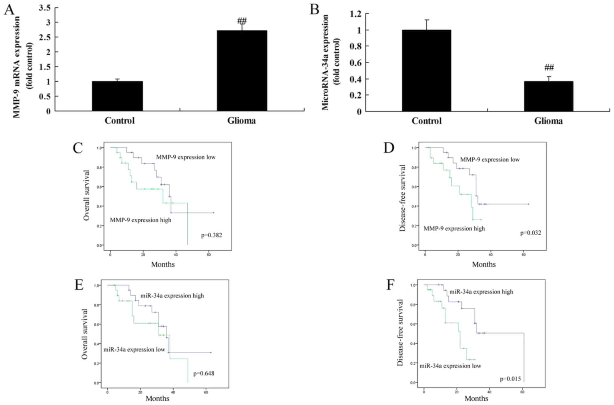
miR-34a and MMP-9 expression
To investigate the role of miR-34a and MMP-9
expression in glioma, the expression of miR-98 in serum from
patients with glioma was examined using RT-qPCR. As presented in
Fig. 1A, MMP-9 expression was
significantly elevated in serum from patients with glioma compared
with the normal group (P<0.01). While miR-34a expression was
significantly lower in serum from patients with glioma compared
with the normal group (P<0.01; Fig.
1B). These results revealed that miR-34a and MMP-9 may be
associated with glioma cell growth. Then, whether miR-34a and MMP-9
affected the OS and DFS of glioma patients was determined. As
presented in Fig. 1C and D, the OS
and DFS of patients with high MMP-9 expression were decreased
compared with those with low MMP-9 expression. In addition, the OS
and DFS of patients with low miR-34a expression were decreased
compared with those with high miR-34a expression (Fig. 1E and F).
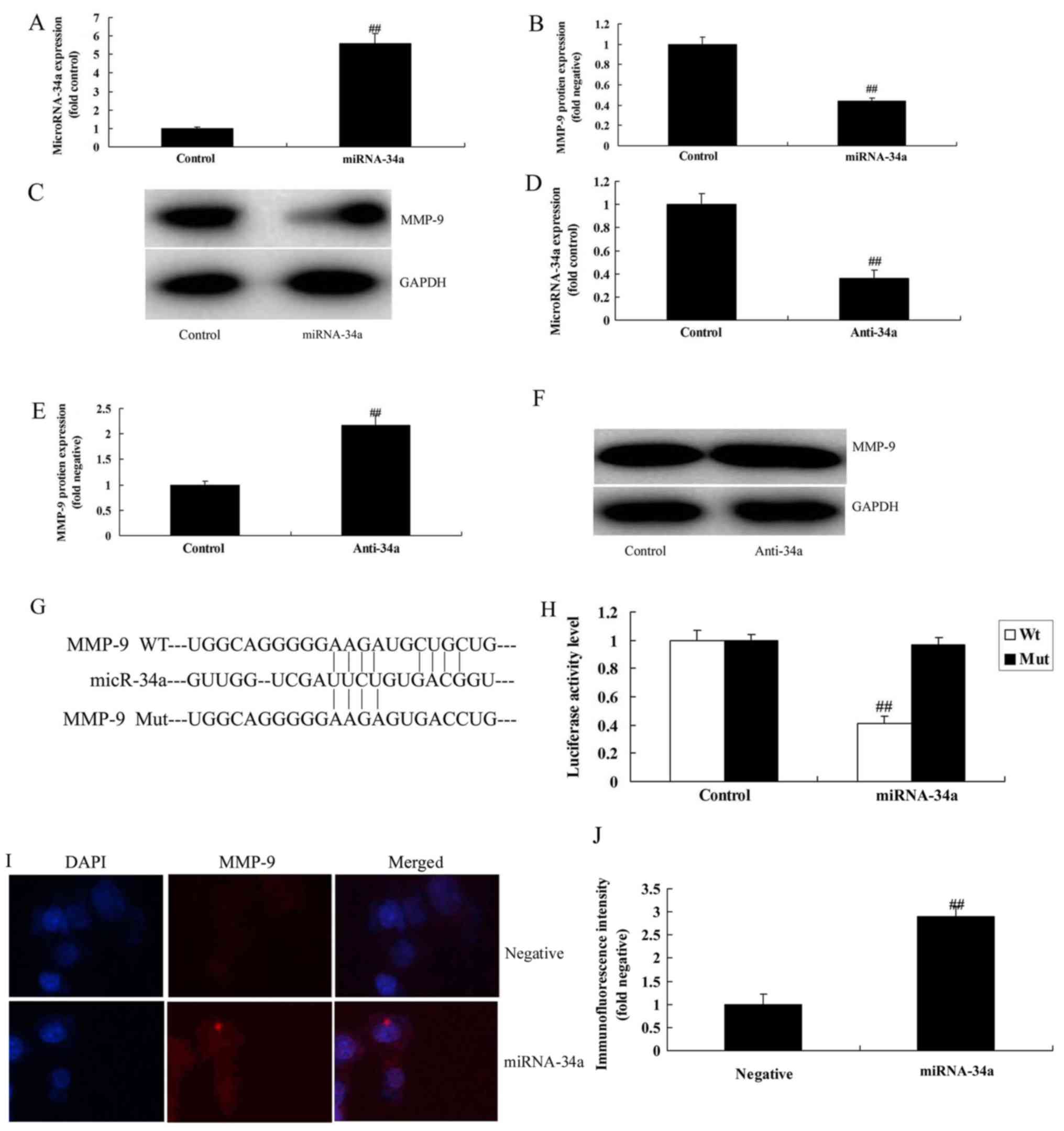
miR-34a regulates MMP-9 protein
expression in glioma cells
To evaluate whether miR-34a regulated the protein
expression of MMP-9 in glioma cells, a RT-qPCR and western blotting
were used to analyze miR-34a and MMP-9 expression in U251-MG glioma
cells transfected with miR-34a mimics and anti-miR-34a. As
presented in Fig. 2A-C, the
miR-34a mimic significantly increased miR-34a expression and
significantly suppressed MMP-9 protein expression in glioma cells,
compared with the negative control group (P<0.01). As presented
in Fig. 2D-F, anti-miR-34a
administration significantly decreased miR-34a expression and
significantly induced MMP-9 protein expression in glioma cells,
compared with the negative control group (P<0.01). Furthermore,
it was demonstrated that miR-34a targeted the 3′-UTR of MMP-9
protein (Fig. 2G), as the
luciferase activity of miR-34a was significantly inhibited in
glioma cells, compared with the negative control group (P<0.01;
Fig. 2H). Immunofluorescence
demonstrated that overexpression of miR-34a significantly
suppressed MMP-9 protein expression in glioma cells, in comparison
with the negative control group (P<0.01; Fig. 2I and J).
Downregulation of miR-34a promotes
cell growth and migration, and inhibits apoptosis in glioma
cells
To investigate the effects of miR-34a on the cell
growth of glioma cells, anti-miR-34a was used. As presented in
Fig. 3, downregulation of miR-34a
significantly promoted cell growth (Fig. 3A), reduced LDH activity levels
(Fig. 3B) and increased migration
(Fig. 3C and D), significantly
inhibited L and apoptosis (Fig. 3E and
F), and significantly decreased caspase-3 and caspase-9
activity levels (Fig. 3G and H) in
glioma cells, compared with the negative control group (all
P<0.01).
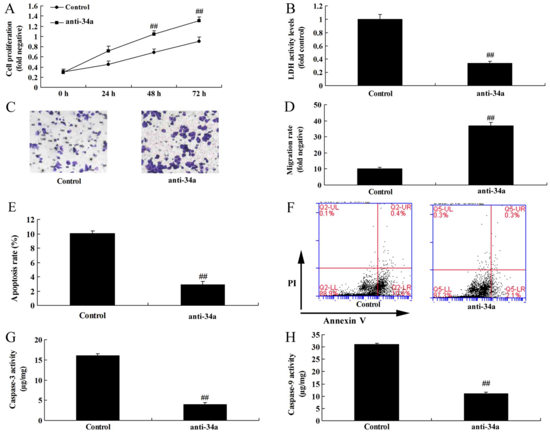 | Figure 3.Downregulation of miRNA-34a promoted
cell growth and migration, and inhibited apoptosis in glioma cells.
(A) Cell growth, (B) LDH activity level, (C) Transwell migration
assay (magnification, ×100) and (D) migration rate, (E) apoptosis
rate and (F) raw flow cytometry data, (G) caspase-3 and (H)
caspase-9 activity. ##P<0.01 vs. the control group.
miRNA-34a, microRNA-34a; Control, negative group; anti-34a,
miRNA-34a inhibitor; LDH, lactate dehydrogenase; PI, propidium
iodide. |
Overexpression of miR-34a inhibits
cell growth and migration, and induces apoptosis in glioma
cells
To further determine the effects of miR-34a on cell
growth of glioma cell, an miR-34a mimic was used in the present
study. As presented in Fig. 4,
overexpression of miR-34a significantly reduced cell growth and
migration, significantly induced apoptosis and LDH activity levels,
and significantly increased caspase-3 and caspase-9 activity levels
in glioma cells, in comparison with the negative control group (all
P<0.01).
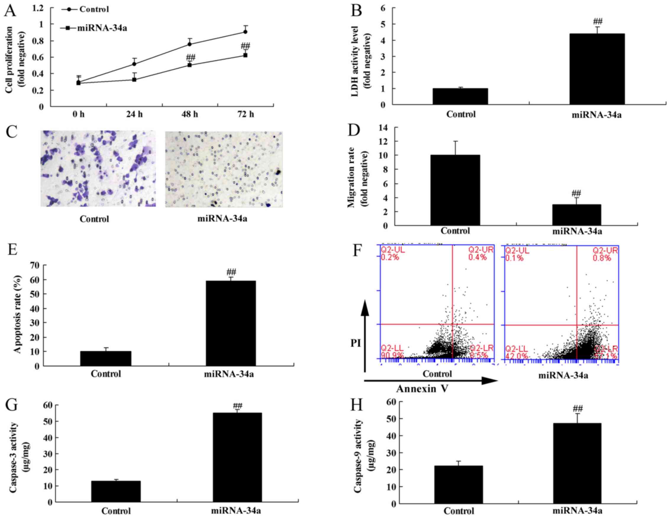 | Figure 4.Overexpression of miRNA-34a inhibited
cell growth and migration, and induced apoptosis in glioma cell.
(A) Cell growth, (B) LDH activity level, (C) Transwell migration
assay (magnification, ×100) and (D) migration rate, (E) apoptosis
rate and (F) raw flow cytometry data, (G) caspase-3 and (H)
caspase-9 activity. ##P<0.01 vs. control group.
miRNA-34a, microRNA-34a; Control, negative group; miRNA-34a,
miRNA-34a mimic overexpression; LDH, lactate dehydrogenase; PI,
propidium iodide. |
MMP-9 inhibitor attenuates the effects
of miR-34a downregulation on glioma cells
SB-3CT (an inhibitor of MMP-9) was used to evaluate
the role of MMP-9 in the effects of miR-34a in glioma cells. As
presented in Fig. 5A and B, the
MMP-9 inhibitor (100 nM) significantly suppressed the protein
expression of MMP-9 in glioma cells following miR-34a
downregulation, compared with the anti-miR-34a group (P<0.01).
The MMP-9 inhibitor attenuated the effects of miR-34a
downregulation on cell growth and migration, apoptosis rate, LDH
activity levels and caspase-3/9 activity level in glioma cells
following miR-34a downregulation, in comparison with the miR-34a
downregulation group (Fig.
5C-J).
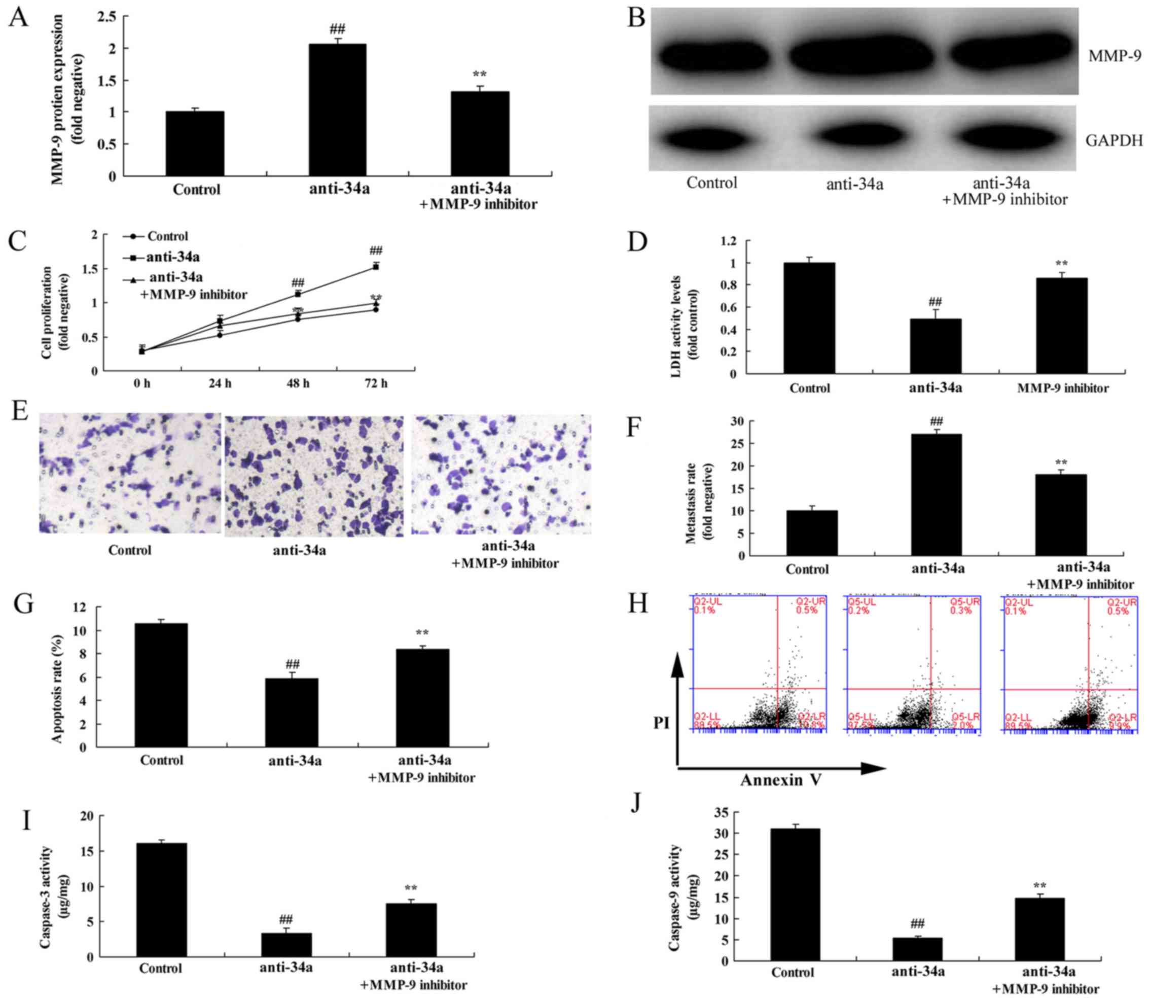 | Figure 5.MMP-9 inhibitor reduced the effects of
miRNA-34a downregulation in glioma cell. (A) MMP-9 protein
expression using western blot analysis and (B) MMP-9 protein
expression by statistical analysis, (C) cell growth, (D) LDH
activity level, (E) Transwell migration assay (magnification, ×100)
and (F) migration rate, (G) apoptosis rate analysis and (H) flow
cytometry data, (I) caspase-3 and (J) caspase-9 activity and.
##P<0.01 vs. control; **P<0.01 vs. anti-34a.
miRNA-34a, microRNA-34a; Control, negative group; anti-34a,
miRNA-34a inhibitor; MMP-9 inhibitor, anti-34a + MMP-9 inhibitor
(100 nM SB-3CT); MMP, matrix metalloproteinase; LDH, lactate
dehydrogenase; PI, propidium iodide. |
Discussion
Glioma is the most common primary intracranial tumor
in adults, which has a high malignant grade, high potential for
recurrence and poor prognosis (13). The results of the present study
suggest that MMP-9 expression was increased, and miRNA-34a was
suppressed in the serum of patients with glioma, compared with the
normal group. The OS and DFS of MMP-9 high-expression groups were
decreased compared with the MMP-9 low-expression group. The OS and
DFS of the miR-34a low-expression group were decreased compared
with the miRNA-34a high-expression group. Overexpression of miR-34a
significantly reduced cell growth and migration, and significantly
increased apoptosis rates, LDH activity levels and caspase-3 and
caspase-9 activity levels in glioma cells. In the present study,
only one cell line U251-MG was used, which is insufficient and more
cell models will be used in future studies.
MMPs are the endogenous zinc ion-dependent proteases
essential for degrading the extracellular matrix (9). They can specifically degrade the
basement membrane components. Gelatinases are the only MMP enzymes
that can degrade the basement membrane and type IV collagen in the
extracellular matrix. MMP-2 and MMP-9 are gelatinases that promote
the break-through of tumor cells past the structural barrier of the
basement membrane. In addition, these MMPs are associated with
angiogenesis, and invasion and metastasis of tumors (9). However, in the present study,
overexpression of miR-34a suppressed MMP-9 protein expression in
glioma cells. Jia et al (11) reported that miR-34a reduced
migration and invasion of tongue squamous cell carcinoma by
targeting MMP-9 and MMP-14.
MMPs serve key roles during tumor metastasis by
degrading multiple proteins in the extracellular matrix (14). Upregulated MMP expression in glioma
can directly or indirectly degrade the extracellular matrix and
basement membrane, which results in glioma invasion and peripheral
tumor tissue edema (14).
Consequently, MMPs are regarded as biomarkers of glioma progression
(15). In addition, they can
promote the structural rigidity, motility and proliferation of
glioma cells (15). MMP inhibitors
can suppress C6 glioma cell invasion in an optic nerve explant.
Furthermore, MMP-2 and MMP-9 secreted by BT5C rat glioma cells can
destroy cultured brain tissue. In addition, research in
vitro suggests that MMPs secreted by glioma cells can decompose
extracellular matrix proteins (14). Additionally, MMPs can promote tumor
cell invasion into normal brain tissue, and enhance glioma invasion
(14). In the present study, an
MMP-9 inhibitor reduced the effects of miR-34a downregulation in
glioma cells. Yang et al (16) indicated that miR-34a inhibits cell
migration and invasion of esophageal squamous cell carcinoma by
targeting MMP-2/MMP-9/fibronectin type III domain-containing
protein 3B.
In conclusion, the present study demonstrated that
MMP-9 expression was increased and miR-34a was suppressed in
patients with glioma. The downregulation of miR-34a promoted cell
growth and migration, and inhibited apoptosis in glioma cells by
activation of MMP-9; thus enhancing the expression of miR-34a may
be a novel strategy to suppress the progression of glioma.
Acknowledgements
Not applicable.
Funding
The present study was supported by funds from the
Education Department of Jilin Province (grant no.
JJKH20170056KJ).
Availability of data and materials
The analyzed data sets generated during the study
are available from the corresponding author on reasonable
request.
Authors' contributions
JP designed the study. XW, XC, LS, XB, HH and LC
performed the experiments. JP and XW analyzed the data. JP wrote
the manuscript.
Ethics approval and consent to
participate
The present study was approved by the Ethics
Committee of Affiliated Hospital of Beihua University. The study
was performed in accordance with the regulations of the
Institutional Review Board of Affiliated Hospital of Beihua
University. Written informed consent was obtained prior to surgery
from all enrolled patients.
Patient consent for publication
Written informed consent was obtained prior to
surgery from all enrolled patients.
Competing interests
The authors declare they have no competing
interests.
References
|
1
|
Wu XL, Huang H, Huang YY, Yuan JX, Zhou X
and Chen YM: Reduced Pumilio-2 expression in patients with temporal
lobe epilepsy and in the lithium-pilocarpine induced epilepsy rat
model. Epilepsy Behav. 50:31–39. 2015. View Article : Google Scholar : PubMed/NCBI
|
|
2
|
Lin XT, Zheng XB, Fan DJ, Yao QQ, Hu JC,
Lian L, Wu XJ, Lan P and He XS: MicroRNA-143 targets ATG2B to
inhibit autophagy and increase inflammatory responses in crohn's
disease. Inflamm Bowel Dis. 24:781–791. 2018. View Article : Google Scholar : PubMed/NCBI
|
|
3
|
He M, Zhan M, Chen W, Xu S, Long M, Shen
H, Shi Y, Liu Q, Mohan M and Wang J: MiR-143-5p deficiency triggers
EMT and metastasis by targeting HIF-1alpha in gallbladder cancer.
Cell Physiol Biochem. 42:2078–2092. 2017. View Article : Google Scholar : PubMed/NCBI
|
|
4
|
Huang FT, Peng JF, Cheng WJ, Zhuang YY,
Wang LY, Li CQ, Tang J, Chen WY, Li YH and Zhang SN: MiR-143
targeting TAK1 attenuates pancreatic ductal adenocarcinoma
progression via MAPK and NF-kappaB pathway in vitro. Dig Dis Sci.
62:944–957. 2017. View Article : Google Scholar : PubMed/NCBI
|
|
5
|
Livak KJ and Schmittgen TD: Analysis of
relative gene expression data using real-time quantitative PCR and
the 2(-Delta Delta C(T)) method. Methods. 25:402–408. 2001.
View Article : Google Scholar : PubMed/NCBI
|
|
6
|
Cui C and Shi X: miR-187 inhibits tumor
growth and invasion by directly targeting MAPK12 in osteosarcoma.
Exp Ther Med. 14:1045–1050. 2017. View Article : Google Scholar : PubMed/NCBI
|
|
7
|
Zhou W, Pal AS, Hsu AY, Gurol T, Zhu X,
Wirbisky-Hershberger SE, Freeman JL, Kasinski AL and Deng Q:
MicroRNA-223 suppresses the canonical NF-kappaB pathway in basal
keratinocytes to dampen neutrophilic inflammation. Cell Rep.
22:1810–1823. 2018. View Article : Google Scholar : PubMed/NCBI
|
|
8
|
Chen M, Zhang H, Shi Z, Li Y, Zhang X, Gao
Z, Zhou L, Ma J, Xu Q, Guan J, et al: The MST4-MOB4 complex
disrupts the MST1-MOB1 complex in the Hippo-YAP pathway and plays a
pro-oncogenic role in pancreatic cancer. J Biol Chem.
293:14455–14469. 2018. View Article : Google Scholar : PubMed/NCBI
|
|
9
|
Cheng J, Jing Y, Kang D, Yang L, Li J, Yu
Z, Peng Z, Li X, Wei Y, Gong Q, et al: The role of Mst1 in
lymphocyte homeostasis and function. Front Immunol. 9:1492018.
View Article : Google Scholar : PubMed/NCBI
|
|
10
|
Huang Y, Huang H, Li M, Zhang X, Liu Y and
Wang Y: MicroRNA-374c-5p regulates the invasion and migration of
cervical cancer by acting on the Foxc1/snail pathway. Biomed
Pharmacother. 94:1038–1047. 2017. View Article : Google Scholar : PubMed/NCBI
|
|
11
|
Jia LF, Wei SB, Mitchelson K, Gao Y, Zheng
YF, Meng Z, Gan YH and Yu GY: miR-34a inhibits migration and
invasion of tongue squamous cell carcinoma via targeting MMP9 and
MMP14. PLoS One. 9:e1084352014. View Article : Google Scholar : PubMed/NCBI
|
|
12
|
Tabouret E, Bertucci F, Pierga JY, Petit
T, Levy C, Ferrero JM, Campone M, Gligorov J, Lerebours F, Roché H,
et al: MMP2 and MMP9 serum levels are associated with favorable
outcome in patients with inflammatory breast cancer treated with
bevacizumab-based neoadjuvant chemotherapy in the BEVERLY-2 study.
Oncotarget. 7:18531–18540. 2016. View Article : Google Scholar : PubMed/NCBI
|
|
13
|
Martin-Alonso A, Cohen A, Quispe-Ricalde
MA, Foronda P, Benito A, Berzosa P, Valladares B and Grau GE:
Differentially expressed microRNAs in experimental cerebral malaria
and their involvement in endocytosis, adherens junctions, FoxO and
TGF-beta signalling pathways. Sci Rep. 8:112772018. View Article : Google Scholar : PubMed/NCBI
|
|
14
|
Chen HX, Xu XX, Tan BZ, Zhang Z and Zhou
XD: MicroRNA-29b inhibits angiogenesis by targeting VEGFA through
the MAPK/ERK and PI3K/Akt signaling pathways in endometrial
carcinoma. Cell Physiol Biochem. 41:933–946. 2017. View Article : Google Scholar : PubMed/NCBI
|
|
15
|
Zhao Q, Ye M, Yang W, Wang M, Li M, Gu C,
Zhao L, Zhang Z, Han W, Fan W and Meng Y: Effect of Mst1 on
endometriosis apoptosis and migration: Role of drp1-related
mitochondrial fission and parkin-required mitophagy. Cell Physiol
Biochem. 45:1172–1190. 2018. View Article : Google Scholar : PubMed/NCBI
|
|
16
|
Yang L, Song X, Zhu J, Li M, Ji Y, Wu F,
Chen Y, Cui X, Hu J, Wang L, et al: Tumor suppressor microRNA-34a
inhibits cell migration and invasion by targeting
MMP-2/MMP-9/FNDC3B in esophageal squamous cell carcinoma. Int J
Oncol. 51:378–388. 2017. View Article : Google Scholar : PubMed/NCBI
|