Introduction
Smoke inhalation injury, a common complication in
patients with thermal injuries, particularly those with flame
burns, is an important cause of increased mortality in patients
with burns (1–3). According to the current view, thermal
injury directly induces respiratory tract damage and the particles
and chemicals contained in smoke strongly exacerbate the
progression of inhalation injury (4). When various particles and chemical
components of smoke, such as dust particles and CO or
SO2 enter the respiratory tract, these components induce
the infiltration of massive numbers of inflammatory cells and the
subsequent release of inflammatory mediators into the respiratory
tract, stimulating a cascade of stress reactions (5,6). The
available evidence consistently indicates that the particulate
matter in smoke induces direct thermal damage and triggers cytokine
production by alveolar macrophages, resulting in a
neutrophil-mediated inflammatory response and activation of the
NF-κB signal pathway, which contributes to lung injury (7). In addition, previous studies have
demonstrated that inducible nitric oxide synthase (iNOS) serves a
role in smoke inhalation injury by promoting the apoptosis of
alveolar epithelial cells via various signal transduction pathways
(8–10). Numerous components of smoke are
strong oxidants that can disrupt the redox balance in the body. In
addition, the excessive accumulation of inflammatory cells in the
lungs leads to the production of excessive reactive oxygen species
(ROS), further aggravating the tissue damage caused by oxidative
stress (11). The activation of
neutrophils and the enhancement of arachidonic acid metabolism
following inhalation injury leads to an increase in the production
of oxygen free radicals and subsequently causes an increase in the
consumption of antioxidant substances (12). Polyunsaturated fatty acids
subsequently undergo overoxidation, causing cell damage and
accelerating apoptosis, resulting in a decrease in superoxide
dismutase (SOD) activity and an increase in malondialdehyde (MDA)
content following smoke inhalation injury (13).
Simvastatin, a widely used cholesterol-lowering
drug, can substantially reduce the incidence of cardiovascular
events in patients with hyperlipidemia (14,15).
Based on clinical analyses, simvastatin also exerts
anti-inflammatory, antioxidant and immunomodulatory effects
independent of its lipid-lowering effect (16–18).
Some clinical studies have reported that patients with inhalation
injury who took statins long-term usually recovered faster
(19). The purpose of the present
study was to investigate the potential of statins in the treatment
of patients with inhalation injury, resolve the underlying
mechanisms by which they inhibit the progression of the disease and
to evaluate the protective effect exerted by simvastatin against
smoke inhalation injury in rats.
Materials and methods
Ethics
The present study was approved by the Animal Care
and Use Committee of Zhengzhou University (Henan, China) and
conducted in accordance with the National Institutes of Health
Guide for the Care and Use of Laboratory Animals (20).
A total of 75 male Sprague-Dawley (SD) rats (aged
6–8 weeks, weighing 220–250 g) were provided by the Laboratory
Animal Center of Henan Province [animal certification: SCXK (Yu)
2015-0004]. The rats were housed under standard conditions, at a
temperature of 20–22°C and 40–60% humidity, with a 12-h light/dark
cycle and free access to water and food. The rats were acclimated
for 1 week before the experiments were performed. The rats were
randomly divided into five groups: A control group (uninjured and
untreated, n=15), a saline group (treated with saline following
smoke exposure, n=15), a low-dose group (treated with 25 mg/kg
simvastatin following smoke exposure, n=15), a middle-dose group
(treated with 50 mg/kg simvastatin following smoke exposure, n=15)
and a high-dose group (treated with 100 mg/kg simvastatin following
smoke exposure, n=15). Within the first 30 min following smoke
exposure, rats in the low-, middle- and high-dose groups were
administered intragastric simvastatin based on their body weight at
12-h intervals and rats in the saline group were administered an
equal volume of normal saline.
Generation of an animal model of smoke
inhalation injury
A total of 15 rats were randomly assigned to the
control group. The remaining 60 rats were exposed to smoke, which
caused smoke inhalation lung injury. The establishment of an animal
model of smoke inhalation injury was based on previous experiments
and relevant literature (21,22).
A temperature monitor was installed inside a special glass
container with a length, width and height of 50, 35 and 45 cm,
respectively, to detect the temperature. Then, 100 g of dry pine
chips and 30 ml of kerosene were thoroughly mixed in a pot equipped
with a heating device at the bottom and the heating device was
switched on 5 min before the experiment. The smoke was wafted from
the pot to the glass container using an air blower. After the glass
container was loaded with smoke, six rats were arranged in a
30×20×15 cm wire cage and placed into the container. Following a
2-min exposure, the rats were removed from the container and
maintained in a normal environment for 5 min. The above procedure
was repeated three times, resulting in a cumulative smoke
inhalation duration of 6 min. Within the first 30 min following
induction of smoke inhalation injury, the low-, middle- and
high-dose groups were administered 25, 50 and 100 mg/kg
intragastric simvastatin, respectively, according to their weight,
every 12 h (23), while the saline
group was administered an equal volume of normal saline. At 24, 48
and 72 h following injury, a 10% solution of chloral hydrate was
administered for anesthesia via intraperitoneal injection at 300
mg/kg according to their weight. None of the rats exhibited signs
of peritonitis, pain or discomfort following administration of
chloral hydrate. Blood samples were collected through puncture of
the left ventricle, allowed to clot for 60 min and centrifuged at
3,000 × g/min at 4°C for 10 min. At that time point, the weight of
each rat was reduced by 10–20 g compared with their previous
weights; the maximum percentage of weight loss that was observed in
the rats from start to endpoint was 7.3%. Rats were euthanized by
exsanguination under anesthesia and the volume of blood collected
by exsanguination was 12–17 ml. The lack of a heartbeat,
respiratory arrest, absent nerve reflex and muscle relaxation were
the parameters used to confirm death before further experiments.
The left lungs from the rats were removed for pathological
examination and scoring. The mRNA levels of iNOS and caspase-3 in
the upper lobes of the right lungs were measured by reverse
transcription-quantitative (RT-q) PCR and the protein levels of
iNOS and caspase-3 in the middle lobes of the right lungs were
measured by western blotting. The activity of SOD and the content
of MDA in serum samples and lung tissues obtained from the lower
lobes of the right lungs were measured.
Pathological examination and
scoring
Lung tissues were fixed in 10% formalin at room
temperature for 24 h, dehydrated through an ascending series of
ethanol solutions (70, 80, 95 and 100% alcohol), embedded in
paraffin, sectioned to 5-µm thickness and stained with hematoxylin
at room temperature for 5 min and followed by eosin at room
temperature for 2 min. Then, two experienced pathologists evaluated
the tissue morphology under a light microscope (magnification,
×200) in a blinded manner. The severity of the observed lung
injuries was scored according to the following criteria: Edema,
neutrophil infiltration, hemorrhage and hyaline membrane formation.
The severity of lung injury was scored from 0 to 4 as follows: 0,
no injury; 1, mild injury; 2, moderate injury; 3, severe injury;
and 4, most severe lung injury. Together, five randomly selected
sections from each pathological specimen were observed and scored
and these scores were added to obtain the lung histopathological
score.
Immunohistochemistry
The immunohistochemical analysis of iNOS and
caspase-3 expression in lung tissues was performed in a two-step
manner. Sections were deparaffnized at 60°C for 1 h, washed in
xylene twice for 10 min and rehydrated in a descending alcohol
series. Antigen retrieval was performed for 12 min in a microwave
and the tissues were then incubated with primary antibody (iNOS,
1:50; cat. no. ab3523 Abcam and caspase-3, 1:50; cat. no. ab4051
Abcam) at room temperature for 16 h, followed by incubation with
horseradish peroxidase-conjugated goat anti-rabbit immunoglobulin G
(IgG) secondary antibody for 1 h (1:2,000, cat. no. TA130024
OriGene Technologies, Inc.) at room temperature. The tissues were
then stained with diaminobenzidine at room temperature for 5 min.
The control sections were subjected to the same procedure but
without primary antibody incubation. The expression levels of iNOS
and caspase-3 in lung tissues were evaluated by two experienced
pathologists under a light microscope (magnification, ×400). A cell
with a brown cytoplasm or a brown nucleus was considered
immunoreactive. All immunoreactive cells in at least five sections
from each specimen were counted and the percentage of positive
cells was determined, expression levels were quantified using
Image-Pro Plus 6.0 (Media Cybernetics, Inc.).
RT-qPCR
The mRNA expression levels of iNOS and caspase-3
were measured using RT-qPCR. Total RNA was extracted from the upper
lobe of the right lung (10–15 g) from each rat using
TRIzol® reagent (Invitrogen; Thermo Fisher Scientific,
Inc.) according to the manufacturer's protocol. The concentration
of RNA was determined using a nanodrop ND-1000 spectrophotometer
(Thermo Fisher Scientific, Inc.). RNA (2 µg) was reverse
transcribed into cDNA for 5 min at 70°C and 5 min at 4°C using a
First-strand cDNA Synthesis kit (Promega Corporation), according to
the manufacturer's protocol. qPCR was performed using a qPCR Master
mix (New England Biolabs, Inc.) and 96-well optical reaction plates
and the CFX96 Real-Time PCR detection system (Bio-Rad Laboratories,
Inc.). The following PCR conditions were used: Predenaturation at
95°C for 60 sec followed by 40 cycles of denaturation at 95°C for
15 sec and extension at 60°C for 45 sec, according to the
manufacturer's protocol. All experiments were performed four times.
The primers were provided by Sangon Biotech (Shanghai) Co., Ltd.
The primer sequences for GAPDH were: Forward:
5′-AACATCCAGAGCTTGACGGTG−3′ and reverse:
5′-TCTTGACCATCCTTGAGAGTGG-3′ and the primer sequences for iNOS were
forward: 5′-AACCCAAGGTCTACGTTCAAG−3′ and reverse:
5′-AAAGTGGTAGCCACATCCCG-3′. The primer sequences for caspase-3 were
forward: 5′-TGGTTCATCCAGTCGCTTTGT−3′ and reverse:
5′-CAAATTCTGTTGCCACCTTTCG-3′. Relative gene expression levels were
calculated using the relative quantification 2−ΔΔCq
method and normalized to GAPDH (24).
Western blotting
The protein expression levels of iNOS and caspase-3
in lung tissues were evaluated using western blotting. The proteins
from the middle lobe tissue from the right lung of each rat were
extracted using RIPA lysis buffer (Biotechwell) supplemented with
phenylmethylsulfonyl fluoride and the supernatant was collected.
The protein concentration was measured with a Bicinchoninic Acid
Assay kit (Biotechwell: http://www.biotechwell.com). Then, 40 µg protein/lane
was separated by 12% SDS-PAGE. The separated proteins were
subsequently transferred onto PVDF membranes (EMD Millipore) and
blocked for 2 h at room temperature with 5% non-fat milk solution.
The membranes were incubated with a rabbit anti-iNOS polyclonal
antibody (1:200, cat. no. ab3523 Abcam), rabbit anti-caspase-3
polyclonal antibody (1:100, cat. no. ab4051 Abcam), or rabbit
anti-β-actin polyclonal antibody (1:500, cat. no. ab6276 Abcam) in
tris-buffered saline and Tween 20 (0.1%) TBST overnight at 4°C. The
membranes were then incubated with horseradish
peroxidase-conjugated goat anti-rabbit IgG secondary antibody
(1:2,000, cat. no. TA130024 OriGene Technologies, Inc.) for 1.5 h
at room temperature. The resulting bands were detected with an
enhanced chemiluminescence detection system, using the
Pierce™ ECL Western Blotting Substrate (Bio-Rad
Laboratories, Inc.). Protein expression was semi-quantified using
Image-Pro Plus version 6.0 software (Media Cybernetics, Inc.) with
β-actin as the loading control.
Measurement of SOD activity and MDA
content
Using precooled saline as the medium, homogenates of
the right lung lower lobes were centrifuged at 3,000 × g/min for 15
min at 4°C and the supernatants were collected. The assays were
performed according to the manufacturer's instructions. The MDA
content in the supernatant and serum was measured using the
thiobarbituric acid method (Nanjing Jiancheng Bioengineering
Institute). The absorbance values used for the MDA measurements
were obtained at 532 nm by microplate reader and converted to
nmol/mg protein or nmol/ml. SOD activity in the supernatant and
serum was measured using the WST-1 method (Nanjing Jiancheng
Bioengineering Institute). The absorbance values used for the
measurements of SOD activity in supernatant and serum were obtained
at 450 nm by microplate reader and converted to U/mg protein or
U/ml.
Statistical analysis
All data were expressed as the mean ± standard
deviation. The data were subjected to statistical analyses using
SPSS version 22 software (IBM, Corp). The homogeneity of variance
was first determined using Levene's test. One way Analysis of
variance was used for overall comparisons between groups and
Tukey's post hoc test when the homogeneity of variance assumption
was met or by the Games-Howell test when the homogeneity of
variance assumption was not met. P<0.05 was considered to
indicate a statistically significant difference.
Results
Pathological examination and
scores
The inflammatory response was significantly reduced
following the administration of simvastatin. Higher pathological
scores were obtained in the saline and low-, middle- and high-dose
groups compared with the control group (P<0.05). In addition,
the pathological scores of the low-, middle- and high-dose groups
were lower compared with the saline group (P<0.05).
Additionally, the scores of the middle- and high-dose groups were
lower compared with the low-dose group (P<0.05). The difference
between the middle- and high-dose groups was non-significant
(P>0.05; Figs. 1, 2 and 3;
statistical data not shown).
Immunohistochemistry
Based on immunohistochemical analysis, iNOS
(Figs. 4, 5 and 6;
statistical data not shown) and caspase-3 (Figs. 7, 8 and 9;
statistical data not shown) expression levels were higher in the
saline and theablr low-, medium- and high-dose groups compared with
the control group (P<0.05); lower in the low-, middle- and
high-dose groups compared with the saline group (P<0.05); and
lower in the middle- and high-dose groups compared with the
low-dose group (P<0.05). The difference between the middle- and
high-dose groups was non-significant (P>0.05).
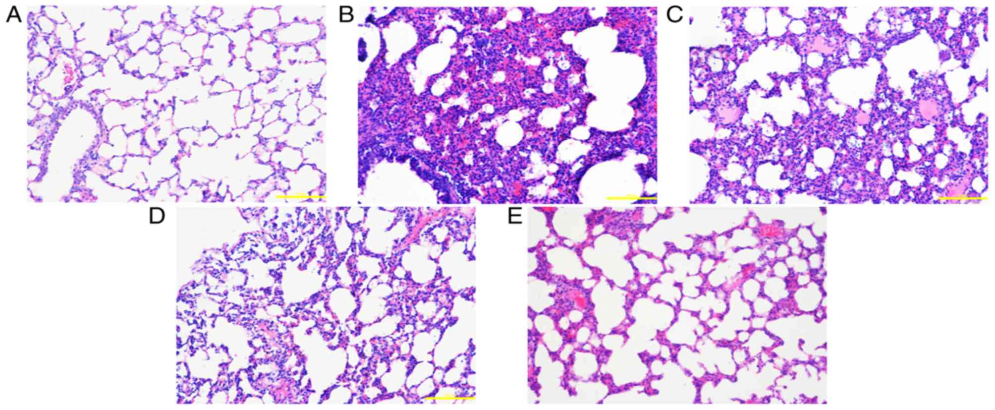
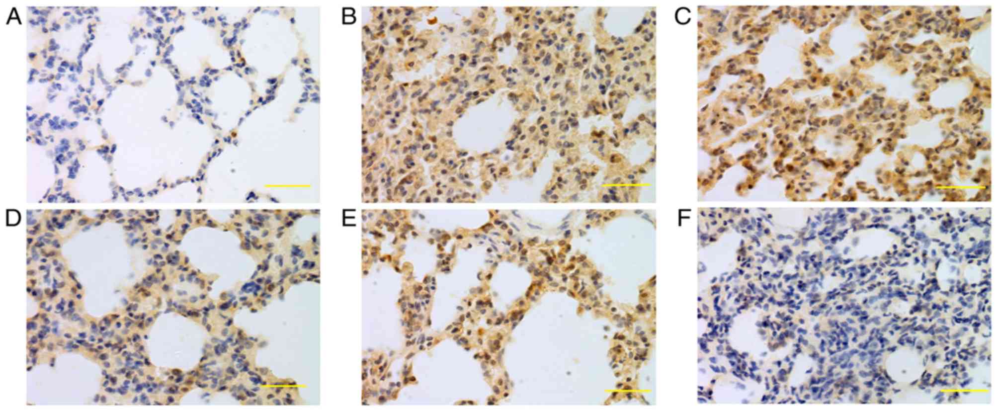
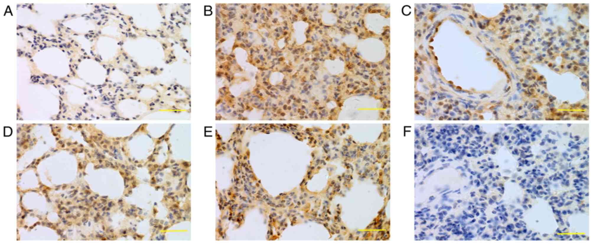
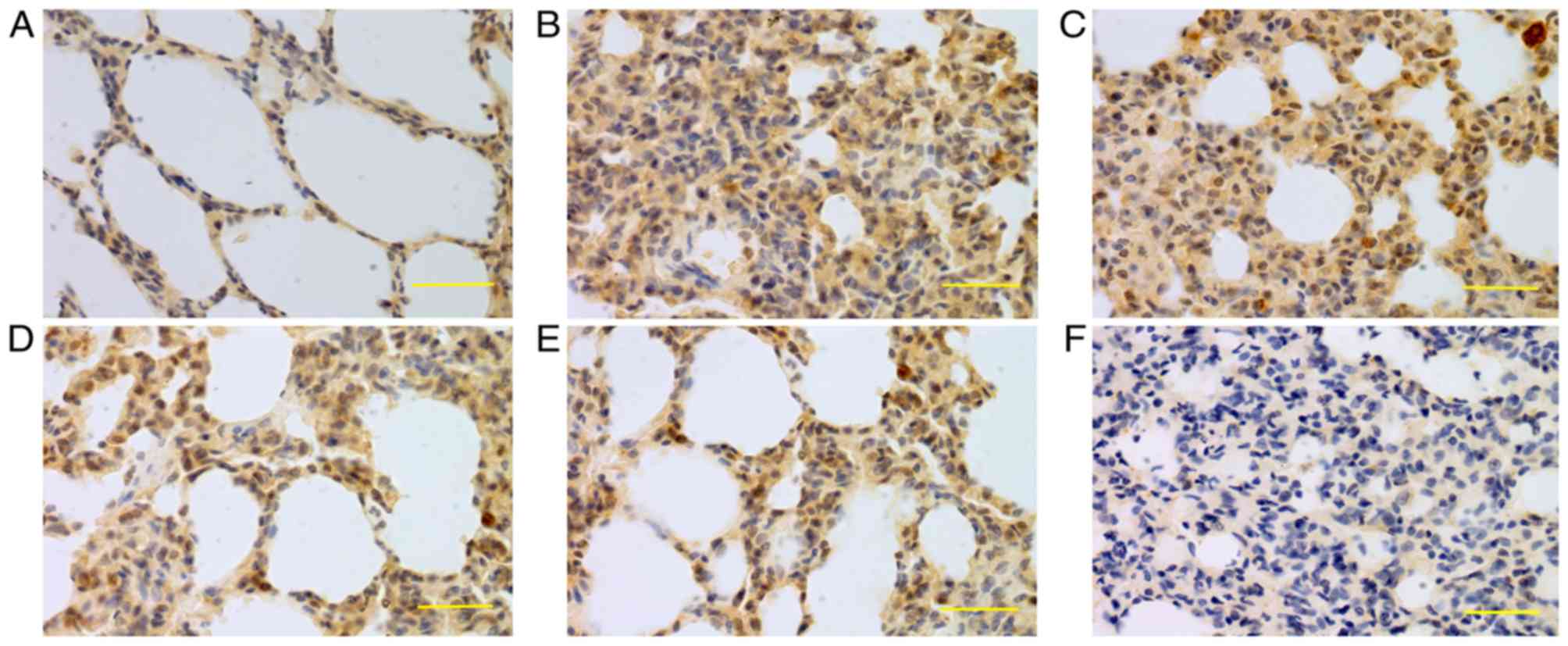 | Figure 4.Expression of iNOS in lung tissue at
24 h. iNOS was mainly expressed in bronchial epithelial cells,
alveolar epithelial cells and vascular and airway smooth muscle
cells. Higher iNOS expression levels were detected in the saline,
low-dose, middle-dose and high-dose groups compared with the
control group. (A) Control group, (B) saline group, (C) low-dose
group, (D) middle-dose group, (E) high-dose group and (F) PBS
group. Scale bars, 100 µm; magnification, ×400; DAB staining. iNOS,
inducible nitric oxide synthase; DAB, 3,3-diaminobenzidine. |
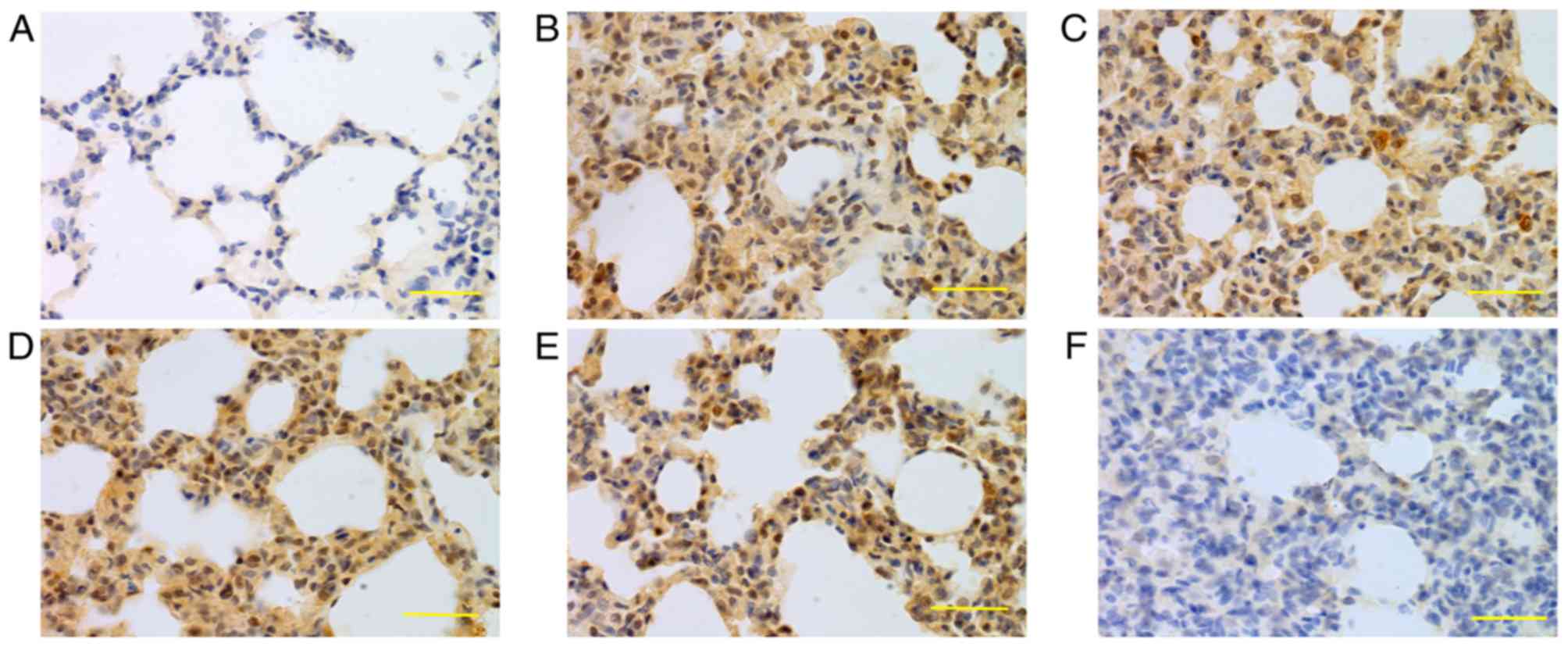
 | Figure 5.Expression of iNOS in lung tissue at
48 h. iNOS was mainly expressed in bronchial epithelial cells,
alveolar epithelial cells and vascular and airway smooth muscle
cells. Higher iNOS expression levels were detected in the saline,
low-dose, middle-dose and high-dose groups compared with the
control group. (A) Control group, (B) saline group, (C) low-dose
group, (D) middle-dose group, (E) high-dose group and (F) PBS
group. Scale bars, 100 µm; magnification, ×400; DAB staining. iNOS,
inducible nitric oxide synthase; DAB, 3,3-diaminobenzidine. |
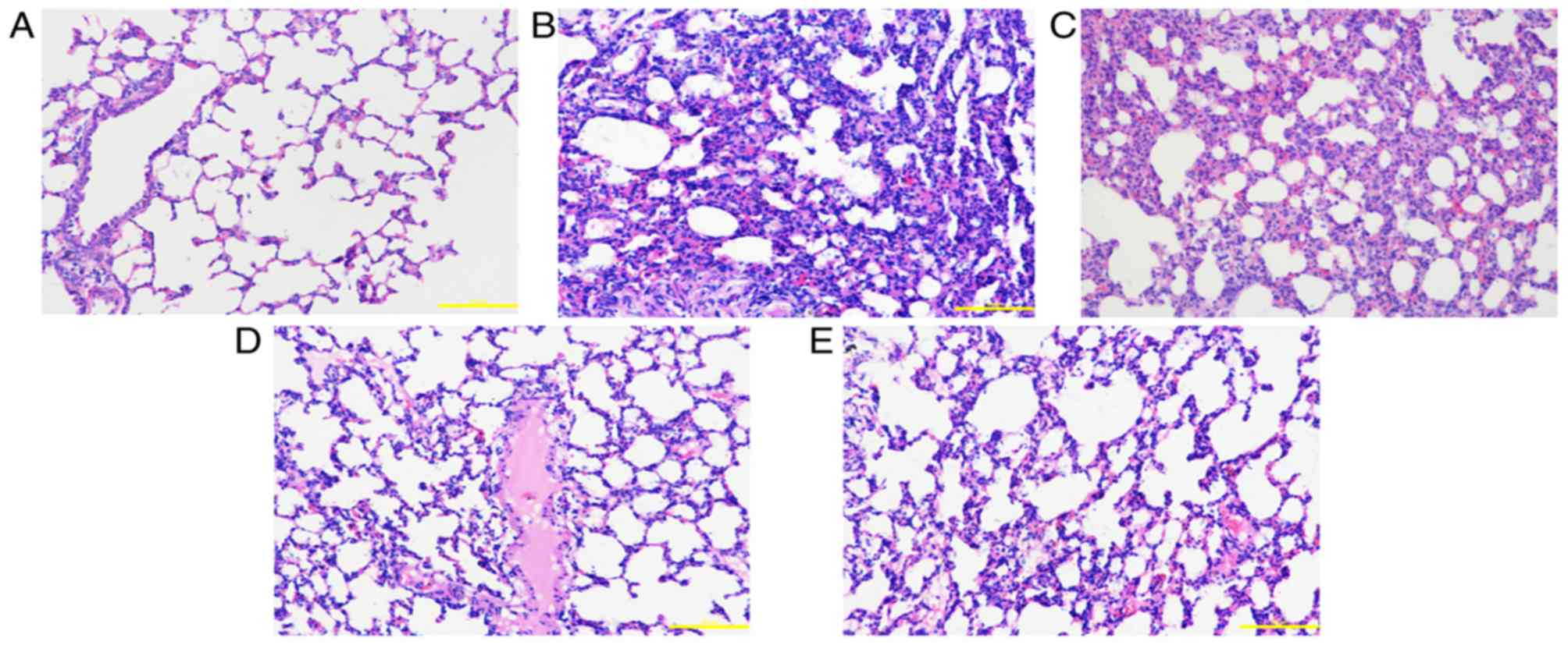
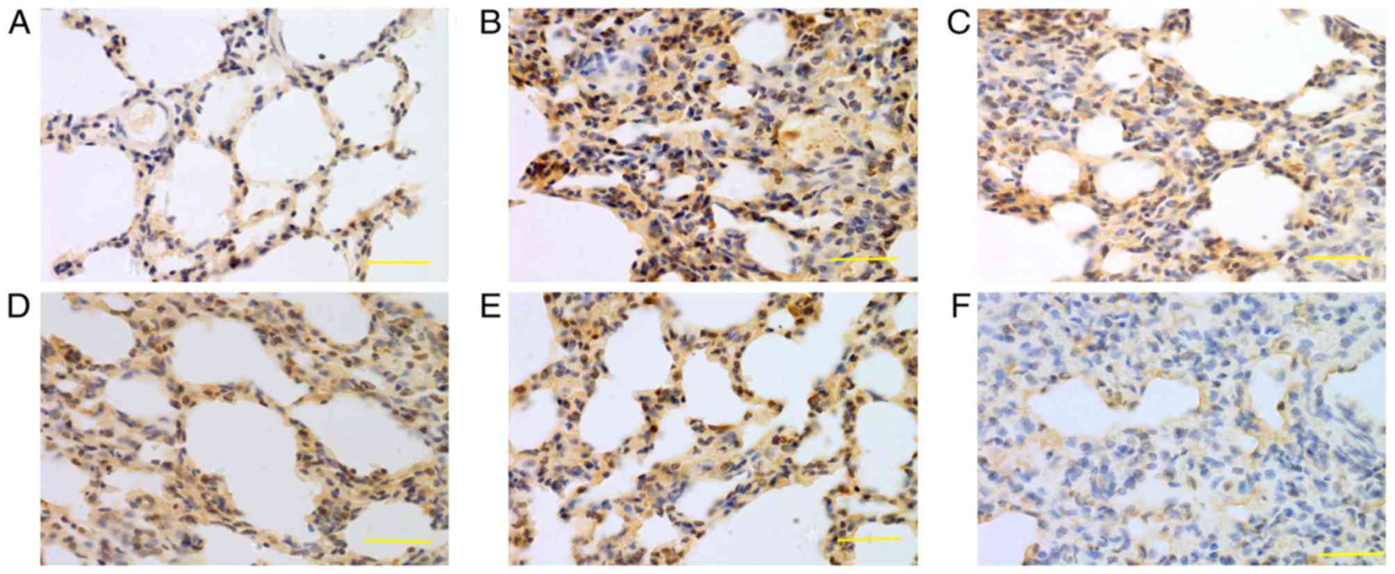
 | Figure 6.Expression of iNOS in lung tissue at
72 h. iNOS was mainly expressed in bronchial epithelial cells,
alveolar epithelial cells and vascular and airway smooth muscle
cells. Higher iNOS expression levels were detected in the saline,
low-dose, middle-dose and high-dose groups compared with the
control group. (A) Control group, (B) saline group, (C) low-dose
group, (D) middle-dose group, (E) high-dose group and (F) PBS
group. Scale bars, 100 µm; magnification, ×400; DAB staining. iNOS,
inducible nitric oxide synthase; DAB, 3,3-diaminobenzidine. |
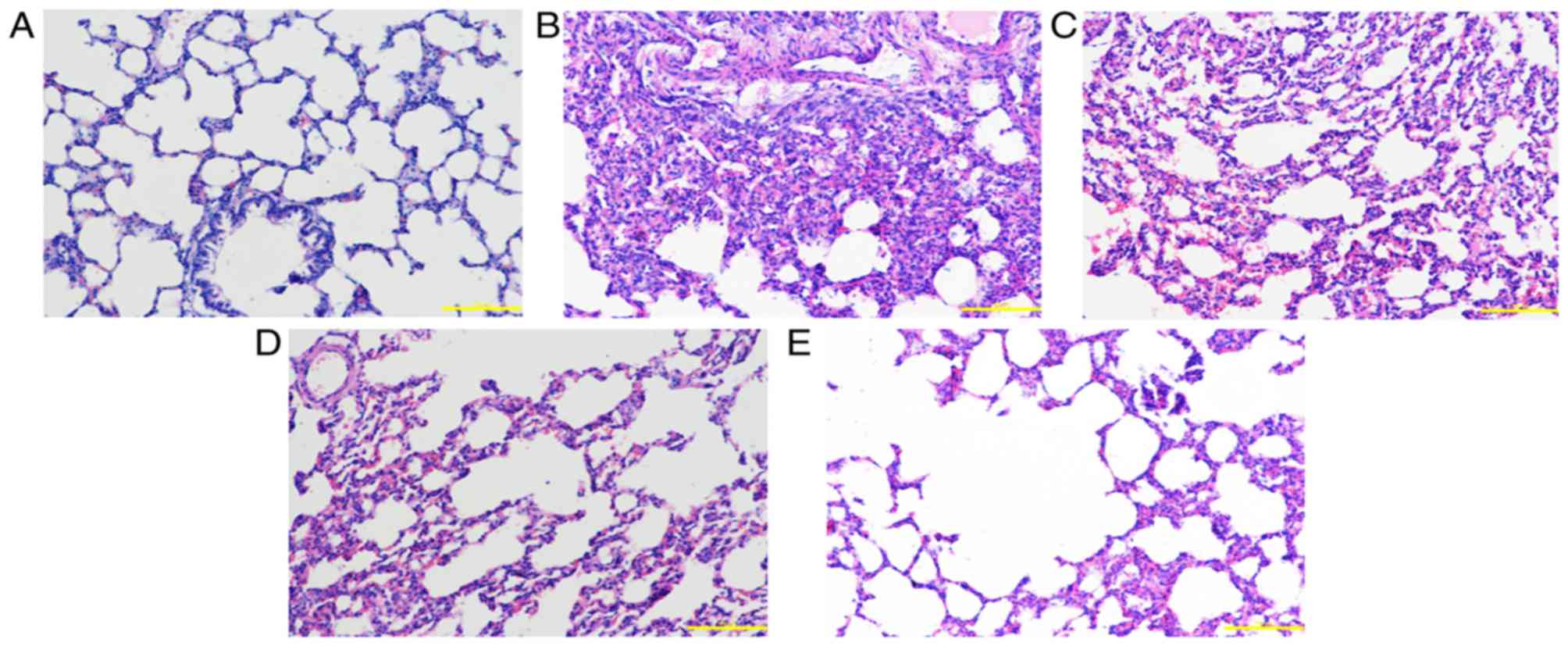
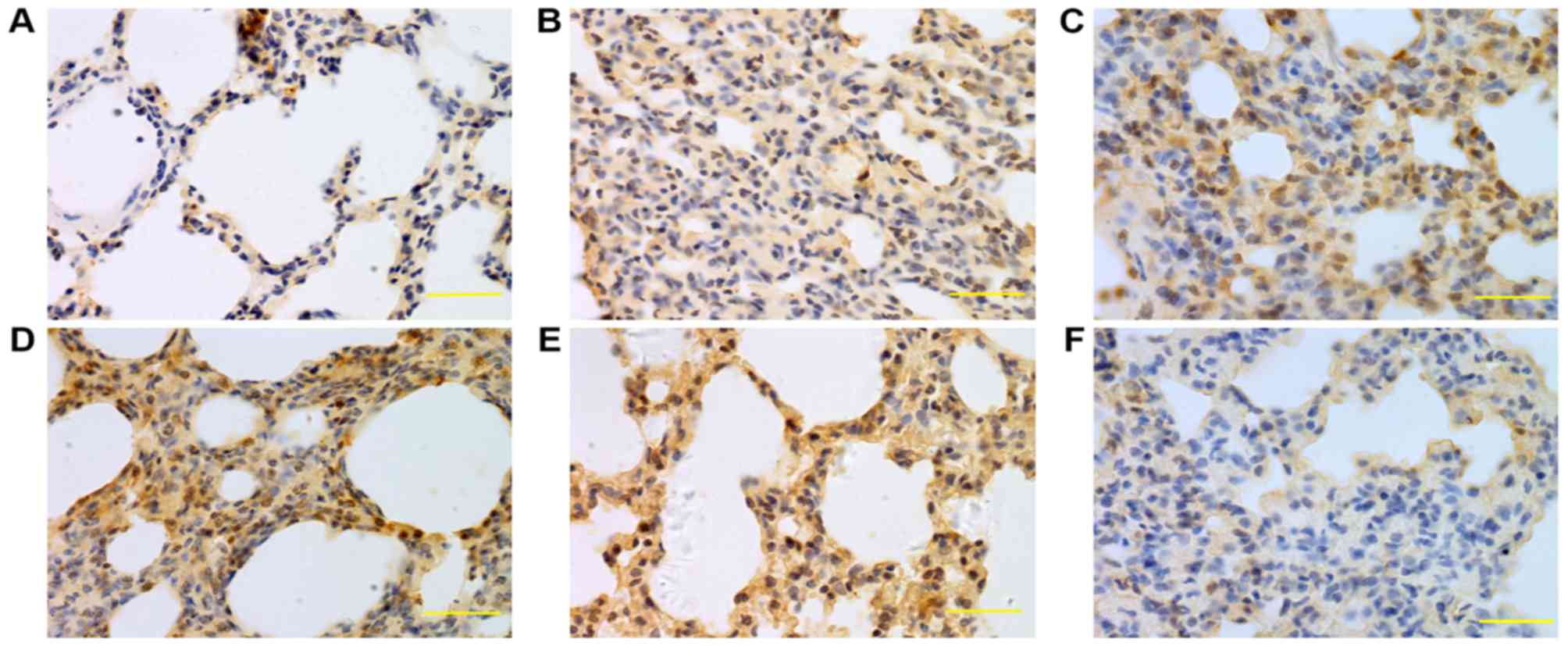
 | Figure 7.Expression of caspase-3 in lung
tissue at 24 h. Caspase-3 was mainly expressed in bronchial
epithelial cells, alveolar epithelial cells and vascular and airway
smooth muscle cells. The caspase-3 expression levels in the saline,
low-, middle- and high-dose groups were higher compared with the
normal group. (A) Control group, (B) saline group, (C) low-dose
group, (D) middle-dose group, (E) high-dose group and (F) PBS
group. Scale bars, 100 µm; magnification, ×400; DAB staining. DAB,
3,3-diaminobenzidine. |
 | Figure 8.Expression of caspase-3 in lung
tissue at 48 h. Caspase-3 was mainly expressed in bronchial
epithelial cells, alveolar epithelial cells and vascular and airway
smooth muscle cells. The caspase-3 expression levels in the saline,
low-, middle- and high-dose groups were higher compared with the
normal group. (A) Control group, (B) saline group, (C) low-dose
group, (D) middle-dose group, (E) high-dose group and (F) PBS
group. Scale bars, 100 µm; magnification, ×400; DAB staining. DAB,
3,3-diaminobenzidine. |
 | Figure 9.Expression of caspase-3 in lung
tissue at 72 h. Caspase-3 was mainly expressed in bronchial
epithelial cells, alveolar epithelial cells and vascular and airway
smooth muscle cells. The caspase-3 expression levels in the saline,
low-, middle- and high-dose groups were higher compared with the
normal group. (A) Control group, (B) saline group, (C) low-dose
group, (D) middle-dose group, (E) high-dose group and (F) PBS
group. Scale bars, 100 µm; magnification, ×400; DAB staining. DAB,
3,3-diaminobenzidine. |
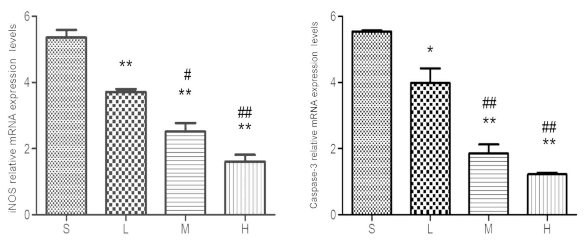
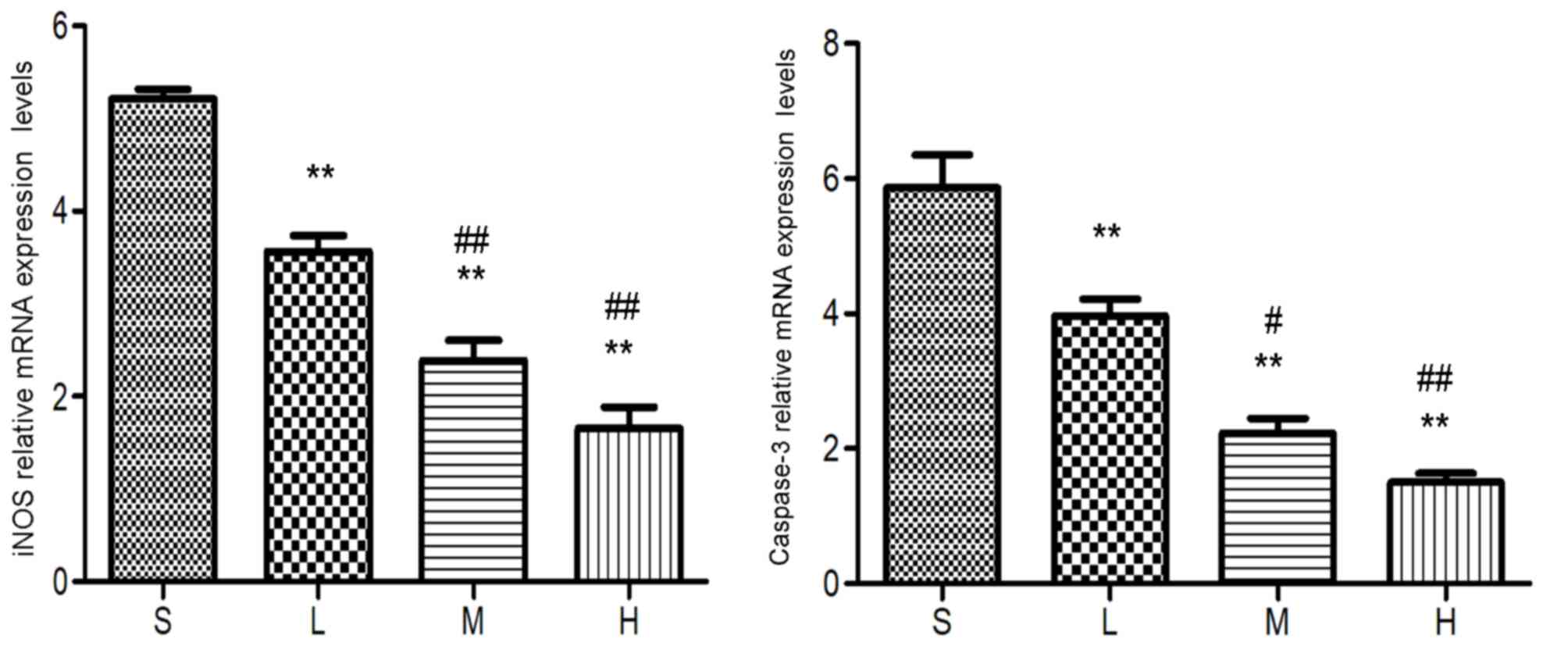
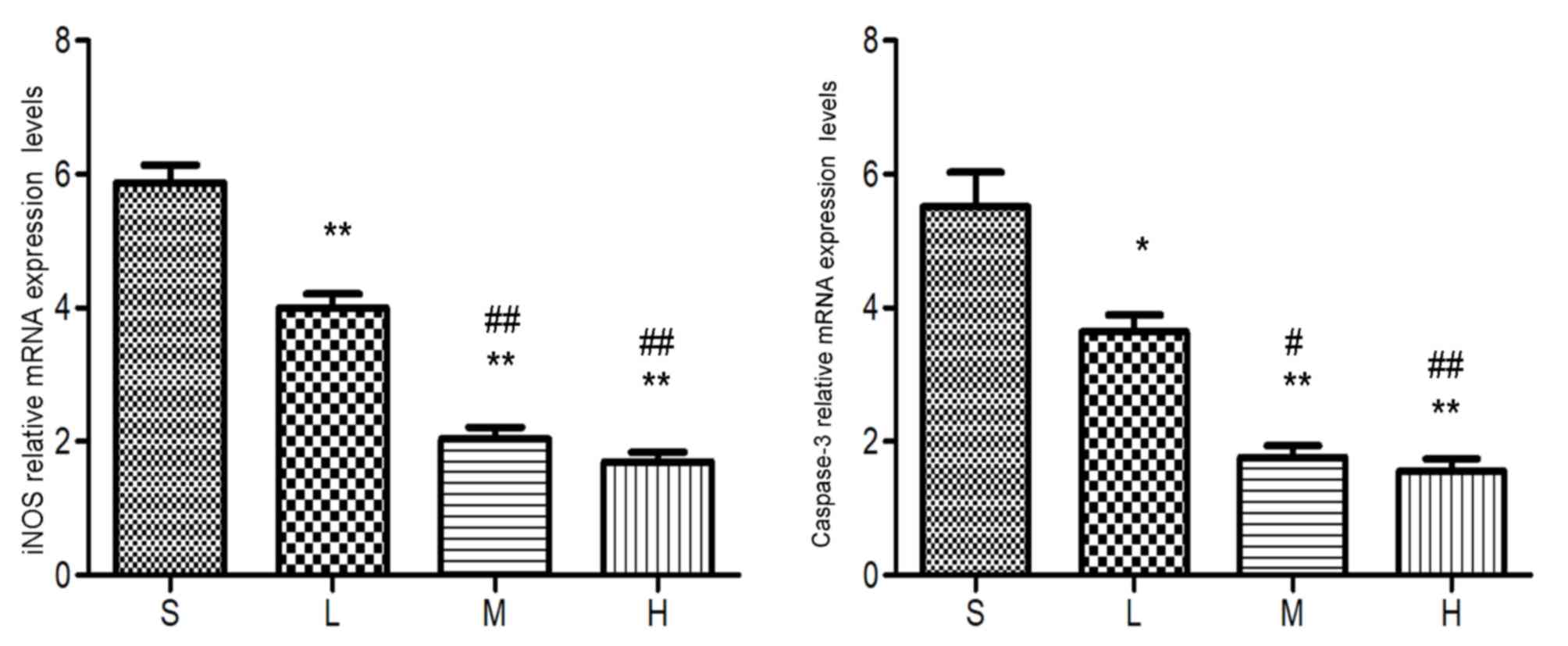
RT-qPCR
RT-qPCR revealed that iNOS and caspase-3 mRNA
expression levels were lower in the low-, middle- and high-dose
groups compared with the saline group (P<0.01 or 0.05) and lower
in the middle- and high-dose groups compared with the low-dose
group (P<0.01 or 0.05). The difference between the middle- and
high-dose groups was not significant (P>0.05; Figs. 10, 11 and 12).
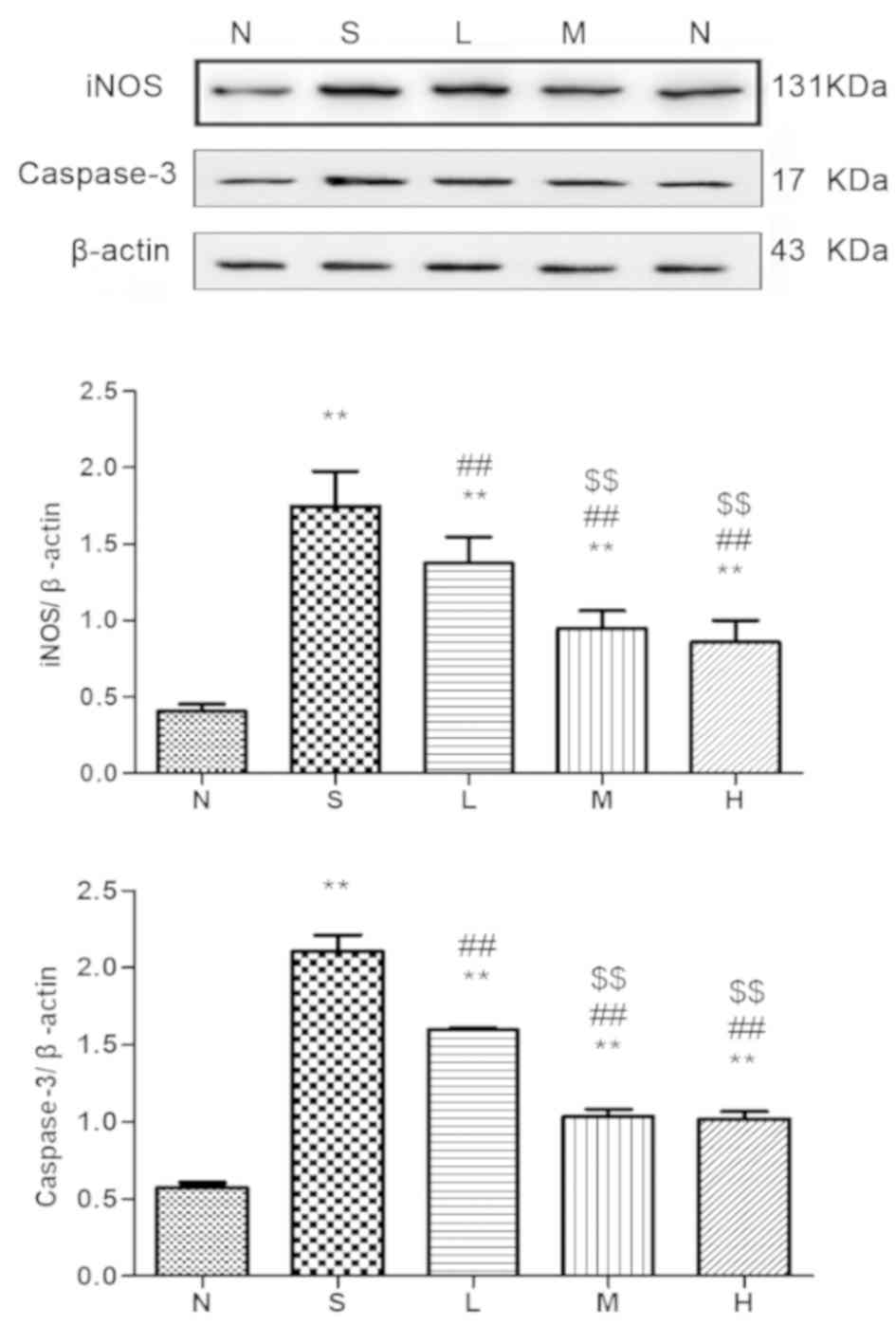
Western blot analysis
Western blot analysis demonstrated that the protein
levels of iNOS and caspase-3 were higher in the saline, low-dose,
middle-dose and high-dose groups compared with the control group
(P<0.01 or P<0.05). Additionally, the protein levels were
lower in the low-, middle- and high-dose groups compared with the
saline group (P<0.01 or P<0.05) and lower in the middle- and
high-dose groups compared with the low-dose group (P<0.01 or
P<0.05). However, no significant difference in the protein
levels of iNOS and caspase-3 was observed between the middle- and
high-dose groups (P>0.05; Figs.
13, 14 and 15).
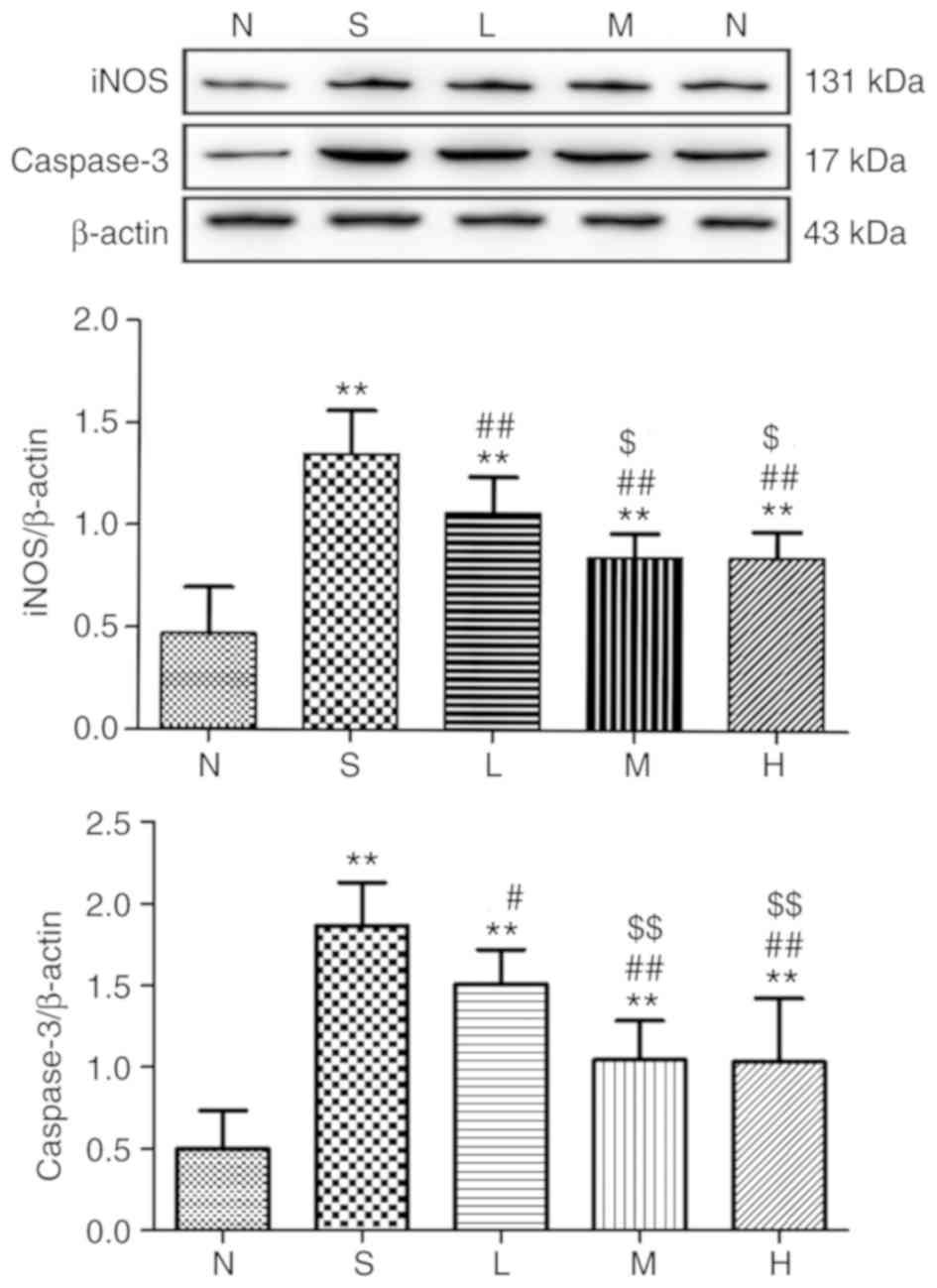 | Figure 13.Relative expression of iNOS and
caspase-3 in lung tissue at 24 h. The levels of iNOS and caspase-3
proteins in rat lungs were detected and immunoreactive bands were
observed at ~131 and 17 kDa, respectively. The levels of the iNOS
and caspase-3 proteins were higher in the saline, low-, middle- and
high-dose groups compared with the control group. **P<0.01 vs.
control group; #P<0.05, ##P<0.01 vs.
saline group; $P<0.05, $$P<0.01 vs.
low-dose group. iNOS, inducible nitric oxide synthase; N, control
group S, saline group; L, low-dose group; M, middle-dose group; H,
high-dose group. |
SOD activity and content of MDA
SOD activity in lung tissues and serum was lower in
the saline group and the low-, middle- and high-dose groups
compared with the control group (P<0.05); significantly higher
in the low-, medium- and high-dose groups compared with the saline
group (P<0.05); and higher in the middle- and high-dose groups
compared with the low-dose group (P<0.05) at 24, 48 and 72 h. No
significant difference in the SOD activity in lung tissues and
serum was observed between the middle- and high-dose groups
(P>0.05), with the exception of at 24 and 72 h in lung tissues.
MDA content in lung tissues and serum was higher in the saline,
low-dose, middle-dose and high-dose groups compared with the
control group (P<0.05). Additionally, the MDA content were lower
in the low-, middle- and high-dose groups compared with the saline
group (P<0.05) and lower in the middle- and high-dose groups
compared with the low-dose group (P<0.05). The difference
between the middle- and high-dose groups was not significant
(P>0.05) except the MDA content in serum at 48 h (Tables I and II).
 | Table I.Activity of superoxide dismutase in
lung tissue (U/mg prot) and serum (U/ml) at different time points
and different doses of simvastatin. |
Table I.
Activity of superoxide dismutase in
lung tissue (U/mg prot) and serum (U/ml) at different time points
and different doses of simvastatin.
|
| 24 h | 48 h | 72 h |
|---|
|
|
|
|
|
|---|
| Groups | Serum | Lung | Serum | Lung | Serum | Lung |
|---|
| Control group | 59.92±8.13 | 250.8±11.27 | 57.83±6.67 | 244.25±11.58 | 60.31±2.29 | 248.03±3.67 |
| Saline group |
16.32±1.67a |
161.45±10.51a |
16.45±1.30a |
164.95±14.97a |
19.96±1.53a |
157.79±4.97a |
| Low-dose group |
23.81±3.53a,b |
183.77±9.22a,b |
28.26±3.09a,b |
189.14±10.01a,b |
27.78±2.03a,b |
197.65±7.57a,b |
| Middle-dose
group |
39.57±6.34a–c |
215.03±9.19a–c |
43.76±5.56a–c |
220.41±6.05a–c |
38.73±3.08a–c |
213.84±6.93a–c |
| High-dose
group |
39.77±4.90a–c |
226.97±7.37a–d |
43.04±5.10a–c |
224.99±1.85a–c |
39.86±4.60a–c |
227.52±10.05a–d |
| Table II.Malondialdehyde in lung tissue
(nmol/mg prot) and serum (nmol/ml) at different time points and
different doses of simvastatin. |
Table II.
Malondialdehyde in lung tissue
(nmol/mg prot) and serum (nmol/ml) at different time points and
different doses of simvastatin.
|
| 24 h | 48 h | 72 h |
|---|
|
|
|
|
|
|---|
| Groups | Serum | Lung | Serum | Lung | Serum | Lung |
|---|
| Control | 4.43±0.65 | 3.59±0.33 | 4.28±0.47 | 3.33±0.27 | 4.58±0.43 | 3.88±0.13 |
| Saline |
11.11±2.14a |
9.68±0.89a |
10.11±0.60a |
10.33±0.87a |
9.95±0.62a |
10.28±0.38a |
| Low-dose |
8.95±0.65a,b |
7.43±0.86a,b |
8.59±0.57a,b |
7.68±1.11a,b |
8.37±0.20a,b |
8.51±0.71a,b |
| Middle-dose |
7.64±0.34a–c |
5.57±0.68a–c |
7.22±0.38a–c |
5.68±0.84a–c |
6.66±0.59a–c |
4.92±0.34a–c |
| High-dose |
6.65±0.37a–c |
5.19±0.37a–c |
6.08±0.68a–d |
5.24±0.29a–c |
6.08±0.66a–c |
4.99±0.26a–c |
Discussion
At present, the pathogenesis of inhaled lung injury
has yet to be fully elucidated and sufficient evidence from basic
research and clinical data is lacking. Previous research on smoke
inhalation injury has primarily focused on inflammation and
oxidative stress (25,26). For example, one study demonstrated
that NF-κB, tumor necrosis factor (TNF) α and interleukin (IL) 6
serve key roles in smoke inhalation injury (27). During the process of inhalation
injury, intracellular quiescent NF-κB is activated as a result of
the phosphorylation and degradation of inhibitor of NF-κB (IκB) and
the two of these processes occur when cells are stimulated with
various cytokines such as TNF-α and IL-6. Activated NF-κB
translocates to the nucleus to regulate the transcription of its
target genes, including cytokines, inflammatory mediators, acute
phase proteins and inducible effector enzymes (28,29).
In previous experimental studies, our research group has
demonstrated that simvastatin can significantly reduce the
mortality of animal models with smoke inhalation lung injury,
alleviate the inflammatory response in lung tissue and
significantly reduce the content of NF-κB (30,31).
Treatment with simvastatin inactivates NF-κB by increasing the
number of IκB molecules, which is regarded as an inhibitor of NF-κB
and decreases the nuclear NF-κB content. The inhibitory effect of
statins might be mediated by isoprenoid-induced intracellular
signal transduction, which involves several key signaling proteins,
including Rho kinase and IκB/NF-κB (32).
NO has become the focus of studies investigating the
mechanism underlying the gradual induction of lung tissue damage in
smoke inhalation injury (33,34).
Endogenous NO is a transduction molecule involved in the
intracellular signaling pathway and NO also affects arterial
dilatation, decreases platelet viscosity and acts as an
anti-inflammatory agent (35).
According to previous studies, NO regulates apoptosis and inhibits
the activities of several caspases (36,37).
The overproduction of NO mediates mitochondrial dysfunction and
triggers apoptosis and, as more in-depth studies are being
performed, the role of NO in the progression of smoke
inhalation-induced lung injury is gradually attracting more
attention (38).
A previous study demonstrated that NO is produced by
three types of NOS: Endothelial NOS, iNOS and neuronal NOS
(39). During smoke inhalation
injury, large quantities of active iNOS catalyze the decomposition
of arginine to produce an excessive amount of NO (40). Simultaneously, excess NO binds to
O2− to form a strong oxidant (ONOO−) that
mediates the production of a large amount of ROS, resulting in
destruction of the cytomembrane and DNA, inducing cell lysis and
necrosis (41). ONOO−
acts mainly through two pathways: i) It affects pulmonary vascular
permeability and the diffusion function of lung tissue, ii) it
destroys the cytomembrane and DNA and iii) affects the energy
metabolism of cells through negative feedback, leading to cell
lysis and necrosis (42). One
subtype of histone deacetylase, sirtuin 1 (SIRT1) serves an
important role in maintaining gene stability, inhibiting apoptosis,
inhibiting oxidative stress and exerting anti-inflammatory effects
(43). However, excessive
production of NO can inhibit SIRT1, thereby inhibiting P53
pathway-dependent apoptosis through a deacetylation pathway and
ultimately promoting apoptosis (44).
However, NO also activates protein kinase G (PKG),
through the NO/cyclic guanosine monophosphate/PKG pathway (45). Additionally, phosphorylated p38
mitogen-activated protein kinase activates caspase-3 (46). Caspase-3, which serves as an
intersection of the endogenous and exogenous apoptotic pathways, is
the main protein with apoptotic activity (47). Therefore, its expression levels can
directly reflect the occurrence of apoptosis (48). Associated inflammatory mediators
and cells regulated by NF-κB can further promote the activation of
NF-κB, TNF-α, IL-6 and iNOS, among other factors, indicating that
NF-κB serves a key role in smoke inhalation injury (49). Pro-inflammatory cytokines, such as
IL-1 and TNF-α, induce the expression of iNOS and consequently the
generation of large quantities of NO, resulting in cell
degeneration and apoptosis (50).
Toll-like receptor 4 (TLR4), one of the most
important proteins involved in cellular signaling, activates the
downstream nuclear factor NF-κB when bound to its ligand (51). Combined with the present results,
it can be hypothesized that simvastatin decreases the expression of
iNOS and caspase-3 by inhibiting the activation of NF-κB, which in
turn is mediated by suppressing TLR4 expression on the surfaces of
CD14+ monocytes in peripheral blood and attenuating downstream
signal pathways. This protects the lungs against cytotoxicity
caused by NF-κB and NO overproduction, and decreasing cell damage
and apoptosis (52). At the same
time, overproduction of NO can block the high levels of
Ca2+ induced by ROS and inhibit apoptosis through the
Bcl-2 signaling pathway (53).
The levels of MDA, a product of the destruction of
polyunsaturated fatty acids in the cell membrane, indirectly
reflects the grade of oxidative damage. SOD acts as an important
antioxidant enzyme by antagonizing and blocking oxygen free
radicals to decrease cell damage and thus speeding the repair of
free radical-induced damage in cells (54,55).
High levels of oxygen free radicals exceed the capacity of the
antioxidant system during smoke inhalation injury, which causes
lipid peroxidation and subsequent destruction of cell membranes
(56). Simvastatin decreases the
activation of neutrophils and arachidonic acid metabolism following
inhalation injury, leading to a decrease in the production of
oxygen free radicals and a consequential consumption of antioxidant
substances. In turn, this increases SOD production and inhibits
oxidative stress in response to smoke inhalation injury, thus
reducing cell damage (57,58).
In the present study, iNOS and caspase-3 expression
levels were significantly increased following smoke inhalation lung
injury (P<0.05), indicating that lung tissue injury was
aggravated, whereas iNOS and caspase-3 expression levels in the
low-, middle- and high-dose groups were significantly lower
compared with the saline group (P<0.05). As an important
antioxidant enzyme in vivo, SOD activity was significantly
decreased following smoke inhalation lung injury (P<0.05) and
MDA content was significantly increased (P<0.05), at 24, 48 and
72 h. In addition, simvastatin treatment increased SOD activity and
decreased the MDA content, and this effect was dose-dependent
within 0–50 mg/kg. Therefore, simvastatin could inhibit the
formation of iNOS and inhibit apoptosis and lung tissue damage
following smoke inhalation, thus exerting a certain protective
effect on lung tissue with smoke inhalation injury. Although the
therapeutic effect of simvastatin on inhalation injury has been
widely recognized (59), the
specific underlying molecular mechanisms and appropriate dosages
remain unclear.
In the present study, simvastatin inhibited iNOS and
caspase-3 expression and decreased NO synthesis and lung cell
apoptosis. In contrast, simvastatin exerted some antioxidant
effects and inhibited excessive oxidative stress in rats with smoke
inhalation injury, thus exerting a protective effect against smoke
inhalation injury.
Acknowledgements
The authors thank Ms Xiu-hua Ren from the School of
Basic Medicine of Zhengzhou University for her help with the
pathological detection and immunohistochemistry experiments.
Funding
The present study was supported by grants from the
National Natural Science Cooperation Foundation of China (grant no.
U1604188) and the 2015 Key Scientific and Technological Projects of
Henan Province (grant no. 152102410065).
Availability of data and materials
The datasets used and/or analyzed during the current
study are available from the corresponding author on reasonable
request.
Authors' contributions
The authors all contributed to the research. ZJC,
ZM, CC, PFG and QNM conceived and designed the study. RQY, YG and
IK performed the experiments. RQY and XBW wrote the paper and
performed analysis or interpretation of data. ZJC reviewed and
edited the manuscript for important intellectual content, and final
approval of the version to be published. All authors read and
approved the final manuscript.
Ethics approval and consent to
participate
The present study was approved by the Animal Care
and Use Committee of Zhengzhou University (Henan, China) and
conducted in accordance with the National Institutes of Health
Guide for the Care and Use of Laboratory Animals.
Patient consent for publication
Not applicable.
Competing interests
The authors declare that they have no competing
interests.
References
|
1
|
Ballard-Croft C, Sumpter LR, Broaddus R,
Alexander J, Wang D and Zwischenberger JB: Ovine smoke/burn ARDS
model: A new ventilator-controlled smoke delivery system. J Surg
Res. 164:3405–e162. 2010. View Article : Google Scholar
|
|
2
|
De Carvalho FO, Silva ÉR, Felipe FA,
Teixeira LGB, Zago LBS, Nunes PS, Shanmugam S, Serafini MR and
Araújo AAS: Natural and synthetic products used for the treatment
of smoke inhalation: A patent review. Expert Opin Ther Pat.
27:877–886. 2017. View Article : Google Scholar : PubMed/NCBI
|
|
3
|
Tanizaki S: Assessing inhalation injury in
the emergency room. Open Access Emerg Med. 7:31–37. 2015.
View Article : Google Scholar : PubMed/NCBI
|
|
4
|
Lee AS and Mellins RB: Lung injury from
smoke inhalation. Paediatr Respir Rev. 7:123–128. 2006. View Article : Google Scholar : PubMed/NCBI
|
|
5
|
Matthay MA and Zemans RL: The acute
respiratory distress syndrome: Pathogenesis and treatment. Annu Rev
Pathol. 6:147–163. 2011. View Article : Google Scholar : PubMed/NCBI
|
|
6
|
Barsanti KC, Luo W, Isabelle LM, Pankow JF
and Peyton DH: Tobacco smoke particulate matter chemistry by NMR.
Magn Reson Chem. 45:167–170. 2007. View
Article : Google Scholar : PubMed/NCBI
|
|
7
|
Wu ZY, Li H and Tang YJ: Effect of
simvastatin on the SIRT2/NF-κB pathway in rats with acute pulmonary
embolism. Pharm Biol. 56:511–518. 2018. View Article : Google Scholar : PubMed/NCBI
|
|
8
|
Cox RA, Jacob S, Oliveras G, Murakami K,
Enkhbaatar P, Traber L, Schmalstieg FC, Herndon DN, Traber DL and
Hawkins HK: Pulmonary expression of nitric oxide synthase isoforms
in sheep with smoke inhalation and burn injury. Exp Lung Res.
35:104–118. 2009. View Article : Google Scholar : PubMed/NCBI
|
|
9
|
Liu X, Ai F, Li H, Xu Q, Mei L, Miao J,
Wen Q, Zhang C, Zhang S, Zhou J, et al: Anti-inflammatory effects
of shenfu injection against acute lung injury through inhibiting
HMGB1-NF-κB pathway in a rat model of endotoxin shock. Evid Based
Complement Alternat Med. 2019:98576832019. View Article : Google Scholar : PubMed/NCBI
|
|
10
|
Hosogi S, Iwasaki Y, Yamada T,
Komatani-Tamiya N, Hiramatsu A, Kohno Y, Ueda M, Arimoto T and
Marunaka Y: Effect of inducible nitric oxide synthase on apoptosis
in Candida-induced acute lung injury. Biomed Res. 29:257–266. 2008.
View Article : Google Scholar : PubMed/NCBI
|
|
11
|
Ray PD, Huang BW and Tsuji Y: Reactive
oxygen species (ROS) homeostasis and redox regulation in cellular
signaling. Cell Signal. 24:981–990. 2012. View Article : Google Scholar : PubMed/NCBI
|
|
12
|
Liu S, Yue Y, Pan P, Zhang L, Su X, Li H,
Li H, Li Y, Dai M, Li Q and Mao Z: IRF-1 intervention in the
classical ROS-dependent release of NETs during LPS-induced acute
lung injury in mice. Inflammation. 42:387–403. 2019. View Article : Google Scholar : PubMed/NCBI
|
|
13
|
Montaño M, Cisneros J, Ramírez-Venegas A,
Pedraza-Chaverri J, Mercado D, Ramos C and Sansores RH:
Malondialdehyde and superoxide dismutase correlate with FEV(1) in
patients with COPD associated with wood smoke exposure and tobacco
smoking. Inhal Toxicol. 22:868–874. 2010. View Article : Google Scholar : PubMed/NCBI
|
|
14
|
Karmali KN, Lloyd-Jones DM, Berendsen MA,
Goff DC Jr, Sanghavi DM, Brown NC, Korenovska L and Huffman MD:
Drugs for primary prevention of atherosclerotic cardiovascular
disease: An overview of systematic reviews. JAMA Cardiol.
1:341–349. 2016. View Article : Google Scholar : PubMed/NCBI
|
|
15
|
Yan YL, Qiu B, Hu LJ, Jing XD, Liu YJ,
Deng SB, Du JL and She Q: Efficacy and safety evaluation of
intensive statin therapy in older patients with coronary heart
disease: A systematic review and meta-analysis. Eur J Clin
Pharmacol. 69:2001–2009. 2013. View Article : Google Scholar : PubMed/NCBI
|
|
16
|
Wang L, Mehta S, Gillis C, Law C and
Taneja R: Modulation of neutrophil apoptosis by murine pulmonary
microvascular endothelial cell inducible nitric oxide synthase.
Biochem Biophys Res Commun. 401:207–212. 2010. View Article : Google Scholar : PubMed/NCBI
|
|
17
|
Aoki T, Kataoka H, Ishibashi R, Nozaki K
and Hashimoto N: Simvastatin suppresses the progression of
experimentally induced cerebral aneurysms in rats. Stroke.
39:1276–1285. 2008. View Article : Google Scholar : PubMed/NCBI
|
|
18
|
Esposito E, Rinaldi B, Mazzon E, Donniacuo
M, Impellizzeri D, Paterniti I, Capuano A, Bramanti P and Cuzzocrea
S: Anti-inflammatory effect of simvastatin in an experimental model
of spinal cord trauma: Involvement of PPAR-alpha. J
Neuroinflammation. 9:812012. View Article : Google Scholar : PubMed/NCBI
|
|
19
|
Shyamsundar M, McKeown ST, O'Kane CM,
Craig TR, Brown V, Thickett DR, Matthay MA, Taggart CC, Backman JT,
Elborn JS and McAuley DF: Simvastatin decreases
lipopolysaccharide-induced pulmonary inflammation in healthy
volunteers. Am J Respir Crit Care Med. 179:1107–1114. 2009.
View Article : Google Scholar : PubMed/NCBI
|
|
20
|
National Research Council Committee for
the Update of the Guide for the Care and Use of Laboratory Animals:
The National Academies Collection: Reports funded by National
Institutes of Health. Guide for the Care and Use of Laboratory
Animals. National Academies Press (US) Copyright© 2011; National
Academy of Sciences, Washington (DC): 2011, PubMed/NCBI
|
|
21
|
Liu PY, Liu YW, Lin LJ, Chen JH and Liao
JK: Evidence for statin pleiotropy in humans: Differential effects
of statins and ezetimibe on rho-associated coiled-coil containing
protein kinase activity, endothelial function and inflammation.
Circulation. 119:131–138. 2009. View Article : Google Scholar : PubMed/NCBI
|
|
22
|
Zhu F, Qiu X, Wang J, Jin Y, Sun Y, Lv T
and Xia Z: A rat model of smoke inhalation injury. Inhal Toxicol.
24:356–364. 2012. View Article : Google Scholar : PubMed/NCBI
|
|
23
|
Garip S and Severcan F: Determination of
simvastatin-induced changes in bone composition and structure by
Fourier transform infrared spectroscopy in rat animal model. J
Pharm Biomed Anal. 52:580–588. 2010. View Article : Google Scholar : PubMed/NCBI
|
|
24
|
Livak KJ and Schmittgen TD: Analysis of
relative gene expression data using real-time quantitative PCR and
the 2(-Delta Delta C(T)) method. Methods. 25:402–408. 2001.
View Article : Google Scholar : PubMed/NCBI
|
|
25
|
de Carvalho FO, Felipe FA, de Melo Costa
AC, Teixeira LG, Silva ÉR, Nunes PS, Shanmugam S, de Lucca Junior
W, Quintans JS and de Souza Araújo AA: Inflammatory mediators and
oxidative stress in animals subjected to smoke inhalation: A
systematic review. Lung. 194:487–499. 2016. View Article : Google Scholar : PubMed/NCBI
|
|
26
|
Lange M, Szabo C, Traber DL, Horvath E,
Hamahata A, Nakano Y, Traber LD, Cox RA, Schmalstieg FC, Herndon DN
and Enkhbaatar P: Time profile of oxidative stress and neutrophil
activation in ovine acute lung injury and sepsis. Shock.
37:468–472. 2012. View Article : Google Scholar : PubMed/NCBI
|
|
27
|
Tan W, Xue-bin C, Tian Z, Xiao-wu C,
Pei-pei H, Zhi-bin C and Bei-sha T: Effects of simvastatin on the
expression of inducible nitric oxide synthase and brain-derived
neurotrophic factor in a lipopolysaccharide-induced rat model of
Parkinson disease. Int J Neurosci. 126:278–286. 2016. View Article : Google Scholar : PubMed/NCBI
|
|
28
|
Lee HJ, Shin JS, Lee WS, Shim HY, Park JM,
Jang DS and Lee KT: Chikusetsusaponin IVa methyl ester isolated
from the roots of achyranthes japonica suppresses LPS-induced iNOS,
TNF-α, IL-6, and IL-1β expression by NF-κB and AP-1 inactivation.
Biol Pharm Bull. 39:657–664. 2016. View Article : Google Scholar : PubMed/NCBI
|
|
29
|
Matsumoto J, Dohgu S, Takata F, Machida T,
Bölükbaşi Hatip FF, Hatip-Al-Khatib I, Yamauchi A and Kataoka Y:
TNF-α-sensitive brain pericytes activate microglia by releasing
IL-6 through cooperation between IκB-NFκB and JAK-STAT3 pathways.
Brain Res. 1692:34–44. 2018. View Article : Google Scholar : PubMed/NCBI
|
|
30
|
Sun JL, Gao Y, Cui ZJ and Guo PF: The
anti-inflammatory effects of simvastatin in a rat model of smoke
inhalation lung injury. Int J Clin Exp Med. 12:12740–12746.
2019.
|
|
31
|
Refaie MMM, El-Hussieny M and Zenhom NM:
Protective role of nebivolol in cadmium-induced hepatotoxicity via
downregulation of oxidative stress, apoptosis and inflammatory
pathways. Environ Toxicol Pharmacol. 58:212–219. 2018. View Article : Google Scholar : PubMed/NCBI
|
|
32
|
Wang X, Luo B, Lu Y, Pang D, Zheng J, Mo
J, Huang H and Feng J: The triggering receptor expressed by myeloid
cells-1 activates TLR4-MyD88-NF-κB-dependent signaling to aggravate
ventilation-induced lung inflammation and injury in mice. Cell
Tissue Res. 374:137–148. 2018. View Article : Google Scholar : PubMed/NCBI
|
|
33
|
Soejima K, Traber LD, Schmalstieg FC,
Hawkins H, Jodoin JM, Szabo C, Szabo E, Virag L, Salzman A and
Traber DL: Role of nitric oxide in vascular permeability after
combined burns and smoke inhalation injury. Am J Respir Crit Care
Med. 163:745–752. 2001. View Article : Google Scholar : PubMed/NCBI
|
|
34
|
Shun-Zhen Q and Hong-Hang Z: The effects
of inhaled nitric oxide on the levels of cGMP plasma and lung
tissue in a canine model of smoke inhalation injury. Burns.
28:299–304. 2002. View Article : Google Scholar : PubMed/NCBI
|
|
35
|
Lange M, Hamahata A, Enkhbaatar P, Cox RA,
Nakano Y, Westphal M, Traber LD, Herndon D and Traber DL:
Beneficial effects of concomitant neuronal and inducible nitric
oxide synthase inhibition in ovine burn and inhalation injury.
Shock. 35:626–631. 2011. View Article : Google Scholar : PubMed/NCBI
|
|
36
|
Rössig L, Fichtlscherer B, Breitschopf K,
Haendeler J, Zeiher AM, Mülsch A and Dimmeler S: Nitric oxide
inhibits caspase-3 by S-nitrosation in vivo. J Biol Chem.
274:6823–6826. 1999. View Article : Google Scholar : PubMed/NCBI
|
|
37
|
Kim YM, Talanian RV and Billiar TR: Nitric
oxide inhibits apoptosis by preventing increases in caspase-3-like
activity via two distinct mechanisms. J Biol Chem. 272:31138–31148.
1997. View Article : Google Scholar : PubMed/NCBI
|
|
38
|
Bai S, Hu Z, Yang Y, Yin Y, Li W, Wu L and
Fang M: Anti-inflammatory and neuroprotective effects of triptolide
via the NF-κB signaling pathway in a rat MCAO model. Anat Rec
(Hoboken). 299:256–266. 2016.20. Enkhbaatar P, Wang J, Saunders F,
Lange M, Hamahata A, Rehberg S, Parkinson JF, Traber LD, Herndon DN
and Traber DL: Mechanistic aspects of inducible nitric oxide
synthase-induced lung injury in burn trauma. Burns 37: 638–645,
2011. View Article : Google Scholar : PubMed/NCBI
|
|
39
|
Li WC, Zou ZJ, Zhou MG, Chen L, Zhou L,
Zheng YK and He ZJ: Effects of simvastatin on the expression of
inducible NOS in acute lung injury in septic rats. Int J Clin Exp
Pathol. 8:15106–15111. 2015.PubMed/NCBI
|
|
40
|
Ahmed AM: Inhibition of inducible nitric
oxide synthase (iNOS) by simvastatin attenuates cardiac hypertrophy
in rats. Folia Morphol (Warsz). 76:15–27. 2017. View Article : Google Scholar : PubMed/NCBI
|
|
41
|
Kim BC, Kim YS, Lee JW, Seo JH, Ji ES, Lee
H, Park YI and Kim CJ: Protective effect of coriolus versicolor
cultivated in citrus extract against nitric oxide-Induced apoptosis
in human neuroblastoma SK-N-MC cells. Exp Neurobiol. 20:100–109.
2011. View Article : Google Scholar : PubMed/NCBI
|
|
42
|
Lange M, Szabo C, Enkhbaatar P, Connelly
R, Horvath E, Hamahata A, Cox RA, Esechie A, Nakano Y, Traber LD,
et al: Beneficial pulmonary effects of a metalloporphyrinic
peroxynitrite decomposition catalyst in burn and smoke inhalation
injury. Am J Physiol Lung Cell Mol Physiol. 300:L167–L175. 2011.
View Article : Google Scholar : PubMed/NCBI
|
|
43
|
Liu X, Jin X, Yu D and Liu G: Suppression
of NLRP3 and NF-κB signaling pathways by α-Cyperone via activating
SIRT1 contributes to attenuation of LPS-induced acute lung injury
in mice. Int Immunopharmacol. 76:1058862019. View Article : Google Scholar : PubMed/NCBI
|
|
44
|
Chung HT, Pae HO, Choi BM, Billiar TR and
Kim YM: Nitric oxide as a bioregulator of apoptosis. Biochem
Biophys Res Commun. 282:1075–1079. 2001. View Article : Google Scholar : PubMed/NCBI
|
|
45
|
Albert M, Corsilli D, Williamson DR,
Brosseau M, Bellemare P, Delisle S, Nguyen AQ and Varin F:
Comparison of inhaled milrinone, nitric oxide and prostacyclin in
acute respiratory distress syndrome. World J Crit Care Med.
6:74–78. 2017. View Article : Google Scholar : PubMed/NCBI
|
|
46
|
Li JT, Wang WQ, Wang L, Liu NN, Zhao YL,
Zhu XS, Liu QQ, Gao CF, Yang AG and Jia L: Subanesthetic isoflurane
relieves zymosan-induced neutrophil inflammatory response by
targeting NMDA glutamate receptor and Toll-like receptor 2
signaling. Oncotarget. 7:31772–31789. 2016. View Article : Google Scholar : PubMed/NCBI
|
|
47
|
Lossi L, Castagna C and Merighi A:
Caspase-3 mediated cell death in the normal development of the
mammalian cerebellum. Int J Mol Sci. 19:39992018. View Article : Google Scholar
|
|
48
|
Li H, Wan A, Xu G and Ye D: Small changes
huge impact: The role of thioredoxin 1 in the regulation of
apoptosis by S-nitrosylation. Acta Biochim Biophys Sin (Shanghai).
45:153–161. 2013. View Article : Google Scholar : PubMed/NCBI
|
|
49
|
Badshah H, Ali T and Kim MO: Osmotin
attenuates LPS-induced neuroinflammation and memory impairments via
the TLR4/NFκB signaling pathway. Sci Rep. 6:244932016. View Article : Google Scholar : PubMed/NCBI
|
|
50
|
Westphal M, Enkhbaatar P, Schmalstieg FC,
Kulp GA, Traber LD, Morita N, Cox RA, Hawkins HK, Westphal-Varghese
BB, Rudloff HE, et al: Neuronal nitric oxide synthase inhibition
attenuates cardiopulmonary dysfunctions after combined burn and
smoke inhalation injury in sheep. Crit Care Med. 36:1196–1204.
2008. View Article : Google Scholar : PubMed/NCBI
|
|
51
|
Sterner JB, Zanders TB, Morris MJ and
Cancio LC: Inflammatory mediators in smoke inhalation injury.
Inflamm Allergy Drug Targets. 8:63–69. 2009. View Article : Google Scholar : PubMed/NCBI
|
|
52
|
Fraunberger P, Grone E, Grone HJ and Walli
AK: Simvastatin reduces endotoxin-induced nuclear factor kappaB
activation and mortality in guinea pigs despite lowering
circulating low-density lipoprotein cholesterol. Shock. 32:159–163.
2009. View Article : Google Scholar : PubMed/NCBI
|
|
53
|
Wassmann S, Laufs U, Bäumer AT, Müller K,
Ahlbory K, Linz W, Itter G, Rösen R, Böhm M and Nickenig G: HMG-CoA
reductase inhibitors improve endothelial dysfunction in
normocholesterolemic hypertension via reduced production of
reactive oxygen species. Hypertension. 37:1450–1457. 2001.
View Article : Google Scholar : PubMed/NCBI
|
|
54
|
Nie Z, Deng S, Zhang L, Chen S, Lu Q and
Peng H: Crocin protects against dexamethasone-induced osteoblast
apoptosis by inhibiting the ROS/Ca2+-mediated
mitochondrial pathway. Mol Med Rep. 20:401–408. 2019.PubMed/NCBI
|
|
55
|
Wang KS, Lv Y, Wang Z, Ma J, Mi C, Li X,
Xu GH, Piao LX, Zheng SZ and Jin X: Imperatorin efficiently blocks
TNF-α-mediated activation of ROS/PI3K/Akt/NF-κB pathway. Oncol Rep.
37:3397–3404. 2017. View Article : Google Scholar : PubMed/NCBI
|
|
56
|
Mondal NK, Saha H, Mukherjee B, Tyagi N
and Ray MR: Inflammation, oxidative stress, and higher expression
levels of Nrf2 and NQO1 proteins in the airways of women
chronically exposed to biomass fuel smoke. Mol Cell Biochem.
447:63–76. 2018. View Article : Google Scholar : PubMed/NCBI
|
|
57
|
Sun A, Wang W, Ye X, Wang Y, Yang X, Ye Z,
Sun X and Zhang C: Protective effects of methane-rich saline on
rats with lipopolysaccharide-induced acute lung injury. Oxid Med
Cell Longev. 2017:74301932017. View Article : Google Scholar : PubMed/NCBI
|
|
58
|
Kanugula AK, Gollavilli PN, Vasamsetti SB,
Karnewar S, Gopoju R, Ummanni R and Kotamraju S: Statin-induced
inhibition of breast cancer proliferation and invasion involves
attenuation of iron transport: Intermediacy of nitric oxide and
antioxidant defence mechanisms. FEBS J. 281:3719–3738. 2014.
View Article : Google Scholar : PubMed/NCBI
|
|
59
|
Ferraro SA, Yakisich JS, Gallo FT and
Tasat DR: Simvastatin pretreatment prevents ambient
particle-induced lung injury in mice. Inhal Toxicol. 23:889–896.
2011. View Article : Google Scholar : PubMed/NCBI
|