Introduction
Endometriosis is a progressive disease that
critically impacts the physical and psychological health of
females. Endometriosis may also lead to, or be a cause of,
infertility. The symptoms of endometriosis can only be temporarily
improved, and endometriosis has a high reoccurrence rate (1). Therefore, it is of great clinical
significance and social value to develop drugs with novel
mechanisms and improved efficacy for endometriosis treatment.
Immunological tolerance is involved in the paroxysm of
endometriosis, and would be induced because the ectopic endometrium
is not eliminated by the immune system (2). Enhanced macrophage activity and
attenuated cellular immune function are commonly observed in
endometriosis, particularly including the dysfunction of the
cytotoxic reaction mediated by T cells and natural killer cells.
The factors released by macrophages, including prostaglandins,
cytokines and growth factors, may induce ectopic endometrium
growth. These results indicated that ectopic endometrial growth may
be inhibited by decreasing macrophage activity (3).
PPARγ is involved in multiple physiological and
pathological processes, including lipid metabolism, glucose
metabolism, cell proliferation and differentiation, tumorigenesis,
inflammation, and immune responses. Recently, the effects of PPARγ
on gynecological disease have gained increasing amounts of
attention. PPARγ is expressed in the endometrium (4), and activating PPARγ may lead to
inflammatory inhibition and anti-angiogenesis effects (5). As endometriosis is associated with
inflammation and angiogenesis, PPARγ may become a novel target for
endometriosis treatment (6). Liu
et al (7) reported that
macrophage activity and proliferation was inhibited by PPARγ
endogenous ligands in a dose-dependent manner (7). Additionally, the inhibitory effect of
PPARγ activation on ectopic endometrial growth is associated with
the inhibition of the activity and proliferation of macrophages
within the endometriosis lesion (4). Rosiglitazone (RSG), a PPARγ agonist,
not only inhibits the progress of endometriosis (8), but also reduces the symptoms (9).
The present study examine the effects of RSG on the
development and progression of endometriosis in a rat model. The
results presented here will allow for deeper understanding of the
mechanism of endometriosis, its treatment, and the identification
of improved clinical diagnostic markers.
Materials and methods
Ethic statements
All animal experiments were authorized by the
Ethical Committee of The First Affiliated Hospital of Nanchang
University and performed following the guidelines for the care and
use of laboratory animals and the principles of laboratory animal
care and protection.
Preparation of an endometriosis model
in rats
Female rats (n=30; weight ~220 g; 3 months) were
purchased from Hunan SJA Laboratory Animal Co., Ltd and housed in a
specific pathogen-free environment that was automatically
maintained at a temperature of 23±2°C, a relative humidity of
45–65%, and with a controlled 12 h light/dark cycle. The animals
had free to access food and water. The endometriosis model was
prepared as previously described (10). The estrous cycles of rats were
confirmed by vaginal smears. The rats in estrus were anesthetized
by isoflurane (5%) and fixed on the operating table. Iodophor was
uniformly smeared over the abdomen, which was then cut open to
locate the uterus. Two sides of the uterus were ligated, along with
the vessels, by twine and ~1 cm was left in the middle. This 1-cm
section of tissue was cut using scissors and placed in a sterile
culture dish containing normal saline. The endometrium was removed
using sterile microscopic tweezers and cut into fragments measuring
5×5 mm. These fragments were attached to the abdominal wall. The
incision was stitched and smeared with iodophor. Rats were places
onto heating pads to regain consciousness quickly.
Experimental groups
Following the induction of endometriosis, the rats
were allowed to recover for 4 weeks, during which time they were
not administered with any medication. Subsequently, 30 rats were
divided into the following five experimental groups: Model, saline
(equal volume of normal saline for each rat); 5 µM RSG; 10 µM RSG;
and 20 µM RSG. The abdomen of each rat was cut open and the
corresponding treatments were administered via one-time treatment
by injection directly into endometriotic implant (100 µl) following
anesthesia with isoflurane (5% induction, 2% maintenance). The
wounds were stitched up and smeared with iodophor, and the animals
were sacrificed 2 weeks after the administration through
decapitation following anesthesia with isoflurane.
Hematoxylin & eosin (H&E)
staining
Endometrium was collected from each animal and fixed
in in 4% paraformaldehyde for ~1 week at 4°C. Subsequently, the
tissues were washed with sterile water for a couple of hours. The
tissue was dehydrated using 70, 80 and 90% ethanol solutions
successively and mixed with equal quantities of ethanol and xylene.
After 15 min incubation, the tissue was mixed with an equal
quantity of xylene for 15 min. These steps were repeated until the
tissue appeared transparent. The tissue was then embedded in
paraffin at room temperature for 5 min and sectioned at a thickness
of 10 µm, and stained with H&E for 3 min at room temperature.
Images were acquired using an inverted microscope (magnification,
×200; Olympus Corporation).
Transmission electron microscopy
The endometrium was collected after the animals were
executed and was fixed with 2.5% glutaraldehyde for over 2 h at
room temperature and washed with 0.1 M phosphoric acid solution
three times. Next, the tissues were fixed with 1% osmic acid for
2–3 h at room temperature and washed with 0.1 M phosphoric acid
three times. The tissues were then washed with 50% ethyl alcohol,
70% ethyl alcohol, 90% ethyl alcohol, 90% ethyl alcohol and 90%
acetone (v:v=1:1), and 90% acetone successively for 15–20 min.
Next, the tissues were incubated with 100% acetone at room
temperature three times for 15–20 min each. Acetone (100%) and the
embedding solution were incubated with the tissues for 3–4 h at
room temperature. Finally, the tissues were successively embedded
in 0.01% epoxy resin at 37°C overnight, 45°C for 12 h, and 60°C for
48 h. The solid tissue was cut into ultrathin sections (100 nm) and
the slides were stained with lead citrate and uranyl acetate at
room temperature for 5 min. Ultrastructural endometrium changes
were observed and images were captured using transmission electron
microscopy (magnification: ×1,000; JEM-1230; JEOL, Ltd.).
Immunohistochemistry
The endometrium was separated and fixed in 4%
paraformaldehyde for ~1 week at 4°C. The tissues were placed into a
plate filled with pre-cooled normal saline. The tissue was embedded
in paraffin, sectioned into 20 µm. The sections were blocking in 5%
bovine serum albumin (Hyclone; Cytiva) at room temperature for 2 h
and incubated with anti-cleaved caspase-3 antibody (dilution,
1:200; cat. no. ab32042; Abcam) and anti-VEGF antibody (dilution,
1:1,000; cat. no. bs-10853R; Bioss) at 4°C overnight. Samples were
then washed with phosphate-buffered saline (PBS), and the slides
were incubated with horseradish peroxidase (HRP)-conjugated goat
anti-rabbit IgG (1:10,000; cat. no. A16104; Thermo Fisher
Scientific, Inc.) at 37°C for 30 min. After washing with PBS, the
slides were dyed with DAB agent for 5–10 min and re-dyed with
hematoxylin for 3 min, both at room temperature. Images were
obtained using an inverted microscope (magnification, ×200; Olympus
Corporation).
Reverse transcription-quantitative
polymerase chain reaction (RT-qPCR)
Total RNA from the collected endometrium cells was
obtained using an RNA Extraction kit (Takara Biotechnology Co.,
Ltd.) and according to the manufacturer's protocols. Extracted RNA
was quantified using a NanoDrop spectrophotometer (NanoDrop
Technologies). cDNA was synthesized at 30°C for 10 min according to
the instructions of the reverse transcriptase kit (CoWin
Biosciences). SYBR Premix Ex Taq™ (Takara Biotechnology Co., Ltd.)
and an Applied Bio-Rad CFX96 Sequence Detection system (Applied
Biosystems) was used for real-time PCR. The cycling protocol was as
follows: Pre-denaturation at 95°C for 10 min, 40 cycles of 95°C for
12 sec, 58°C for 30 sec and 72°C for 30 sec. PPARγ and
MAT2A expression levels were determined by the threshold
cycle (Ct), and relative expression levels were calculated using
the 2−ΔΔCq method after normalization with reference to
the expression of U6 small nuclear RNA (11). GAPDH expression levels were
used as negative controls. Primer information is listed in Table I.
 | Table I.Primer sequences. |
Table I.
Primer sequences.
| Genes | Sequences
(5′-3′) | Length of the primer
(bp) | Length of the product
(bp) | Annealing temperature
(°C) |
|---|
| PPARγ F |
CCCAGGTTTGCTGAATGTG | 18 | 197 | 57.8 |
| PPARγ R |
TGTCTGTCTCCGTCTTCTTGAT | 20 |
|
|
| MAT2A F |
TTGTGCCTGCGAAATACCT | 19 | 102 | 57 |
| MAT2A R |
CCCCAACCGCCATAAGT | 17 |
|
|
| GAPDH F |
CAATGACCCCTTCATTGACC | 20 | 106 | 57.2 |
| GAPDH R |
GAGAAGCTTCCCGTTCTCAG | 20 |
|
|
Western blot analysis
The Nuclear and Cytoplasmic Protein Extraction kit
(Beyotime Institute of Biotechnology) was used to isolate proteins
from the tissues. After determining the concentration using the BCA
method, approximately 35 µg protein per lane was separated on 12%
SDS-polyacrylamide gels. The separated proteins were transferred
onto a polyvinylidene difluoride (PVDF) membrane (EMD Millipore).
The membrane was blocked with 5% skimmed dry milk in TBST (Tris
buffered saline/0.1% Tween-20, pH 7.4) for 1 h at room temperature
and incubated overnight with primary rabbit anti-human antibodies
against PPARγ (cat. no. AF6284; Affinity Biosciences; dilution
1:1,500) and MAT2A (cat. no. ab189208, Abcam; dilution 1:1,000). A
horseradish peroxidase-conjugated antibody against rabbit IgG (cat.
no. ZB-2305, OriGene Technologies, Inc.; dilution 1:5,000) was used
as a secondary antibody. Blots were incubated with ECL reagents
(Beyotime Institute of Biotechnology) and exposed using a Tanon
5200-multi to detect protein expression.
Statistical analysis
The data are expressed as the mean and standard
deviation with six repeats. Statistically significant differences
for continuous variables were determined using a one-way analysis
of variance with the least significant difference test for normally
distributed data. All testing was performed using GraphPad Prism 5
software (GraphPad Software, Inc.). P<0.05 was considered to
indicate a statistically significant difference.
Results
Effects of RSG on the pathological
states of the endometrium
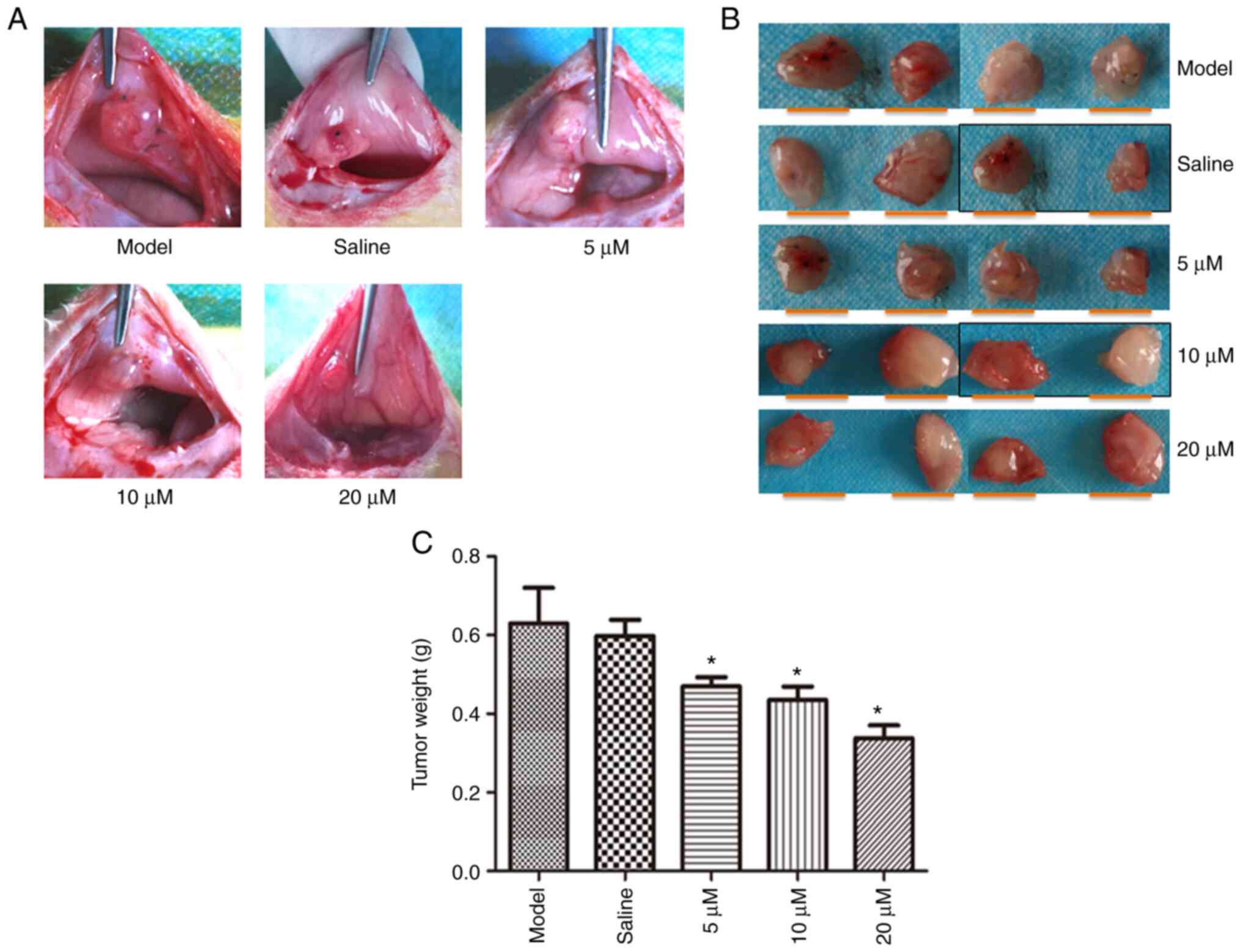
The endometriosis rat model in each group is shown
in Fig. 1. Compared with the model
group, the tumor weight in the RGS group was significantly
decreased (vs. model; P<0.05). This decrease was more
significant at 10 and 20 µM doses of RSG.
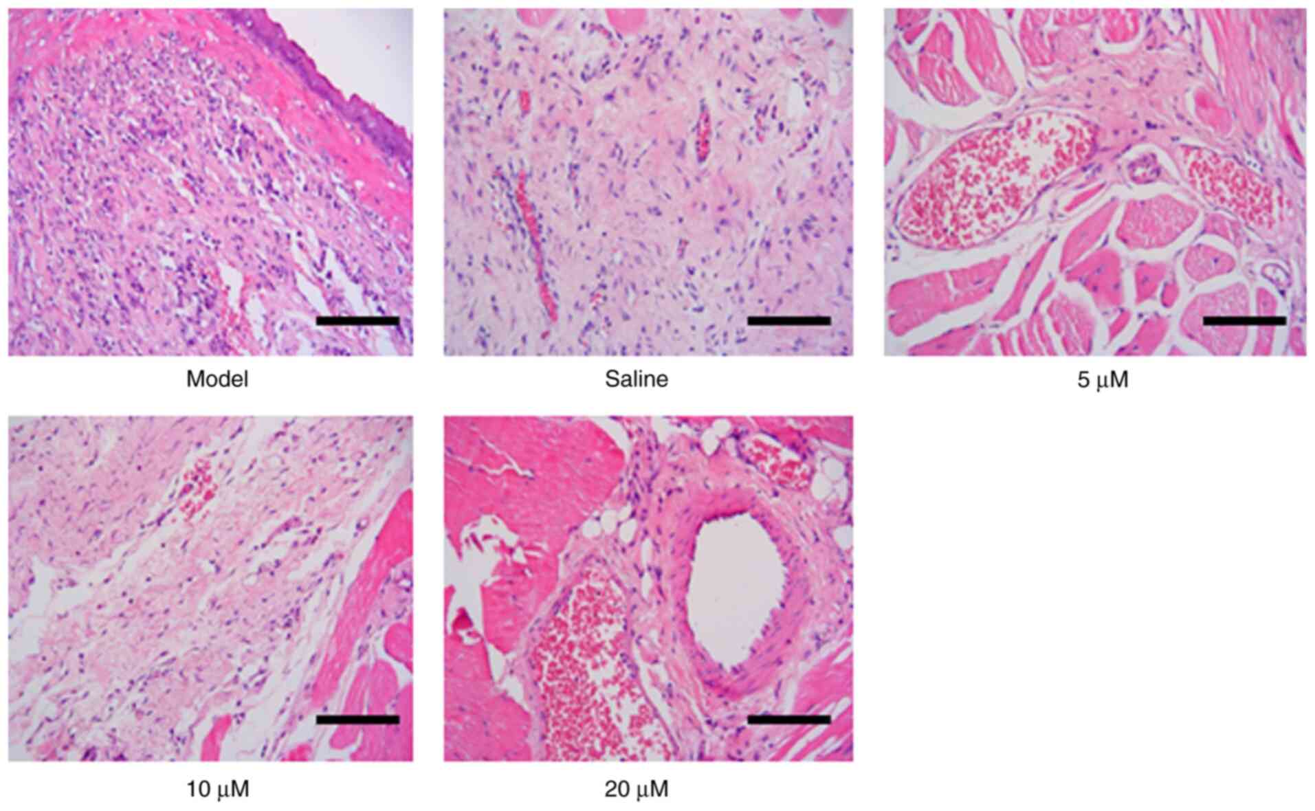
To investigate the impact of RSG on the pathological
changes of endometria extracted from experimental rats, H&E
staining was performed. Three RSG dosages were used: 5, 10 and 20
µM. Transparent and red nodules were observed in the model group,
and vasoganglion were present all over the nodules (Fig. 2). Columnar shaped endometrial
epithelial hyperplasia was observed in the model and saline groups.
Increased numbers of interstitial cells, with compact structure and
abundant blood supply, were detected in model and NS groups.
Compared with the model group, incomplete epithelial structure with
sparse interstitial cells and loose structure was observed in the
pathological images from RSG-treated groups. Inflammatory cells
were involved in the tissues, and mainly included neutrophil
granulocytes. The cells were filled with edema, with poor blood
supply. The gland was filled with more vacuolar cells. These
pathological results indicated that the endometrium was notably
damaged by different RSG dosages.
Effects of RSG on the physiological
structure of the endometrium
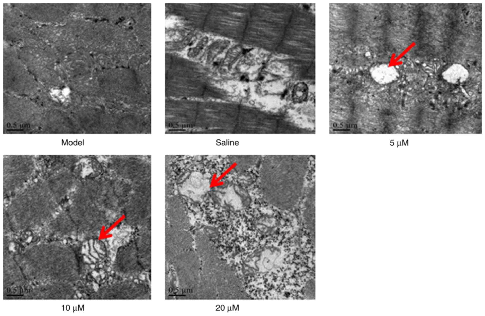
To investigate the effects of RSG on the growth of
the endometrium, the subcellular structure in the tissues was
observed using transmission electron microscopy. The results
demonstrated that the tissue structure was integrated in the model
and saline-treated groups (Fig. 3).
Numerous vacuoles were formed within the endometrial tissue and
classical morphological changes in apoptotic cells were observed in
the RSG-treated groups.
Effects of RSG on VEGF and caspase-3
expression levels in the endometrium
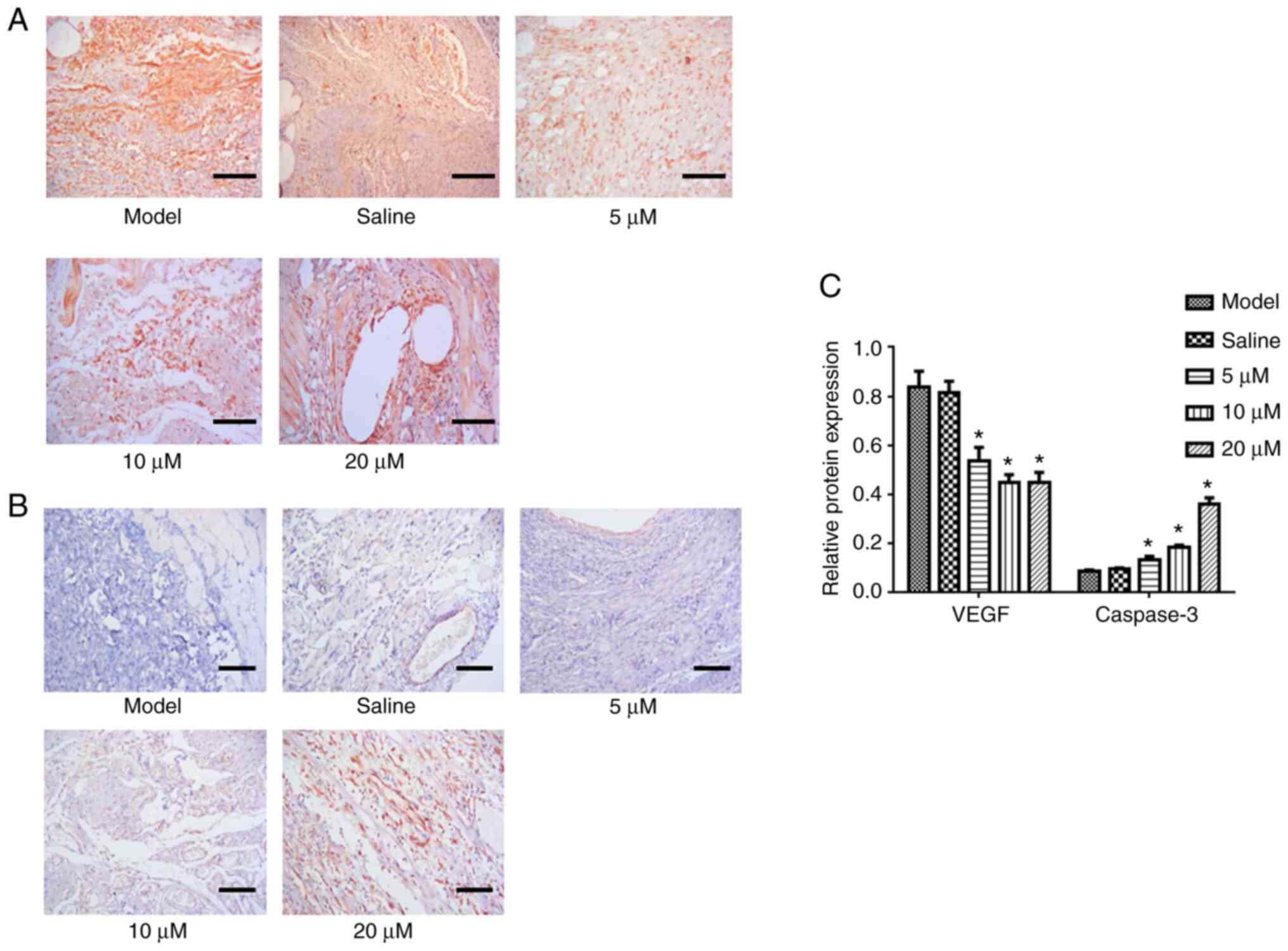
To further investigate the potential mechanism of
RSG on endometriosis, VEGF and cleaved caspase-3 expression levels
were assessed by immunohistochemistry. As shown in Fig. 4, compared with the model group,
expression of VEGF in endometria from rats treated with different
RSG dosages decreased significantly (vs. model; P<0.05) and
caspase-3 expression increased (vs. model; P<0.05; Fig. 4).
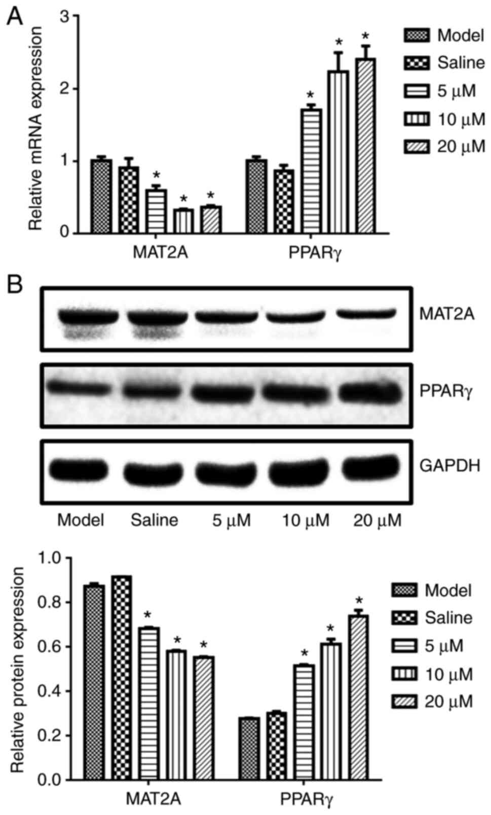
Effects of RSG on PPARγ and MAT2A
expression levels in the endometrium
To investigate the effects of RSG on the
endometriosis at the molecular level, the expression of MAT2A, a
major clinical biomarker for endometriosis, was assessed using
RT-qPCR and western blotting. Compared with the model group, the
PPARγ expression in the endometria of rats treated with RSG
increased significantly (vs. model, P<0.05; Fig. 5). These results indicated that RSG,
which is a PPARγ agonist, may upregulate PPARγ expression in this
tissue. By contrast, MAT2A expression was decreased in the
endometrium of rats treated with RSG, compared with that observed
in the model group (P<0.05).
Discussion
Numerous factors, including immunoreactions,
inflammation and angiogenesis, contribute toward the development
and progression of endometriosis (12). Therapeutically, the formation and
progression of endometriosis may be inhibited by regulating these
factors (13,14). Given the difficulties and limits to
reflecting the integrity of biological processes, including
endometriosis, in isolated cells, a rat endometriosis model was
utilized in this study (10). This
approach enabled the assessment of the general pattern of
transplanted endometrial growth in the abdomen, and investigation
of the optimal action time of drugs by observing the dynamic
pathological and structural changes in the endometrium of the
endometriosis animal model. SD rats were selected because of their
fast reproductive cycle and high level of homology with humans.
Endometriosis was established in rats by transplanting the
endometrium to their abdominal wall. Clear endometriosis lesions,
including protuberances, were observed in the abdomen. Along with
variations in H&E staining results in the endometrium tissues,
the results of the present study indicated that the endometriosis
model was successfully established in SD rats.
PPARγ agonists are potential inhibitors of cell
proliferation (15) and may induce
apoptosis (16). PPARγ agonists
also exert anti-angiogenesis properties by downregulating the
expression of vascular growth factors (17). Research using the endometriosis
animal model revealed that application of the RSG PPARγ agonist may
significantly inhibit angiogenesis within the endometrium and
shrink endometriosis lesions (8,18,19).
In the present study, to investigate the impact of RSG on
endometriosis, an endometriosis model was successfully established
in rats. The results indicated that the size of the endometrium was
diminished as the RSG concentration increased. The observation of
pathological states and sub-cellular structure revealed numerous
vacuoles and the classic morphological changes of apoptotic cells
in the endometrium following RSG treatment. The degeneration of
endometrium may be induced by PPARγ activation (20). Nenicu et al reported that the
therapeutic effects of Telmisartan on endometriosis in mice were
associated with angiotensin II receptor inhibition and PPARγ signal
pathway activation (19). In a
mouse model of endometriosis, the volume of the endometriosis
injury site in mice treated with RSG was much smaller than that in
model mice (21). Lebovic et
al reported that the growth of ectopic endometrial tissue was
significantly inhibited in baboons by oral administration of RSG
(22). These reports indicated that
the growth of ectopic endometrium may be suppressed by PPARγ
activation. These results demonstrated that the PPARγ signal
pathway was activated by treatment with RSG, which inhibited the
expression of MAT2A.
Immune cells, adhesion molecules, extracellular
matrix metalloproteinase and pro-inflammatory cytokines
activate/alter the peritoneal microenvironment, creating conditions
for differentiation, adhesion, proliferation and survival of
ectopic endometrial cells (23–25).
Meanwhile, angiogenesis is an important factor that induces the
paroxysm of endometriosis. The establishment and maintenance of the
blood supply between ectopic endometrium and surrounding are a
precondition for the establishment of endometriosis. The factors
involved in angiogenesis within ectopic endometrium include
epidermal growth factor (EGF), vascular endothelial growth factor
(VEGF), angiogenin-II and transformed growth factor β (TGFβ). Among
these factors, VEGF is of great importance and may induce
angiogenesis directly. VEGF is highly expressed in ectopic
endometrial and mesenchymal cells (26) and VEGF expression in ectopic
endometrial cells is significantly decreased by RSG treatment
(27). RSG treatment also decreases
the vessel density within the ectopic endometrium (21). In the present study, VEGF expression
in ectopic endometrial tissues markedly declined following RSG
treatment. Taken together, these results implied that the
inhibition of ectopic endometrial growth by PPARγ activation may be
associated with the decreased VEGF expression.
Caspase-3 is a key apoptotic protease in mammals,
and serves a central role in the apoptotic cascade reaction pathway
(28). Caspase-3 is activated when
cells encounter apoptotic stimulants, which then activate other
proteins in the apoptotic pathway to induce apoptosis (29). The therapeutic effects of
cyclooxygenase-2 on endometriosis are associated with apoptosis
mediated by caspase-3 activation (30). The ectopic endometrial cells
refluxed to the pelvic cavity were still active because of the
inhibition of ectopic endometrial cell apoptosis, which was
beneficial to the plantation and growth of ectopic endometrium and
induced endometriosis (31). In the
present study, caspase-3 expression levels significantly increased
following RSG treatment. These results indicated that the
therapeutic effects of RSG on endometriosis may be associated with
apoptosis in ectopic endometrial cells mediated by the activation
of caspase-3. Nevertheless, the exact signaling pathways involved
in RSG-induced apoptosis and more persuasive evidence to support
the fact that angiogenesis should be tested in future.
In conclusion, RSG impacts the development and
progression of endometriosis, likely by inhibiting angiogenesis and
inducing apoptosis.
Acknowledgements
Not applicable.
Funding
The present study was supported by grants from the
National Natural Science Foundation of China (grant no.
81560247).
Availability of data and materials
The datasets used and/or analyzed during the current
study are available from the corresponding author on reasonable
request.
Authors' contributions
SZ, LZ, QL, XY, QM and MC performed the experiments
and analyzed the data. SZ and QC designed the study and wrote the
manuscript. All authors read and approved the final manuscript.
Ethics approval and consent to
participate
All animal experiments were authorized by the
Ethical Committee of The First Affiliated Hospital of Nanchang
University and performed following the guidelines for the care and
use of laboratory animals and the principles of laboratory animal
care and protection.
Patient consent for publication
Not applicable.
Competing interests
The authors declare that they have no competing
interests.
References
|
1
|
Arablou T and Kolahdouz-Mohammadi R:
Curcumin and endometriosis: Review on potential roles and molecular
mechanisms. Biomed Pharmacother. 97:91–97. 2018. View Article : Google Scholar : PubMed/NCBI
|
|
2
|
Zhao RH: Strategies for activating blood
circulation-regulating gan (Liver)-tonifying shen (Kidney)
sequential therapy of endometriosis-associated infertility. Chin J
Integr Med. 25:243–245. 2019. View Article : Google Scholar : PubMed/NCBI
|
|
3
|
Zhu H, Cao XX, Liu J and Hua H:
MicroRNA-488 inhibits endometrial glandular epithelial cell
proliferation, migration, and invasion in endometriosis mice via
Wnt by inhibiting FZD7. J Cell Mol Med. 23:2419–2430. 2019.
View Article : Google Scholar : PubMed/NCBI
|
|
4
|
Lebovic DI, Kavoussi SK, Lee J, Banu SK
and Arosh JA: PPARγ activation inhibits growth and survival of
human endometriotic cells by suppressing estrogen biosynthesis and
PGE2 signaling. Endocrinology. 154:4803–4813. 2013. View Article : Google Scholar : PubMed/NCBI
|
|
5
|
Clemenza S, Sorbi F, Noci I, Capezzuoli T,
Turrini I, Carriero C, Buffi N, Fambrini M and Petraglia F: From
pathogenesis to clinical practice: Emerging medical treatments for
endometriosis. Best Pract Res Clin Obstet Gynaecol. 51:92–101.
2018. View Article : Google Scholar : PubMed/NCBI
|
|
6
|
Jafarabadi M, Salehnia M and Sadafi R:
Evaluation of two endometriosis models by transplantation of human
endometrial tissue fragments and human endometrial mesenchymal
cells. Int J Reprod Biomed. 15:21–32. 2017.PubMed/NCBI
|
|
7
|
Liu X, Yu H, Yang L, Li C and Li L:
15-Deoxy-D (12,14)-prostaglandin J(2) attenuates the biological
activities of monocyte/macrophage cell lines. Eur J Cell Biol.
91:654–661. 2012. View Article : Google Scholar : PubMed/NCBIPubMed/NCBI
|
|
8
|
Demirturk F, Aytan H, Caliskan AC, Aytan P
and Koseoglu DR: Effect of peroxisome proliferator-activated
receptor-gamma agonist rosiglitazone on the induction of
endometriosis in an experimental rat model. J Soc Gynecol Investig.
13:58–62. 2006. View Article : Google Scholar : PubMed/NCBI
|
|
9
|
Dworzanski T, Celinski K, Korolczuk A,
Slomka M, Radej S, Czechowska G, Madro A and Cichoz-Lach H:
Influence of the peroxisome proliferator-activated receptor gamma
(PPAR-γ) agonist, rosiglitazone and antagonist,
biphenol-A-diglicydyl ether (BADGE) on the course of inflammation
in the experimental model of colitis in rats. J Physiol Pharmacol.
61:683–693. 2010.PubMed/NCBI
|
|
10
|
Li Z, Liu H, Lang J, Zhang G and He Z:
Effects of cisplatin on surgically induced endometriosis in a rat
model. Oncol Lett. 16:5282–5290. 2018.PubMed/NCBI
|
|
11
|
Livak KJ and Schmittgen TD: Analysis of
relative gene expression data using real-time quantitative PCR and
the 2(-Delta Delta C(T)) method. Methods. 25:402–408. 2001.
View Article : Google Scholar : PubMed/NCBI
|
|
12
|
Zubrzycka A, Zubrzycki M, Perdas E and
Zubrzycka M: Genetic, epigenetic, and steroidogenic modulation
mechanisms in endometriosis. J Clin Med. 9:13092020.
|
|
13
|
Zhou WJ, Yang HL, Shao J, Mei J, Chang KK,
Zhu R and Li MQ: Anti-inflammatory cytokines in endometriosis. Cell
Mol Life Sci. 76:2111–2132. 2019. View Article : Google Scholar : PubMed/NCBI
|
|
14
|
Zani ACT, Valerio FP, Meola J, da Silva
AR, Nogueira AA, Candido-Dos-Reis FJ, Poli-Neto OB and Rosa-E-Silva
JC: Impact of bevacizumab on experimentally induced endometriotic
lesions: Angiogenesis, invasion, apoptosis, and cell proliferation.
Reprod Sci. 27:1943–1950. 2020.
|
|
15
|
Loy CJ, Evelyn S, Lim FK, Liu MH and Yong
EL: Growth dynamics of human leiomyoma cells and inhibitory effects
of the peroxisome proliferator-activated receptor-gamma ligand,
pioglitazone. Mol Hum Reprod. 11:561–566. 2005. View Article : Google Scholar : PubMed/NCBI
|
|
16
|
Heaney AP, Fernando M and Melmed S:
PPAR-gamma receptor ligands: Novel therapy for pituitary adenomas.
J Clin Invest. 111:1381–1388. 2003. View Article : Google Scholar : PubMed/NCBI
|
|
17
|
Panigrahy D, Huang S, Kieran MW and
Kaipainen A: PPARgamma as a therapeutic target for tumor
angiogenesis and metastasis. Cancer Biol Ther. 4:687–693. 2005.
View Article : Google Scholar : PubMed/NCBI
|
|
18
|
Aytan H, Caliskan AC, Demirturk F, Aytan P
and Koseoglu DR: Peroxisome proliferator-activated receptor-gamma
agonist rosiglitazone reduces the size of experimental
endometriosis in the rat model. Aust N Z J Obstet Gynaecol.
47:321–325. 2007. View Article : Google Scholar : PubMed/NCBI
|
|
19
|
Nenicu A, Korbel C, Gu Y, Menger MD and
Laschke MW: Combined blockade of angiotensin II type 1 receptor and
activation of peroxisome proliferator-activated receptor-γ by
telmisartan effectively inhibits vascularization and growth of
murine endometriosis-like lesions. Hum Reprod. 29:1011–1024. 2014.
View Article : Google Scholar : PubMed/NCBI
|
|
20
|
Chang HJ, Lee JH, Hwang KJ, Kim MR and Yoo
JH: Peroxisome proliferator-activated receptor g agonist suppresses
human telomerase reverse transcriptase expression and aromatase
activity in eutopic endometrial stromal cells from endometriosis.
Clin Exp Reprod Med. 40:67–75. 2013. View Article : Google Scholar : PubMed/NCBI
|
|
21
|
Olivares C, Ricci A, Bilotas M, Barañao RI
and Meresman G: The inhibitory effect of celecoxib and
rosiglitazone on experimental endometriosis. Fertil Steril.
96:428–433. 2011. View Article : Google Scholar : PubMed/NCBI
|
|
22
|
Lebovic DI, Mwenda JM, Chai DC, Santi A,
Xu X and D'Hooghe T: Peroxisome proliferator-activated
receptor-(gamma) receptor ligand partially prevents the development
of endometrial explants in baboons: A prospective, randomized,
placebo-controlled study. Endocrinology. 151:1846–1852. 2010.
View Article : Google Scholar : PubMed/NCBI
|
|
23
|
Lagana AS, Garzon S, Götte M, Viganò P,
Franchi M, Ghezzi F and Martin DC: The pathogenesis of
endometriosis: Molecular and cell biology insights. Int J Mol Sci.
20:56152019. View Article : Google Scholar
|
|
24
|
Lagana AS, Salmeri FM, Ban Frangez H,
Ghezzi F, Vrtacnik-Bokal E and Granese R: Evaluation of M1 and M2
macrophages in ovarian endometriomas from women affected by
endometriosis at different stages of the disease. Gynecol
Endocrinol. 36:441–444. 2020. View Article : Google Scholar : PubMed/NCBI
|
|
25
|
Lagana AS, Salmeri FM, Vitale SG, Triolo O
and Götte M: Stem cell trafficking during endometriosis: May
epigenetics play a pivotal role? Reprod Sci. 25:978–979. 2018.
View Article : Google Scholar : PubMed/NCBI
|
|
26
|
Rashidi BH, Sarhangi N, Aminimoghaddam S,
Haghollahi F, Naji T, Amoli MM and Shahrabi-Farahani M: Association
of vascular endothelial growth factor (VEGF) Gene polymorphisms and
expression with the risk of endometriosis: A case-control study.
Mol Biol Rep. 46:3445–3450. 2019. View Article : Google Scholar : PubMed/NCBI
|
|
27
|
Cazzaniga A, Locatelli L, Castiglioni S
and Maier J: The contribution of EDF1 to PPARγ transcriptional
activation in VEGF-treated human endothelial cells. Int J Mol Sci.
19:18302018. View Article : Google Scholar
|
|
28
|
Li J, Yang S and Zhu G: Postnatal calpain
inhibition elicits cerebellar cell death and motor dysfunction.
Oncotarget. 8:87997–88007. 2017. View Article : Google Scholar : PubMed/NCBI
|
|
29
|
Wang Y, Gao W, Shi X, Ding J, Liu W, He H,
Wang K and Shao F: Chemotherapy drugs induce pyroptosis through
caspase-3 cleavage of a gasdermin. Nature. 547:99–103. 2017.
View Article : Google Scholar : PubMed/NCBI
|
|
30
|
Das I and Saha T: Effect of garlic on
lipid peroxidation and antioxidation enzymes in DMBA-induced skin
carcinoma. Nutrition. 25:459–471. 2009. View Article : Google Scholar : PubMed/NCBI
|
|
31
|
Park JH, Lee SK, Kim MK, Lee JH, Yun BH,
Park JH, Seo SK, Cho SH and Choi YS: Saponin extracts induced
apoptosis of endometrial cells from women with endometriosis
through modulation of miR-21-5p. Reprod Sci. 25:292–301. 2018.
View Article : Google Scholar : PubMed/NCBI
|