Introduction
Coronary heart disease (CHD) is one of the most
common causes of death worldwide. Between 2000 and 2012, the death
rate of CHD rose by 33.8% in men, and by 22.8% in women (1). Atherosclerosis (AS) is the basis of
CHD and can cause coronary plaque formation, vascular stenosis or
obstruction, resulting in myocardial ischemia, hypoxia or necrosis
(2). Lipoprotein accumulation in
macrophages causes cells to become foam cells; this process is one
of the leading causes of arterial plaque formation (3,4).
Previous studies have demonstrated that oxidized low-density
lipoprotein (ox-LDL) is a key factor in the initiation and
progression of AS as it induces vascular cells to recruit monocytes
and promotes their differentiation into macrophages, which then
transform into foam cells (5,6).
The PLA2G7 gene in humans encodes
lipoprotein-associated phospholipase A2 (Lp-PLA2), also
known as platelet-activating factor acetylhydrolase, which is an
independent risk factor for the initiation and progression of
cardiovascular disease (7).
Lp-PLA2 catalyses the hydrolysis of oxidized
phospholipids in ox-LDL, leading to the release of downstream
inflammatory mediators, such as lysophosphatidylcholine and
oxidized fatty acids (8). Oxidized
phospholipids in ox-LDL can also promote the conversion of
macrophages into foam cells and further increase the expression
level of Lp-PLA2 (9,10).
Finally, lysophosphatidylcholine induces the production of reactive
oxygen species, thereby exacerbating the formation of
atherosclerosis and destabilizing plaques (11,12).
Therefore, Lp-PLA2 serves a crucial role in the
pathogenesis of AS.
Hydrogen sulfide (H2S) is a key gas
signaling molecule in the cardiovascular system and exerts notable
cardiovascular protective effects (13). H2S has been shown to
exhibit anti-inflammatory and antioxidative stress effects, as well
as blocks the development of AS by protecting the vascular
endothelium, inhibiting vascular smooth muscle cell proliferation
and foam cell formation (14,15)
and decreasing endothelial dysfunction by decreasing p38MAPK
expression levels.
To the best of our knowledge, no studies have yet
investigated the association between H2S and
ox-LDL-induced Lp-PLA2 expression levels and its
potential underlying mechanisms. The present study used ox-LDL to
induce THP1 macrophage to establish a foam cell model. The aims of
the present study were to determine whether H2S can
decrease the expression level of Lp-PLA2 in THP1 cells
induced by ox-LDL; to determine whether H2S improves the
formation of foam cells caused by ox-LDL by decreasing the
expression level of Lp-PLA2; and to identify potential
signalling pathways that promote H2S to decrease
Lp-PLA2 expression levels.
Materials and methods
Reagents
THP-1 cells were obtained from the Cell Bank of the
Chinese Academy of Sciences (cat. no. SCSP-567). RPMI-1640 cell
culture medium (cat. no. 72400120; Thermo Fisher Scientific, Inc.)
and FBS were obtained from Gibco (cat. no.12483020; Thermo Fisher
Scientific, Inc.). ox-LDL, the p38MAPK specific inhibitors SB203580
(cat. no. S8307) and SB202190 (cat. no. S7067), NaHS (cat. no.
161527) and DL-propargylglycine (PPG; cat. no. P7888) were
purchased from Sigma-Aldrich (Merck KGaA). An Lp-PLA2
primary antibody (cat. no. 160603) and enzyme activity detection
kit (cat. no. 760901) were obtained from Cayman Chemical Company.
ox-LDL was purchased from BIOSS (cat. no. bs-1698P). The total
(t)-p38MAPK primary antibody (cat. no. ab31828) and phosphorylated
(p)-p38MAPK primary antibody (cat. no. ab4822) were obtained from
Abcam. Secondary antibodies [HRP-labeled Goat Anti-Rabbit IgG(H+L);
cat. no. A0208] were purchased from Beyotime Institute of
Biotechnology. Cystathionine γ-lyase (CSE) primary antibody was
purchased from BIOSS (cat. no. bs-9515R). β-actin primary antibody
was purchased from Beyotime Institute of Biotechnology (cat. no.
AF5006).
Cell transfection
Cells were seeded in 6-well plates (4×10⁵
cells/well) and transfected the following day with human
Lp-PLA2 small interfering (si)RNA (30 nM; cat. no.
AM16708; Invitrogen; Thermo Fisher Scientific, Inc.) using
Lipofectamine® 2000 (cat. no. 11668019; Invitrogen;
Thermo Fisher Scientific, Inc.), according to the following method:
24 h prior to transfection, the cells were seeded in 6-well plates
and ~2 ml RPMI-1640 medium was added into each well, so that the
cell density was 4×10⁵ cells/well at the time of
transfection, and 2 ml antibiotic-free medium was replaced. Then,
Lipofectamine® 2000 (4 µl/well) was shaken gently and
diluted with 245 µl opti-MEM (cat. no. 51985034; Thermo Fisher
Scientific, Inc.), and the mixture was incubated at room
temperature for 5 min. A total of 30 nM Lp-PLA2
siRNA/negative control (NC) siRNA (cat. no. AM16708; Thermo Fisher
Scientific, Inc.) was diluted with 245 µl opti-MEM and mixed
gently. The two mixtures with mixed together and allowed to stand
at room temperature for 20 min in order to form a mixture of
Lp-PLA2 siRNA/NC siRNA transfection reagents. This
mixture (500 µl) was added to the well containing cells, along with
2 ml antibiotic-free medium. Cells were incubated in 5%
CO2 at 37°C for 6 h and the medium was replaced with
RPMI-1640 complete medium-containing serum. After transfection for
24 h, the cells were exposed to 50 µM ox-LDL at 37°C in a
humidified atmosphere with 5% CO2 for 24 h. The
Lp-PLA2 siRNA transfection and validation, followed the
method of Zheng et al (16).
Cell culture
THP-1 cells were maintained in RPMI-1640 medium
supplemented with 10% FBS at 37°C in a humidified atmosphere with
5% CO2. Before performing the experiments, the medium
was replaced with RPMI-1640 medium containing fresh serum unless
otherwise indicated. Cells were divided into the following groups:
Control (THP-1 cells treated with RPMI-1640 medium supplemented
with 10% FBS); ox-LDL [THP-1 cells treated with ox-LDL (50 µg/ml)
for 24 h]; ox-LDL + SB203580 [THP-1 cells pretreated with SB203580
(20 µM) for 30 min before being treated with ox-LDL (50 µg/ml) for
24 h]; ox-LDL + SB202190 [THP-1 cells pretreated with SB202190 (20
µM) for 30 min before being treated with ox-LDL (50 µg/ml) for 24
h]; ox-LDL + NaHS [THP-1 cells pretreated with the exogenous
H2S donor, NaHS, at different concentrations (0, 50, 100
or 200 µM) for different times (0, 6, 12 or 24 h) in the presence
of ox-LDL (50 µg/ml)]; ox-LDL + PPG [THP-1 cells pretreated with
PPG (3 mM) for 2 h before being treated with ox-LDL (50 µg/ml) for
24 h]; and ox-LDL + Lp-PLA2 siNRA [THP-1 cells
pretreated with Lp-PLA2 siNRA (30 nM) for 48 h before
being treated with ox-LDL (50 µg/ml) for 24 h].
Western blot analysis
Following treatment, cells were collected by
centrifugation (300 × g for 10 min at 4°C), then resuspended with
appropriate volume of PBS buffer, centrifuged at 300 × g for 10 min
at 4°C, and the supernatant removed. The above operations were
repeated twice to collect cell precipitates. The cells were lysed
in mammalian cell lysis buffer (cat. no. AS1004; Aspen
Biotechnology Co., Ltd.) on ice for 30 min. A pipette was used to
blow repeatedly and ensure that the cells were completely lysed
(8). The resulting cell lysates
were clarified by centrifugation at 12,000 × g for 15 min at 4°C.
BCA protein concentration assay kit (cat. no. AS1086; Aspen
Biotechnology Co., Ltd.) was used to determine the protein
concentration of samples. According to the concentration of the
sample, the loading amount was determined to ensure that the total
protein loading amount of each sample was 40 µg. The appropriate
amount of 5X protein loading buffer was added to the protein
sample, which was placed in a boiling water bath at 95–100°C for 5
min. The supernatants were subjected to 10% SDS-PAGE and then
transferred onto nitrocellulose membranes. The membranes were
blocked with 3% non-fat milk in TBS-Tween-20 buffer (50 mM Tris,
250 mM NaCl, and 0.1% Tween-20; pH 7.5) and then probed with
antibodies against β-actin (1:2,500), CSE (1:400),
Lp-PLA2 (1:200), t-p38MAPK (1:500) and p-p38MAPK
(1:1,000) in a sealed plastic bag on a shaker at room temperature
for 4 h, during which the bag was turned frequently. After three
washes in TBST, the membranes were incubated with the appropriate
secondary antibodies for 1 h at room temperature. The Developer and
Fixer kit for Black and White Film and Papers (cat. no. P0019;
Beyotime Institute of Biotechnology) was used to prepare the
developer and fixing solution and the film was finally exposed to
X-rays. The results were analyzed using Quantity One software
(version 4.6.6; Bio-Rad Laboratories, Inc.) to determine the ratio
of the grey value, and the β-actin represent the corresponding
protein expression level.
Reverse transcription (RT)-PCR
Cells were collected by centrifugation at 300 × g
for 6 min at 4°C, an appropriate volume of cold PBS buffer added to
resuspend, centrifuged at 300 × g for 10 min at 4°C and the
supernatant aspirated. This operation was repeated twice to collect
the cell pellet. TRIzol® solution (1 ml; cat. no.
15596-026; Thermo Fisher Scientific, Inc.), was pipetted repeatedly
to fully pipette the cells into TRIzol, 250 µl of chloroform added
and the mixture stood on ice for 5 min. The mixed solution was
centrifuged at 10,000 × g for 10 min at 4°C. Supernatant (500 µl)
was pipetted into a 1.5 ml EP tube, an equal volume of 4°C
pre-cooled isopropanol added and the mixture stood at −20°C for 15
min. The solution was centrifuged at 4°C and 10,000 × g for 10 min,
the liquid discarded, 1 ml of 75% ethanol pre-cooled added at 4°C,
the RNA precipitate washed and centrifuged at 4°C and 10,000 × g
for 5 min and the supernatant discarded. RNase-free water (10 µl)
was added to fully dissolve the RNA. First-strand cDNA synthesis
was performed using PrimeScript™ RT reagent kit (cat. no. RR047A;
Takara Biotechnology Co., Ltd.) with gDNA Eraser. The reaction
solution (5X gDNA Eraser Buffer 2 µl, gDNA Eraser 1 µl and RNA 10
µl) was prepared on ice to remove genomic DNA and configure the
reaction The PCR machine was placed at 42°C for 2 min and then
cooled to 4°C. The reverse transcription reaction was performed
again and the reaction solution configured on ice (5X gDNA Eraser
Buffer 2 µl, gDNA Eraser 1 µl, RNA 10 µl, PrimeScript RT Enzyme Mix
I 1 µl, RT Primer Mix 1 µl, 5X PrimeScript Buffer 2 4 µl and RNase
Free dH2O 4 µl). Following configuration of the reaction
system, it was placed on the PCR machine at 37°C for 15 min, 85°C
for 5 sec and then cooled to 4°C. PCR was completed on the StepOne
Real-Time PCR instrument from Thermo Fisher Scientific, Inc., each
sample was placed into 3 replicate wells and the SYBR®
Premix Ex Taq™ kit (cat. no. RR420A; Takara Biotechnology Co.,
Ltd.) was used as follows: Pre-denaturation 95°C, 1 min, 40 cycles,
95°C for 15 sec, 58°C for 20 sec, 72°C for 45 sec and extension at
72°C for 5 min. The cDNA for Lp-PLA2, CSE and β-actin
was amplified using specific primers. The PCR products were
subjected to electrophoresis on 1% agarose gels and visualized with
ethylene bromide. The specific primers used were as follows: CSE,
forward 5′-GAGGGAAGTCTTGGAAATGGC-3′ and reverse
5′-CGCAACATTTCATTTCCCG-3′; Lp-PLA2, forward
5′-CGTAAGATCTCCAGACTTCCTACTGCAATCAG-3′ and reverse
5′-GACTTAGATCTTCTCGCCGACAGCACTG-3′; and β-actin, forward
5′-ATCCTCACCCTGAAGTACC-3′ and reverse
5′-CTCCTTAATGTCACGCACG-3′.
Lp-PLA2 enzyme activity
assay
Following treatment, cells were collected according
to the manufacturer's instructions of the PAF Acetylhydrolase Assay
kit (cat. no. 760901; Cayman Chemical Company). The samples were
loaded as follows: Blank wells (no-enzyme control) contained 10 µl
Ellman's reagent (DTNB), 15 µl assay buffer and 200 µl substrate
solution; positive control wells (human Lp-PLA2)
contained 10 µl DTNB, 5 µl assay buffer, 10 µl PAF-AH and 200 µl
substrate solution; and sample wells contained 10 µl DTNB, 5 µl
assay buffer, 10 µl sample and 200 µl substrate solution. The
absorbance was read once every min at 405 nm using a plate reader
to obtain ≥5 time points.
Measurement of CSE activity
Following treatment, THP-1 cells were collected and
homogenized in 50 mM ice-cold potassium phosphate buffer (pH 6.8).
Each 1 ml of the reaction mixture contained potassium phosphate
buffer (100 mM; pH 7.4), L-cysteine (10 mM), pyridoxal 5′-phosphate
(2 mM) and cell lysis solution. In the central pool, 1% zinc
acetate (400 µl) was added to trap the evolved H2S. The
reaction was performed in tightly stoppered cryovial test tubes in
a shaking water bath at 37°C. After incubating for 120 min, the
zinc acetate was collected, and N,N-dimethyl-p-phenylenediamine
sulfate (20 mM; 40 µl) in 7.2 mol/l HCl was added, which was
immediately followed by the addition of FeCl3 (30 mM; 40
µl) in 1.2 mol/l HCl. Subsequently, the absorbance at 670 nm was
measured using a microplate reader. According to the standard
curve, the protein concentrations of control group and ox-LDL group
were calculated, and the release of endogenous H2S was
calculated. As CSE is the synthetase of endogenous H2S
in THP-1, the determination of H2S content is also a
direct comparison of CSE activity. The experiment was repeated ≥3
times, consistent with previous studies (17).
Oil Red O (ORO) staining
THP-1 cells were pretreated with
phorbol-12-myristate-13-acetate (PMA, 100 nM, cat. no. P1585;
Sigma-Aldrich; Merck KGaA.) at 37°C in a humidified atmosphere with
5% CO2 for 72 h to differentiate cells into macrophages,
which were then washed with PBS three times and fixed with 10%
formalin at 37°C in a humidified atmosphere with 5% CO2
for 10 min. After being washed for a further three times with PBS,
the cells were incubated with ORO solution (0.6 g/l in 60%
isopropanol) for at 37°C in a humidified atmosphere with 5%
CO2 for 30 min and washed with 70% methanol for 10 min.
The number of ORO-positive cells was observed under a microscope
(Olympus Corporation) at a magnification ×40. Red areas from six
random fields were analysed using ImageJ v1.41o software (National
Institutes of Health), and the positive area of ORO (%) was used to
represent the lipid accumulation.
Statistical analysis
Each assay was performed ≥3 times, and all data are
presented as the mean ± SEM. Statistical analysis was performed
using unpaired Student's t-test for comparisons between two groups.
One-way ANOVA followed by post hoc Tukey's test was used to
evaluate the expression levels of Lp-PLA2 and p38MAPK
and the positive area of ORO (%). P<0.05 was considered to
indicate a statistically significant difference. All statistical
analyses were performed using GraphPad Prism 6 (v6.01; GraphPad
Software, Inc.). The number of repeats performed for each assay is
indicated in the figure legends.
Results
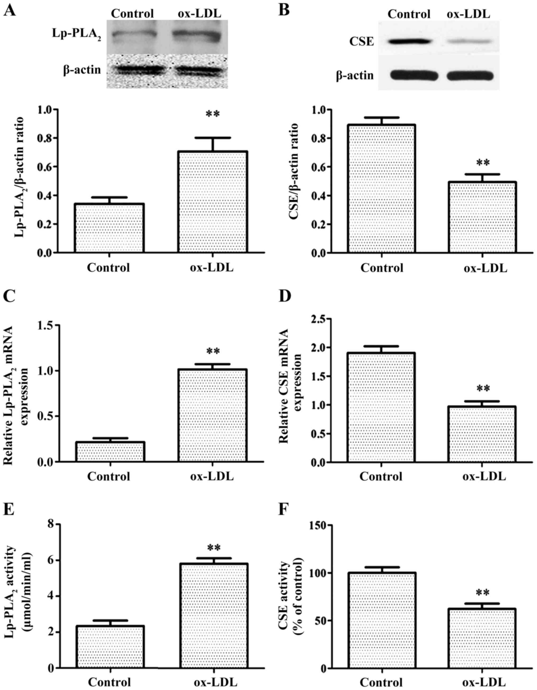
ox-LDL increases the expression level
of Lp-PLA2 but decreases the expression of CSE
First, to determine whether ox-LDL can increase the
levels of Lp-PLA2, RT-PCR, western blotting and
Lp-PLA2 activity assays were used to examine mRNA
levels, protein expression levels and Lp-PLA2 activity,
respectively, in THP-1 cells treated with or without ox-LDL.
Control THP-1 cells only exhibited weak expression levels and
decreased activity of Lp-PLA2, whereas treatment with
ox-LDL for 24 h significantly increased the expression level and
activity of Lp-PLA2 (Fig.
1A, C and E).
The production of H2S primarily involves
three constitutively expressed enzymes: CSE, cystathionine
β-synthase and 3-mercaptopyruvate sulfurtransferase (18). CSE is present specifically in the
tissues of the cardiovascular system (19). It was then determined whether ox-LDL
treatment of THP-1 cells affects CSE expression levels. Under
normal conditions, THP-1 exhibited high CSE expression levels and
activity, whereas after ox-LDL treatment for 24 h, CSE expression
levels and activity were significantly decreased (Fig. 1B, D and F).
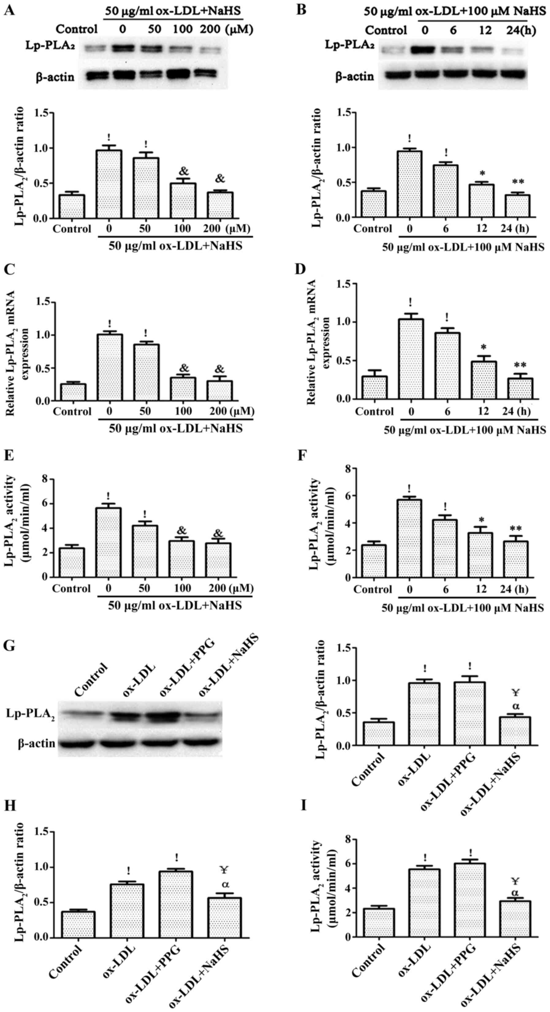
Role of H2S in the
expression level of Lp-PLA2 induced by ox-LDL
In order to confirm the effect of H2S on
Lp-PLA2 expression levels and activity in ox-LDL-induced
THP-1 cells, THP-1 cells were pretreated with NaHS from exogenous
H2S donors for different time points (0, 6, 12 and 24 h)
or different concentration (0, 50, 100 and 200 µM), followed by
treatment with ox-LDL for an additional 24 h. The expression level
and activity of Lp-PLA2 was detected via RT-PCR, western
blotting and Lp-PLA2 activity assays. It was found that
pretreatment of THP-1 cells with NaHS decreased the expression
level and activity of ox-LDL-induced Lp-PLA2 in a time-
and concentration-dependent manner (Fig. 2A-F). In addition, pretreatment with
PPG for 2 h significantly increased ox-LDL-induced
Lp-PLA2 expression levels and activity compared with the
control group (Fig. 2G-I).
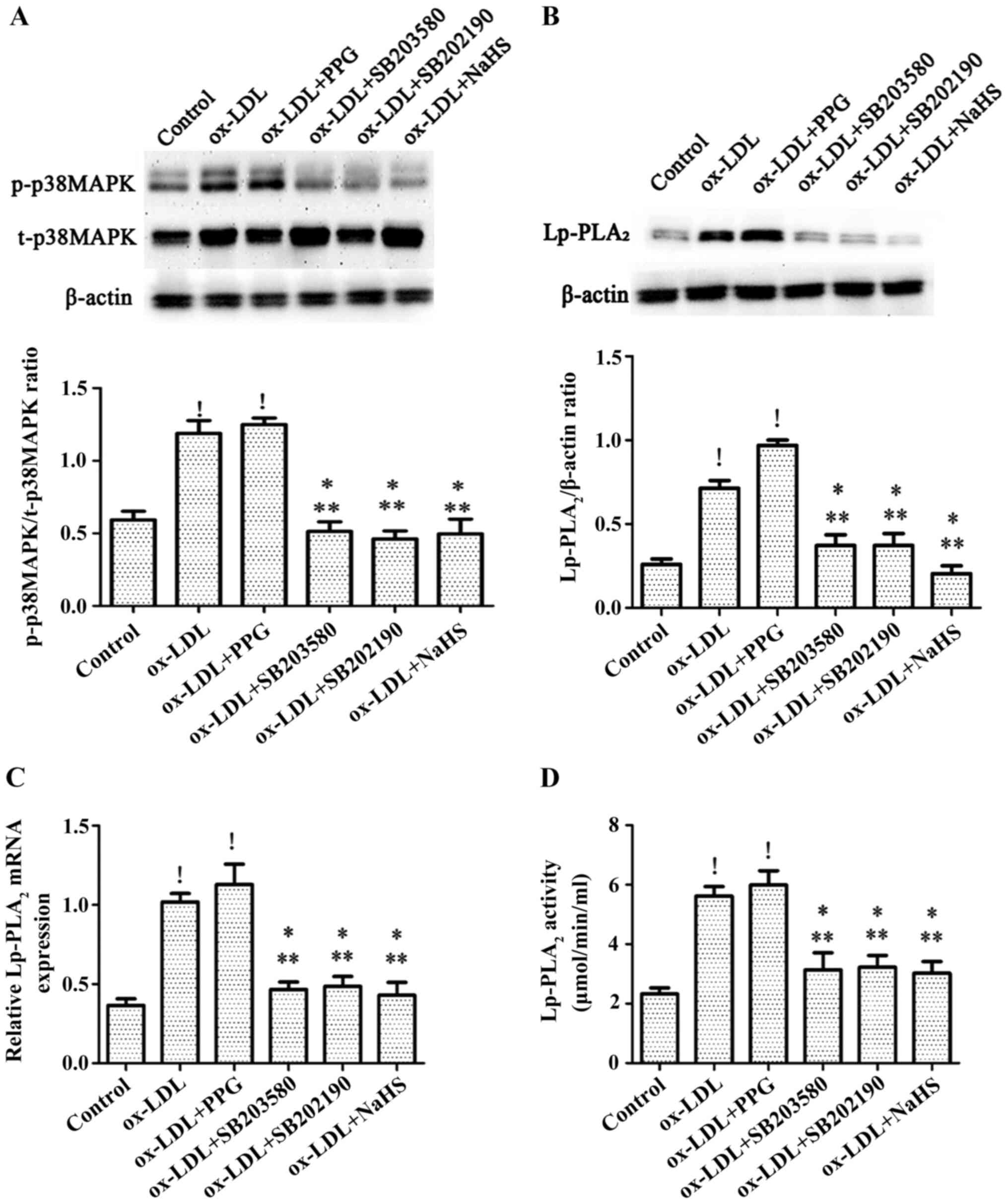
H2S inhibits ox-LDL-induced
expression levels of Lp-PLA2 by blocking p38MAPK
In order to determine whether H2S can
decrease the expression of Lp-PLA2 in THP-1 cells by
inhibiting the p38MAPK pathway, cells were treated with the p38MAPK
specific inhibitors SB203580 and SB202190 for 30 min prior to
incubation with 50 µg/ml ox-LDL for 24 h, following H2S
preincubation for 24 h, or following PPG preincubation for 2 h then
incubation with 50 µg/ml ox-LDL for 24 h. It was observed that
treatment with ox-LDL and pretreatment with PPG followed by ox-LDL
treatment significantly increased the expression level of p38MAPK
compared with the control group (Fig.
3A). Pretreatment with NaHS significantly decreased the
expression level of p38MAPK in THP-1 cells induced by ox-LDL to a
level equivalent to that observed following pretreatment of cells
with the p38MAPK specific inhibitors SB203580 and SB202190, which
also decreased ox-LDL-induced p38MAPK effects (Fig. 3A). In addition, NaHS, SB203580 and
SB202190 significantly decreased the expression level and activity
of Lp-PLA2 in THP-1 cells induced by ox-LDL, whereas
treatment with ox-LDL and pretreatment with PPG prior to ox-LDL
treatment increased the expression level and activity of
Lp-PLA2 in THP-1 cells induced by ox-LDL (Fig. 3B-D).
H2S decreases lipid
accumulation in macrophages by inhibiting the activity of
Lp-PLA2
In order to determine whether H2S
decreases lipid accumulation in macrophages by decreasing
Lp-PLA2 expression levels, THP-1 cells were incubated
with PMA for 72 h to establish a macrophage model, after which the
cells were treated with ox-LDL + SB203580, ox-LDL + SB202190,
ox-LDL + PPG or ox-LDL + Lp-PLA2 siRNA. The transfection
efficiency of the Lp-PLA2 siRNA was verified in
macrophages vs. the NC siRNA, both in the absence and presence of
ox-LDL (Fig. 4I-L). It was found
that Lp-PLA2 siRNA downregulated macrophage and
ox-LDL-induced macrophage Lp-PLA2 expression. ORO
staining was used to assess the uptake of ox-LDL by THP-1 cells.
The results demonstrated that both the ox-LDL and ox-LDL + PPG
treatments increased intracellular lipid accumulation (Fig. 4A-H). By contrast, NaSH, SB253580,
SB202190 and Lp-PLA2 siRNA inhibited intracellular lipid
accumulation. These results indicate that inhibition of
Lp-PLA2 activity can decrease lipid deposition in
macrophages (Fig. 4).
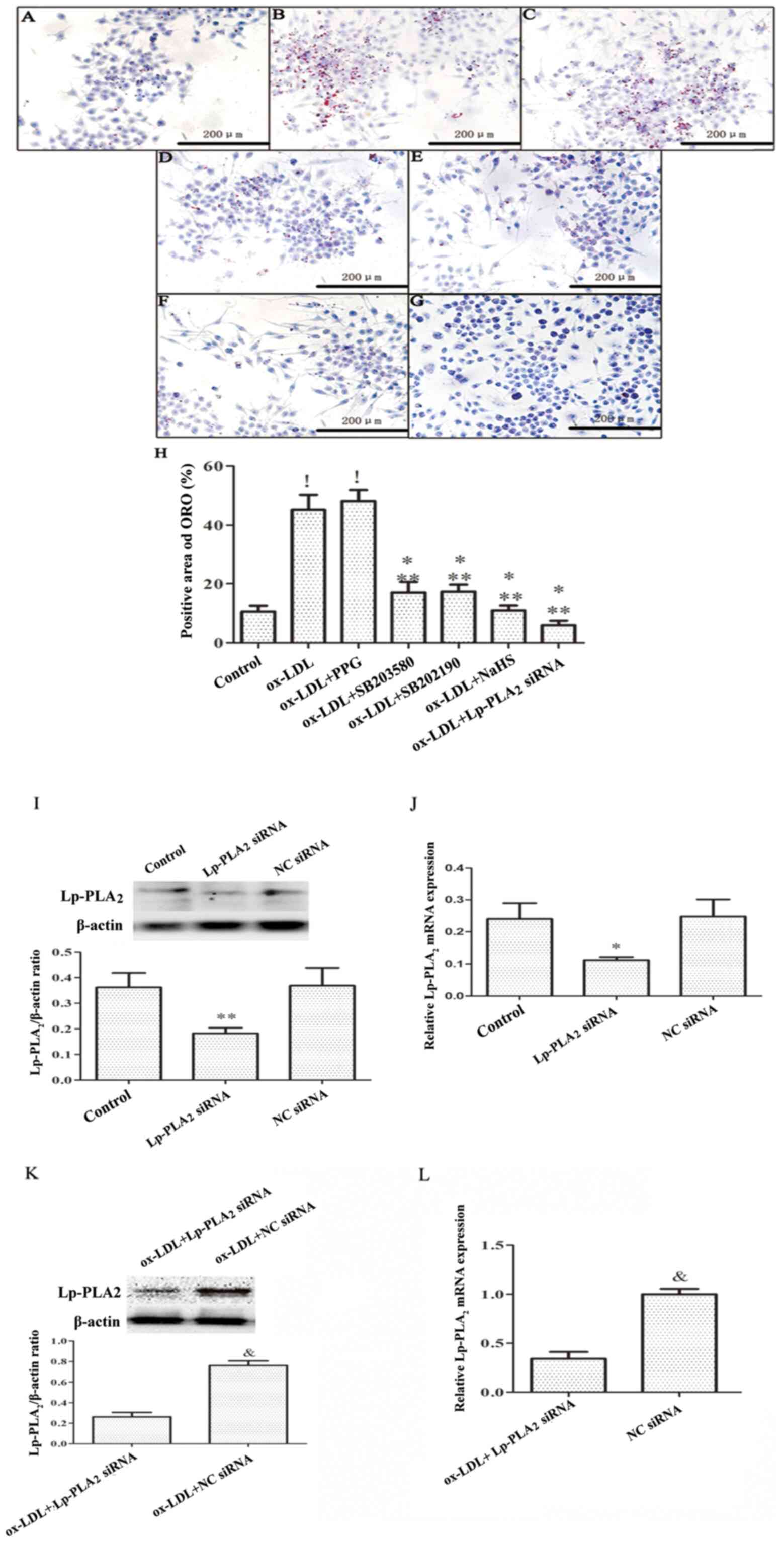 | Figure 4.H2S decreases lipid
accumulation in macrophages by inhibiting the activity of
Lp-PLA2. THP-1 cells were preincubated with PMA for 72 h
to establish a macrophage model. THP-1 macrophages were pretreated
with SB203580 (20 mM) and SB202190 (20 mM) for 30 min, PPG (3 mM)
for 2 h, NaHS (100 µM) for 24 h or Lp-PLA2 siRNA (30 nM)
for 48 h, prior to incubation with ox-LDL (50 µg/ml) for 24 h.
Cells exhibiting lipid accumulation were observed and counted via
light microscopy following ORO staining (magnification, ×40) in the
(A) Control, (B) ox-LDL, (C) ox-LDL + PPG, (D) ox-LDL + SB203580,
(E) ox-LDL + SB202190, (F) ox-LDL + NaHS and (G) ox-LDL +
Lp-PLA2 siRNA groups. Images are representative of six
independent repeats. (H) Quantitative analysis of the positive area
of ORO (%) in each group. Detection of Lp-PLA2 siRNA
transfection by (I) western blotting and (J) RT-PCR. THP-1
macrophages were pretreated with Lp-PLA2 siRNA (30 nM)
or NC siRNA(30 nM) for 48 h. (K) Western blotting and (L) RT-PCR
were conducted to detect the effect of Lp-PLA2 siRNA on
inhibiting the expression of Lp-PLA2 in THP-1
macrophages induced by ox-LDL. !P<0.01 vs. control;
*P<0.05 vs. ox-LDL; **P<0.01 vs. ox-LDL + PPG;
&P<0.01 vs. ox-LDL + Lp-PLA2 siRNA.
Lp-PLA2, lipoprotein-associated phospholipase A2; PMA,
phorbol-12-myristate-13-acetate; PPG, DL-propargylglycine; ox-LDL,
oxidized low-density lipoprotein; ORO, oil red O; RT, reverse
transcription; siRNA, small interfering RNA; NC, negative
control. |
Discussion
Lp-PLA2 is an independent risk factor for
the development of AS and is notably associated with the occurrence
of numerous types of cardiovascular events, such as acute
myocardial infarction (20) and
unstable angina (21). Therefore,
Lp-PLA2 is considered to be a potential therapeutic
target for AS. In the vessel wall, the interaction between
Lp-PLA2 and ox-LDL produces oxidized fatty acids and
lysophosphatidylcholine, which are potent atherogenic factors
(20). Moreover, H2S
exerts notable cardiovascular protective effects, particularly in
AS (15,22). The present study investigated the
effects of H2S on the expression level of
Lp-PLA2 in ox-LDL-treated THP-1 macrophages to elucidate
its effect on the formation of foam cells.
Previous studies have demonstrated that a poor lipid
profile (i.e., high levels of ox-LDL and triacylglycerol) in
circulating blood can significantly increase the expression level
and activity of Lp-PLA2 (23,24).
The present study demonstrated that exposing THP-1 cells to ox-LDL
significantly inhibited the expression level and activity of
Lp-PLA2, which is consistent with the results of Wang
et al (8). CSE is not only
important for the vascular system but is also the primary
H2S-producing enzyme in macrophages. Knocking out CSE
can decrease plasma H2S levels and inhibit the
CSE-H2S pathway in macrophages, accelerating the
progression of AS in apoE−/− mice (25,26).
Based on previous research, the present study demonstrated that
ox-LDL can decrease the expression level and activity of CSE in
THP-1 cells.
H2S is the third most crucial
cardiovascular mediator after nitric oxide and carbon monoxide, and
has been shown to prevent AS (27).
When NaHS is dissolved in water, it acts as an exogenous
H2S donor, releasing HS− and forming
H2S with H+, with the H2S
concentration ~33% of the initial mass of NaHS (28). Exogenous NaHS supplementation can
prevent the conversion of macrophages into foam cells by increasing
the level of H2S, and interfering with the production
inflammatory cytokines and the occurrence of oxidative stress
(29,30). Lp-PLA2 is involved in
vascular inflammation and oxidative stress in macrophages, which
contributes to macrophage-derived foam cell formation (16). In the present study, H2S
downregulated the expression level and activity of
Lp-PLA2 in THP-1 monocytes induced by ox-LDL, whereas
PPG significantly upregulated the effect of ox-LDL on the
expression level and activity of Lp-PLA2 in THP-1
monocytes.
Previous reports demonstrated that ox-LDL can
upregulate Lp-PLA2 expression levels due to its oxidized
phospholipids (oxPCs) (8), with
ox-LDL/oxPCs increasing the generation of inflammatory cytokines in
multiple types of inflammatory responses (31). oxPCs have been shown to activate
numerous signaling pathways, such as the p38MAPK and JNK pathways
(32). The p38MAPK pathway
participates in a number of cellular processes, including
inflammation, differentiation, cell growth, cell cycle and cell
death (33). A number of studies
have revealed that p38MAPK pathway activation mediates numerous
mechanisms that enhance the pathogenesis of chronic inflammatory
disease development, particularly in macrophages with respect to
inhibiting the hardening of arteries in AS (34,35).
The p38MAPK pathway has been shown to serve a key role in ox-LDL
induction by upregulating Lp-PLA2 expression levels and
decreasing the uptake of ox-LDL in THP-1 cells (8). Therefore, effective blocking of the
p38MAPK pathway can decrease the activity of Lp-PLA2.
H2S can also decrease cardiovascular inflammation by
decreasing p38MPAK pathway activity (36). In addition, inflammation and
apoptosis can result in the production of oxPCs by activating cAMP
response elements (37); this
effect can be blocked by H2S (38). The results of the present study
demonstrate that H2S can decrease the expression levels
and activity of Lp-PLA2 in THP-1 cells induced by ox-LDL
by inhibiting the p38 MAPK pathway.
H2S can destroy lipid hydroperoxides
(LOOHs) in ox-LDL, attenuate ox-LDL-induced oxidative stress,
improve atherosclerosis and decrease the levels of ox-LDL (39). Previous studies have reported that
LOOH is an essential component of oxPCs and serves a key role in
the upregulation of Lp-PLA2 expression levels (8,40,41).
The use of PPG to decrease endogenous H2S production can
increase plasma lipid levels and oxidative stress, thereby
aggravating the occurrence of AS (42). In the present study, ORO staining
was performed to assess the lipid content in foam cells and it was
observed that treatment with H2S and p38MAPK inhibitors,
as well as with Lp-PLA2 siRNA, significantly decreased
the lipid content in foam cells. The results of the present study,
combined with those of Zhao et al (29) and Wang et al (43), indicate that H2S can
decrease the activity of Lp-PLA2 in macrophages induced
by ox-LDL by blocking the p38MAPK pathway, thereby decreasing lipid
accumulation.
In summary, these results provide novel insights
into the association between H2S and Lp-PLA2
in AS. H2S significantly decreased the expression level
and activity of Lp-PLA2 in THP-1 cells induced by
ox-LDL. Secondly, H2S may decrease lipid accumulation in
ox-LDL-induced macrophages by decreasing Lp-PLA2
activity. Finally, H2S was shown to decrease the
expression level and activity of Lp-PLA2 in THP-1 cells
induced by ox-LDL by inhibiting the p38MAPK signalling pathway.
Although the differential effects of H2S appear to be
mediated by a typical signal cascade, the complexity of the
signaling route has yet to be elucidated. Thus, the underlying
mechanism of the anti-AS activity of H2S should be the
focus of future investigations.
There were limitations to the present study; only
cell changes were observed, and no animal experiments were
performed. Therefore, it is not clear that the effect of hydrogen
sulfide on atherosclerosis by regulating the expression of
Lp-PLA2 in the overall model is worthy of further
study.
Acknowledgements
Not applicable.
Funding
The present work was supported by grants from the
National Natural Science Foundation of China (grant no. 81700306),
the Natural Science Foundation of Hunan Province (grant no.
2018JJ3469) and the China Postdoctoral Science Foundation (grant
no. 2017M622588).
Availability of data and materials
The datasets used and/or analyzed during the current
study are available from the corresponding author on reasonable
request.
Authors' contributions
HJH and ZSJ conceived and led the experimental
design and participated in the writing of the manuscript. HJH, JQ,
CZ, ZHT and SLQ performed the experiments. HJH and JQ conducted
literature searches and completed the verification and revision of
important knowledge content. HJH and ZSJ performed the final
verification and proofreading of the article and are responsible
for confirming the authenticity of the data in this section. All
authors have read and approved the final manuscript.
Ethics approval and consent to
participate
Not applicable.
Patient consent for publication
Not applicable.
Competing interests
The authors declare that they have no competing
interests.
References
|
1
|
Arroyo-Quiroz C, O'Flaherty M,
Guzman-Castillo M, Capewell S, Chuquiure-Valenzuela E,
Jerjes-Sanchez C and Barrientos-Gutierrez T: Explaining the
increment in coronary heart disease mortality in Mexico between
2000 and 2012. PLoS One. 15:e02429302020. View Article : Google Scholar : PubMed/NCBI
|
|
2
|
Cai X, Zhang Y, Li M, Wu JH, Mai L, Li J,
Yang Y, Hu Y and Huang Y: Association between prediabetes and risk
of all cause mortality and cardiovascular disease: Updated
meta-analysis. BMJ. 370:m22972020. View Article : Google Scholar : PubMed/NCBI
|
|
3
|
Zalewski A and Macphee C: Role of
lipoprotein-associated phospholipase A2 in atherosclerosis:
Biology, epidemiology, and possible therapeutic target.
Arterioscler Thromb Vasc Biol. 25:923–931. 2005. View Article : Google Scholar : PubMed/NCBI
|
|
4
|
Libby P: Inflammation in atherosclerosis.
Nature. 420:868–874. 2002. View Article : Google Scholar : PubMed/NCBI
|
|
5
|
Robichaux WG III, Mei FC, Yang W, Wang H,
Sun H, Zhou Z, Milewicz DM, Teng BB and Cheng X: Epac1 (Exchange
Protein Directly Activated by cAMP 1) upregulates LOX-1 (Oxidized
Low-Density Lipoprotein Receptor 1) to promote foam cell formation
and atherosclerosis development. Arterioscler Thromb Vasc Biol.
40:e322–e335. 2020. View Article : Google Scholar : PubMed/NCBI
|
|
6
|
Wang Y, Li Z, Liu B, Wu R, Gong H, Su Z
and Zhang S: Isoborneol attenuates low-density lipoprotein
accumulation and foam cell formation in macrophages. Drug Des Devel
Ther. 14:167–173. 2020. View Article : Google Scholar : PubMed/NCBI
|
|
7
|
Hou L, Chen S, Yu H, Lu X, Chen J, Wang L,
Huang J, Fan Z and Gu D: Associations of PLA2G7 gene polymorphisms
with plasma lipoprotein-associated phospholipase A2 activity and
coronary heart disease in a Chinese Han population: The Beijing
atherosclerosis study. Hum Genet. 125:11–20. 2009. View Article : Google Scholar : PubMed/NCBI
|
|
8
|
Wang WY, Li J, Yang D, Xu W, Zha RP and
Wang YP: OxLDL stimulates lipoprotein-associated phospholipase A2
expression in THP-1 monocytes via PI3K and p38 MAPK pathways.
Cardiovasc Res. 85:845–852. 2010. View Article : Google Scholar : PubMed/NCBI
|
|
9
|
Keleşoğlu M, Kızılay F, Barutçuoğlu B,
Başol G, Saraç F, Mutaf I and Semerci B: The relationship between
lipoprotein-associated phospholipase A2 with cardiovascular risk
factors in testosterone deficiency. Turk J Urol. 44:103–108. 2018.
View Article : Google Scholar : PubMed/NCBI
|
|
10
|
Nègre-Salvayre A, Augé N, Camaré C,
Bacchetti T, Ferretti G and Salvayre R: Dual signaling evoked by
oxidized LDLs in vascular cells. Free Radic Biol Med. 106:118–133.
2017. View Article : Google Scholar : PubMed/NCBI
|
|
11
|
Paapstel K, Kals J, Eha J, Tootsi K, Ottas
A, Piir A, Jakobson M, Lieberg J and Zilmer M: Inverse relations of
serum phosphatidylcholines and lysophosphatidylcholines with
vascular damage and heart rate in patients with atherosclerosis.
Nutr Metab Cardiovasc Dis. 28:44–52. 2018. View Article : Google Scholar : PubMed/NCBI
|
|
12
|
Uchida Y and Kameda N: Visualization of
lipid components in human coronary plaques using color fluorescence
angioscopy. Circ J. 74:2181–2186. 2010. View Article : Google Scholar : PubMed/NCBI
|
|
13
|
Teoh JP, Li X, Simoncini T, Zhu D and Fu
X: Estrogen-mediated gaseous signaling molecules in cardiovascular
disease. Trends Endocrinol Metab. 31:773–784. 2020. View Article : Google Scholar : PubMed/NCBI
|
|
14
|
Mao Z, Huang Y, Zhang Z, Yang X, Zhang X,
Huang Y, Sawada N, Mitsui T, Takeda M and Yao J: Pharmacological
levels of hydrogen sulfide inhibit oxidative cell injury through
regulating the redox state of thioredoxin. Free Radic Biol Med.
134:190–199. 2019. View Article : Google Scholar : PubMed/NCBI
|
|
15
|
Wen YD, Wang H and Zhu YZ: The Drug
developments of hydrogen sulfide on cardiovascular disease. Oxid
Med Cell Longev. 2018:40103952018. View Article : Google Scholar : PubMed/NCBI
|
|
16
|
Zheng H, Cui D, Quan X, Yang W, Li Y,
Zhang L and Liu E: Lp-PLA2 silencing protects against
ox-LDL-induced oxidative stress and cell apoptosis via Akt/mTOR
signaling pathway in human THP1 macrophages. Biochem Biophys Res
Commun. 477:1017–1023. 2016. View Article : Google Scholar : PubMed/NCBI
|
|
17
|
Wang XY, Yang CT, Zheng DD, Mo LQ, Lan AP,
Yang ZL, Hu F, Chen PX, Liao XX and Feng JQ: Hydrogen sulfide
protects H9c2 cells against doxorubicin-induced cardiotoxicity
through inhibition of endoplasmic reticulum stress. Mol Cell
Biochem. 363:419–426. 2012. View Article : Google Scholar : PubMed/NCBI
|
|
18
|
Kimura H: Hydrogen sulfide: Its
production, release and functions. Amino Acids. 41:113–121. 2011.
View Article : Google Scholar : PubMed/NCBI
|
|
19
|
Olson KR: The therapeutic potential of
hydrogen sulfide: Separating hype from hope. Am J Physiol Regul
Integr Comp Physiol. 301:R297–R312. 2011. View Article : Google Scholar : PubMed/NCBI
|
|
20
|
Chen BF, Deng Y, Xu X, Ma SC, Tang LQ,
Chen JF, Sun WQ, Liu SF and Liang JR: Effect of selective thrombus
aspiration on serum lipoprotein-associated phospholipase A2 in
patients with ST-elevation myocardial infarction undergoing primary
percutaneous coronary intervention with high thrombus burden. Acta
Cardiol Sin. 34:233–241. 2018.PubMed/NCBI
|
|
21
|
Yang L, Cong HL, Wang SF and Liu T:
AMP-activated protein kinase mediates the effects of
lipoprotein-associated phospholipase A2 on endothelial dysfunction
in atherosclerosis. Exp Ther Med. 13:1622–1629. 2017. View Article : Google Scholar : PubMed/NCBI
|
|
22
|
Wang ZJ, Wu J, Guo W and Zhu YZ:
Atherosclerosis and the hydrogen sulfide signaling
pathway-therapeutic approaches to disease prevention. Cell Physiol
Biochem. 42:859–875. 2017. View Article : Google Scholar : PubMed/NCBI
|
|
23
|
Nima B, Nasli-Esfahani E, Djafarian K,
Qorbani M, Hedayati M, Mishani MA, Faghfoori Z, Ahmaripour N and
Hosseini S: The beneficial effects of alpha lipoic acid
supplementation on Lp-PLA2 mass and its distribution between HDL
and apoB-containing lipoproteins in type 2 diabetic patients: A
randomized, double-blind, placebo-controlled trial. Oxid Med Cell
Longev. 2020:58508652020.PubMed/NCBI
|
|
24
|
Ayşegül KT, Sema U, Yalçin AU, Sahin G,
Temiz G, Kara M, Temel HE, Demirkan ES, Colak E and Colak O:
Effects of lipoprotein-associated phospholipase A2 on
arginase/nitric oxide pathway in hemodialysis patients. Ren Fail.
34:738–743. 2012. View Article : Google Scholar : PubMed/NCBI
|
|
25
|
Mani S, Li H, Untereiner A, Wu L, Yang G,
Austin RC, Dickhout JG, Lhoták Š, Meng QH and Wang R: Decreased
endogenous production of hydrogen sulfide accelerates
atherosclerosis. Circulation. 127:2523–2534. 2013. View Article : Google Scholar : PubMed/NCBI
|
|
26
|
Du HP, Li J, You SJ, Wang YL, Wang F, Cao
YJ, Hu LF and Liu CF: DNA methylation in cystathionine-gamma-lyase
(CSE) gene promoter induced by ox-LDL in macrophages and in apoE
knockout mice. Biochem Biophys Res Commun. 469:776–782. 2016.
View Article : Google Scholar : PubMed/NCBI
|
|
27
|
Lv B, Chen S, Tang C, Jin H, Du J and
Huang Y: Hydrogen sulfide and vascular regulation-An update. J Adv
Res. 27:85–97. 2020. View Article : Google Scholar : PubMed/NCBI
|
|
28
|
Beauchamp RO Jr, Bus JS, Popp JA, Boreiko
CJ and Andjelkovich DA: A critical review of the literature on
hydrogen sulfide toxicity. Crit Rev Toxicol. 13:25–97. 1984.
View Article : Google Scholar : PubMed/NCBI
|
|
29
|
Zhao ZZ, Wang Z, Li GH, Wang R, Tan JM,
Cao X, Suo R and Jiang ZS: Hydrogen sulfide inhibits
macrophage-derived foam cell formation. Exp Biol Med (Maywood).
236:169–176. 2011. View Article : Google Scholar : PubMed/NCBI
|
|
30
|
Liu Z, Han Y, Li L, Lu H, Meng G, Li X,
Shirhan M, Peh MT, Xie L, Zhou S, et al: The hydrogen sulfide
donor, GYY4137, exhibits anti-atherosclerotic activity in high fat
fed apolipoprotein E(−/-) mice. Br J Pharmacol. 169:1795–1809.
2013. View Article : Google Scholar : PubMed/NCBI
|
|
31
|
Pidkovka NA, Cherepanova OA, Yoshida T,
Alexander MR, Deaton RA, Thomas JA, Leitinger N and Owens GK:
Oxidized phospholipids induce phenotypic switching of vascular
smooth muscle cells in vivo and in vitro. Circ Res. 101:792–801.
2007. View Article : Google Scholar : PubMed/NCBI
|
|
32
|
Loidl A, Sevcsik E, Riesenhuber G, Deigner
HP and Hermetter A: Oxidized phospholipids in minimally modified
low density lipoprotein induce apoptotic signaling via activation
of acid sphingomyelinase in arterial smooth muscle cells. J Biol
Chem. 278:32921–32928. 2003. View Article : Google Scholar : PubMed/NCBI
|
|
33
|
Kukkonen-Macchi A, Sicora O, Kaczynska K,
Oetken-Lindholm C, Pouwels J, Laine L and Kallio MJ: Loss of
p38gamma MAPK induces pleiotropic mitotic defects and massive cell
death. J Cell Sci. 124:216–227. 2011. View Article : Google Scholar : PubMed/NCBI
|
|
34
|
Kim KS, Cui X, Lee DS, Sohn JH, Yim JH,
Kim YC and Oh H: Anti-inflammatory effect of neoechinulin a from
the marine fungus eurotium sp. SF-5989 through the Suppression of
NF-кB and p38 MAPK pathways in lipopolysaccharide-stimulated
RAW264.7 macrophages. Molecules. 18:13245–13259. 2013. View Article : Google Scholar : PubMed/NCBI
|
|
35
|
Zhai C, Cong H, Hou K, Hu Y, Zhang J,
Zhang Y, Zhang Y and Zhang H: Effects of miR-124-3p regulation of
the p38MAPK signaling pathway via MEKK3 on apoptosis and
proliferation of macrophages in mice with coronary atherosclerosis.
Adv Clin Exp Med. 29:803–812. 2020. View Article : Google Scholar : PubMed/NCBI
|
|
36
|
Wu Z, Peng H, Du Q, Lin W and Liu Y:
GYY4137, a hydrogen sulfidereleasing molecule, inhibits the
inflammatory response by suppressing the activation of nuclear
factorkappa B and mitogenactivated protein kinases in Coxsackie
virus B3infected rat cardiomyocytes. Mol Med Rep. 11:1837–1844.
2015. View Article : Google Scholar : PubMed/NCBI
|
|
37
|
Kronke G, Bochkov VN, Huber J, Gruber F,
Blüml S, Fürnkranz A, Kadl A, Binder BR and Leitinger N: Oxidized
phospholipids induce expression of human heme oxygenase-1 involving
activation of cAMP-responsive element-binding protein. J Biol Chem.
278:51006–51014. 2003. View Article : Google Scholar : PubMed/NCBI
|
|
38
|
Yong QC, Pan TT, Hu LF and Bian JS:
Negative regulation of beta-adrenergic function by hydrogen
sulphide in the rat hearts. J Mol Cell Cardiol. 44:701–710. 2008.
View Article : Google Scholar : PubMed/NCBI
|
|
39
|
Muellner MK, Schreier SM, Laggner H,
Hermann M, Esterbauer H, Exner M, Gmeiner BM and Kapiotis S:
Hydrogen sulfide destroys lipid hydroperoxides in oxidized LDL.
Biochem J. 420:277–281. 2009. View Article : Google Scholar : PubMed/NCBI
|
|
40
|
Fang L, Harkewicz R, Hartvigsen K, Wiesner
P, Choi SH, Almazan F, Pattison J, Deer E, Sayaphupha T, Dennis EA,
et al: Oxidized cholesteryl esters and phospholipids in zebrafish
larvae fed a high cholesterol diet: Macrophage binding and
activation. J Biol Chem. 285:32343–32351. 2010. View Article : Google Scholar : PubMed/NCBI
|
|
41
|
Ibusuki D, Nakagawa K, Asai A, Oikawa S,
Masuda Y, Suzuki T and Miyazawa T: Preparation of pure lipid
hydroperoxides. J Lipid Res. 49:2668–2677. 2008. View Article : Google Scholar : PubMed/NCBI
|
|
42
|
Chen ZF, Zhao B, Tang XY, Li W, Zhu LL,
Tang CS, DU JB and Jin HF: Hydrogen sulfide regulates vascular
endoplasmic reticulum stress in apolipoprotein E knockout mice.
Chin Med J (Engl). 124:3460–3467. 2011.PubMed/NCBI
|
|
43
|
Wang XH, Wang F, You SJ, Cao YJ, Cao LD,
Han Q, Liu CF and Hu LF: Dysregulation of cystathionine gamma-lyase
(CSE)/hydrogen sulfide pathway contributes to ox-LDL-induced
inflammation in macrophage. Cell Signal. 25:2255–2262. 2013.
View Article : Google Scholar : PubMed/NCBI
|