Introduction
Rotator cuff tears (RCTs) represent a prevalent
orthopedic condition that exhibits notable morbidity and affects
the quality of life of numerous individuals, particularly the
elderly (1). The rotator cuff
comprises the tendons of the teres minor, supraspinatus,
infraspinatus and subscapularis muscles. It plays an important role
in regulating the stability and movement of the shoulder joint, and
injury to the rotator cuff may lead to pain, weakness and impaired
joint mobility (2). Treatment for
RCTs typically involves surgical repair, but the healing process
after surgery is complex and may be hindered by various factors,
including the age of the patient, fat infiltration, tear size, and
the quality of the repaired tendons and bones (3).
One of the predominant challenges in rotator cuff
repair is the poor healing ability of the tendon-bone interface
(TBI), which is important for restoring the structural and
functional integrity of the rotator cuff (4). The TBI has a unique structure that is
difficult to replicate and conventional surgery often leads to poor
healing and a high risk of re-tear (5). Metal organic frameworks (MOFs)
represent a category of porous materials constructed from metal
ions or clusters that are connected to each other by organic
ligands through coordination bonds (6). Due to their high surface area,
adjustable pore size and capacity for modification, MOFs have
attracted notable attention in the biomedical field (7). MOFs can be designed to deliver
various therapeutic agents to the site of injury in a controlled
manner (8), and can also serve as
scaffolds to promote cell adhesion, proliferation and
differentiation, which are important processes for TBI healing
(9). In addition, MOFs have been
shown to serve a notable role in alleviating the local accumulation
of reactive oxygen species (ROS), which is also considered to be
important for promoting TBI repair (10–12).
Furthermore, recent studies have highlighted the ability of MOFs to
biomineralize biomolecules, offering an additional layer of
bioactive protection that further supports cellular functions, and
improves the stability and therapeutic efficacy of encapsulated
cargo (13,14).
Strontium (Sr) exhibits notable osteogenic
properties that enhance TBI healing (15,16).
In the present study, a hydrogel was prepared by combining silk
fibroin (SF) and Sr-based MOFs (Sr/MOFs) loaded with alendronate
sodium (Aln) (Sr/MOF-Aln). It was hypothesized that this hydrogel
would exhibit notable osteogenic activity and promote healing at
the TBI in animal models, thus resulting in a structure closer to
the natural TBI.
Materials and methods
Preparation of MOFs
Sr/MOFs were synthesized using a previously reported
method (17). SrCl2 ·
6H2O (0.58 g; 2.2 mmol), H2BDC (0.32 g; 1.93
mmol) and 56 ml dimethylformamide (DMF) were mixed together and
stirred. This solution was then stored in a
polytetrafluoroethylene-lined stainless-steel container at 100°C
for 2 days. After cooling to room temperature, the synthesized MOFs
were filtered, washed with DMF and vacuum-dried at 60°C.
Preparation of SF hydrogel
An SF hydrogel was prepared as described previously
(18). A silkworm cocoon was
boiled in 2 l 0.02 M sodium carbonate solution for 1 h, and
subsequently boiled in 1 l distilled water for 30 min. The cocoon
was then rinsed with fresh distilled water and dried at room
temperature in a culture dish for 2 days. Purified SF was dissolved
in 9.3 M lithium bromide at 70°C for 1 h, which was then filtered
and dialyzed with distilled water at room temperature for 48 h.
After being concentrated with polyethylene glycol (20,000 g/mol),
the dialysis tube was cleaned and SF was collected and stored at
4°C. SF hydrogel was prepared by mixing 1 ml SF solution, 100 µl
HRP solution (Beijing Solarbio Science & Technology Co., Ltd.)
and 65 µl hydrogen peroxide (H2O2) solution
(Shanghai Aladdin Biochemical Technology Co., Ltd.).
Sample characteristics
Sr/MOF morphology was characterized via field
emission scanning electron microscopy (SEM; Sigma 500; Zeiss GmbH)
after being sputter-coated with a thin layer of gold (19). High angle annular dark field
scanning transmission electron microscopy combined with energy
dispersive spectroscopy was used for elemental mapping (20). The powder samples were dispersed in
ethanol via ultrasonication (40 kHz for 5 min at 25°C) and
drop-cast onto a carbon-coated copper grid prior to observation.
The X-ray diffraction (XRD) pattern was recorded on an equipped
X-ray diffractometer (D8 Discover; Bruker Corporation) (21). To determine the optimal MOF
concentration, a series of MOF suspensions with varying
concentrations (200, 300, 400, 500, 600, 700 and 800 µg/ml) were
prepared. The concentration of Sr from these different groups was
evaluated at predetermined time points (days 1, 4, 7, 14 and 21)
using inductively coupled plasma mass spectrometry (ICP-MS) (iCAP
RQ; Thermo Scientific; Thermo Fisher Scientific, Inc.) (22).
Aln loading
A total of 30.5 mM (8.2 g) Aln was dissolved in 50
ml methanol. Subsequently, 2 ml Aln solution (1.22 mM) was mixed
with 100 mg prepared Sr/MOFs and placed on a magnetic stirrer for
48 h (37°C; 600 rpm) (23). The
stirred mixture was then transferred to a centrifuge and
centrifuged for 10 min at 12,000 × g at room temperature. This was
followed by the removal of the supernatant to obtain the
centrifuged product. After repeated washing with deionized water
and methanol, the reactants were dried overnight in a vacuum freeze
dryer to remove the solvent and subsequently stored at room
temperature for further experiments. The success of Aln loading was
confirmed via high-performance liquid chromatography (HPLC) (1260
LC system; Agilent Technologies, Inc.). The chromatographic
separation was performed at a column temperature of 40°C using an
ACQUITY UPLC HSS T3 column (1.8 µm, 2.1×100 mm; Waters
Corporation). The mobile phase consisted of solvent A (10 mM
ammonium acetate containing 0.1% formic acid) and solvent B
(acetonitrile) at a flow rate of 0.300 ml/min, with a sample
injection quantity of 10.0 µl. A total of 400 µg/ml Sr/MOF-Aln was
dissolved in distilled water and HPLC analysis was performed at
different time points (0, 4, 14, 15 and 18 min) to detect the
sustained release of Aln.
Isolation and culture of bone marrow
mesenchymal stem cells (BMSCs)
Following induction of anesthesia with 4%
isoflurane, which was maintained with 2% isoflurane, 6 female
Sprague Dawley (SDa) rats (age, 3–4 weeks; weight, 40–60 g; Charles
River Laboratories, Inc.) were euthanized by cervical dislocation.
The animals were housed in a controlled environment with a
temperature of 22±2°C, relative humidity of 50±10% and under a 12-h
light/dark cycle. The rats were provided ad libitum access
to standard rodent chow and water throughout the duration of the
study. Primary rat BMSCs were subsequently isolated from the
femoral and tibial bone marrow cavities of euthanized rats
(24). BMSCs were cultured in a
complete culture medium comprising DMEM (Gibco; Thermo Fisher
Scientific, Inc.) supplemented with 1% penicillin-streptomycin
(Gibco; Thermo Fisher Scientific, Inc.) and 10% fetal bovine serum
(Wisent Biotechnology). BMSCs were cultured in an incubator at 37°C
with 5% CO2 and the culture medium was changed every 3
days. The BMSCs were obtained for the present study only.
Cell proliferation
Firstly, 96-well plates were pre-coated with 50
µl/well of the respective hydrogels (SF, SF + Sr/MOF and SF +
Sr/MOF-Aln) and incubated at 37°C for 24 h. Subsequently, the wells
were washed with a PBS solution. Primary BMSCs (passage 3) were
then harvested, and a total of 1×10³ BMSCs were added to
each well corresponding to the different treatment groups and
cultured at 37°C in a 5% CO2 incubator. The culture
medium was changed on days 1, 3 and 5. Subsequently, the original
culture medium was discarded and the cells were incubated with
culture medium containing 10% Cell Counting Kit-8 (CCK-8) solution
(Abbkine Scientific Co., Ltd.) for 1 h at 1, 4 and 7 days of
culture. Following this incubation, 100 µl culture medium was taken
from each well and added to a new 96-well plate, after which,
optical density values were detected using a microplate reader
(Model 680; Bio-Rad Laboratories, Inc.) at 450 nm absorbance
(25).
Cytotoxicity assay
BMSCs were seeded into 96-well plates, which were
pre-coated with SF hydrogel at 37°C for 24 h, at a density of
1×10³ cells/well. After 48 h of incubation at 37°C, the
cell survival rate was measured using the Calcein AM/PI dual
staining kit (cat. no. C2015S; Beyotime Biotechnology) according to
the manufacturer's instructions. The culture medium was replaced
with a working solution and incubated at 37°C with 5%
CO2. Subsequently, an inverted fluorescence microscope
(MF53-N; Guangzhou Micro-shot Technology Co., Ltd.) was used to
observe the cells and capture images.
Measurement of intracellular ROS
BMSCs were seeded into 96-well plates, which were
pre-coated with SF hydrogel at 37°C for 24 h, at a density of
1×10³ cells/well. Subsequently, 100 µM
H2O2 was added to the culture medium and
cells were stimulated at 37°C for 24 h, whereas cells in the
control group were cultured in normal medium without
H2O2 treatment. Cells were incubated with a
2′,7′-dichlorodihydrofluorescein diacetate (DCFH-DA) probe
(Beyotime Biotechnology) at 37°C in 5% CO2 for 20 min
and ROS levels were measured according to the manufacturer's
instructions. Fluorescence images were obtained for each group
using an inverted fluorescence microscope (MF53-N; Guangzhou
Micro-shot Technology Co., Ltd.).
In vitro osteogenic test
To determine whether MOF-based hydrogels promoted
osteogenesis, alkaline phosphatase (ALP) activity (at weeks 1 and
2) and extracellular-matrix mineralization (at weeks 2 and 3) were
evaluated using ALP (cat. no. P0321S; Beyotime Biotechnology) and
Alizarin Red staining kits (cat. no. C0148S; Beyotime
Biotechnology), respectively, according to the manufacturer's
instructions. For quantification, a total of 100 µl of the
respective reaction solution from each sample was transferred to a
new 96-well plate. Subsequently, a microplate reader (Model 680;
Bio-Rad Laboratories, Inc.) was utilized to measure the absorbance
at 410 nm for ALP activity and 560 nm for mineralization.
Reverse transcription-quantitative PCR
(RT-qPCR)
BMSCs were seeded into 96-well plates pre-coated
with SF hydrogel at 37°C for 7 days. Total RNA was extracted from
cells using TRIzol® reagent (Invitrogen; Thermo Fisher
Scientific, Inc.) according to the manufacturer's instructions.
cDNA was synthesized from 500 ng total RNA using the PrimeScript™
RT Reagent Kit (Takara Bio, Inc.) according to the manufacturer's
protocol. Subsequently, qPCR was performed using SYBR Green Master
Mix (Takara Bio, Inc.) on a StepOnePlus™ Real-Time PCR System
(Applied Biosystems; Thermo Fisher Scientific, Inc.) to detect the
expression levels of runt-related transcription factor 2 (Runx2),
osteocalcin (OCN), collagen type I α1 chain (COL1A1) and collagen
type II α1 chain (COL2A1). The primers used are presented in
Table I. The amplification
protocol consisted of an initial denaturation step at 95°C for 30
sec, followed by 50 cycles of denaturation at 95°C for 5 sec and
amplification at 60°C for 30 sec. GAPDH was used as the internal
reference gene. All reactions were conducted in triplicate and
relative gene expression levels were calculated using the
2−ΔΔCq method and normalized to the control group
(26).
 | Table I.Primers used for reverse
transcription-quantitative PCR. |
Table I.
Primers used for reverse
transcription-quantitative PCR.
| Primer | Sequence,
5′-3′ |
|---|
| GAPDH |
|
| F |
GGAATCCACTGGCGTCTTCA |
| R |
GGTTCACGCCCATCACAAAC |
| Runx2 |
|
| F |
CCACAGAGCTATTAAAGTGA |
| R |
AACAAACTAGGTTTAGAGTCATCAAGC |
| OCN |
|
| F |
GGTGCAGACCTAGCAGACACCA |
| R |
AGGTAGCGCCGGAGTCTATTCA |
| COL1A1 |
|
| F |
GCATCAGGGTTTCAGAGCA |
| R |
CGTTGGGTCATTTCCACAT |
| COL2A1 |
|
| F |
CCCCTGCAGTACATGCGG |
| R |
CTCGACGTCATGCTGTCTCAAG |
Surgical treatment of the rat RCT
model
A rat RCT model was established to evaluate the
ability of MOF-based hydrogels to promote RCT repair in vivo
according to a previously described method (27). In the present study, 48 female SDa
rats (age, 4–5 months; weight, 300–350 g; Charles River
Laboratories, Inc.) were randomly divided into the following four
groups: i) Control group (n=12); ii) SF group (n=12); iii) SF +
Sr/MOF group (n=12); and iv) SF + Sr/MOF-Aln group (n=12). This
sample size was consistent with previously reported studies on rat
models of RCT and TBI healing (28,29).
The allocation of rats to each group was concealed using opaque
envelopes to ensure that the researchers conducting the experiment
were blinded to group assignments, preventing any bias in the
outcome assessment.
Initially, rats were anesthetized with isoflurane
(induction, 4%; maintenance, 2%). Subsequently, the deltoid muscle
was exposed under sterile conditions and incised to access the
supraspinatus muscle. The supraspinatus tendon was then carefully
dissected from its insertion site on the humeral bone using a
surgical blade. A tunnel was drilled into the greater tubercle of
the humerus and, for experimental groups, the relevant hydrogel was
inserted into the tunnel. Finally, the supraspinatus tendon was
secured back to the greater tubercle using a non-absorbable suture.
Rats in the control group underwent simple suturing of the
supraspinatus tendon to the humerus without the addition of any
hydrogel. Rats in the SF group were subject to suturing with the
incorporation of SF hydrogel at the interface. Similarly, the SF +
Sr/MOF group received suturing with the addition of the SF + Sr/MOF
hydrogel at the interface, whereas the SF + Sr/MOF-Aln group were
sutured with insertion of the SF + Sr/MOF-Aln hydrogel at the
interface. Rats were administered aspirin (100 mg/kg; once daily
postoperatively for 3 days) via oral gavage to manage pain
(30).
The rats were housed in a controlled environment
that was maintained at a temperature of 22±2°C, relative humidity
of 50±10% and subject to a 12-h light/dark cycle. All rats were
provided ad libitum access to standard rodent chow and water
throughout the duration of the study. Rats exhibiting signs of
notable weight loss (>20% baseline body weight), an inability to
move or infection were humanely euthanized immediately. After 8
weeks, the rats were sacrificed by cervical dislocation under
anesthesia, which was induced with 4% isoflurane and maintained
with 2% isoflurane. Of these, 6 rats per group were allocated for
micro-MRI, micro-CT and biomechanical testing, whereas samples from
the remaining 6 rats per group were used exclusively for
histological analysis.
Micro-MRI evaluation
Imaging of the supraspinatus tendon-bone complex was
performed using a 9.4 T BioSpec 94/30 MRI scanner and positron
emission tomography insert (Bruker Corporation). Image acquisition
and reconstruction were processed using ParaVision software
(version 6.0.1; Bruker Corporation). MRI images of each sample were
independently evaluated and graded by two radiologists who were
both blinded to the group allocation of the animals. MRI images of
the supraspinatus tendon-bone complex were assessed using a
semi-quantitative grading system, which included the following
three parameters: i) Signal intensity; ii) tendon thickness; and
iii) tendon retraction. The detailed grading criteria for sample
signal intensity, tendon thickness and tendon retraction are
provided in Table SI (31). To ensure the reliability of MRI
scoring, inter- and intra-observer consistency were evaluated by
calculating the intraclass correlation coefficient based on the
assessments of two independent, blinded observers.
Micro-CT evaluation
Imaging of shoulder joint samples was performed
using a micro-CT system (Quantum GX2; PerkinElmer, Inc.) with a
scanning resolution of 18 µm at a voltage of 50 kV and a current of
100 µA. Image reconstruction and analysis were performed using the
Quantum GX2 software (version 2.0; PerkinElmer, Inc.). The
parameters analyzed included bone volume/tissue volume (BV/TV) and
bone mineral density (BMD), which were quantified according to a
previously described method (32).
Biomechanical testing
Samples of the supraspinatus tendon-humerus complex
were collected from SDa rats for biomechanical testing using a
universal testing machine (UTM6104; Shenzhen SUNS Technology Co.
Ltd.) (27). The maximum load (N)
and load deformation curve at the time of failure were recorded.
The stiffness (N/mm) of each specimen was determined by the slope
of the linear region of the load deformation curve until the point
of maximum load failure (33).
Biomechanical parameters were intentionally reported without
normalization to the cross-sectional area of the samples, as
successful TBI healing is typically accompanied by an increase in
tissue volume; therefore, non-normalized values more accurately
reflect the functional repair outcome (33).
Histopathological observation
On the day of sacrifice in week 8, supraspinatus TBI
samples from each group were fixed in 4% neutral buffered formalin
at room temperature for 48 h. Following dehydration, the samples
underwent decalcification for ~60 days. Subsequently, specimens
were vertically sectioned and embedded in paraffin wax to obtain
tissue blocks with a thickness of ~5 µm using a Leica SP1600
microtome (Leica Microsystems, Inc.). Tissue sections were stained
with toluidine blue at room temperature for 5 min to evaluate
morphological changes in newly formed fibrocartilage (33). Additional sections were stained
with hematoxylin and eosin (H&E) at room temperature for 5 min
to assess the maturation of the repaired rotator cuff tissues.
Images were captured using a light microscope (Leica TCS SP2; Leica
Microsystems, Inc.). Finally, tendon maturation scores were
determined using a modified scoring system adapted from a previous
study (28). Specifically, this
system evaluates five distinct histological parameters:
Cellularity, vascularity, continuity, fibrocartilage and tidemark.
Each parameter is graded on a scale of 0 to 4, yielding a
cumulative total score with a maximum possible value of 20 for each
sample.
Statistical analysis
All data in the present study were analyzed using
SPSS 17.0 statistical software (SPSS, Inc.). Continuous data are
presented as the mean ± standard deviation, and comparisons between
groups were conducted via one-way ANOVA followed by Tukey's post
hoc test. Categorical data, such as the histological maturation
scores and semi-quantitative MRI scores, are presented as the
median and interquartile range, and were evaluated using the
non-parametric Kruskal-Wallis test followed by Dunn's post hoc test
for multiple comparisons. All experiments were independently
repeated three times to ensure the reliability of the results.
P<0.05 was considered to indicate a statistically significant
difference.
Results
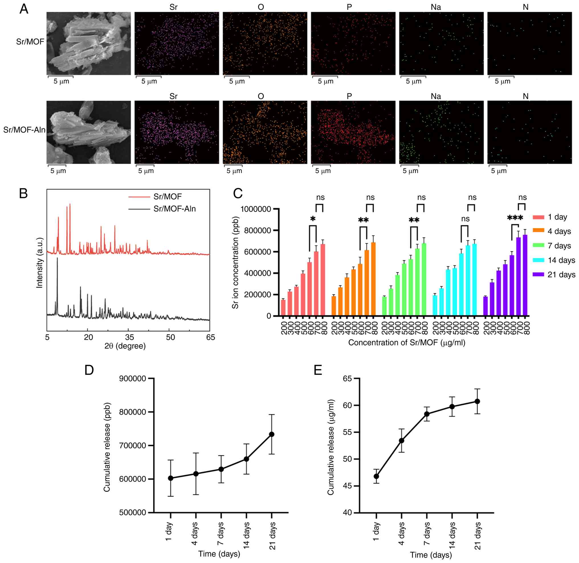
Characteristics of MOFs
In order to obtain information regarding the
microstructure of Sr/MOFs, both Sr/MOF and Sr/MOF-Aln were analyzed
by SEM, as shown in Fig. 1A.
Sr/MOFs were found to be rod-shaped, indicating that they exhibited
notable crystalline morphology. Elemental mapping illustrated the
uniform distribution of each element detected within the Sr/MOFs.
When loaded with Aln, the phosphorus signal of Sr/MOFs markedly
increased, indicating that the loading of Aln onto MOFs was
successful (Fig. 1A). The XRD
pattern of the synthesized Sr/MOFs also reflected a notable crystal
structure, indicating that functional MOFs had been successfully
synthesized (Fig. 1B). Upon
loading with Aln, no shifts in diffraction peaks were observed.
However, observed changes in the Bragg reflectance intensity ratio
may have been caused by variations in interatomic distance and bond
angle or the preferential orientation of Aln particles. The ICP-MS
results demonstrated a general concentration-dependent increase in
Sr ion release as the Sr/MOF concentration rose from 200 to 700
µg/ml. For visual conciseness, only the statistical comparisons
between the higher concentration groups (600 vs. 700 µg/ml and 700
vs. 800 µg/ml) are explicitly indicated in Fig. 1C. Specifically, significant
differences were observed between the 600 and 700 µg/ml groups at
days 1, 4,7 and 21. Notably, there was no statistical difference in
Sr ion release between the 700 and 800 µg/ml groups at days 1, 4,
7, 14 and 21, indicating that the ion release reached a distinct
plateau at concentrations above 700 µg/ml (Fig. 1C). Therefore, a MOF concentration
of 700 µg/ml was selected for use in SF hydrogels in later
experiments. HPLC results revealed that MOFs loaded with Aln
exhibited the same pattern of peaks as Aln, indicating that the
MOFs were successfully loaded with Aln (Fig. S1). Further analysis indicated that
the drug loading capacity of Sr/MOFs with Aln reached 39% (data not
shown). Subsequently, Sr2+ ion release was detected via
HPLC. The cumulative release of Sr2+ ions increased
progressively over the 21-day period, with larger increases in
cumulative release observed on days 14 and 21 compared with earlier
time points (Fig. 1D). The
cumulative release of Aln from Sr/MOF-Aln also increased over the
21-day period; however, this release became more gradual at later
time points compared with earlier time points (Fig. 1E).
 | Figure 1.(A) Scanning electron microscopy and
corresponding elemental mapping of Sr/MOF and Sr/MOF-Aln. (B) X-ray
diffraction patterns of Sr/MOF and Sr/MOF-Aln. (C) Inductively
coupled plasma mass spectrometry analysis of Sr ion concentration
at different Sr/MOF concentrations. (D) Cumulative release of
Sr2+ ions from 700 µg/ml Sr/MOF was measured at
different time points over a 21-day period. (E) Cumulative release
of Aln from Sr/MOF-Aln was measured at different time points over a
21-day period. **P<0.01 and ***P<0.001. Aln, alendronate
sodium; a.u., arbitrary units; MOF, metal organic framework; N,
nitrogen; Na, sodium; ns, no statistically significant difference;
O, oxygen; P, phosphorus; ppb, parts per billion; Sr,
strontium. |
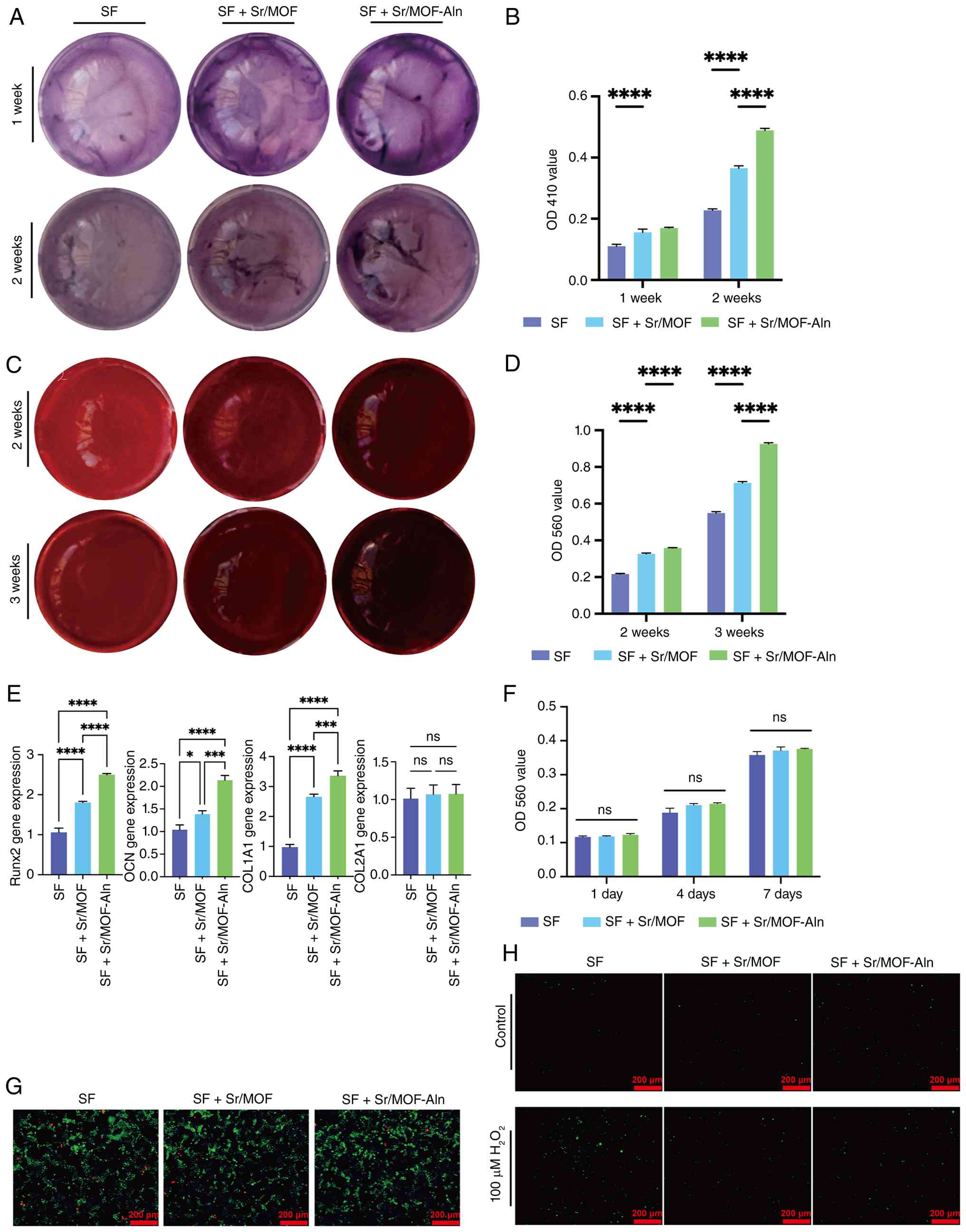
Hydrogel promotes osteogenic
differentiation of BMSCs in vitro
The osteogenic properties of the SF + Sr/MOF group
were investigated by measuring the ALP activity of BMSCs. The
present study found that ALP staining increased in intensity
between 1 and 2 weeks, demonstrating that the degrees of
mineralization and osteogenic differentiation increased with time
(Fig. 2A). In addition, compared
with in the SF group, the ALP activity of BMSCs in the SF + Sr/MOF
group significantly increased after 1 and 2 weeks of co-culture,
and ALP activity in the SF + Sr/MOF-Aln group was significantly
higher than the SF + Sr/MOF group after 2 weeks (Fig. 2B). In addition, the mineralization
activity was evaluated to detect osteogenic differentiation status
in weeks 2 and 3, with the staining intensity of cells gradually
increasing over time (Fig. 2C).
Furthermore, quantitative analysis confirmed a significantly higher
mineralization degree in the SF + Sr/MOF group at both time points,
as evidenced by the increased OD values (Fig. 2D). Additionally, the SF +
Sr/MOF-Aln group exhibited denser mineral nodules and significantly
higher mineralization levels compared with in the SF + Sr/MOF group
at both time points. Furthermore, the expression levels of Runx2,
OCN and COL1A1 gradually increased across the three experimental
groups (Fig. 2E). Specifically,
the SF + Sr/MOF group exhibited significantly increased gene
expression levels compared with the SF group, whereas the SF +
Sr/MOF-Aln group demonstrated the highest expression levels. By
contrast, COL2A1 expression remained consistent across all
treatment groups, demonstrating no statistically significant
differences.
 | Figure 2.(A) ALP staining of BMSCs after
co-culture with hydrogels for 1 and 2 weeks. (B) Quantification of
ALP activity at 1 and 2 weeks. (C) Alizarin Red staining of BMSCs
to detect extracellular matrix mineralization after co-culture of
cells with hydrogels for 2 and 3 weeks. (D) Quantitative
mineralization analysis. (E) Relative mRNA expression levels of
Runx2, OCN, COL1A1 and COL2A1 in different experimental groups. (F)
Analysis of cell proliferation via Cell Counting Kit-8 assay after
co-culture of BMSCs with hydrogels for 1, 4 and 7 days. (G)
Live/dead staining of BMSCs using Calcein AM/PI following a 48-h
incubation with SF hydrogels. (H) Representative images of
2′,7′-dichlorodihydrofluorescein diacetate staining for detection
of intracellular reactive oxygen species. *P<0.05, ***P<0.001
and ****P<0.0001. Aln, alendronate sodium; ALP, alkaline
phosphatase; BMSCs, bone marrow mesenchymal stem cells; COL1A1,
collagen type I α1 chain; COL2A1, collagen type II α1 chain;
H2O2, hydrogen peroxide; MOF, metal organic
framework; ns, no statistically significant difference; OCN,
osteocalcin; Runx2, runt-related transcription factor 2; SF, silk
fibroin; Sr, strontium. |
Biocompatibility of hydrogel
According to CCK-8 assay results, compared with in
the SF group, the SF + Sr/MOF and SF + Sr/MOF-Aln groups
demonstrated no significant differences in proliferation rate after
incubation for 1, 4 and 7 days (Fig.
2F). Additionally, according to the results of calcein AM/PI
staining, no notable differences in cell survival rate were
observed between groups after 48 h of treatment (Fig. 2G).
Antioxidant properties of
hydrogels
The present study subsequently investigated whether
SF + Sr/MOF or SF + Sr/MOF-Aln could reduce the increase in ROS
caused by H2O2. Analysis of DCFH-DA staining
revealed that the proportion of cells that stained positive for ROS
following H2O2 treatment were increased
compared with in the untreated control group. In addition, compared
with the H2O2 + SF group, the relative number
of BMSCs that stained positively for ROS was markedly reduced in
the H2O2 + SF + Sr/MOF and SF + Sr/MOF-Aln
groups (Fig. 2H).
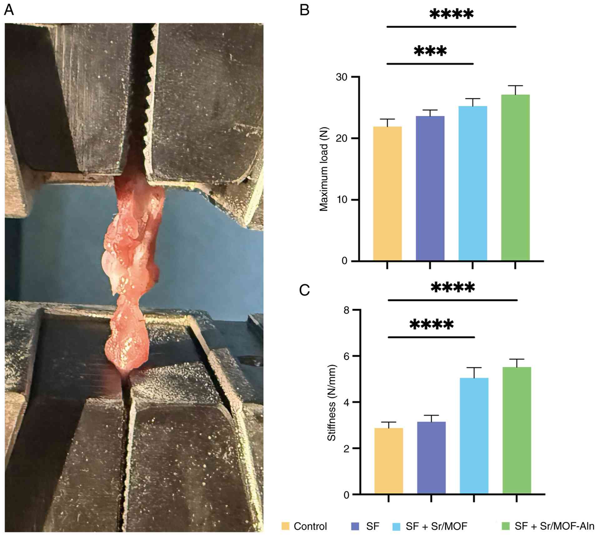
Biomechanical testing
Biomechanical testing of the collected SDa rat
supraspinatus tendon-humerus complexes was performed via a
universal testing machine (Fig.
3A). After 8 weeks of rotator cuff tendon repair, the maximum
load values in the SF + Sr/MOF and SF + Sr/MOF-Aln groups were
significantly increased compared with in the control group
(Fig. 3B). The stiffness of
rotator cuffs across the four groups also showed similar results;
the stiffness values gradually increased from the control group to
the SF + Sr/MOF-Aln group, and a significant difference was
observed between the control group and the SF + Sr/MOF group
(Fig. 3C). In addition, a
significant increase in rotator cuff stiffness was observed between
the control group and the SF + Sr/MOF-Aln group.
Imaging evaluation
A total of 8 weeks after rotator cuff surgery, rats
were examined using a micro-MRI system to observe the healing of
the TBI (Fig. 4A). Subsequently,
CT imaging of rat shoulder joints was used to examine bone repair
at the TBI (Fig. 4C). Analysis of
the regeneration status of tendons was evaluated using an MRI
scoring system, in which higher MRI scores corresponded with poorer
TBI healing. The control group scored the highest, followed by the
SF and SF + Sr/MOF groups, whereas the SF + Sr/MOF-Aln group had
the lowest MRI score (Fig. 4B).
Notably, compared with in the control group, the MRI scores of the
SF + Sr/MOF and SF + Sr/MOF-Aln groups were significantly
decreased. However, the difference between the SF group and the
control group was not statistically significant, and no significant
differences were observed among the three treatment groups (SF, SF
+ Sr/MOF and SF + Sr/MOF-Aln) (Fig.
4B). Micro-CT images were used to generate 3D reconstructions
of the humerus bones of rats in order to evaluate the growth of new
bone tissue in each treatment group. There was no statistically
significant difference in BV/TV and BMD between the control group
and the SF group (Fig. 4D and E).
However, the BV/TV and BMD values of the SF + Sr/MOF group were
significantly increased compared with in the control group and the
BMD of the SF + Sr/MOF-Aln group was significantly increased
compared with the SF + Sr/MOF group, which indicated that the
loading of Sr/MOFs with Aln was therapeutically effective.
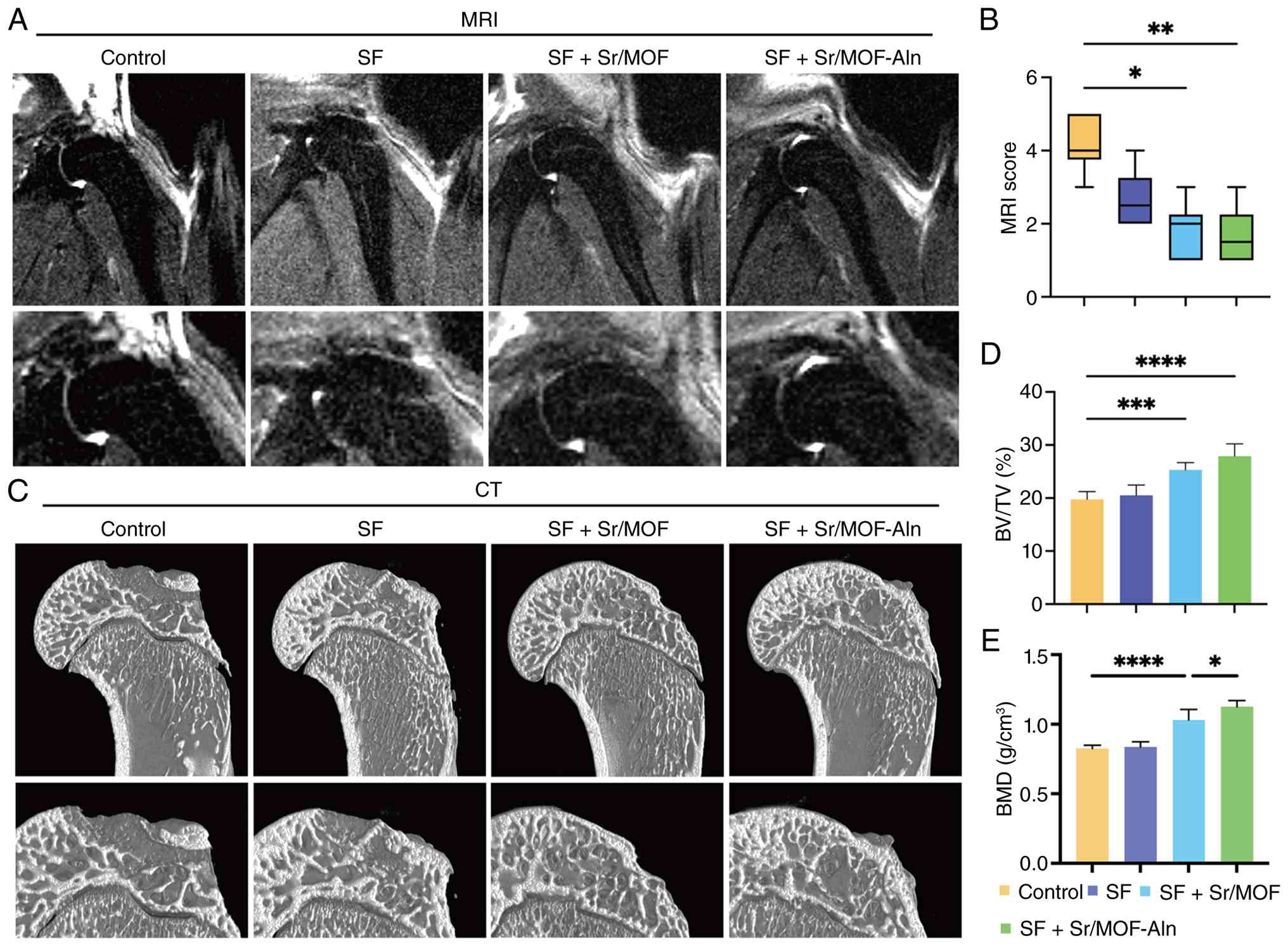 | Figure 4.(A) Representative micro-MRI images
of the supraspinatus tendon-bone complex and (B) MRI scores in the
control, SF, SF + Sr/MOF and SF + Sr/MOF-Aln groups, with higher
scores indicating poorer healing. (C) Representative micro-CT
images of new bone formation close to the hydrogel implantation
site. Quantitative analysis of (D) BV/TV and (E) BMD. *P<0.05,
**P<0.01, ***P<0.001 and ****P<0.0001. Aln, alendronate
sodium; BMD, bone mineral density; BV/TV, bone volume/tissue
volume; MOF, metal organic framework; SF, silk fibroin; Sr,
strontium. |
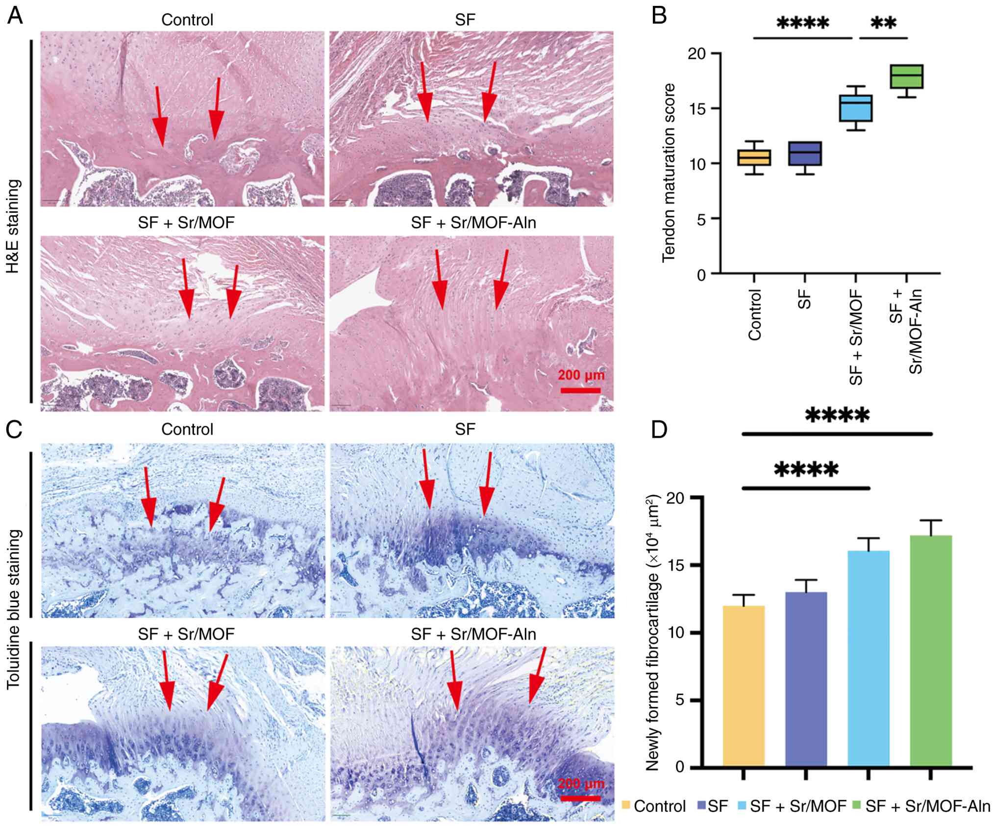
Histological analysis
In order to observe the microstructure after
surgery, the local tissues were stained and analyzed. H&E
staining indicated that collagen fibers in the SF + Sr/MOF-Aln
group appeared to be more organized compared with the other
treatment groups (Fig. 5A).
According to tendon maturation score, the SF + Sr/MOF group showed
a significant increase in tendon maturity 8 weeks after RCT surgery
compared with in the control group (Fig. 5B). Toluidine blue staining was used
to evaluate newly formed fibrocartilage, as shown in Fig. 5C. While cartilage matrix deposition
was limited in the control group, the SF + Sr/MOF and SF +
Sr/MOF-Aln groups exhibited a robust and continuous fibrocartilage
layer. After 8 weeks, the fibrocartilage area of the SF + Sr/MOF
and SF + Sr/MOF-Aln groups was significantly increased compared
with the control group (Fig. 5D).
These results indicated that, compared with in the other treatment
groups, the SF + Sr/MOF-Aln group may have demonstrated an improved
ability to repair RCTs.
Discussion
In the present study, an SF-based hydrogel was
prepared and combined with Sr/MOFs loaded with or without Aln for
use in RCT repair. The therapeutic efficacy of different hydrogel
treatments was validated through comprehensive in vitro and
in vivo evaluations.
Sr/MOFs exhibited a clear rod-shaped morphology,
uniform element distribution and stable crystal structure, as well
as high Aln-loading efficiency and controlled Sr2+ ion
release. In vitro experiments revealed that SF + Sr/MOF and
SF + Sr/MOF-Aln hydrogels exhibited notable biocompatibility,
enhanced osteogenic differentiation and demonstrated marked
antioxidant activity. The results of the present study suggested
that SF + Sr/MOF hydrogels may have primarily mediated RCT repair
by influencing osteogenesis and fibrocartilage formation rather
than chondrogenesis. Therefore, these results highlighted the
functional impact of Sr/MOF-Aln in enhancing bone regeneration at
the TBI.
In vivo, compared with in the SF and control
groups, the SF + Sr/MOF-Aln group achieved higher maximum load and
stiffness, improved bone microstructure, and enhanced organization
of collagen fibers and fibrocartilage at the TBI. Several studies
have investigated the use of Sr-based biomaterials and drug-loaded
systems for promoting TBI healing (29,34,35).
The present study offers unique insights by integrating Sr/MOFs
with a SF hydrogel. The present study was, to the best of our
knowledge, the first study to apply Sr/MOFs for RCT repair and the
results supported the efficacy of Sr/MOFs in promoting TBI healing
by utilizing the notable osteogenic properties of MOFs and
Sr2+ ions. In vitro, Sr2+ ions were
continuously released from Sr/MOF for 21 days, preventing sudden
ion release and ensuring sustained osteogenic stimulation. In
vivo, this sustained release is transformed into enhanced
growth of new bone and formation of fibrocartilage at the TBI
(36). The osteogenic effect of Sr
has been fully supported by previous studies (15,16).
Sr2+ ions can activate the Smad/Runx2 signaling pathway
and upregulate osteogenic gene expression. Other studies (37,38)
have confirmed the ability of Sr to balance the activity of
osteoblasts and osteoclasts, which is necessary for preventing bone
resorption at the repair site. Additionally, Sr exerts notable
antioxidant effects by mitigating ROS-related damage, particularly
in contexts relevant to musculoskeletal tissue repair (10,12),
which is important for TBI healing (11).
MOFs have demonstrated considerable therapeutic
advantages when compared with traditional drug carriers in RCT
repair. The high porosity and notable specific surface area of MOFs
have been shown to facilitate their high drug-loading capacity and
adjustable release kinetics (39),
and MOFs have been reported to maintain their crystal structure
under physiological conditions (39). In addition, MOFs have been shown to
lack cytotoxicity towards mammalian cells, which is consistent with
the in vitro biocompatibility results observed in the
present study (40).
Gao et al (29) explored the ability of Sr-doped
mesoporous bioglass nanoparticles (Sr-MBG) in electrospun fiber
scaffolds to promote healing at the TBI. The approach adopted in
the aforementioned study highlighted the immune-modulatory effects
of Sr-MBG, which promoted macrophage polarization towards the M2
phenotype, and increased the osteogenic and chondrogenic
differentiation of mesenchymal stem cells, improving biomechanical
strength at the TBI. By contrast, the present study demonstrated
that Sr/MOFs integrated with SF hydrogel offer an alternative
approach to TBI healing by providing sustained Sr2+ ion
release and bioactive protection, similarly enhancing regeneration
and improving mechanical properties at the TBI. Additionally, Sun
et al (34) used
dissolvable microneedle patches loaded with diacerein nanoparticles
to mitigate oxidative stress and promote macrophage polarization
towards the M2 phenotype. The results of the aforementioned study
aligned with the findings of the present study, demonstrating
Sr2+ ion-mediated mitigation of ROS-induced damage,
enhancing tendon-bone healing. Furthermore, Baker et al
(35) explored the pharmacological
mobilization of endogenous mesenchymal stem cells for rotator cuff
repair. The findings of the aforementioned study aligned with the
findings of the present study, which showed that Sr/MOF-based
hydrogels could enhance cell differentiation and tissue
regeneration without the need for exogenous cells, suggesting the
therapeutic potential of integrating these hydrogels with
mesenchymal stem cell mobilization strategies for synergistic
effects in tendon-bone healing.
The combination of Aln and Sr/MOFs further enhanced
the efficacy of RCT repair. This was related to the function of
MOFs as drug carriers (39,41),
which may enable localized, sustained delivery of therapeutic
agents directly to the TBI, thereby maintaining a high local drug
concentration while minimizing dissemination to non-target tissues.
In the present study, the synergistic effect of Aln and
Sr2+ ions facilitated bone remodeling and improved the
biomechanical strength of regenerated bone, which was measured
in vivo using micro-CT and biomechanical tests. Yin et
al (42) provided evidence
that Aln treatment in a rat RCT model of osteoporosis improved bone
microstructure and reduced the receptor activator of NF-κB
ligand/osteoprotegerin ratio, thereby inhibiting excessive
osteoclast activity and stabilizing the TBI. This was consistent
with the in vivo results of the present study, which
demonstrated that the SF + Sr/MOF-Aln hydrogel increased bone
growth compared with the SF + Sr/MOF group. Abdalla and Pendegrass
(43) demonstrated that
bisphosphonates can promote TBI healing by reducing bone loss at
tendon attachment points, which is important for preventing suture
anchor dislodgement. In addition, another study emphasized that
when released through local carriers, Aln can regulate local bone
metabolism without producing systemic side effects, thereby
addressing the limitations of systemic Aln administration (44). In general, the aforementioned
studies provided notable evidence supporting the role of Aln in
enhancing RCT healing. The findings of the present study supported
this role by revealing the synergistic effects of Aln and Sr/MOFs
in the hydrogel system.
Although the results of the present study are
encouraging, there are several limitations that need to be
addressed in future research. Notably, the present study focused
primarily on the osteogenic potential of the Sr/MOF-Aln hydrogel
and did not assess tenogenic markers, such as scleraxis and
tenomodulin, which are important for evaluating tendon regeneration
(45). The absence of these
markers from the analyses of the present study therefore limited
the elucidation of cellular events involved in TBI healing.
Although the present study generated quantitative release profiles
for both Sr2+ ions and Aln over a 21-day period, a
correlation analysis between ion or drug release and biological
outcomes was not performed. Future studies should incorporate
correlation analyses to further elucidate how the release kinetics
of Sr2+ ions and Aln influence biological responses and
to establish a clearer material-function relationship. These
analyses would provide valuable insights into the sustained
osteogenic stimulation and therapeutic efficacy of the hydrogel
system used in the present study. Notably, the in vivo
experiments of the present study were only established over 8
weeks. However, other studies have evaluated the effectiveness of
RCT repair at 12 weeks postoperatively to assess long-term
mechanical durability and tissue remodeling at the TBI (29,43).
Furthermore, the present study used a healthy rat model of RCTs,
but patients with RCT often exhibit osteoporosis, which is a key
risk factor for poor TBI healing (42). Testing hydrogels in an RCT model of
osteoporosis is necessary to support their applicability in
high-risk groups. The present study provided new insights into RCT
repair, demonstrating that Sr/MOFs may not only promote bone
formation but also may load drugs to further enhance TBI
regeneration, establishing avenues for the development of Sr/MOFs
that promote TBI healing.
The present study successfully developed
multifunctional composite hydrogels by integrating Sr/MOFs, Aln and
SF, and systematically verified the efficacy of these hydrogels in
RCT repair at the TBI. Both in vitro and in vivo
evaluations systematically confirmed their enhanced efficacy
compared with the control and SF-only groups in cytocompatibility,
osteogenesis, ROS scavenging and subsequent TBI regeneration. To
the best of our knowledge, the present study represented the first
application of Sr/MOFs in RCT repair, leveraging the synergistic
effects of Sr ions, Aln and the drug-delivery capability of MOFs on
TBI healing to address the notable challenge of RCT treatment. The
composite SF + Sr/MOF-Aln hydrogel provided a novel, efficient and
biocompatible strategy for promoting rotator cuff repair, laying a
foundation for the development of advanced biomaterials for
promoting TBI regeneration and demonstrating the promising
translational potential of Sr/MOF hydrogels for use in clinical
orthopedic practice.
Supplementary Material
Supporting Data
Supporting Data
Acknowledgements
The authors would like to thank Dr Jiahao Luo (Hebei
University of Technology) for his assistance in the
experiments.
Funding
The present study was supported by the Foundation of Tianjin
Union Medical Center (grant no. 2020YJ009) and the Tianjin Science
and Technology Major Special Projects (grant nos. 21ZXJBSY00080 and
25ZXSWSY00370).
Availability of data and materials
The raw ICP-MS data generated in the present study
have been deposited in the iProX repository under accession number
PXD077411 or at the following URL: https://www.iprox.cn/page/project.html?id=IPX0016720000.
The other data generated in the present study may be requested from
the corresponding author.
Authors' contributions
MT and TY designed the study. TY and HZ performed
the experiments. ML, YR, YS, WH and MT analyzed data. MT and TY
confirm the authenticity of all the raw data. All authors
contributed to the writing of the manuscript. MT and TY revised the
manuscript. All authors read and approved the final manuscript.
Ethics approval and consent to
participate
All animal procedures were examined and approved by
The Animal Ethics Committee of Nankai University (approval no.
2025-SYDWLL-000288).
Patient consent for publication
Not applicable.
Competing interests
The authors declare that they have no competing
interests.
References
|
1
|
Yamamoto A, Takagishi K, Osawa T, Yanagawa
T, Nakajima D, Shitara H and Kobayashi T: Prevalence and risk
factors of a rotator cuff tear in the general population. J
Shoulder Elbow Surg. 19:116–120. 2010. View Article : Google Scholar : PubMed/NCBI
|
|
2
|
Neviaser A, Andarawis-Puri N and Flatow E:
Basic mechanisms of tendon fatigue damage. J Shoulder Elbow Surg.
21:158–163. 2012. View Article : Google Scholar : PubMed/NCBI
|
|
3
|
Quinlan NJ, Hillyard B, Wheelwright JC,
Miller M, Kawakami J, Tashjian RZ and Chalmers PN: Footprint size
matters: Wider coronal greater tuberosity width is associated with
increased rates of healing after rotator cuff repair. JSES Int.
5:486–492. 2021. View Article : Google Scholar : PubMed/NCBI
|
|
4
|
Zhao S, Su W, Shah V, Hobson D, Yildirimer
L, Yeung KWK, Zhao J, Cui W and Zhao X: Biomaterials based
strategies for rotator cuff repair. Colloids Surf B Biointerfaces.
157:407–416. 2017. View Article : Google Scholar : PubMed/NCBI
|
|
5
|
Turcotte JJ, Kelly M, West M, Lashgari C,
Petre BM and Redziniak DE: Rates of medial and lateral row failure
and risk factors for Re-tear in arthroscopic double row rotator
cuff repair. J Clin Orthop Trauma. 36:1020832022. View Article : Google Scholar : PubMed/NCBI
|
|
6
|
Nguyen LM, Wang Y, Quynh Vu GT, Hoai Ta
QT, Tran DL, Nguyen NH, Van Tran T, Zhang C and Nguyen DH: The
synergy of metal-organic frameworks and biomaterials for bone
tissue engineering: Recent advances, challenges, and future
recommendations. Nanoscale Adv. 7:5479–5500. 2025. View Article : Google Scholar : PubMed/NCBI
|
|
7
|
Yang R, Zheng Y, Zhang Y, Li G, Xu Y,
Zhang Y, Xu Y, Zhuang C, Yu P, Deng L, et al: Bipolar metal
flexible electrospun fibrous membrane based on metal-organic
framework for gradient healing of tendon-to-bone interface
regeneration. Adv Healthc Mater. 11:e22000722022. View Article : Google Scholar : PubMed/NCBI
|
|
8
|
Khafaga DSR, El-Morsy MT, Faried H, Diab
AH, Shehab S, Saleh AM and Ali GAM: Metal-organic frameworks in
drug delivery: Engineering versatile platforms for therapeutic
applications. RSC Adv. 14:30201–30229. 2024. View Article : Google Scholar : PubMed/NCBI
|
|
9
|
Li X, Shu X, Shi Y, Li H and Pei X: MOFs
and bone: Application of MOFs in bone tissue engineering and bone
diseases. Chinese Chemical Letters. 34:1079862023. View Article : Google Scholar
|
|
10
|
Han C, Zhang M, Xu S, Wang C, Li B and
Zhao W: Strontium ranelate-loaded human hair keratin-hyaluronic
acid hydrogel accelerates wound repair with anti-inflammatory and
antioxidant properties. Int J Biol Macromol. 281((Pt 4)):
1365362024. View Article : Google Scholar : PubMed/NCBI
|
|
11
|
Kim RJ, An SH, Gwark JY and Park HB:
Antioxidant effects on hypoxia-induced oxidative stress and
apoptosis in rat rotator cuff fibroblasts. Eur Cell Mater.
41:680–693. 2021. View Article : Google Scholar : PubMed/NCBI
|
|
12
|
Shen X, Fang K, Ru Yie KH, Zhou Z, Shen Y,
Wu S, Zhu Y, Deng Z, Ma P, Ma J and Liu J: High proportion
strontium-doped micro-arc oxidation coatings enhance early
osseointegration of titanium in osteoporosis by anti-oxidative
stress pathway. Bioact Mater. 10:405–419. 2021.PubMed/NCBI
|
|
13
|
Zhou JN, Liu C, Wang Y, Guo Y, Xu XY,
Vuorimaa-Laukkanen E, Koivisto O, Filppula AM, Ye J and Zhang H:
Biomineralize mitochondria in metal-organic frameworks to promote
mitochondria transplantation from non-tumorigenic cells into cancer
cells. Smart Med. 4:e1342025. View Article : Google Scholar : PubMed/NCBI
|
|
14
|
Faheem A, Lawrence MC, Bushra GA, Meli MV
and Blight BA: Metal-organic frameworks as anchors for giant
unilamellar vesicle immobilization. J Mater Chem B. 13:2317–2326.
2025. View Article : Google Scholar : PubMed/NCBI
|
|
15
|
Kuang GM, Yau WP, Lu WW and Chiu KY: Local
application of strontium in a calcium phosphate cement system
accelerates healing of soft tissue tendon grafts in anterior
cruciate ligament reconstruction: Experiment using a rabbit model.
Am J Sports Med. 42:2996–3002. 2014. View Article : Google Scholar : PubMed/NCBI
|
|
16
|
Kuang GM, Yau WP, Lu WW and Chiu KY: Use
of a strontium-enriched calcium phosphate cement in accelerating
the healing of soft-tissue tendon graft within the bone tunnel in a
rabbit model of anterior cruciate ligament reconstruction. Bone
Joint J. 95-B:923–928. 2013. View Article : Google Scholar : PubMed/NCBI
|
|
17
|
Li Z, Peng Y, Xia X, Cao Z, Deng Y and
Tang B: Sr/PTA metal organic framework as a drug delivery system
for osteoarthritis treatment. Sci Rep. 9:175702019. View Article : Google Scholar : PubMed/NCBI
|
|
18
|
Pierantoni L, Ribeiro VP, Costa L, Pina S,
da Silva Morais A, Silva-Correia J, Kundu SC, Motta A, Reis RL and
Oliveira JM: Horseradish peroxidase-crosslinked calcium-containing
silk fibroin hydrogels as artificial matrices for bone cancer
research. Macromol Biosci. 21:e20004252021. View Article : Google Scholar : PubMed/NCBI
|
|
19
|
Wei X, Wang M, Dong X, He Y, Nan W, Ji S,
Zhao M, Chang H, Wei H, Ding D and Chen H: Internal-External
homologous drug-loaded exosome-like nanovesicles released from
Semi-IPN hydrogel enhancing wound healing of
chemoradiotherapy-induced oral mucositis. Int J Nanomedicine.
20:4105–4121. 2025. View Article : Google Scholar : PubMed/NCBI
|
|
20
|
Liu S, Dun C, Yang F, Tung KL, Wierzbicki
D, Ghose S, Chen K, Chen L, Ciora R, Khan MA, et al: A general
flame aerosol route to kinetically stabilized metal-organic
frameworks. Nat Commun. 15:93652024. View Article : Google Scholar : PubMed/NCBI
|
|
21
|
Donnadio A, Paul G, Barbalinardo M,
Ambrogi V, Pettinacci G, Posati T, Bisio C, Vivani R and Nocchetti
M: Immobilization of alendronate on zirconium phosphate
nanoplatelets. Nanomaterials (Basel). 13:7422023. View Article : Google Scholar : PubMed/NCBI
|
|
22
|
Reiner T, Sorbi R, Müller M, Nees T,
Kretzer JP, Rickert M and Moradi B: Blood metal ion release after
primary total knee arthroplasty: A prospective study. Orthop Surg.
12:396–403. 2020. View Article : Google Scholar : PubMed/NCBI
|
|
23
|
Xue X, Yu J, Lu F, Jiang H and Wang X:
Enhancement of cancer chemotherapeutic efficacy via bone-targeted
drug delivery carrier in bone metastases. Drug Des Devel Ther.
15:4455–4468. 2021. View Article : Google Scholar : PubMed/NCBI
|
|
24
|
Zhang W, Zheng L, Yan Y and Shi W: Facile
preparation of multifunctional hydrogels with sustained resveratrol
release ability for bone tissue regeneration. Gels. 10:4292024.
View Article : Google Scholar : PubMed/NCBI
|
|
25
|
Ren J, Duan H, Dong H, Wu S, Du Y, Zhang G
and Zhang A: TAT nanobody exerts antiviral effect against PRRSV in
vitro by targeting viral nucleocapsid protein. Int J Mol Sci.
24:19052023. View Article : Google Scholar : PubMed/NCBI
|
|
26
|
Livak KJ and Schmittgen TD: Analysis of
relative gene expression data using real-time quantitative PCR and
the 2(−Delta Delta C(T)) method. Methods. 25:402–408. 2001.
View Article : Google Scholar : PubMed/NCBI
|
|
27
|
Ren Y, Zhang S, Wang Y, Jacobson DS,
Reisdorf RL, Kuroiwa T, Behfar A, Moran SL, Steinmann SP and Zhao
C: Effects of purified exosome product on rotator cuff tendon-bone
healing in vitro and in vivo. Biomaterials. 276:1210192021.
View Article : Google Scholar : PubMed/NCBI
|
|
28
|
Wu B, Zhang T, Chen H, Shi X, Guan C, Hu J
and Lu H: Exosomes derived from bone marrow mesenchymal stem cell
preconditioned by low-intensity pulsed ultrasound stimulation
promote bone-tendon interface fibrocartilage regeneration and
ameliorate rotator cuff fatty infiltration. J Orthop Translat.
48:89–106. 2024. View Article : Google Scholar : PubMed/NCBI
|
|
29
|
Gao H, Wang L, Lin Z, Jin H, Lyu Y, Kang
Y, Zhu T, Zhao J and Jiang J: Bi-lineage inducible and
immunoregulatory electrospun fibers scaffolds for synchronous
regeneration of tendon-to-bone interface. Mater Today Bio.
22:1007492023. View Article : Google Scholar : PubMed/NCBI
|
|
30
|
The Forsyth Institute, . Administration of
Analgesia in Rats and Mice. The Forsyth Institute; Somerville, MA:
2021, https://www.google.com/url?sa=t&source=web&rct=j&opi=89978449&url=https://forsyth.org/wp-content/uploads/2025/05/Analgesia.pdf&ved=2ahUKEwiso9zGpfeUAxX6hf0HHaJYLz8QFnoECBoQAQ&usg=AOvVaw0XXKs-mz3aP7JdLpbt8-qI
|
|
31
|
Ross D, Maerz T, Kurdziel M, Hein J, Doshi
S, Bedi A, Anderson K and Baker K: The effect of granulocyte-colony
stimulating factor on rotator cuff healing after injury and repair.
Clin Orthop Relat Res. 473:1655–1664. 2015. View Article : Google Scholar : PubMed/NCBI
|
|
32
|
Wang HT, Li J, Ma ST, Feng WY, Wang Q,
Zhou HY, Zhao JM and Yao J: A study on the prevention and treatment
of murine calvarial inflammatory osteolysis induced by
ultra-high-molecular-weight polyethylene particles with
neomangiferin. Exp Ther Med. 16:3889–3896. 2018.PubMed/NCBI
|
|
33
|
Renhao Y, Gen L, Chengyu Z, Yang R, Li G,
Zhuang C, Yu P, Ye T, Zhang Y, Shang P, et al: Gradient bimetallic
ion-based hydrogels for tissue microstructure reconstruction of
tendon-to-bone insertion. Sci Adv. 7:eabg38162021. View Article : Google Scholar
|
|
34
|
Sun J, Chen QZ, Hong AZ, Ju F, Wang HL,
Zhang B, Liu W, Sun YC, Tan J, Yang QQ and Zhou YL: Dissolvable
microneedle-assisted transdermal administration of diacerein
nanoparticles achieved satisfactory therapeutic effects in
tendon-bone insertion repair by reducing oxidative stress and
inflammation. Mater Today Bio. 33:1019992025. View Article : Google Scholar : PubMed/NCBI
|
|
35
|
Baker KC, Fleischer M, Newton MD, Galasso
L, Cavinatto L, Weisz KM, Hartner S, Maerz T, Lammlin L, Baker EA,
et al: Pharmacologic mobilization and chemokine-directed
recruitment of mesenchymal stromal cells to the surgically repaired
rotator cuff. Am J Sports Med. 53:1806–1816. 2025. View Article : Google Scholar : PubMed/NCBI
|
|
36
|
Thomopoulos S, Matsuzaki H, Zaegel M,
Gelberman RH and Silva MJ: Alendronate prevents bone loss and
improves tendon-to-bone repair strength in a canine model. J Orthop
Res. 25:473–479. 2007. View Article : Google Scholar : PubMed/NCBI
|
|
37
|
Ma P, Chen T, Wu X, Hu Y, Huang K, Wang Y
and Dai H: Effects of bioactive strontium-substituted
hydroxyapatite on osseointegration of polyethylene terephthalate
artificial ligaments. J Mater Chem B. 9:6600–6613. 2021. View Article : Google Scholar : PubMed/NCBI
|
|
38
|
Chen T, Wu X, Zhang P, Wu W, Dai H and
Chen S: Strontium-doped hydroxyapatite coating improves
osteo/angiogenesis for ameliorative graft-bone integration via the
macrophage-derived cytokines-mediated integrin signal pathway. ACS
Appl Mater Interfaces. 16:15687–15700. 2024. View Article : Google Scholar : PubMed/NCBI
|
|
39
|
Xie X, Wang Y, Li Z, Yang G, Cheng G, Qin
S, Wang H and Zhu L: Recent advances in gradient biomimetic
scaffolds for tendon-bone interface regeneration. Front Bioeng
Biotechnol. 13:16298162025. View Article : Google Scholar : PubMed/NCBI
|
|
40
|
Zhu C, Lv Y, Qian C, Qian H, Jiao T, Wang
L and Zhang F: Proliferation and osteogenic differentiation of rat
BMSCs on a novel Ti/SiC metal matrix nanocomposite modified by
friction stir processing. Sci Rep. 6:388752016. View Article : Google Scholar : PubMed/NCBI
|
|
41
|
Guan H, Ding F, Xue Y and Zhao J:
Engineering the mechanical characteristics of regenerated silk
fibroin materials: the impact of chemical and physical modification
strategies. Front Chem. 13:16069952025. View Article : Google Scholar : PubMed/NCBI
|
|
42
|
Yin H, Ding Y, Feng Z, Yan Z and Guo A:
Quantitative computed tomography analysis of bone microarchitecture
is associated with rotator cuff healing. J Orthop Surg Res.
20:6702025. View Article : Google Scholar : PubMed/NCBI
|
|
43
|
Abdalla AA and Pendegrass CJ: Biological
approaches to the repair and regeneration of the rotator cuff
tendon-bone enthesis: A literature review. Biomater Transl.
4:85–103. 2023.PubMed/NCBI
|
|
44
|
Song L, Li N and Chen J: Research advances
in mesenchymal stem cells and related therapies for rotator cuff
tendon-to-bone healing. Front Bioeng Biotechnol. 13:16479092025.
View Article : Google Scholar : PubMed/NCBI
|
|
45
|
Kubo Y, Hoffmann B, Goltz K, Schnakenberg
U, Jahr H, Merkel R, Schulze-Tanzil G, Pufe T and Tohidnezhad M:
Different frequency of cyclic tensile strain relates to
anabolic/catabolic conditions consistent with immunohistochemical
staining intensity in tenocytes. Int J Mol Sci. 21:10822020.
View Article : Google Scholar : PubMed/NCBI
|