Introduction
Cancer remains a leading cause of morbidity and
mortality worldwide and continues to pose a notable burden on
global health systems (1).
Epidemiological projections have indicated that the incidence of
cancer is expected to rise markedly in the coming decades, with ~35
million new cases annually by 2050 (1,2). A
defining feature of malignant diseases is their pronounced
heterogeneity, encompassing distinct molecular architectures,
clinical trajectories and responses to therapy across cancer types
and even among patients with the same diagnosis (3,4).
Therapeutic strategies for cancer have evolved markedly, extending
beyond traditional modalities such as surgery, chemotherapy and
radiotherapy to include targeted agents and immune-based
interventions, which have achieved durable benefits in selected
patient populations (5,6). Despite this, treatment resistance,
tumor plasticity and immune escape remain major obstacles that
limit long-term efficacy and frequently result in disease relapse
(7). Increasing evidence has
suggested that these difficulties are associated with the dynamic
interactions between tumor cells and the surrounding immune
microenvironment, highlighting the urgent need to identify novel
molecular determinants that can inform more effective therapeutic
strategies (8–10).
In recent years, pan-cancer analytical frameworks
have emerged as important tools for elucidating shared and
context-specific molecular features across numerous tumor types. By
integrating large-scale genomic, transcriptomic and epigenomic
datasets, pan-cancer approaches enable systematic comparisons that
are not feasible within single-cancer studies, thereby facilitating
the identification of conserved oncogenic programs as well as
lineage-restricted alterations (11). Beyond nucleic acid-based analyses,
contemporary pan-cancer investigations have begun to increasingly
incorporate proteomic, metabolomic and immunological data, allowing
for a more nuanced depiction of tumor biology and its regulatory
networks (12–14). Such integrative strategies have
contributed to the refinement of molecular classification systems
and have supported a shift toward precision oncology paradigms that
prioritize molecular characteristics over the tissue of origin
(15).
Ubiquitin conjugating enzyme E2A (UBE2A), also known
as RAD6A, is encoded on the Xq24 locus and functions as a core
component of the ubiquitin-proteasome system (UPS). Through its
interaction with specific E3 ligases, UBE2A participates in
ubiquitin transfer reactions that are key in maintaining cellular
homeostasis, including the regulation of DNA damage responses, cell
cycle progression and protein turnover (16–18).
Dysregulation of UPS-associated enzymes has been increasingly
recognized as a contributor to malignant transformation and tumor
progression (19,20). Accumulating studies have implicated
aberrant UBE2A activity in a number of malignancies. For example,
recurrent mutations in UBE2A have been reported in blast crisis
chronic myeloid leukemia, where they disrupt myeloid
differentiation and facilitate disease advancement (21). In hepatocellular carcinoma, UBE2A
overexpression driven by the hsa_circ_0001394/miR-527 regulatory
axis has been associated with enhanced tumor aggressiveness and
adverse clinical outcomes (22).
Despite these observations, the expression landscape, prognostic
importance and biological functions of UBE2A across human cancer
types have not been systematically investigated. In addition, its
potential involvement in tumor immune regulation and the cellular
contexts in which UBE2A is preferentially expressed remain largely
unclear.
Therefore, the present study conducted a
comprehensive pan-cancer analysis to characterize the expression
landscape, prognostic importance and molecular features of UBE2A
across human malignancies. Its associations with the tumor immune
microenvironment were further explored through analyses of immune
infiltration patterns, immune checkpoint-associated genes and
immunoregulatory signatures. In addition, single-cell
transcriptomic datasets were analyzed to investigate the cellular
distribution of UBE2A and its potential functional states within
the tumor microenvironment. Functional enrichment analyses and
protein-protein interaction (PPI) network analyses were performed
to elucidate the biological processes associated with UBE2A.
Finally, the oncogenic role of UBE2A was experimentally validated
in breast cancer cells. Collectively, the present study aimed to
provide a comprehensive overview of UBE2A in cancer and offers
insights into its potential roles in tumor progression and
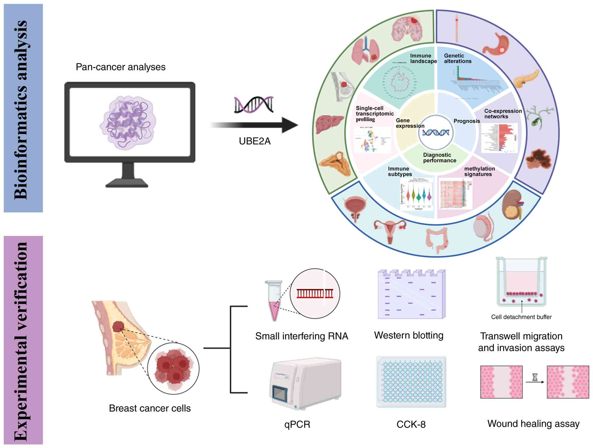
tumor-immune interactions. The overall study design is illustrated
in Fig. 1.
Materials and methods
Gene expression analysis
UBE2A expression across human cancer types was
analyzed using RNA-sequencing (RNA-seq) data obtained from The
Cancer Genome Atlas (TCGA; http://portal.gdc.cancer/gov/). Transcript per million
(TPM)-format expression matrices processed using the STAR pipeline
were downloaded and normalized by log2 (TPM+1)
transformation. Differential expression analyses between tumor and
normal tissues were performed in R software (version 4.2.1; Posit
Software, PBC) using the Wilcoxon rank-sum test and data
visualization was conducted using the ‘ggplot2’ package (version
3.4.4) (23). For tumor types
lacking sufficient normal tissue controls in TCGA, the pan-cancer
differential expression module of SangerBox (http://sangerbox.com/) was additionally used, which
integrates TCGA and Genotype-Tissue Expression (GTEx; http://www.gtexportal.org/home/) datasets
(24). To validate the expression
pattern of UBE2A, two independent breast cancer cohorts [GSE29044
(25) and GSE65194 (26)] were downloaded from the Gene
Expression Omnibus (GEO; http://www.ncbi.nlm.nih.gov/geo/) and differential
expression analyses were performed between tumor and normal tissues
using the same statistical methods. In addition, the associations
between UBE2A expression and immune or molecular subtypes across
cancers were explored using the Tumor-Immune System Interaction
Database (http://cis.hku.hk/TISIDB/) (27).
Clinicopathological correlation
analysis
To investigate the association between UBE2A
expression and clinicopathological characteristics across cancers,
normalized pan-cancer transcriptomic data (TCGA Pan-Cancer;
n=10,535) were downloaded from the UCSC Xena database (https://xenabrowser.net/). UBE2A expression data were
extracted from the dataset and samples derived from primary tumors
were included for further analysis. Expression values were
transformed using log2 (x + 0.001) normalization prior
to statistical analysis. Differences in UBE2A expression among
different clinicopathological stages, including T stage, N stage
and pathological stage, were analyzed using R software. Unpaired
Student's t-tests were used for pairwise comparisons, while one-way
ANOVAs were performed for comparisons among multiple groups.
Diagnostic performance analysis
Diagnostic performance of UBE2A expression in
distinguishing tumor tissues from normal tissues was evaluated
using receiver operating characteristic (ROC) curve analysis.
RNA-seq expression data in TPM format and corresponding clinical
information were downloaded from TCGA. Gene expression values were
transformed using log2 (TPM + 1) normalization prior to
analysis. ROC analyses were performed in R software using the
‘pROC’ package (version 1.18.0) (28) and visualization was generated using
the ‘ggplot2’ package (version 3.4.4) (23). Tumor samples were defined as the
positive group, whereas normal tissues served as the reference
group for binary classification analysis. The ‘pROC’ package
automatically determined the direction of comparison to ensure an
upward ROC curve orientation. The area under the ROC curve (AUC),
sensitivity, specificity and optimal cut-off values were calculated
to assess diagnostic efficacy. The optimal cut-off threshold was
determined based on the maximum Youden index. An AUC value closer
to 1.0 indicated superior discriminative performance. Pan-cancer
analyses were performed across TCGA datasets and cancer types with
significant diagnostic performance are presented in the
results.
Survival prognosis analysis
Prognostic value of UBE2A was evaluated using the
GEPIA2 (http://gepia2.cancer-pku.cn/#analysis) and
Kaplan-Meier Plotter (http://kmplot.com/analysis/) databases. GEPIA2, which
integrates transcriptomic and clinical data from TCGA, was used to
assess the associations between UBE2A expression and overall
survival (OS) as well as disease-free survival (DFS) using the
log-rank test (29). Kaplan-Meier
Plotter, which incorporates survival data from TCGA and GEO
cohorts, was used to further validate the prognostic importance of
UBE2A in breast cancer, including OS, distant metastasis-free
survival (DMFS) and relapse-free survival (RFS). Patients were
stratified into high- and low-expression groups using the default
cut-off settings of each platform. Hazard ratios (HRs), 95% CIs and
log-rank P-values were used to evaluate prognostic importance
(30).
Genetic alteration analysis
Genetic alteration data for UBE2A were obtained from
the cBioPortal platform, which provides access to TCGA-based cancer
genomics datasets (31).
Alteration frequency, mutation type, mutation distribution and copy
number variation of UBE2A were assessed across multiple cancer
types. In addition, the associations between UBE2A genetic
alterations and clinical outcomes were evaluated.
Co-expression analysis and gene set
enrichment analysis (GSEA)
Genes co-expressed with UBE2A were identified using
the LinkedOmics database, which contains multi-omics data from TCGA
cohorts (32). Pearson correlation
analysis was performed to determine genes significantly associated
with UBE2A expression. The top 500 positively and negatively
correlated genes were subsequently subjected to Gene Ontology (GO)
and Kyoto Encyclopedia of Genes and Genomes (KEGG) enrichment
analyses using the ‘clusterProfiler’ R package (version 4.14.6)
(33–35). In addition, GSEA was performed in
the TCGA-BRCA cohort by comparing samples with high and low UBE2A
expression (median cut-off). Genes were ranked according to
differential expression and significantly enriched pathways were
visualized using the ‘enrichplot’ package (version 1.26.6)
(34).
PPI network analysis
A PPI network centered on UBE2A was constructed
using the GeneMANIA platform (http://genemania.org) (36). Genes functionally associated with
UBE2A were identified based on numerous evidence sources, including
co-expression and physical interactions, to explore the potential
molecular functions of UBE2A.
DNA methylation and RNA modification
gene correlation analysis
Epigenetic modifications, including DNA methylation
and RNA methylation, serve key roles in cancer development and
progression (37,38). Promoter DNA methylation levels of
UBE2A across 33 TCGA cancer types were evaluated using TCGA
HumanMethylation450 BeadChip data accessed through the UALCAN
platform (http://ualcan.path.uab.edu) (39). In addition, correlations between
UBE2A expression and RNA modification-associated regulators,
including genes involved in m6A, m5C and m1A modification pathways,
were analyzed to explore potential epigenetic regulatory
mechanisms.
Immune infiltration analysis
Immune cell infiltration levels were estimated using
the Cell-type Identification by Estimating Relative Subsets of RNA
Transcripts algorithm (40) based
on TCGA transcriptomic data. The relative proportions of 22 immune
cell subsets were quantified and Spearman's correlation analysis
was performed to evaluate the associations between UBE2A expression
and immune cell infiltration levels across cancer types. The
results are visualized as heatmaps. To improve the robustness of
immune infiltration analyses, multiple immune deconvolution
algorithms available in the SangerBox platform (http://www.sangerbox.com) were applied, including
Tumor Immune Estimation Resource (TIMER) (41), Estimating the Proportions of Immune
and Cancer cells (EPIC) (42),
Quantification of the Tumor Immune Contexture from Human RNA-seq
Data (quanTIseq) (43) and xCell
(44). Correlations between UBE2A
expression and immune cell infiltration were evaluated using these
algorithms.
Tumor mutational burden (TMB) and
microsatellite instability analysis (MSI)
TCGA transcriptomic data, TMB data and MSI data were
integrated for pan-cancer analysis. Spearman's correlation analysis
was performed to evaluate the associations between UBE2A expression
and TMB or MSI across different cancer types. Correlation
coefficients and corresponding P-values were calculated using R
software and the results are visualized as radar plots.
Single-cell analysis
Single-cell transcriptomic analysis was performed
using the Tumor Immune Single-Cell Hub 2 (TISCH2) database, a
comprehensive single-cell RNA-seq (scRNA-seq) resource for tumor
microenvironment analysis (45).
TISCH2 provides uniformly processed scRNA-seq datasets with
standardized quality control, clustering, batch-effect correction
and hierarchical cell-type annotation using the Model-based
Analyses of Single-cell Transcriptome and Regulome pipeline
(45). In the present study, four
breast cancer datasets, including BRCA_GSE114727 (46), BRCA_GSE110686 (47), BRCA_GSE148673 (48) and BRCA_GSE176078 (49), were analyzed to investigate the
single-cell expression pattern of UBE2A in different cell
populations within the tumor microenvironment. Cell annotations
curated by TISCH2 were used at the malignancy, major-lineage and
minor-lineage levels to ensure consistency and comparability across
datasets. Gene expression distributions and cell-type-specific
expression patterns were directly obtained and visualized through
the TISCH2 platform.
Cell culture
MCF-10A, the human mammary epithelial cell line and
breast cancer cell lines MCF-7, MDA-MB-231, MDA-MB-468 and Hs578T
were purchased from Procell Life Science & Technology Co., Ltd.
MCF-10A cells were cultured in DMEM/F12 medium, whereas breast
cancer cell lines were maintained in DMEM medium. All media were
supplemented with 10% FBS (Biological Industries; Sartorius AG) and
1% penicillin-streptomycin (Beijing Solarbio Science &
Technology Co., Ltd.). Cells were cultured at 37°C in a humidified
incubator containing 5% CO2.
Reverse transcription-quantitative PCR
(RT-qPCR) analysis
Total RNA was extracted from cultured breast
epithelial and breast cancer cells lines, including MCF-10A, MCF-7,
MDA-MB-231, MDA-MB-468 and Hs578T, using the Cell/Tissue Total RNA
Kit (Suzhou Xinsaimei Biotechnology Co., Ltd.) following the
manufacturer's protocol. RT was performed using HiScript III RT
SuperMix (Vazyme Biotech Co., Ltd.) according to the manufacturer's
protocol. RT-qPCR was carried out on the qTOWER3G system (Analytik
Jena GmbH + Co. KG) with ChamQ Universal SYBR qPCR Master Mix
(Vazyme Biotech Co., Ltd.) under the following thermocycling
conditions: Initial denaturation at 95°C for 30 sec, followed by 40
cycles of denaturation at 95°C for 10 sec and annealing/extension
at 60°C for 30 sec. β-actin was used as the internal reference gene
for normalization and relative expression levels were calculated
using the 2−ΔΔCq method (50). The primer sequences are listed in
Table SI.
Small interfering RNA (siRNA)
transfection of cells
Targeted silencing of UBE2A was achieved using
specific siRNA and corresponding scrambled negative control siRNA,
all synthesized by Hanbio Biotechnology Co., Ltd. For siRNA
transfection, MCF-7 and MDA-MB-231 cells in 6-well plates were
transfected with 100 pmol siRNA per well using Lipofectamine™ 3000
(Invitrogen; Thermo Fisher Scientific, Inc.) according to the
manufacturer's protocol. Following 48 h of transfection, knockdown
efficiency was validated at both mRNA and protein levels through
qPCR and western blotting, respectively. The siRNA sequences are
detailed in Table SII.
Cell Counting Kit-8 (CCK-8) assay
MCF-7 and MDA-MB-231 cells were seeded in 96-well
plates at a density of 3,000 cells/well (100 µl medium/well) with 5
technical replicates per group. After cell attachment (0 h), the
medium was replaced with 100 µl fresh complete medium containing
10% CCK-8 reagent (Vazyme Biotech Co., Ltd.) at 0, 24, 48 and 72 h,
followed by incubation under standard culture conditions for 2 h.
Absorbance was measured at 450 nm using a microplate reader
(BioTek; Agilent Technologies, Inc.). Cell viability was calculated
and growth curves were plotted based on optical density values.
Wound healing assay
To evaluate cell migration capacity, pre-marked
reference lines were drawn on the bottom of 6-well plates prior to
cell seeding. MCF-7 and MDA-MB-231 cells were cultured in complete
medium until reaching 95–100% confluence. A wound was created by
scratching the monolayer with a 200 µl sterile pipette tip
perpendicular to the reference lines. After washing with PBS to
remove detached cells, fresh serum-free medium was added. Wound
areas were photographed at 0 and 24 h post-scratching using an
inverted phase-contrast microscope (Olympus-IX71; Olympus
Corporation). ImageJ (National Institutes of Health; version 1.53a)
was used to calculate the scratch area.
Transwell assay
For migration assays, MCF-7 and MDA-MB-231 cells
(5×10⁴ cells per well) were suspended in 100 µl
serum-free medium and seeded into the upper chamber of Transwell
inserts (Corning, Inc.). The lower chamber was filled with 800 µl
complete medium containing 10% FBS (Biological Industries;
Sartorius AG) as a chemoattractant. For invasion assays, transwell
inserts were pre-coated with Matrigel (Shanghai Yeasen
Biotechnology Co., Ltd.) and incubated at 37°C for 2 h before cell
seeding. Cells were then incubated at 37°C in a humidified
atmosphere containing 5% CO2 for 24 h. After incubation,
non-migrated cells were removed from the upper surface of the
membrane using cotton swabs. Cells that had migrated or invaded to
the lower surface were fixed with fixed with 4% paraformaldehyde
(Beijing Solarbio Science & Technology Co., Ltd.) at room
temperature for 20 min and stained with 0.1% crystal violet
(Beyotime Biotechnology) at room temperature for 20 min. Images
were captured using an inverted microscope (Olympus IX73; Olympus
Corporation) and cell numbers were quantified using ImageJ
software.
Western blotting
Total cellular proteins were extracted from MCF-7
and MDA-MB-231 cells using a column-based total protein extraction
kit (Suzhou Xinsaimei Biotechnology Co., Ltd.) and protein
concentrations were determined by a BCA assay (Beytome
Biotechnology). Equal quantities of protein (30 µg per lane) were
separated by 10% SDS-PAGE and subsequently transferred onto PVDF
membranes (MilliporeSigma; Merck KGaA). Following transfer,
membranes were blocked with 5% non-fat milk at room temperature for
1 h and then incubated with the indicated primary antibodies at 4°C
overnight. After washing, membranes were probed with HRP-conjugated
secondary antibodies for 1 h at room temperature. Immunoreactive
signals were detected using an ECL detection system (Thermo Fisher
Scientific, Inc.) and captured with an imaging system from e-BLOT
Life Science (Shanghai) Co., Ltd. The antibodies used included
anti-UBE2A (1:1,000; cat. no. sc-365507; Santa Cruz Biotechnology,
Inc.), anti-β-actin (1:20,000; cat. no. 66009-1-Ig; Proteintech
Group, Inc.), HRP-conjugated goat anti-rabbit IgG (1:10,000; cat.
no. SA00001-2; Proteintech Group, Inc.) and HRP-conjugated goat
anti-mouse IgG (1:10,000 cat. no. SA00001-1; Proteintech Group,
Inc.).
Statistical analysis
Statistical analyses were performed using GraphPad
Prism (version 10.1.2; Dotmatics) and R software. Differences
between groups were evaluated using unpaired Student's t-test or
one-way ANOVA, as appropriate. Unless otherwise specified,
two-sided P<0.05 was considered to indicate a statistically
significant difference. For bioinformatic analyses, statistical
testing and multiple-testing correction were performed according to
the corresponding analytical pipelines and false discovery
rate-adjusted P-values were applied in enrichment analyses.
Results
UBE2A shows differential expression
across several cancers
To characterize the expression profile of UBE2A
across human cancers, TCGA transcriptomic datasets were analysed
and UBE2A expression was compared between tumor tissues and
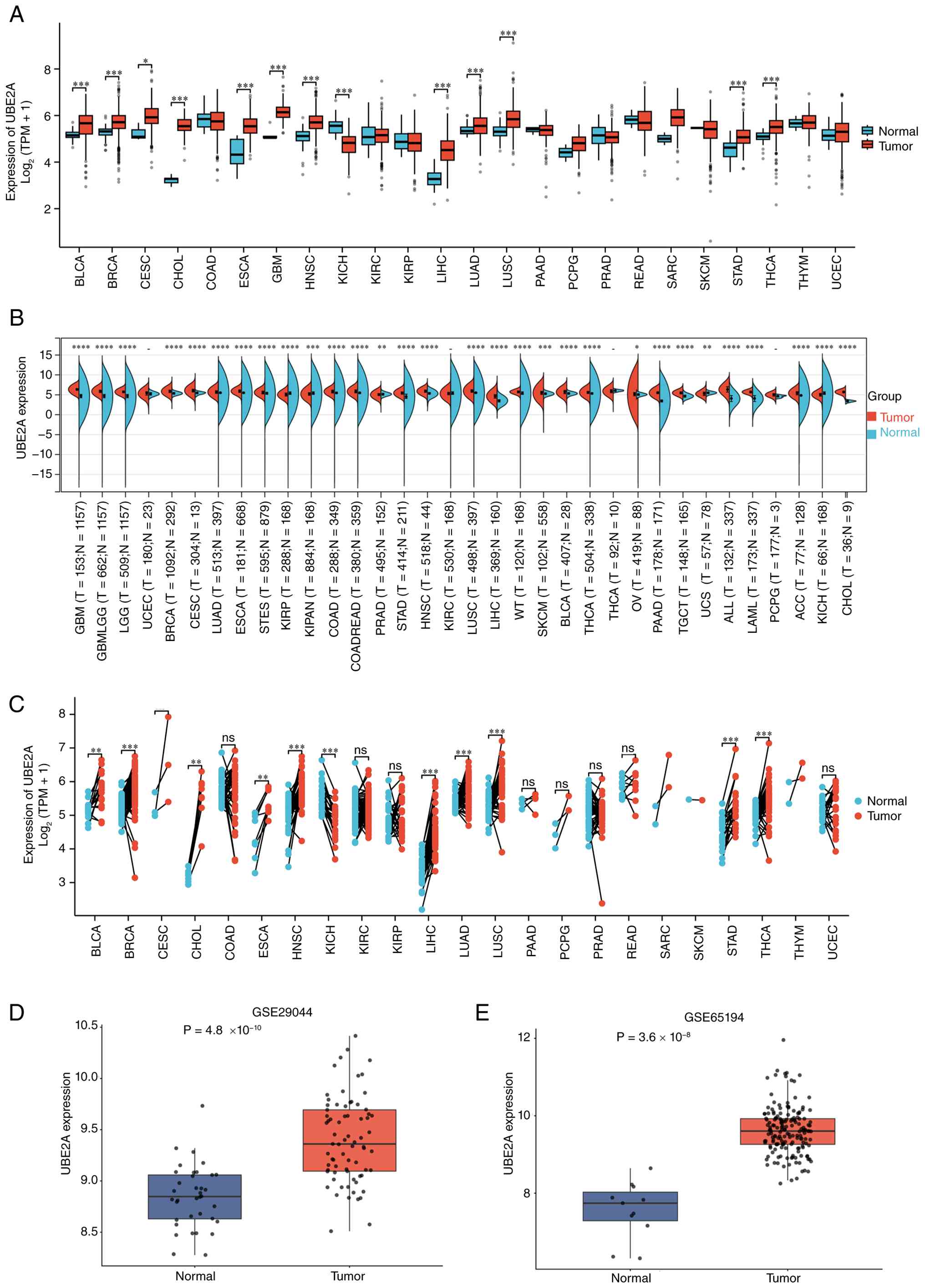
corresponding normal tissues. As shown in Fig. 2A, UBE2A expression was found to be
significantly elevated in bladder urothelial carcinoma (BLCA),
BRCA, cervical squamous cell carcinoma and endocervical
adenocarcinoma (CESC), cholangiocarcinoma (CHOL), esophageal
carcinoma (ESCA), glioblastoma multiforme (GBM), head and neck
squamous cell carcinoma (HNSC), liver hepatocellular carcinoma
(LIHC), lung adenocarcinoma (LUAD), lung squamous cell carcinoma
(LUSC), stomach adenocarcinoma (STAD) and thyroid carcinoma (THCA)
compared with corresponding normal tissues. By contrast, kidney
chromophobe (KICH) exhibited significantly lower UBE2A expression.
To further validate these findings, an integrated analysis
combining TCGA tumor samples and GTEx normal tissues was performed.
Consistent with the TCGA-based analysis, UBE2A remained
significantly upregulated in BLCA, BRCA, CESC, CHOL, ESCA, GBM,
HNSC, LIHC, LUAD, LUSC, STAD and THCA. In addition, further cancer
types with significant dysregulation were identified after
incorporating GTEx normal tissues, resulting in elevated UBE2A
expression in 25 cancer types and reduced expression in five cancer
types overall (Fig. 2B). In
addition, paired tumor-normal analyses within TCGA cohorts further
demonstrated significantly elevated UBE2A expression in BLCA, BRCA,
CHOL, ESCA, HNSC, LIHC, LUAD, LUSC, STAD and THCA, while
significantly reduced expression was observed in KICH (Fig. 2C). By contrast, no significant
differences were detected in CESC, COAD, KIRC, KIRP, PAAD, PCPG,
PRAD, READ or UCEC.
To further assess the robustness of these
observations, two independent breast cancer cohorts from the GEO
database (GSE29044 and GSE65194) were analyzed. Consistent with the
TCGA results, UBE2A expression was found to be significantly
increased in breast cancer tissues compared with normal breast
tissues in both datasets (Fig. 2D and
E). Collectively, these findings demonstrate that UBE2A is
aberrantly upregulated in a broad spectrum of human cancer types,
particularly in breast cancer, suggesting that UBE2A may serve an
important role in tumor development and progression.
UBE2A is associated with
clinicopathological features in pan-cancer
To investigate the clinical relevance of UBE2A, its
association with clinicopathological characteristics was evaluated
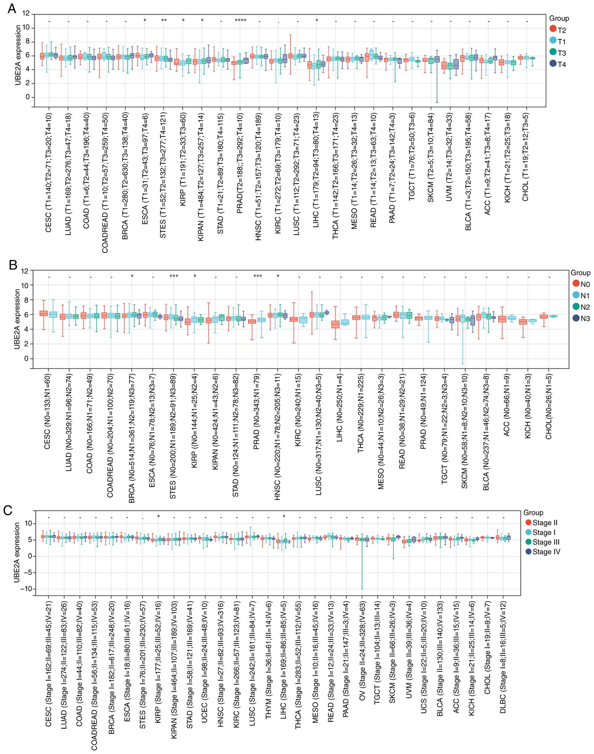
across numerous cancer types. Analysis of tumor invasion status
revealed significant differences in UBE2A expression among T-stage
subgroups in ESCA, STES, KIRP, pan-kidney cohort, PRAD and LIHC
(Fig. 3A). With regard to lymph
node metastasis, UBE2A expression was found to be significantly
associated with N stage in BRCA, STES, KIRP, PRAD and HNSC
(Fig. 3B). Furthermore,
significant correlations between UBE2A expression and overall
pathological stage were observed in KIRP and LIHC (Fig. 3C). These findings suggest that
UBE2A expression is associated with tumor progression in numerous
malignancies. Notably, the significant association between UBE2A
expression and N stage in BRCA indicates a potential role for UBE2A
in breast cancer progression and metastasis.
UBE2A exhibits diagnostic value across
multiple cancer types
Diagnostic values of UBE2A expression were evaluated
across a number of cancer types using ROC curve analysis. UBE2A
exhibited good discriminatory ability between tumor and normal
tissues in 15 cancer types (Fig.
4A-O). Notably, excellent diagnostic performance was observed
in CHOL, esophageal adenocarcinoma (ESAD), ESCA, GBM, KICH and
LIHC, with AUC values exceeding 0.90. These findings indicate that
UBE2A expression may serve as a potential diagnostic biomarker in
selected malignancies.
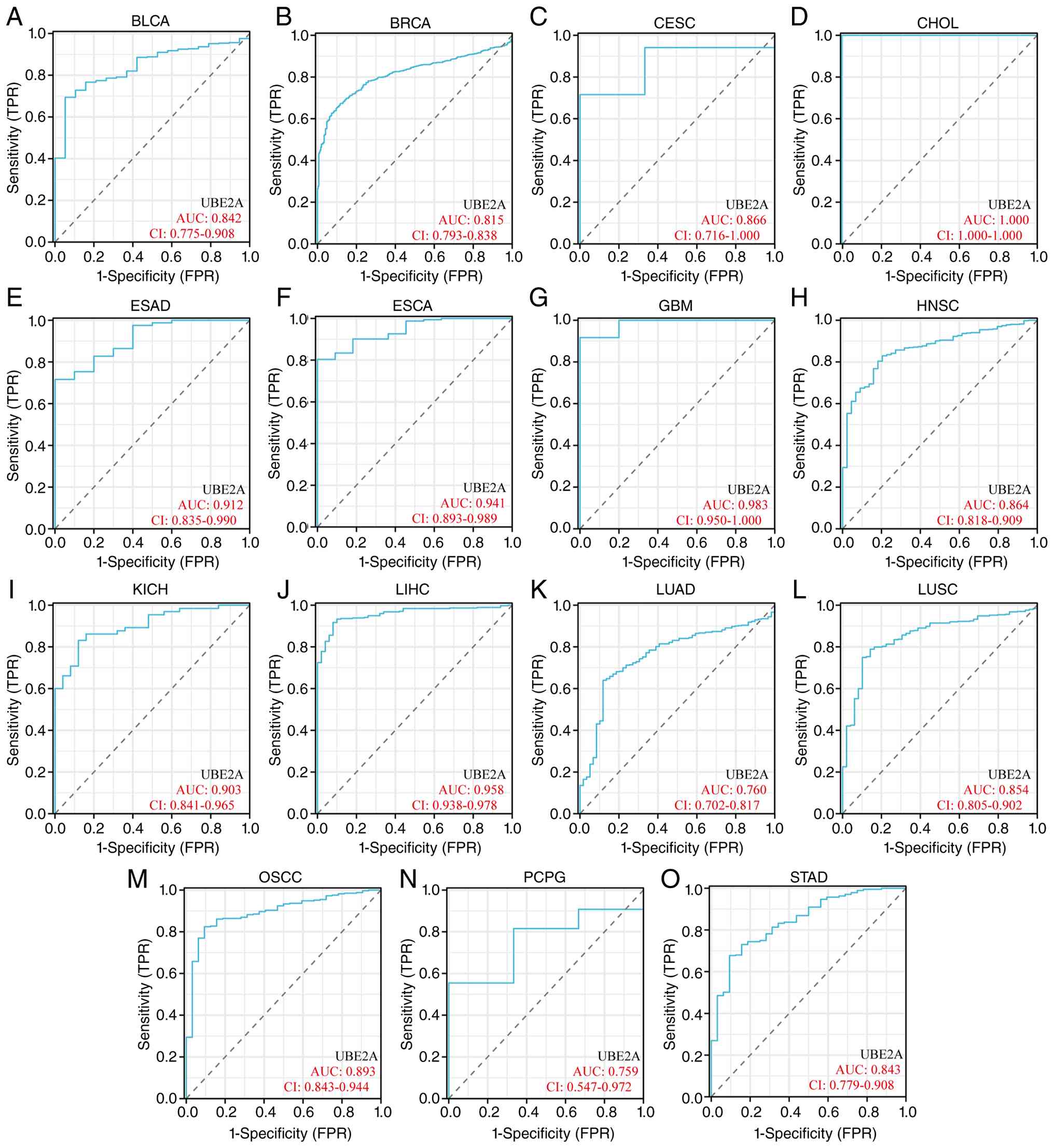 | Figure 4.Diagnostic performance of UBE2A
expression across cancer types. Receiver operating characteristic
(ROC) curves evaluating the ability of UBE2A expression to
distinguish tumor tissues from normal tissues in (A) BLCA, (B)
BRCA, (C) CESC, (D) CHOL, (E) ESAD, (F) ESCA, (G) GBM, (H) HNSC,
(I) KICH, (J) LIHC, (K) LUAD, (L) LUSC, (M) OSCC, (N) PCPG and (O)
STAD. UBE2A, ubiquitin conjugating enzyme E2A; ROC, receiver
operating characteristic; AUC, area under the curve; FPR, false
positive rate; TRP, true positive rate. |
UBE2A is associated with prognosis
across human cancers
To evaluate the prognostic relevance of UBE2A across
cancers, survival analyses were performed using OS and DFS
endpoints. Elevated UBE2A expression was significantly associated
with shorter OS in BRCA, low-grade glioma (LGG), LIHC,
mesothelioma, sarcoma (SARC) and uveal melanoma (UVM; Fig. 5A-G). Similarly, patients with high
UBE2A expression exhibited significantly worse DFS in LGG, LIHC and
UVM (Fig. 5H-K). To further
validate these findings, independent BRCA cohorts were analyzed
using the Kaplan-Meier Plotter platform. Consistent with the
pan-cancer findings, elevated UBE2A expression was associated with
unfavorable OS, RFS and DMFS (Fig.
5L-N). Collectively, these results suggest that UBE2A may
represent a prognostic indicator in a number of malignancies, with
reproducible associations observed across different survival
endpoints and analytical platforms.
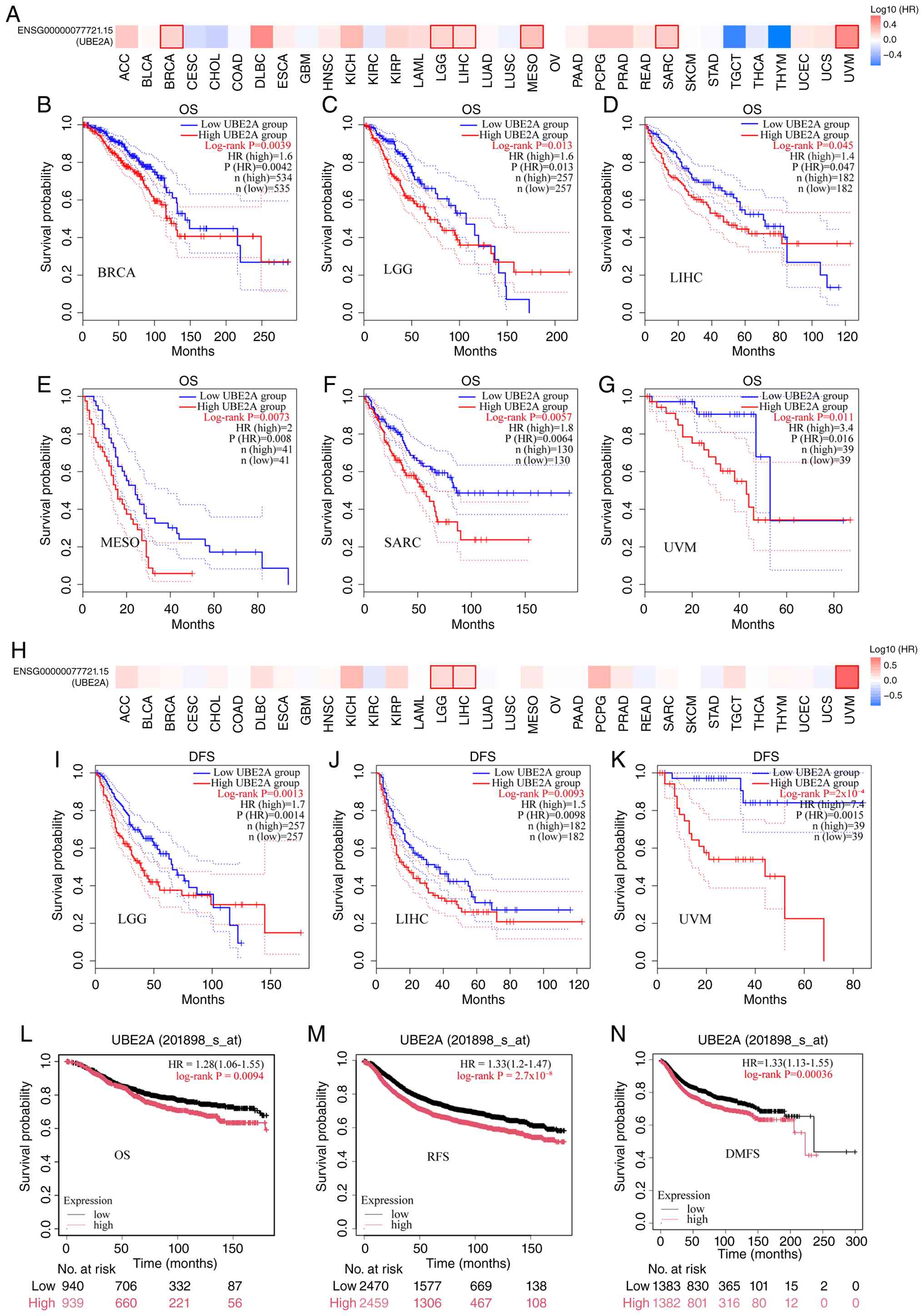 | Figure 5.Prognostic relevance of UBE2A
expression across human cancers. (A) Overview of the associations
between UBE2A expression and OS across TCGA cancer types.
Kaplan-Meier analysis of OS in (B) BRCA, (C) LGG, (D) LIHC, (E)
MESO, (F) SARC and (G) UVM. (H) Overview of the associations
between UBE2A expression and DFS across TCGA cancer types.
Kaplan-Meier analysis of DFS in (I) LGG, (J) LIHC and (K) UVM. (L)
Kaplan-Meier Plotter validation of OS in BRCA. (M) Kaplan-Meier
Plotter validation of RFS in BRCA. (N) Kaplan-Meier Plotter
validation of DMFS in BRCA. UBE2A, ubiquitin conjugating enzyme
E2A; OS, overall survival; DFS, disease-free survival; RFS,
relapse-free survival; HR, hazard ratio. |
UBE2A displays distinct genetic
alteration patterns across pan-cancer
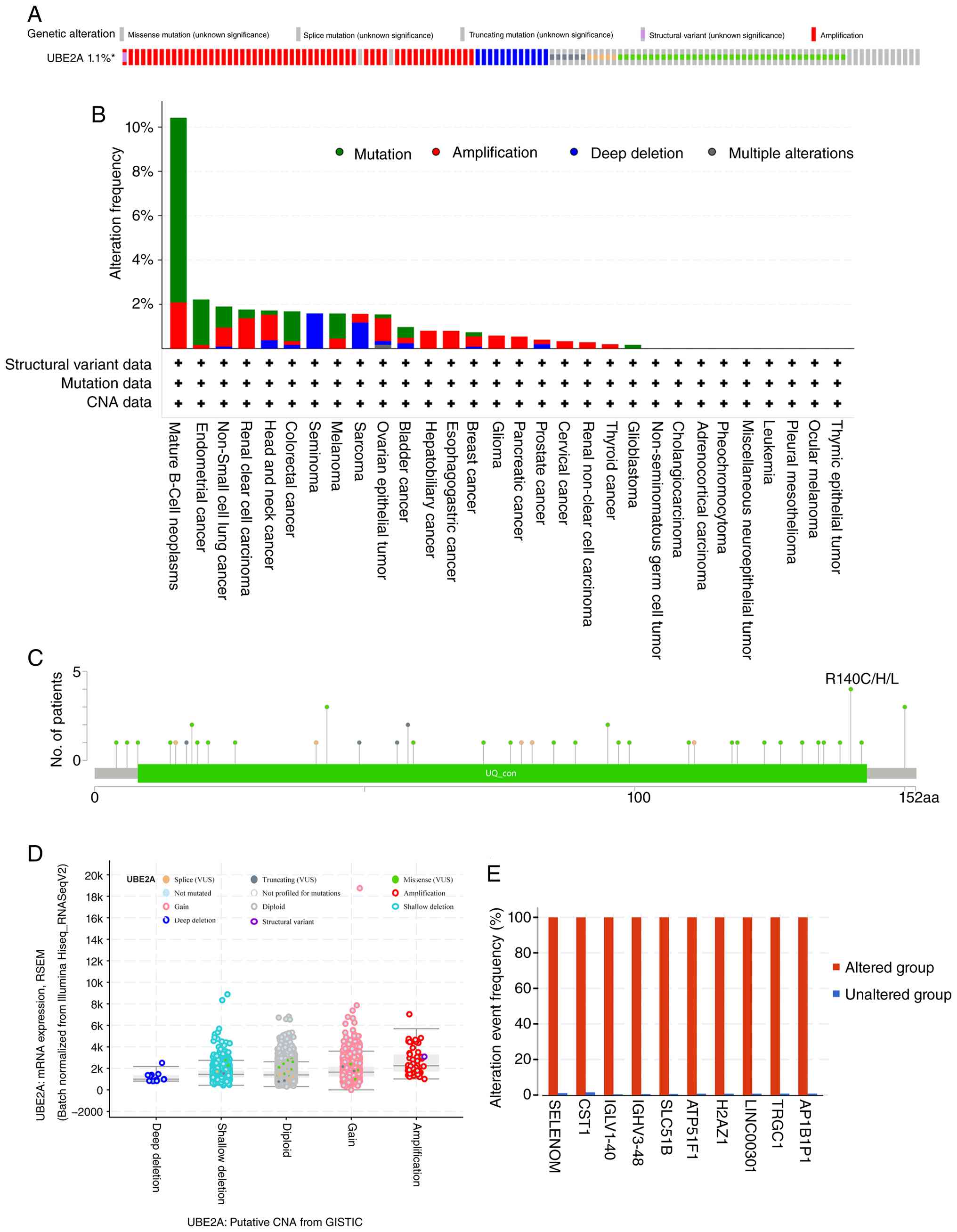
Genomic alteration landscapes of UBE2A were next
investigated across human cancers. UBE2A alterations were detected
in ~1.1% of all analyzed tumors, although notable heterogeneity was
observed among cancer types (Fig.
6A). The highest alteration frequency occurred in mature B-cell
neoplasms (>10%), followed by endometrial carcinoma and
non-small cell lung cancer, each exhibiting alteration frequencies
of ~2% (Fig. 6B). Analysis of
mutation patterns revealed that missense mutations were the
predominant alteration type, with recurrent mutations affecting the
R140 residue identified as hotspot events (Fig. 6C). Copy number gain represented the
most common copy number alteration (Fig. 6D). Furthermore, tumors harboring
UBE2A alterations exhibited significantly higher mutation
frequencies of cystatin SN, SLC51 subunit β, ATPase inhibitory
factor 1 and a number of additional genes compared with UBE2A
wild-type tumors (Fig. 6E),
suggesting that UBE2A alterations may co-occur with specific
genomic events during tumor evolution.
UBE2A is linked to cancer-associated
co-expression and protein interaction networks
To gain insight into the biological functions
associated with UBE2A, genes correlated with UBE2A expression were
identified and analyzed. The top 50 positively and negatively
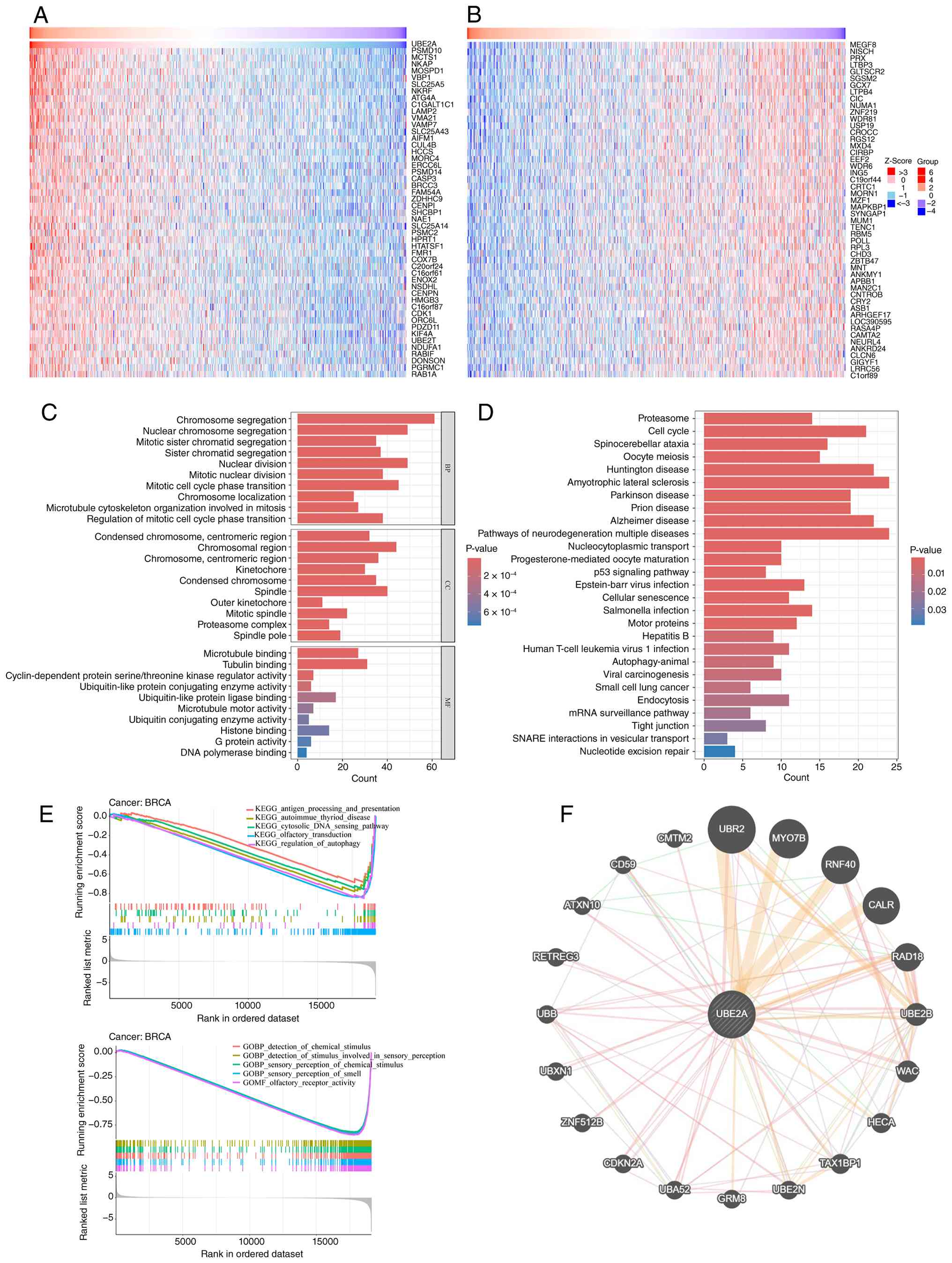
correlated genes are visualized as heatmaps (Fig. 7A and B). Functional enrichment
analyses based on the top 500 UBE2A-associated genes revealed
significant enrichment in ‘chromosome segregation’, ‘mitotic cell
cycle phase transition’ and other cell cycle-associated biological
processes (Fig. 7C). Consistently,
KEGG pathway analysis identified significant enrichment of
‘proteasome’ and ‘cell cycle’ pathways (Fig. 7D).
To further characterize biological pathways
associated with UBE2A expression, GSEA was performed in BRCA.
GO-based GSEA identified enrichment of
‘GOMG_OLFACTORY_RECEPTOR_ACTIVITY’, amongst other terms, whereas
KEGG-based GSEA demonstrated significant enrichment of
‘KEGG_ANTIGEN_PROCESSING_AND_PRESENTATION’ as well as
‘KEGG_CYTOSOLIC_DNA_SENSING_PATHWAY’ in the UBE2A high-expression
group (Fig. 7E). In addition, a
PPI network centered on UBE2A revealed numerous closely connected
interacting proteins and functional modules (Fig. 7F), supporting the potential
involvement of UBE2A in cell cycle regulation and
proteostasis-associated processes.
UBE2A is associated with DNA and RNA
methylation patterns in pan-cancer
To investigate potential epigenetic mechanisms
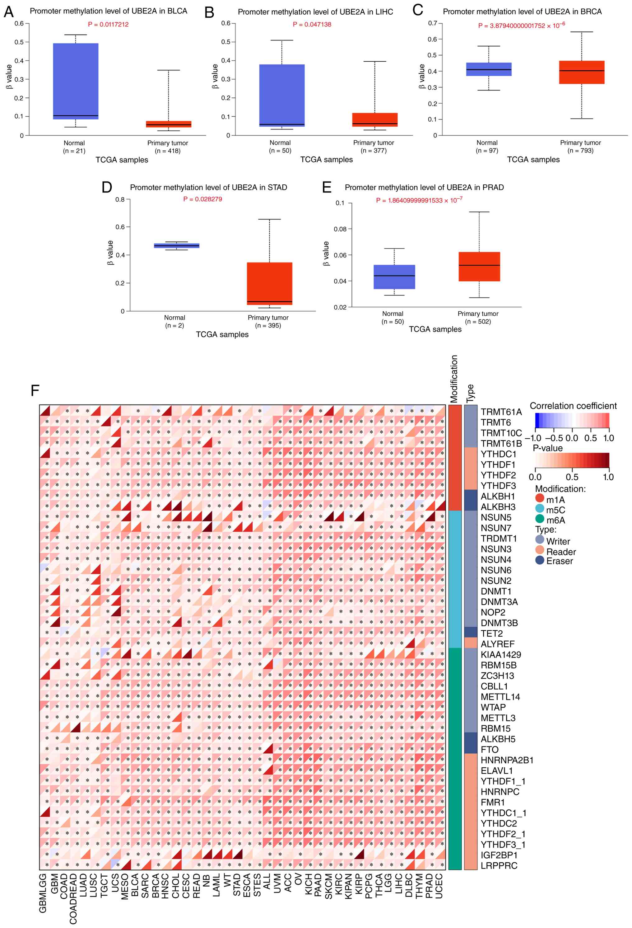
underlying UBE2A dysregulation, promoter methylation patterns were
analyzed across multiple cancer types. Compared with normal
tissues, a significantly lower promoter methylation levels of UBE2A
was observed in BLCA, LIHC, BRCA and STAD (Fig. 8A-D). By contrast, significantly
increased promoter methylation was detected in PRAD (Fig. 8E). The association between UBE2A
expression and RNA modification regulators was further examined.
UBE2A expression exhibited significant correlations with numerous
RNA modification-associated genes across cancer types, particularly
a number m6A regulators, including methyltransferase-like 14 and
Wilms tumor 1-associating protein (Fig. 8F). Collectively, these findings
suggest that epigenetic mechanisms may contribute to the
dysregulated expression of UBE2A in human cancers.
Differential expression of UBE2A among
immune and molecular subtypes in 33 malignancies
Whether UBE2A expression differed among distinct
molecular and immune subtypes across cancers was next examined.
Significant differences in UBE2A expression among molecular
subtypes were observed in BRCA, COAD, HNSC, KIRP, LGG, LUSC,
ovarian cancer (OV) and pheochromocytoma and paraganglioma
(Fig. 9A-H). Similarly, UBE2A
expression varied markedly among immune subtypes in 12 cancer types
(Fig. 9I-T). These observations
indicated that UBE2A expression may be associated with tumor
heterogeneity at both molecular and immune levels.
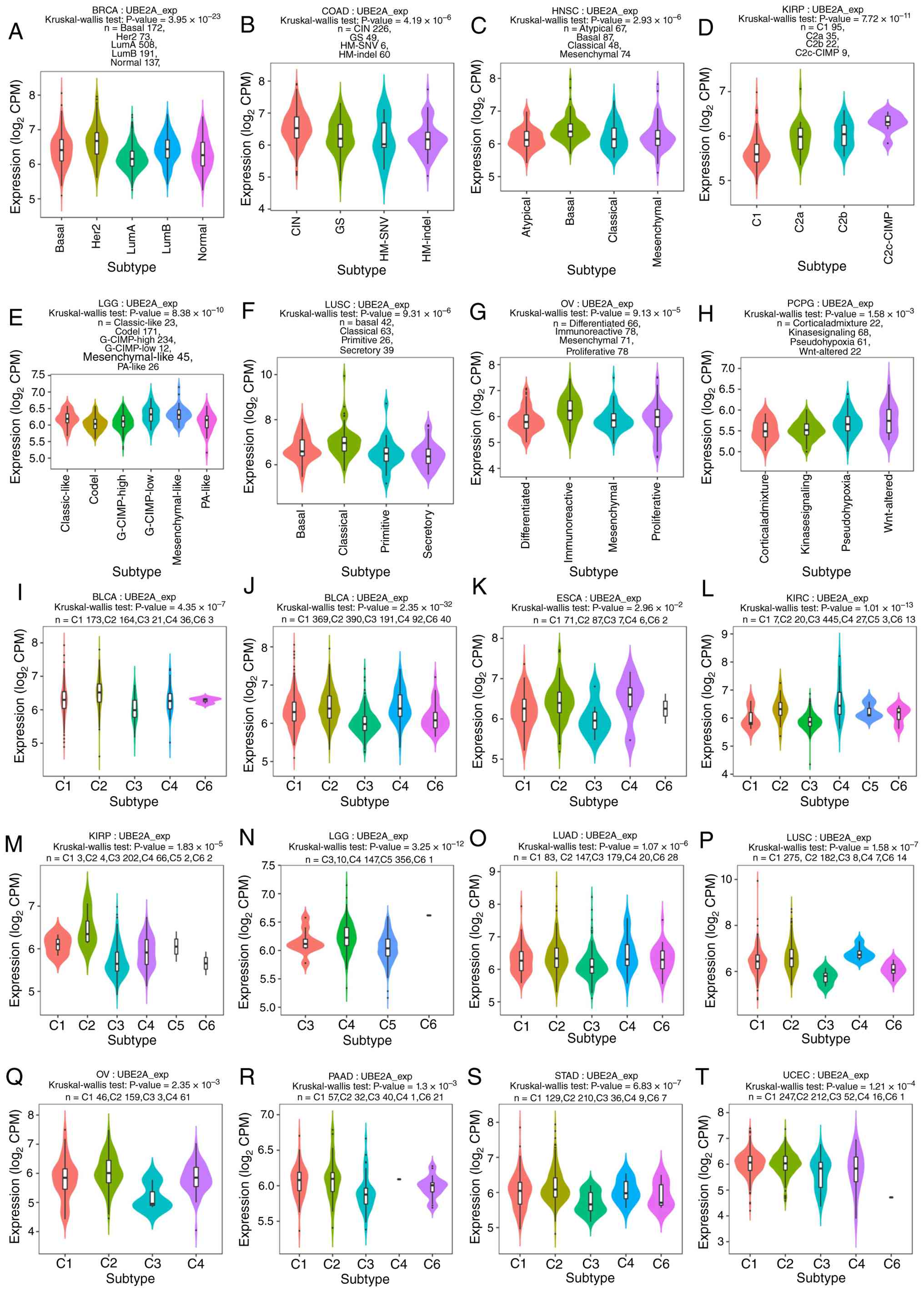 | Figure 9.UBE2A expression patterns across
molecular and immune subtypes in 33 cancer types. (A-H) UBE2A
expression among molecular subtypes in (A) BRCA, (B) COAD, (C)
HNSC, (D) KIRP, (E) LGG and (F) LUSC, (G) OV and (H) PCPG. (I-T)
UBE2A expression profiles across immune subtypes in (I) BLBA, (J)
BRCA, (K) ESCA, (L) KIRC, (M) KIRP, (N) LGG, (O) LUAD, (P) LUSC,
(Q) OV, (R) PAAD, (S) STAD and (T) UCEC. UBE2A, ubiquitin
conjugating enzyme E2A; Lum, luminal; CIN, chromosomal instability;
GS, genomically stable; HM, hypermutated; SNV, single nucleotide
variant; indel, insertion/deletion; G-CIMP, glioma CpG island
methylator phenotype. |
UBE2A is associated with immune
infiltration and genomic instability in pan-cancer
To explore potential associations between UBE2A
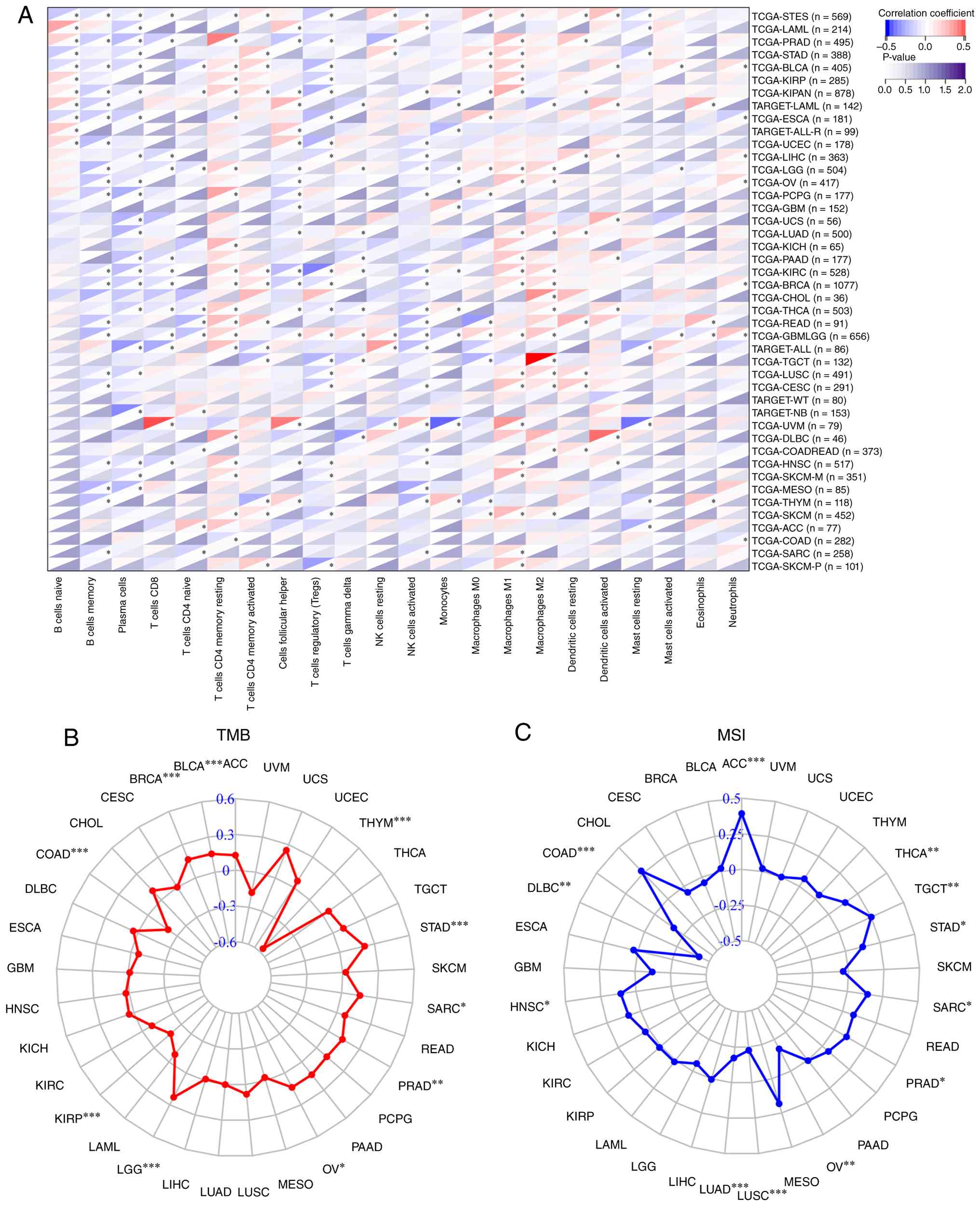
expression and the tumor immune microenvironment, correlations
between UBE2A expression and immune cell infiltration were
evaluated across cancers. UBE2A expression exhibited significant
correlations with numerous immune cell populations in different
tumor types, including resting memory CD4+ T cells,
activated memory CD4+ T cells, activated NK cells and M2
macrophages (Fig. 10A). Similar
trends were observed when additional immune deconvolution
algorithms, including EPIC, QUANTISEQ, TIMER and xCELL, were
applied (Fig. S1A-D). The
associations between UBE2A expression and immune regulatory genes
were further examined. UBE2A expression exhibited widespread
correlations with numerous immunomodulatory molecules, including
C-X-C motif chemokine ligand 10 and IL-10, across cancer types
(Fig. S2). In addition,
significant associations were observed between UBE2A expression and
a number of immune checkpoint-associated genes, including IL-12A
and TGF-β1 (Fig. S3).
Given the clinical relevance of genomic instability
in immunotherapy, correlations between UBE2A expression and TMB or
MSI were subsequently analyzed (51,52).
UBE2A expression was found to be positively associated with TMB in
BLCA, BRCA, LGG, OV, PRAD, SARC and STAD, whereas negative
correlations were observed in COAD, KIRP and thymoma (Fig. 10B). For MSI, positive correlations
were detected in adrenocortical carcinoma, HNSC, PRAD, SARC, STAD,
tenosynovial giant cell tumor and THCA, while negative associations
were observed in COAD, diffuse large B-cell lymphoma, LUAD, LUSC
and OV (Fig. 10C). Collectively,
these findings suggest that UBE2A expression may be associated with
distinct immune and genomic features in a cancer type-dependent
manner.
Single-cell analysis reveals
cell-type-specific expression of UBE2A
Single-cell omics technologies enable
high-resolution profiling of tumor heterogeneity by capturing
cell-to-cell variability that is masked by traditional bulk
sequencing, thereby advancing precision oncology and improving our
understanding of tumor biology (53,54).
To further characterize the cellular distribution of UBE2A within
the tumor microenvironment, single-cell RNA-seq datasets from four
independent BRCA cohorts were analyzed. Across all datasets, UBE2A
expression was found to be predominantly enriched in the
proliferative T-cell population (Fig.
11A-E). Consistent expression patterns across independent
datasets indicated a potential association between UBE2A expression
and proliferative T-cell states within the breast tumor
microenvironment. Notably, these observations were derived from
transcriptomic datasets and therefore should be considered
hypothesis-generating findings that require further experimental
validation.
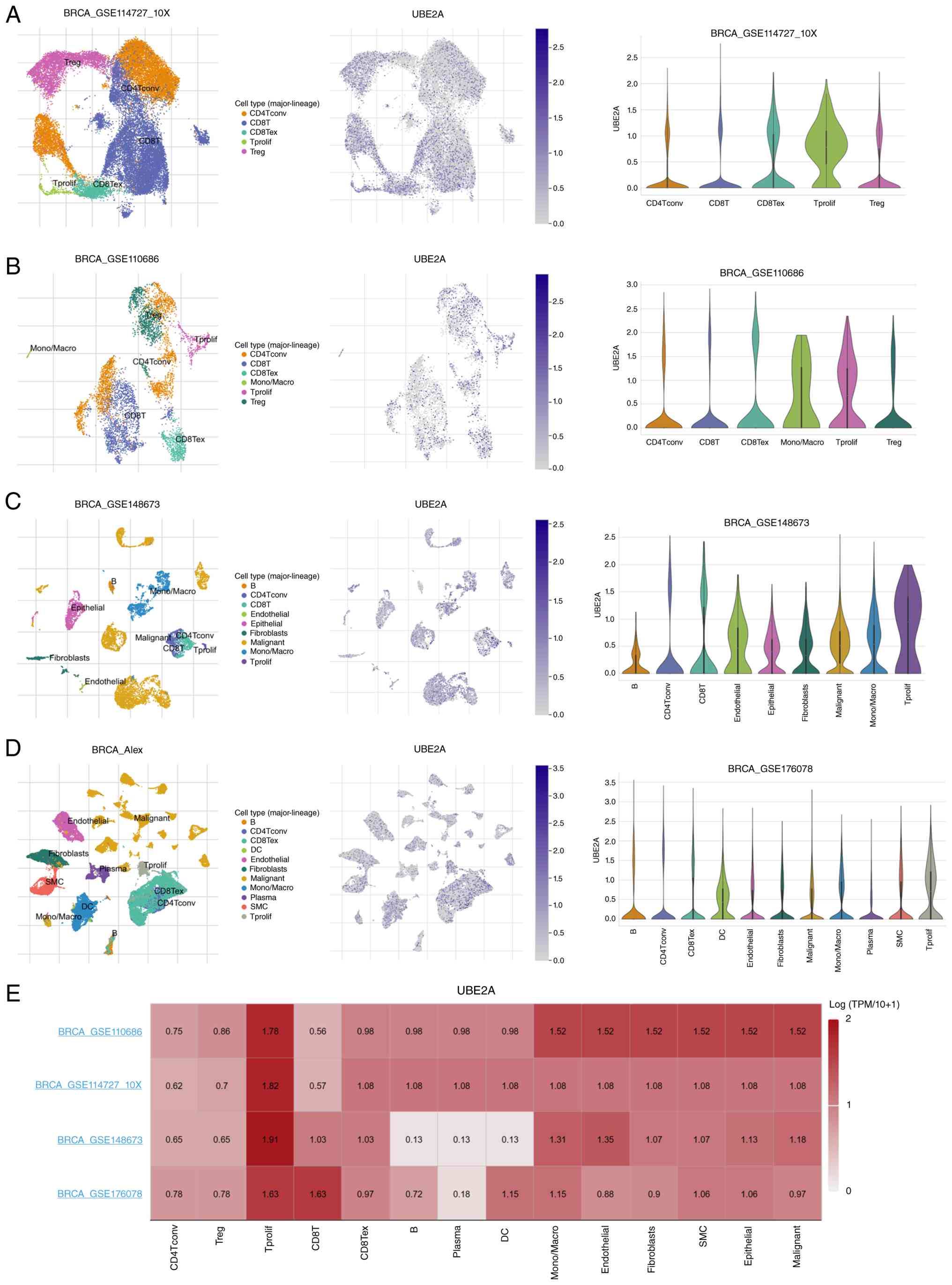 | Figure 11.Single-cell transcriptomic
characterization of UBE2A expression in breast cancer. Uniform
Manifold Approximation and Projection and Violin plots showing the
expression levels of UBE2A across different cellular subpopulations
in BRCA single-cell RNA sequencing datasets (A) GSE114727, (B)
GSE110686, (C) GSE148673 and (D) GSE176078. (E) UBE2A expression
landscape across cellular subpopulations, highlighting preferential
expression in proliferative T-cell populations. UBE2A, ubiquitin
conjugating enzyme E2A; TPM, transcripts per million; DC, dendritic
cell; SMC, smooth muscle cell; conv, conventional T cell; ex,
exhausted T cell; Tprolif, proliferative T-cell; Treg, regulatory
T-cell. |
UBE2A deficiency suppresses migration,
proliferation and invasion of breast cancer cells in vitro
Based on the pan-cancer analysis results, breast
cancer was selected for functional validation as UBE2A exhibited
significant upregulation and prognostic relevance in BRCA. In
addition, the present bioinformatic analyses suggested a potential
involvement of UBE2A in tumor progression and tumor
microenvironment regulation in breast cancer. Therefore, the
biological functions of UBE2A in breast cancer cell lines were
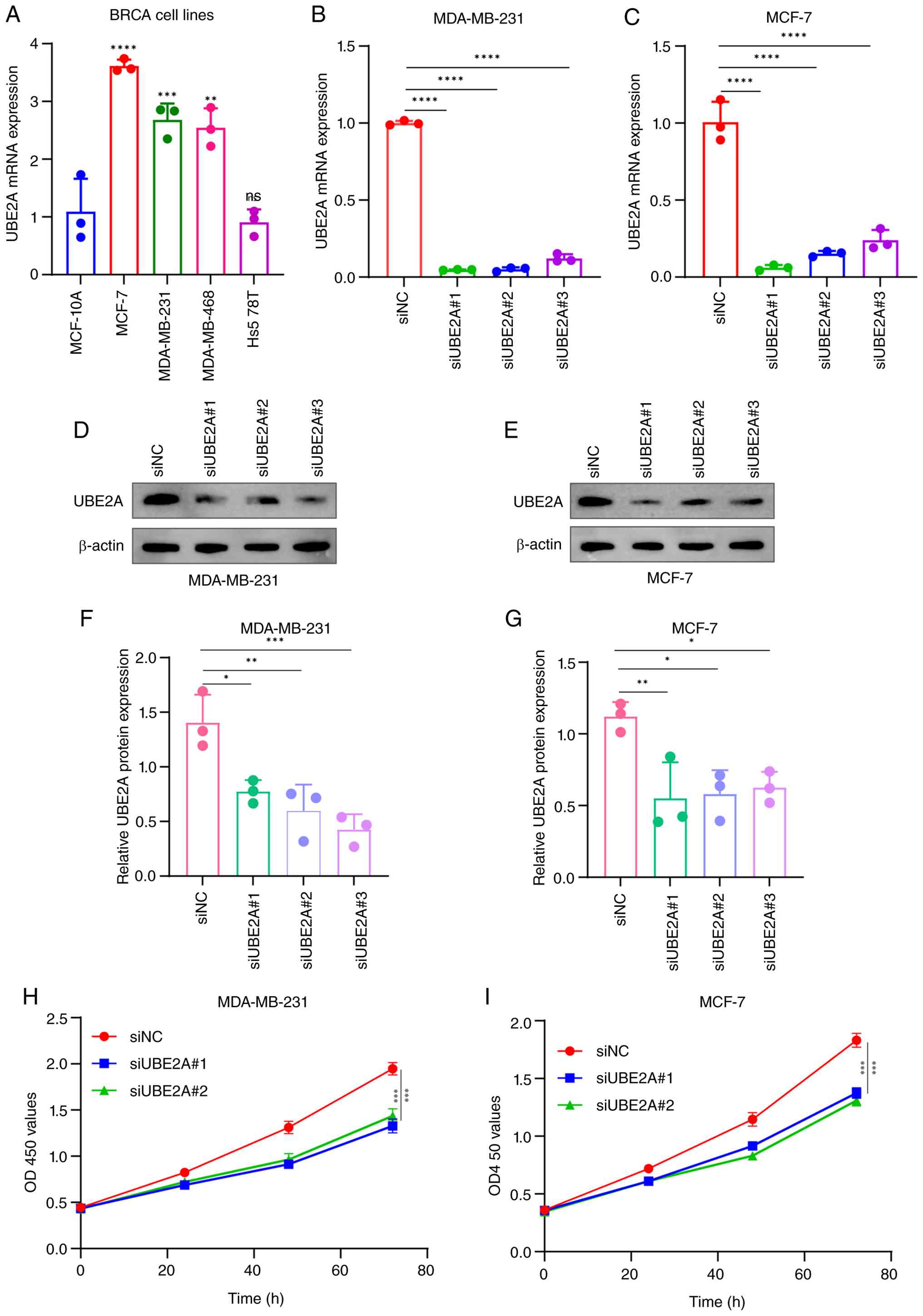
further investigated. qPCR analysis demonstrated differential UBE2A
expression among breast cancer cell lines. Compared with the normal
breast epithelial cell line MCF-10A, UBE2A expression was
significantly elevated in MCF-7, MDA-MB-231 and MDA-MB-468 cells
(Fig. 12A). Based on their
relatively high expression levels, MCF-7 and MDA-MB-231 cells were
selected for subsequent functional experiments.
A total of three siRNAs targeting UBE2A were
designed and transfected into MCF-7 and MDA-MB-231 cells. qPCR and
western blotting analyses demonstrated that all three siRNAs
significantly reduced UBE2A expression (Fig. 12B-G), with siUBE2A#1 and siUBE2A#2
exhibiting the strongest knockdown efficiency; thus, these two
sequences were chosen for subsequent experiments. CCK-8 assays
revealed that UBE2A silencing significantly inhibited cell
proliferation in both lines, compared with the negative control
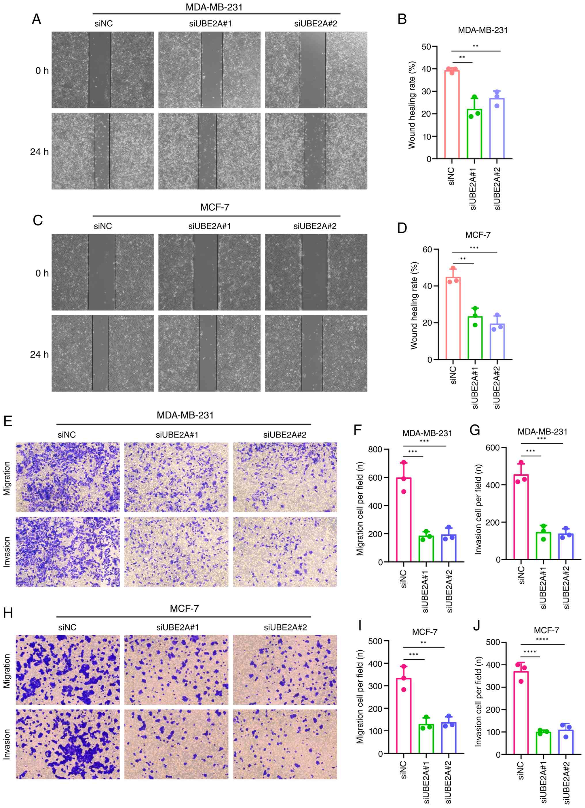
(Fig. 12H and I). Wound healing
assays further indicated a significant reduction in migration
capacity upon UBE2A knockdown, compared with the negative control
(Fig. 13A-D). Lastly, Transwell
assays demonstrated that UBE2A knockdown significantly impaired
both migration and invasion abilities in MCF-7 and MDA-MB-231 cells
(Fig. 13E-J).
Discussion
Cancer continues to represent a major global health
challenge owing to its high morbidity, mortality and biological
complexity (55,56). Despite notable advances in early
detection and therapeutic interventions, numerous aspects of tumor
initiation and progression remain insufficiently defined, largely
due to the extensive inter- and intra-tumoral heterogeneity
observed across cancer types (57). This complexity poses marked
barriers to durable therapeutic efficacy and underscores the
importance of identifying molecular determinants that are conserved
across cancers while retaining tumor-type specificity. Pan-cancer
analytical strategies provide a valuable framework for addressing
these challenges by enabling the systematic interrogation of
oncogenic processes across a number of malignancies, encompassing
tumor heterogeneity, evolutionary trajectories and interactions
with the tumor microenvironment (11). By integrating multi-omics datasets,
pan-cancer analyses can uncover shared molecular programs as well
as context-dependent features that may not be apparent in
single-cancer investigations. Within this framework, the present
study sought to characterize the pan-cancer landscape of UBE2A and
to explore its potential biological and clinical relevance.
Although UBE2A has been implicated in the
progression of a number of malignancies, including HCC, OV,
melanoma and multiple myeloma (58–61),
its expression profile and regulatory features across a broad
spectrum of cancers have not been systematically evaluated. Using
integrated TCGA and GTEx datasets, it was observed that UBE2A
expression exhibited marked heterogeneity across tumor types, with
notable upregulation in the majority of cancers, including GBM,
BRCA and STAD, as well as downregulation in a limited subset such
as KIRC. This pattern suggests that UBE2A expression may be subject
to tissue- or lineage-specific regulatory constraints. Epigenetic
analyses further indicated that reduced promoter DNA methylation
was associated with elevated UBE2A expression in certain cancer
types, including BLCA, LIHC, BRCA and STAD, consistent with the
established role of promoter hypomethylation in transcriptional
activation (62). However, in
other tumor types, differential expression of UBE2A occurred in the
absence of corresponding methylation changes, implying that
additional regulatory mechanisms, such as histone modifications,
transcription factor binding or post-transcriptional regulation,
may contribute to its context-dependent expression. These findings
highlight the complexity of UBE2A regulation and suggest that
epigenetic control alone does not fully account for its
dysregulation in cancer.
From a clinical perspective, elevated UBE2A
expression was found to be associated with unfavorable OS and DFS
in numerous malignancies, including breast cancer. While previous
studies have reported the prognostic relevance of UBE2A in
individual cancer types (18,22),
the present pan-cancer approach extended these observations by
providing a broader and more systematic assessment across diverse
tumor contexts. In addition to its prognostic implications, UBE2A
demonstrated notable diagnostic performance in a number of cancer
types, with strong discriminatory capacity observed in CHOL, ESAD,
ESCA, GBM, KICH and LIHC. These findings suggest that aberrant
UBE2A expression may reflect underlying oncogenic processes that
are detectable at the transcriptomic level. Collectively, these
results support the potential utility of UBE2A as a prognostic and
diagnostic biomarker in selected malignancies, although its
clinical applicability may vary depending on tumor type.
Somatic mutations affecting oncogenes and tumor
suppressor genes are central drivers of malignant transformation
and tumor evolution (63,64). Despite the overall mutation
frequency of UBE2A across cancers being relatively low in the
present analysis, a notable enrichment of UBE2A mutations was
observed in mature B-cell neoplasms, whereby mutation rates were
>10%. This tumor-type-specific pattern contrasts with the low
prevalence of UBE2A alterations in the majority of solid tumors.
These findings are consistent with previous reports suggesting that
UBE2A mutations, while uncommon, may exert disproportionate
functional effects in specific hematological malignancies (21,65).
Such context-dependent mutational roles underscore the importance
of considering tissue-specific molecular environments when
interpreting the oncogenic importance of UBE2A alterations and
warrant further functional investigation in hematologic cancer
models.
The tumor microenvironment constitutes a highly
dynamic network in which immune cells, stromal components and tumor
cells interact to influence disease progression and therapeutic
response (66,67). Increasing evidence has indicated
that dysregulation of immune infiltration and immune signaling
pathways contributes to immune evasion and resistance to therapy.
In the present study, numerous immune infiltration algorithms were
applied to minimize methodological bias and to comprehensively
evaluate the association between UBE2A expression and immune cell
composition across cancers. The present analyses revealed that
higher UBE2A expression was frequently associated with increased
infiltration of CD8+ T cells, dendritic cells,
neutrophils and macrophage subsets in a number of tumor types. In
addition, UBE2A expression correlated positively with numerous
immune checkpoint and immunoregulatory genes, including IL-12A and
CD276, which are known to modulate anti-tumor immunity and immune
homeostasis within the tumor microenvironment (68). These associations suggest that
UBE2A may participate in shaping immune-associated signaling
networks, potentially contributing to an immunosuppressive or
immune-dysregulated microenvironment that favors tumor progression.
However, it should be noted that these observations were primarily
derived from computational analyses of transcriptomic datasets and
therefore do not establish a direct causal role for UBE2A in
regulating anti-tumor immunity. Consequently, the immune-associated
findings of the present study should be interpreted as
hypothesis-generating evidence that requires further experimental
validation. TMB and MSI have emerged as clinically relevant
biomarkers for predicting responsiveness to immune checkpoint
inhibitors (51,52). The observed correlations between
UBE2A expression and both TMB and MSI across numerous cancer types
raise the possibility that UBE2A expression may reflect underlying
genomic instability and tumor immunogenicity, thereby providing
complementary information for immunotherapy stratification.
Notably, single-cell transcriptomic analysis
revealed relatively high UBE2A expression in proliferative T-cell
populations within the tumor microenvironment. Proliferative T
cells are generally considered an activated immune cell subset
associated with ongoing anti-tumor immune responses, but persistent
proliferative signaling may also reflect T-cell dysfunction or
exhaustion under chronic tumor-associated immune stimulation
(69). Therefore, the elevated
expression of UBE2A in proliferative T cells suggests that UBE2A
may participate in immune regulatory processes within the tumor
microenvironment. Despite this, the present study did not directly
investigate the biological function of UBE2A in T cells. Future
studies employing immune-cell models, co-culture systems and
immunocompetent animal models should aim to determine whether UBE2A
directly influences T-cell activation, proliferation, exhaustion or
anti-tumor immune responses. Although the present study primarily
focused on the tumor-intrinsic functions of UBE2A in breast cancer
cells, these findings suggest a potential immunomodulatory role of
UBE2A that warrants further investigation through dedicated
immune-associated functional experiments, such as T-cell co-culture
systems and immune phenotype analyses.
Mechanistically, UBE2A has been reported to
facilitate p53 degradation through the ubiquitin-proteasome pathway
(70) thereby attenuating
p53-mediated cell cycle arrest and apoptotic signaling. Due to the
central role of p53 in tumor suppression, dysregulation of this
axis may represent a key mechanism through which UBE2A promotes
malignant phenotypes. Beyond the p53 axis, the comparable
suppressive phenotypes observed following UBE2A silencing in both
wild-type p53 (MCF-7) and mutant p53 (MDA-MB-231) cells suggest
that additional p53-independent mechanisms may be involved. As a
ubiquitin-conjugating enzyme, UBE2A participates in
ubiquitin-mediated protein turnover and may influence the stability
of a number of proteins beyond p53, including heterochromatin
protein 1 (HP1) and cyclin D1 (16,71).
The present enrichment analyses revealed significant
associations between UBE2A and cell-cycle regulation, chromosome
segregation, mitotic progression and proteasome-associated
pathways. Consistent with these observations, previous studies have
demonstrated that RAD6A (UBE2A) promotes homologous
recombination-mediated DNA double-strand break repair through
ubiquitin-dependent degradation of heterochromatin protein HP1α,
thereby facilitating chromatin remodeling and maintaining genome
stability (71). In addition,
RAD6A (UBE2A) has been reported to enhance DNA damage tolerance and
repair signaling while inducing the expression of
stemness-associated genes through histone H2B
ubiquitination-mediated transcriptional regulation, ultimately
contributing to tumor progression and acquired chemoresistance
(18). These findings raise the
possibility that UBE2A may facilitate tumor cell proliferation
through the regulation of key cell-cycle regulators and mitotic
proteins, thereby promoting G1/S transition, maintaining
mitotic fidelity and supporting efficient chromosome segregation
during cell division. Dysregulation of these processes is a
well-established driver of genomic instability and tumor
progression (16,18,71).
In addition, the enrichment of proteasome-associated pathways
suggests that UBE2A may broadly regulate protein homeostasis
through ubiquitin-dependent degradation mechanisms. Therefore, its
oncogenic effects may not be restricted to p53 turnover but may
also involve the modulation of additional cell-cycle, DNA repair or
signaling proteins. Notably, antigen-processing pathways were also
enriched among UBE2A-associated genes. Given that
ubiquitin-mediated proteolysis serves a key role in generating
peptides for MHC class I antigen presentation, aberrant UBE2A
expression may influence tumor immune recognition and contribute to
the immune-related associations observed in the present pan-cancer
analyses. Collectively, these findings support a broader functional
role of UBE2A beyond p53 regulation and suggest that both
p53-dependent and p53-independent mechanisms may contribute to its
tumor-promoting activities.
A previous study in hepatocellular carcinoma
reported that UBE2A is regulated by an upstream non-coding RNA
network and promotes tumor progression (22). Consistent with these observations,
functional enrichment analyses of UBE2A co-expressed genes in the
present study highlighted strong associations with cell cycle
regulation and mitotic processes, as supported by GO and KEGG
annotations. Notably, GSEA further revealed enrichment of antigen
processing and presentation pathways, suggesting a potential
association between UBE2A activity and tumor immunogenicity. Among
the analyzed tumor types, BRCA was selected for functional
validation as UBE2A exhibited significant upregulation, strong
diagnostic performance and unfavorable prognostic associations in
BRCA within the pan-cancer analysis. Although the highest mutation
frequency of UBE2A was observed in mature B-cell neoplasms and
single-cell analyses demonstrated relatively high expression in
proliferative T-cell populations, the primary objective of the
present study was to evaluate the tumor-intrinsic role of UBE2A in
a solid tumor model. Breast cancer was therefore selected as a
representative cancer type with robust transcriptomic evidence and
accessible experimental systems. Consequently, the potential
functions of UBE2A in immune cells and hematological malignancies
remain key topics for future investigations. In addition,
single-cell transcriptomic analyses revealed distinct UBE2A
expression patterns in the breast cancer tumor microenvironment,
further supporting its potential biological relevance in BRCA
progression. Complementing these bioinformatic findings,
loss-of-function experiments demonstrated that UBE2A silencing
significantly impaired the proliferative, migratory and invasive
capacities of breast cancer cells in vitro. Collectively,
these results indicate that UBE2A may contribute to tumor
progression through coordinated regulation of cell cycle dynamics
and immune-associated processes.
A number of limitations of the present study should
be considered. First, although external validation was performed
using independent GEO cohorts, the majority of pan-cancer analyses
were still based on publicly available datasets and may therefore
be influenced by dataset-specific biases. Additional validation
using larger prospective clinical cohorts is warranted. Second, the
associations identified between UBE2A expression and
immune-associated characteristics were primarily derived from
bioinformatic analyses and remain correlative in nature. Third,
despite the enrichment of UBE2A expression in proliferative T-cell
populations revealed by single-cell analyses, the experimental
validation in the present study was restricted to breast cancer
epithelial cells and direct evidence supporting an immunomodulatory
role of UBE2A is currently lacking. Fourth, the functional
experiments relied on transient siRNA-mediated knockdown, which may
be associated with potential off-target effects. Future studies
using stable gene modulation strategies, such as shRNA- or
CRISPR/Cas9-mediated approaches, together with rescue experiments
and immunocompetent animal models, are necessary to further
validate the biological functions of UBE2A.
In conclusion, the present study provided a
systematic pan-cancer evaluation of UBE2A, outlining its expression
characteristics and clinical relevance across a broad range of
malignancies. The results indicated that aberrant UBE2A expression
was frequently associated with adverse clinical outcomes and
displays measurable diagnostic and prognostic utility in selected
cancer types. In addition, UBE2A expression showed consistent
associations with immune cell infiltration patterns and the
expression of immune regulatory molecules, suggesting its
involvement in tumor-immune interactions. In breast cancer, UBE2A
was significantly upregulated and correlated with unfavorable
prognosis and functional assays further demonstrated that UBE2A
depletion attenuated malignant phenotypes in vitro.
Collectively, these findings supported a role for UBE2A in cancer
progression and suggested a potential association between UBE2A and
immune-association processes, thereby underscoring the need for
further mechanistic studies to clarify its biological functions and
potential therapeutic value.
Supplementary Material
Supporting Data
Supporting Data
Acknowledgements
Not applicable.
Funding
Funding: No funding was received.
Availability of data and materials
The data generated in the present study may be
requested from the corresponding author.
Authors' contributions
YW conceived and designed the study, performed
bioinformatics analyses, conducted the cell experiments, collected
and analyzed the data, generated the figures and visualizations,
validated the experimental and computational results, and drafted
the manuscript. PL supervised the study, provided research
resources and technical support, participated in data
interpretation, critically reviewed and revised the manuscript and
managed the project. YW and PL confirm the authenticity of all the
raw data. All authors read and approved the final version of the
manuscript.
Ethics approval and consent to
participate
Not applicable.
Patient consent for publication
Not applicable.
Competing interests
The authors declare that they have no competing
interests.
Use of artificial intelligence tools
During the preparation of this work, artificial
intelligence tools were used to improve the readability and
language of the manuscript or to generate images, and subsequently,
the authors revised and edited the content produced by the
artificial intelligence tools as necessary, taking full
responsibility for the ultimate content of the present
manuscript.
Glossary
Abbreviations
Abbreviations:
|
UBE2A
|
ubiquitin conjugating enzyme E2A
|
|
TCGA
|
The Cancer Genome Atlas
|
|
GTEx
|
Genotype-Tissue Expression Project
|
|
ROC
|
receiver operating characteristic
|
|
AUC
|
area under the curve
|
|
OS
|
overall survival
|
|
DFS
|
disease-free survival
|
|
DMFS
|
distant metastasis-free survival
|
|
RFS
|
relapse-free survival
|
|
GSEA
|
Gene Set Enrichment Analysis
|
|
KEGG
|
Kyoto Encyclopedia Of Genes And
Genomes
|
|
GO
|
Gene Ontology
|
|
TMB
|
tumor mutational burden
|
|
MSI
|
microsatellite instability
analysis
|
|
CCK-8
|
Cell Counting Kit-8
|
|
BLCA
|
bladder urothelial carcinoma
|
|
CESC
|
cervical squamous cell carcinoma
|
|
CHOL
|
cholangiocarcinoma
|
|
ESCA
|
esophageal carcinoma
|
|
GBM
|
glioblastoma multiforme
|
|
HNSC
|
head-neck squamous cell carcinoma
|
|
LIHC
|
liver hepatocellular carcinoma
|
|
LUAD
|
lung adenocarcinoma
|
|
LUSC
|
lung squamous cell carcinoma
|
|
STAD
|
stomach adenocarcinoma; gastric
adenocarcinoma
|
|
THCA
|
thyroid carcinoma
|
|
COAD
|
colon adenocarcinoma
|
|
KICH
|
kidney chromophobe
|
|
KIRC
|
kidney renal clear cell carcinoma
|
|
KIRP
|
kidney renal papillary cell
carcinoma
|
|
PRAD
|
prostate adenocarcinoma
|
|
STES
|
stomach and esophageal carcinoma
|
|
ESAD
|
esophageal adenocarcinoma
|
|
UVM
|
uveal melanoma
|
|
SARC
|
sarcoma
|
|
THYM
|
thymoma
|
|
TISCH2
|
Tumor Immune Single Cell Hub 2
|
References
|
1
|
Bray F, Laversanne M, Sung H, Ferlay J,
Siegel RL, Soerjomataram I and Jemal A: Global cancer statistics
2022: GLOBOCAN estimates of incidence and mortality worldwide for
36 cancers in 185 countries. CA Cancer J Clin. 74:229–263.
2024.PubMed/NCBI
|
|
2
|
Bizuayehu HM, Ahmed KY, Kibret GD, Dadi
AF, Belachew SA, Bagade T, Tegegne TK, Venchiarutti RL, Kibret KT,
Hailegebireal AH, et al: Global disparities of cancer and its
projected burden in 2050. JAMA Netw Open. 7:e24431982024.
View Article : Google Scholar : PubMed/NCBI
|
|
3
|
Tanaka M, Lum L, Hu KH, Chaudhary P,
Hughes S, Ledezma-Soto C, Samad B, Superville D, Ng K, Chumber A,
et al: Tumor cell heterogeneity drives spatial organization of the
intratumoral immune response. J Exp Med. 222:e202422822025.
View Article : Google Scholar : PubMed/NCBI
|
|
4
|
MacDonald WJ, Purcell C, Pinho-Schwermann
M, Stubbs NM, Srinivasan PR and El-Deiry WS: Heterogeneity in
cancer. Cancers. 17:4412025. View Article : Google Scholar : PubMed/NCBI
|
|
5
|
Wang J, Chen Q, Shan Q, Liang T, Forde P
and Zheng L: Clinical development of immuno-oncology therapeutics.
Cancer Lett. 617:2176162025. View Article : Google Scholar : PubMed/NCBI
|
|
6
|
Zhang M, Liu C, Tu J, Tang M, Ashrafizadeh
M, Nabavi N, Sethi G, Zhao P and Liu S: Advances in cancer
immunotherapy: Historical perspectives, current developments, and
future directions. Mol Cancer. 24:1362025. View Article : Google Scholar : PubMed/NCBI
|
|
7
|
Saw PE, Liu Q, Wong PP and Song E: Cancer
stem cell mimicry for immune evasion and therapeutic resistance.
Cell Stem Cell. 31:1101–1112. 2024. View Article : Google Scholar : PubMed/NCBI
|
|
8
|
Barkley D, Moncada R, Pour M, Liberman DA,
Dryg I, Werba G, Wang W, Baron M, Rao A, Xia B, et al: Cancer cell
states recur across tumor types and form specific interactions with
the tumor microenvironment. Nat Genet. 54:1192–1201. 2022.
View Article : Google Scholar : PubMed/NCBI
|
|
9
|
Wang X, Wang J, Wang Q, Ding G, Huang Y
and Feng Y: Precision cytokine modulation to overcome tumor
microenvironment-driven resistance to immune checkpoint blockade.
Biochim Biophys Acta Rev Cancer. 1881:1895362026. View Article : Google Scholar : PubMed/NCBI
|
|
10
|
Wickramasekara R, Jones V, Zhao Y, Luo S,
Sun G and Huang RP: Mechanisms of therapy resistance in the tumor
microenvironment: Insights from antibody array-based cytokine
profiling. Drug Resist Updat. 86:1013782026. View Article : Google Scholar : PubMed/NCBI
|
|
11
|
Cen X, Lan Y, Zou J, Chen R, Hu C, Tong Y,
Zhang C, Chen J, Wang Y, Zhou R, et al: Pan-cancer analysis shapes
the understanding of cancer biology and medicine. Cancer Commun
(Lond). 45:728–746. 2025. View Article : Google Scholar : PubMed/NCBI
|
|
12
|
Knol JC, Lyu M, Böttger F, Nunes Monteiro
M, Pham TV, Rolfs F, Vallés-Martí A, Schelfhorst T, de Goeij-de
Haas RR, Bijnsdorp IV, et al: The pan-cancer proteome atlas, a mass
spectrometry-based landscape for discovering tumor biology,
biomarkers, and therapeutic targets. Cancer Cell. 43:1328–1346.e8.
2025. View Article : Google Scholar : PubMed/NCBI
|
|
13
|
Bai L, Lyu J, Feng J, Qiao X, Qu Y, Yang
G, Zhu Y, Liao L, Gao H, Zang A, et al: Cancer biomarkers
discovered using pan-cancer plasma proteomic profiling. Nat Biomed
Eng. 10:16–38. 2025. View Article : Google Scholar : PubMed/NCBI
|
|
14
|
Hao Q, Zhou T, Yan H, Ren Z, Mao W, Huang
LB, Chen YY, Wang Y, Xiao M, He Y, et al: A pan-cancer single cell
landscape reveals heterogeneity and functional diversity of
double-negative T cells. Mol Cancer. 25:242026. View Article : Google Scholar : PubMed/NCBI
|
|
15
|
Lusby R, Demirdizen E, Inayatullah M,
Kundu P, Maiques O, Zhang Z, Terp MG, Sanz-Moreno V and Tiwari VK:
Pan-cancer drivers of metastasis. Mol Cancer. 24:22025. View Article : Google Scholar : PubMed/NCBI
|
|
16
|
Cai F, Chen P, Chen L, Biskup E, Liu Y,
Chen PC, Chang JF, Jiang W, Jing Y, Chen Y, et al: Human RAD6
promotes G1-S transition and cell proliferation through
upregulation of cyclin D1 expression. PLoS One. 9:e1137272014.
View Article : Google Scholar : PubMed/NCBI
|
|
17
|
Hong JH, Kaustov L, Coyaud E, Srikumar T,
Wan J, Arrowsmith C and Raught B: KCMF1 (potassium channel
modulatory factor 1) Links RAD6 to UBR4 (ubiquitin N-recognin
domain-containing E3 ligase 4) and lysosome-mediated degradation.
Mol Cell Proteomics. 14:674–685. 2015. View Article : Google Scholar : PubMed/NCBI
|
|
18
|
Somasagara RR, Spencer SM, Tripathi K,
Clark DW, Mani C, Madeira da Silva L, Scalici J, Kothayer H,
Westwell AD, Rocconi RP and Palle K: RAD6 promotes DNA repair and
stem cell signaling in ovarian cancer and is a promising
therapeutic target to prevent and treat acquired chemoresistance.
Oncogene. 36:6680–6690. 2017. View Article : Google Scholar : PubMed/NCBI
|
|
19
|
Barnsby-Greer L, Mabbitt PD, Dery MA,
Squair DR, Wood NT, Lamoliatte F, Lange SM and Virdee S: UBE2A and
UBE2B are recruited by an atypical E3 ligase module in UBR4. Nat
Struct Mol Biol. 31:351–363. 2024. View Article : Google Scholar : PubMed/NCBI
|
|
20
|
Ramatenki V, Potlapally SR, Dumpati RK,
Vadija R and Vuruputuri U: Homology modeling and virtual screening
of ubiquitin conjugation enzyme E2A for designing a novel selective
antagonist against cancer. J Recept Signal Transduct Res.
35:536–549. 2015. View Article : Google Scholar : PubMed/NCBI
|
|
21
|
Magistroni V, Mauri M, D'Aliberti D,
Mezzatesta C, Crespiatico I, Nava M, Fontana D, Sharma N, Parker W,
Schreiber A, et al: De novo UBE2A mutations are recurrently
acquired during chronic myeloid leukemia progression and interfere
with myeloid differentiation pathways. Haematologica.
104:1789–1797. 2019. View Article : Google Scholar : PubMed/NCBI
|
|
22
|
Yan Y, Nie Y, Peng C, Xing F, Ji S, Liu H
and Zhu C: The circular RNA hsa_circ_0001394 promotes
hepatocellular carcinoma progression by targeting the miR-527/UBE2A
axis. Cell Death Discov. 8:812022. View Article : Google Scholar : PubMed/NCBI
|
|
23
|
Wickham H: ggplot2: Elegant Graphics for
Data Analysis. Springer-Verlag; New York, NY: 2016, Available
from:. https://ggplot2.tidyverse.org
|
|
24
|
Chen D, Xu L, Xing H, Shen W, Song Z, Li
H, Zhu X, Li X, Wu L, Jiao H, et al: Sangerbox 2: Enhanced
functionalities and update for a comprehensive clinical
bioinformatics data analysis platform. Imeta. 3:e2382024.
View Article : Google Scholar : PubMed/NCBI
|
|
25
|
Colak D, Nofal A, Albakheet A, Nirmal M,
Jeprel H, Eldali A, Al-Tweigeri T, Tulbah A, Ajarim D, Malik OA, et
al: Age-specific gene expression signatures for breast tumors and
cross-species conserved potential cancer progression markers in
young women. PLoS One. 8:e632042013. View Article : Google Scholar : PubMed/NCBI
|
|
26
|
Maire V, Némati F, Richardson M,
Vincent-Salomon A, Tesson B, Rigaill G, Gravier E, Marty-Prouvost
B, De Koning L, Lang G, et al: Polo-like kinase 1: A potential
therapeutic option in combination with conventional chemotherapy
for the management of patients with triple-negative breast cancer.
Cancer Res. 73:813–823. 2012. View Article : Google Scholar : PubMed/NCBI
|
|
27
|
Ru B, Wong CN, Tong Y, Zhong JY, Zhong
SSW, Wu WC, Chu KC, Wong CY, Lau CY, Chen I, et al: TISIDB: An
integrated repository portal for tumor-immune system interactions.
Bioinformatics. 35:4200–4202. 2019. View Article : Google Scholar : PubMed/NCBI
|
|
28
|
Robin X, Turck N, Hainard A, Tiberti N,
Lisacek F, Sanchez JC and Müller M: pROC: An open-source package
for R and S+ to analyze and compare ROC curves. BMC Bioinformatics.
12:772011. View Article : Google Scholar : PubMed/NCBI
|
|
29
|
Tang Z, Kang B, Li C, Chen T and Zhang Z:
GEPIA2: An enhanced web server for large-scale expression profiling
and interactive analysis. Nucleic Acids Res. 47:W556–W560. 2019.
View Article : Google Scholar : PubMed/NCBI
|
|
30
|
Lánczky A and Győrffy B: Web-based
survival analysis tool tailored for medical research (KMplot):
Development and implementation. J Med Internet Res. 23:e276332021.
View Article : Google Scholar : PubMed/NCBI
|
|
31
|
Gao J, Aksoy BA, Dogrusoz U, Dresdner G,
Gross B, Sumer SO, Sun Y, Jacobsen A, Sinha R, Larsson E, et al:
Integrative analysis of complex cancer genomics and clinical
profiles using the cBioPortal. Sci Signal. 6:pl12013. View Article : Google Scholar : PubMed/NCBI
|
|
32
|
Vasaikar SV, Straub P, Wang J and Zhang B:
LinkedOmics: Analyzing multi-omics data within and across 32 cancer
types. Nucleic Acids Res. 46:D956–D963. 2018. View Article : Google Scholar : PubMed/NCBI
|
|
33
|
Balakrishnan R, Harris MA, Huntley R, Van
Auken K and Cherry JM: A guide to best practices for Gene Ontology
(GO) manual annotation. Database (Oxford). 2013:bat0542013.
View Article : Google Scholar : PubMed/NCBI
|
|
34
|
Kanehisa M and Goto S: KEGG: Kyoto
encyclopedia of genes and genomes. Nucleic Acids Res. 28:27–30.
2000. View Article : Google Scholar : PubMed/NCBI
|
|
35
|
Yu G, Wang LG, Han Y and He QY:
clusterProfiler: An R package for comparing biological themes among
gene clusters. OMICS. 16:284–287. 2012. View Article : Google Scholar : PubMed/NCBI
|
|
36
|
Franz M, Rodriguez H, Lopes C, Zuberi K,
Montojo J, Bader GD and Morris Q: GeneMANIA update 2018. Nucleic
Acids Res. 46:W60–W64. 2018. View Article : Google Scholar : PubMed/NCBI
|
|
37
|
Lee AV, Nestler KA and Chiappinelli KB:
Therapeutic targeting of DNA methylation alterations in cancer.
Pharmacol Ther. 258:1086402024. View Article : Google Scholar : PubMed/NCBI
|
|
38
|
Xu C, Xu X, Huang Y, Shang S and Ma L: RNA
methylation: A new promising biomaker in cancer liquid biopsy.
Biochim Biophys Acta Rev Cancer. 1880:1893372025. View Article : Google Scholar : PubMed/NCBI
|
|
39
|
Chandrashekar DS, Karthikeyan SK, Korla
PK, Patel H, Shovon AR, Athar M, Netto GJ, Qin ZS, Kumar S, Manne
U, et al: UALCAN: An update to the integrated cancer data analysis
platform. Neoplasia. 25:18–27. 2022. View Article : Google Scholar : PubMed/NCBI
|
|
40
|
Newman AM, Liu CL, Green MR, Gentles AJ,
Feng W, Xu Y, Hoang CD, Diehn M and Alizadeh AA: Robust enumeration
of cell subsets from tissue expression profiles. Nat Methods.
12:453–457. 2015. View Article : Google Scholar : PubMed/NCBI
|
|
41
|
Li T, Fan J, Wang B, Traugh N, Chen Q, Liu
JS, Li B and Liu XS: TIMER: A web server for comprehensive analysis
of tumor-infiltrating immune cells. Cancer Res. 77:e108–e110. 2017.
View Article : Google Scholar : PubMed/NCBI
|
|
42
|
Racle J and Gfeller D: EPIC: A tool to
estimate the proportions of different cell types from bulk gene
expression data. Methods Mol Biol. 2120:233–248. 2020. View Article : Google Scholar : PubMed/NCBI
|
|
43
|
Plattner C, Finotello F and Rieder D:
Deconvoluting tumor-infiltrating immune cells from RNA-seq data
using quanTIseq. Methods Enzymol. 636:261–285. 2019.PubMed/NCBI
|
|
44
|
Aran D, Hu Z and Butte AJ: xCell:
Digitally portraying the tissue cellular heterogeneity landscape.
Genome Biol. 18:2202017. View Article : Google Scholar : PubMed/NCBI
|
|
45
|
Han Y, Wang Y, Dong X, Sun D, Liu Z, Yue
J, Wang H, Li T and Wang C: TISCH2: Expanded datasets and new tools
for single-cell transcriptome analyses of the tumor
micro-environment. Nucleic Acids Res. 51:D1425–D1431. 2023.
View Article : Google Scholar : PubMed/NCBI
|
|
46
|
Azizi E, Carr AJ, Plitas G, Cornish AE,
Konopacki C, Prabhakaran S, Nainys J, Wu K, Kiseliovas V, Setty M,
et al: Single-cell map of diverse immune phenotypes in the breast
tumor microenvironment. Cell. 174:1293–1308.e36. 2018. View Article : Google Scholar : PubMed/NCBI
|
|
47
|
Savas P, Virassamy B, Ye C, Salim A,
Mintoff CP, Caramia F, Salgado R, Byrne DJ, Teo ZL, Dushyanthen S,
et al: Single-cell profiling of breast cancer T cells reveals a
tissue-resident memory subset associated with improved prognosis.
Nat Med. 24:986–993. 2018. View Article : Google Scholar : PubMed/NCBI
|
|
48
|
Gao R, Bai S, Henderson YC, Lin Y, Schalck
A, Yan Y, Kumar T, Hu M, Sei E, Davis A, et al: Delineating copy
number and clonal substructure in human tumors from single-cell
transcriptomes. Nat Biotechnol. 39:599–608. 2021. View Article : Google Scholar : PubMed/NCBI
|
|
49
|
Wu SZ, Al-Eryani G, Roden DL, Junankar S,
Harvey K, Andersson A, Thennavan A, Wang C, Torpy JR, Bartonicek N,
et al: A single-cell and spatially resolved atlas of human breast
cancers. Nat Genet. 53:1334–1347. 2021. View Article : Google Scholar : PubMed/NCBI
|
|
50
|
Livak KJ and Schmittgen TD: Analysis of
relative gene expression data using real-time quantitative PCR and
the 2(−Delta Delta C(T)) method. Methods. 25:402–408. 2001.
View Article : Google Scholar : PubMed/NCBI
|
|
51
|
Zheng M: Super-high tumor mutational
burden predicts complete remission following immunotherapy: From
Peto's paradox to druggable cancer hallmark. J Immunother Cancer.
13:e0104862025. View Article : Google Scholar : PubMed/NCBI
|
|
52
|
Landre T and Des Guetz G: Microsatellite
instability-high status as a pan-cancer biomarker for immunotherapy
efficacy. Cancer Immunol Immunother. 74:1222025. View Article : Google Scholar : PubMed/NCBI
|
|
53
|
Cheng X, Peng T, Chu T, Yang Y, Liu J, Gao
Q, Cao C and Wei J: Application of single-cell and spatial omics in
deciphering cellular hallmarks of cancer drug response and
resistance. J Hematol Oncol. 18:702025. View Article : Google Scholar : PubMed/NCBI
|
|
54
|
Tirosh I and Suva ML: Cancer cell states:
Lessons from ten years of single-cell RNA-sequencing of human
tumors. Cancer Cell. 42:1497–1506. 2024. View Article : Google Scholar : PubMed/NCBI
|
|
55
|
Siegel RL, Giaquinto AN and Jemal A:
Cancer statistics, 2024. CA Cancer J Clin. 74:12–49.
2024.PubMed/NCBI
|
|
56
|
Diao X, Guo C, Jin Y, Li B, Gao X, Du X,
Chen Z, Jo M, Zeng Y, Ding C, et al: Cancer situation in China: An
analysis based on the global epidemiological data released in 2024.
Cancer Commun (Lond). 45:178–197. 2025. View Article : Google Scholar : PubMed/NCBI
|
|
57
|
Cordani M, Dando I, Ambrosini G and
González-Menéndez P: Signaling, cancer cell plasticity, and
intratumor heterogeneity. Cell Commun Signal. 22:2552024.
View Article : Google Scholar : PubMed/NCBI
|
|
58
|
Shen JD, Fu SZ, Ju LL, Wang YF, Dai F, Liu
ZX, Ji HZ, Shao JG and Bian ZL: High expression of
ubiquitin-conjugating enzyme E2A predicts poor prognosis in
hepatocellular carcinoma. Oncol Lett. 15:7362–7368. 2018.PubMed/NCBI
|
|
59
|
Zou R, Xu H, Li F, Wang S and Zhu L:
Increased expression of UBE2T predicting poor survival of
epithelial ovarian cancer: Based on comprehensive analysis of
UBE2s, clinical samples, and the GEO database. DNA Cell Biol.
40:36–60. 2021. View Article : Google Scholar : PubMed/NCBI
|
|
60
|
Rosner K, Mehregan DR, Kirou E, Abrams J,
Kim S, Campbell M, Frieder J, Lawrence K, Haynes B and Shekhar MP:
Melanoma development and progression are associated with Rad6
Upregulation and β-catenin relocation to the cell membrane. J Skin
Cancer. 2014:4392052014. View Article : Google Scholar : PubMed/NCBI
|
|
61
|
Zamani-Ahmadmahmudi M, Nassiri SM and
Soltaninezhad F: Development of an RNA sequencing-based prognostic
gene signature in multiple myeloma. Br J Haematol. 192:310–321.
2021. View Article : Google Scholar : PubMed/NCBI
|
|
62
|
Berdasco M and Esteller M: Clinical
epigenetics: Seizing opportunities for translation. Nat Rev Genet.
20:109–127. 2018. View Article : Google Scholar : PubMed/NCBI
|
|
63
|
Wang Z, Strasser A and Kelly GL: Should
mutant TP53 be targeted for cancer therapy? Cell Death Differ.
29:911–120. 2022. View Article : Google Scholar : PubMed/NCBI
|
|
64
|
Hauseman ZJ, Stauffer F, Beyer KS, Mollé
S, Cavicchioli E, Marchand JR, Fodor M, Viscomi J, Dhembi A, Katz
S, et al: Targeting the SHOC2-RAS interaction in RAS-mutant
cancers. Nature. 642:232–241. 2025. View Article : Google Scholar : PubMed/NCBI
|
|
65
|
de Miranda NFCC, Georgiou K, Chen L, Wu C,
Gao Z, Zaravinos A, Lisboa S, Enblad G, Teixeira MR, Zeng Y, et al:
Exome sequencing reveals novel mutation targets in diffuse large
B-cell lymphomas derived from Chinese patients. Blood.
124:2544–2553. 2014. View Article : Google Scholar : PubMed/NCBI
|
|
66
|
de Visser KE and Joyce JA: The evolving
tumor microenvironment: From cancer initiation to metastatic
outgrowth. Cancer Cell. 41:374–403. 2023. View Article : Google Scholar : PubMed/NCBI
|
|
67
|
Zeng D, Yu Y, Qiu W, Ou Q, Mao Q, Jiang L,
Wu J, Wu J, Luo H, Luo P, et al: Immunotyping the tumor
microenvironment reveals molecular heterogeneity for personalized
immunotherapy in cancer. Adv Sci (Weinh). 12:e24175932025.
View Article : Google Scholar : PubMed/NCBI
|
|
68
|
Cheng M, Chen S, Li K, Wang G, Xiong G,
Ling R, Zhang C, Zhang Z, Han H, Chen Z, et al: CD276-dependent
efferocytosis by tumor-associated macrophages promotes immune
evasion in bladder cancer. Nat Commun. 15:28182024. View Article : Google Scholar : PubMed/NCBI
|
|
69
|
Baessler A and Vignali DAA: T cell
exhaustion. Annu Rev Immunol. 42:179–206. 2024. View Article : Google Scholar : PubMed/NCBI
|
|
70
|
Chen S, Wang DL, Liu Y, Zhao L and Sun FL:
RAD6 regulates the dosage of p53 by a combination of
transcriptional and posttranscriptional mechanisms. Mol Cell Biol.
32:576–587. 2012. View Article : Google Scholar : PubMed/NCBI
|
|
71
|
Chen S, Wang C, Sun L, Wang DL, Chen L,
Huang Z, Yang Q, Gao J, Yang XB, Chang JF, et al: RAD6 promotes
homologous recombination repair by activating the
autophagy-mediated degradation of heterochromatin protein HP1. Mol
Cell Biol. 35:406–416. 2014. View Article : Google Scholar : PubMed/NCBI
|