Spandidos Publications Logo
  • About
    • About Spandidos
    • Aims and Scopes
    • Abstracting and Indexing
    • Editorial Policies
    • Reprints and Permissions
    • Job Opportunities
    • Terms and Conditions
    • Contact
  • Journals
    • All Journals
    • Oncology Letters
      • Oncology Letters
      • Information for Authors
      • Editorial Policies
      • Editorial Board
      • Aims and Scope
      • Abstracting and Indexing
      • Bibliographic Information
      • Archive
    • International Journal of Oncology
      • International Journal of Oncology
      • Information for Authors
      • Editorial Policies
      • Editorial Board
      • Aims and Scope
      • Abstracting and Indexing
      • Bibliographic Information
      • Archive
    • Molecular and Clinical Oncology
      • Molecular and Clinical Oncology
      • Information for Authors
      • Editorial Policies
      • Editorial Board
      • Aims and Scope
      • Abstracting and Indexing
      • Bibliographic Information
      • Archive
    • Experimental and Therapeutic Medicine
      • Experimental and Therapeutic Medicine
      • Information for Authors
      • Editorial Policies
      • Editorial Board
      • Aims and Scope
      • Abstracting and Indexing
      • Bibliographic Information
      • Archive
    • International Journal of Molecular Medicine
      • International Journal of Molecular Medicine
      • Information for Authors
      • Editorial Policies
      • Editorial Board
      • Aims and Scope
      • Abstracting and Indexing
      • Bibliographic Information
      • Archive
    • Biomedical Reports
      • Biomedical Reports
      • Information for Authors
      • Editorial Policies
      • Editorial Board
      • Aims and Scope
      • Abstracting and Indexing
      • Bibliographic Information
      • Archive
    • Oncology Reports
      • Oncology Reports
      • Information for Authors
      • Editorial Policies
      • Editorial Board
      • Aims and Scope
      • Abstracting and Indexing
      • Bibliographic Information
      • Archive
    • Molecular Medicine Reports
      • Molecular Medicine Reports
      • Information for Authors
      • Editorial Policies
      • Editorial Board
      • Aims and Scope
      • Abstracting and Indexing
      • Bibliographic Information
      • Archive
    • World Academy of Sciences Journal
      • World Academy of Sciences Journal
      • Information for Authors
      • Editorial Policies
      • Editorial Board
      • Aims and Scope
      • Abstracting and Indexing
      • Bibliographic Information
      • Archive
    • International Journal of Functional Nutrition
      • International Journal of Functional Nutrition
      • Information for Authors
      • Editorial Policies
      • Editorial Board
      • Aims and Scope
      • Abstracting and Indexing
      • Bibliographic Information
      • Archive
    • International Journal of Epigenetics
      • International Journal of Epigenetics
      • Information for Authors
      • Editorial Policies
      • Editorial Board
      • Aims and Scope
      • Abstracting and Indexing
      • Bibliographic Information
      • Archive
    • Medicine International
      • Medicine International
      • Information for Authors
      • Editorial Policies
      • Editorial Board
      • Aims and Scope
      • Abstracting and Indexing
      • Bibliographic Information
      • Archive
  • Articles
  • Information
    • Information for Authors
    • Information for Reviewers
    • Information for Librarians
    • Information for Advertisers
    • Conferences
  • Language Editing
Spandidos Publications Logo
  • About
    • About Spandidos
    • Aims and Scopes
    • Abstracting and Indexing
    • Editorial Policies
    • Reprints and Permissions
    • Job Opportunities
    • Terms and Conditions
    • Contact
  • Journals
    • All Journals
    • Biomedical Reports
      • Information for Authors
      • Editorial Policies
      • Editorial Board
      • Aims and Scope
      • Abstracting and Indexing
      • Bibliographic Information
      • Archive
    • Experimental and Therapeutic Medicine
      • Information for Authors
      • Editorial Policies
      • Editorial Board
      • Aims and Scope
      • Abstracting and Indexing
      • Bibliographic Information
      • Archive
    • International Journal of Epigenetics
      • Information for Authors
      • Editorial Policies
      • Editorial Board
      • Aims and Scope
      • Abstracting and Indexing
      • Bibliographic Information
      • Archive
    • International Journal of Functional Nutrition
      • Information for Authors
      • Editorial Policies
      • Editorial Board
      • Aims and Scope
      • Abstracting and Indexing
      • Bibliographic Information
      • Archive
    • International Journal of Molecular Medicine
      • Information for Authors
      • Editorial Policies
      • Editorial Board
      • Aims and Scope
      • Abstracting and Indexing
      • Bibliographic Information
      • Archive
    • International Journal of Oncology
      • Information for Authors
      • Editorial Policies
      • Editorial Board
      • Aims and Scope
      • Abstracting and Indexing
      • Bibliographic Information
      • Archive
    • Medicine International
      • Information for Authors
      • Editorial Policies
      • Editorial Board
      • Aims and Scope
      • Abstracting and Indexing
      • Bibliographic Information
      • Archive
    • Molecular and Clinical Oncology
      • Information for Authors
      • Editorial Policies
      • Editorial Board
      • Aims and Scope
      • Abstracting and Indexing
      • Bibliographic Information
      • Archive
    • Molecular Medicine Reports
      • Information for Authors
      • Editorial Policies
      • Editorial Board
      • Aims and Scope
      • Abstracting and Indexing
      • Bibliographic Information
      • Archive
    • Oncology Letters
      • Information for Authors
      • Editorial Policies
      • Editorial Board
      • Aims and Scope
      • Abstracting and Indexing
      • Bibliographic Information
      • Archive
    • Oncology Reports
      • Information for Authors
      • Editorial Policies
      • Editorial Board
      • Aims and Scope
      • Abstracting and Indexing
      • Bibliographic Information
      • Archive
    • World Academy of Sciences Journal
      • Information for Authors
      • Editorial Policies
      • Editorial Board
      • Aims and Scope
      • Abstracting and Indexing
      • Bibliographic Information
      • Archive
  • Articles
  • Information
    • For Authors
    • For Reviewers
    • For Librarians
    • For Advertisers
    • Conferences
  • Language Editing
Login Register Submit
  • This site uses cookies
  • You can change your cookie settings at any time by following the instructions in our Cookie Policy. To find out more, you may read our Privacy Policy.

    I agree
Search articles by DOI, keyword, author or affiliation
Search
Advanced Search
presentation
Oncology Letters
Join Editorial Board Propose a Special Issue
Print ISSN: 1792-1074 Online ISSN: 1792-1082
Journal Cover
November-December 2011 Volume 2 Issue 6

Full Size Image

Sign up for eToc alerts
Recommend to Library

Journals

International Journal of Molecular Medicine

International Journal of Molecular Medicine

International Journal of Molecular Medicine is an international journal devoted to molecular mechanisms of human disease.

International Journal of Oncology

International Journal of Oncology

International Journal of Oncology is an international journal devoted to oncology research and cancer treatment.

Molecular Medicine Reports

Molecular Medicine Reports

Covers molecular medicine topics such as pharmacology, pathology, genetics, neuroscience, infectious diseases, molecular cardiology, and molecular surgery.

Oncology Reports

Oncology Reports

Oncology Reports is an international journal devoted to fundamental and applied research in Oncology.

Experimental and Therapeutic Medicine

Experimental and Therapeutic Medicine

Experimental and Therapeutic Medicine is an international journal devoted to laboratory and clinical medicine.

Oncology Letters

Oncology Letters

Oncology Letters is an international journal devoted to Experimental and Clinical Oncology.

Biomedical Reports

Biomedical Reports

Explores a wide range of biological and medical fields, including pharmacology, genetics, microbiology, neuroscience, and molecular cardiology.

Molecular and Clinical Oncology

Molecular and Clinical Oncology

International journal addressing all aspects of oncology research, from tumorigenesis and oncogenes to chemotherapy and metastasis.

World Academy of Sciences Journal

World Academy of Sciences Journal

Multidisciplinary open-access journal spanning biochemistry, genetics, neuroscience, environmental health, and synthetic biology.

International Journal of Functional Nutrition

International Journal of Functional Nutrition

Open-access journal combining biochemistry, pharmacology, immunology, and genetics to advance health through functional nutrition.

International Journal of Epigenetics

International Journal of Epigenetics

Publishes open-access research on using epigenetics to advance understanding and treatment of human disease.

Medicine International

Medicine International

An International Open Access Journal Devoted to General Medicine.

Journal Cover
November-December 2011 Volume 2 Issue 6

Full Size Image

Sign up for eToc alerts
Recommend to Library

  • Article
  • Citations
    • Cite This Article
    • Download Citation
    • Create Citation Alert
    • Remove Citation Alert
    • Cited By
  • Similar Articles
    • Related Articles (in Spandidos Publications)
    • Similar Articles (Google Scholar)
    • Similar Articles (PubMed)
  • Download PDF
  • Download XML
  • View XML
Case Report

The rare case of giant cell tumor occuring in the axial skeleton after 15 years of follow-up: Case report

  • Authors:
    • Ye-Soo Park
    • Jin Kyu Lee
    • Seung-Wook Baek
    • Chan-Kum Park
  • View Affiliations / Copyright

    Affiliations: Department of Orthopaedic Surgery, Guri Hospital, Hanyang University College of Medicine, Seoul 249-1, Republic of Korea, Department of Pathology, Hanyang University College of Medicine, Seoul 133-791, Republic of Korea
  • Pages: 1323-1326
    |
    Published online on: September 2, 2011
       https://doi.org/10.3892/ol.2011.402
  • Expand metrics +
Metrics: Total Views: 0 (Spandidos Publications: | PMC Statistics: )
Metrics: Total PDF Downloads: 0 (Spandidos Publications: | PMC Statistics: )
Cited By (CrossRef): 0 citations Loading Articles...

This article is mentioned in:


Abstract

The majority of giant cell tumors (GCTs) occur in the ends of the long bones. The presence of more than one GCT in the axial skeleton is rare. A GCT is capable of remaining clinically latent following treatment and becoming active a number of years later. We report an extremely rare case of GCT occurring in the axial skeleton, involving the sacrum, thoracic spine and parieto-occipital skull in more than 15 years of follow-up.

Introduction

Giant cell tumors (GCTs) are benign tumors commonly occurring at the ends of the long bones, representing approximately 5% of bone tumors (1). GCTs rarely occur in the spine, with 2–5% of tumors found in the spine above the sacrum (2,3). When occurring in the spine, these tumors most commonly present with localized pain and swelling of the affected side, and may also result in neurological deficit (3,4).

In this study, the authors present an extremely rare case of GCT occurring in the axial skeleton, involving the sacrum, thoracic spine and parieto-occipital skull in more than 15 years of follow up. The study was approved by the Institutional Review Board (IRB) for Human Subjects Research and Ethics Committees of Hanyang University Guri Hospital, Korea.

Case report

In March 1993, a 24-year-old male without a significant past medical history presented with a several-month history of localized pain in the right buttock area. There was induration on the buttock and a firm, palpable mass.

Neurological examination revealed mild weakness of the flexor hallucis longus and flexor digitorum longus with grade 4/5 power. The patient experienced diminished sensation in the perineal region, and sphincter tone was slightly diminished. 
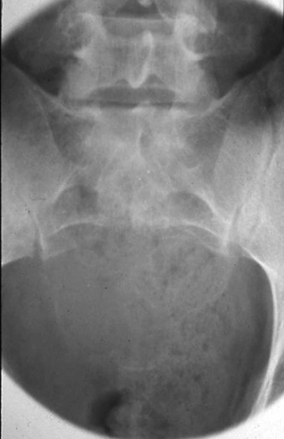
Radiographs revealed a large, osteolytic lesion involving almost the entire sacrum (Fig. 1). Computed tomography (CT) also revealed a destructive sacral mass involving almost the entire sacrum below the S1 vertebral body. 
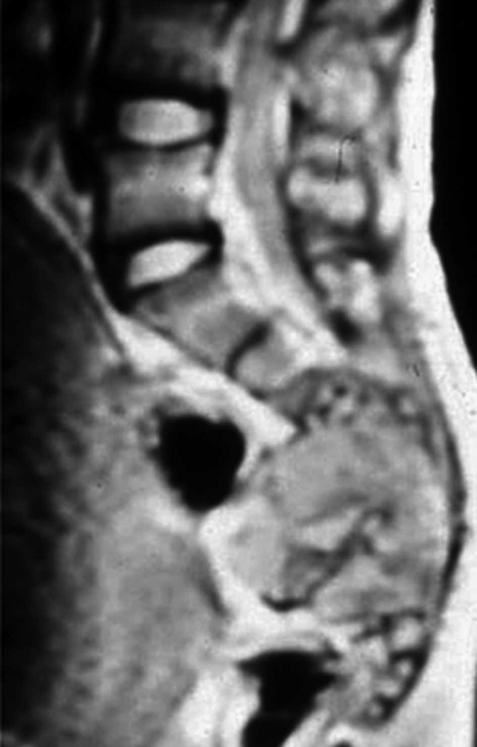
T2-weighted magnetic resonance imaging (MRI) showed an expansile soft tissue mass with a heterogeneous signal density. The tumor infiltrated into the spinal and sacral canal, and into the presacral area above S2 (Fig. 2). 
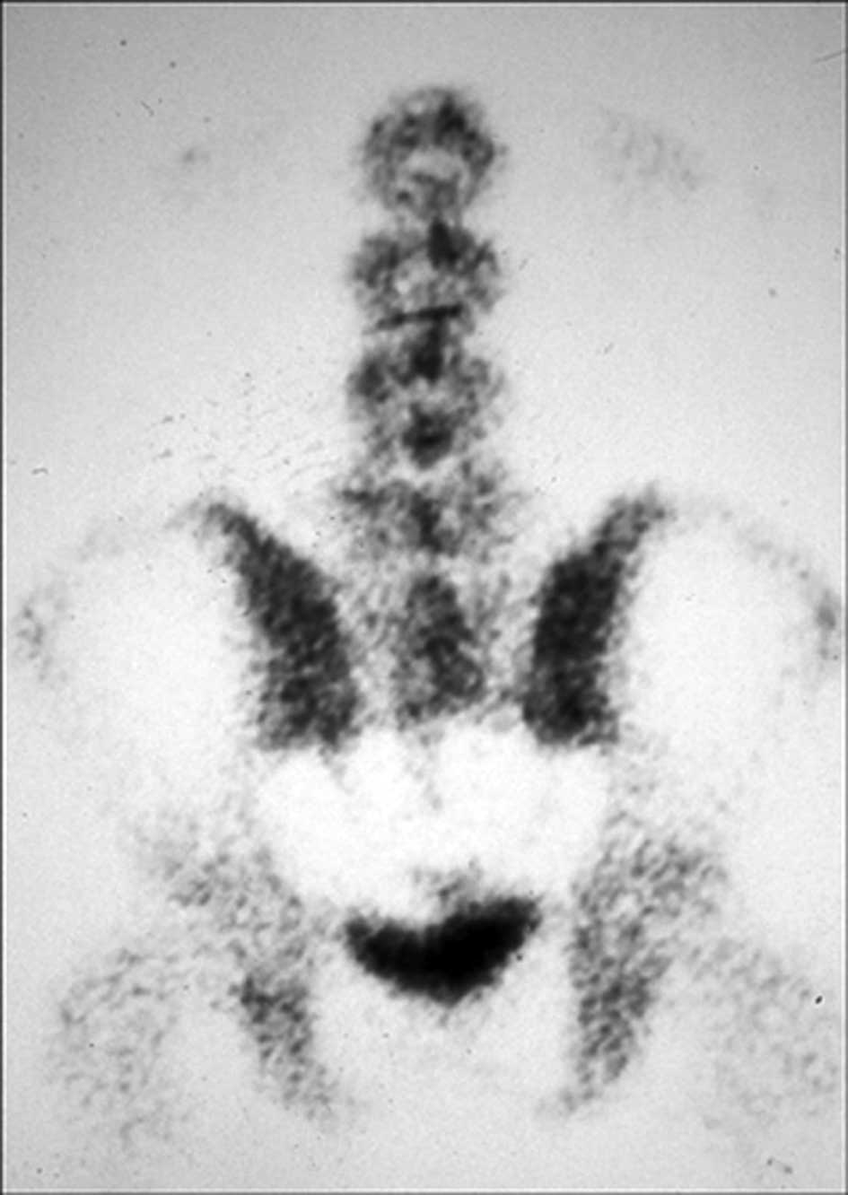
The technetium-99m methylene diphosphonate whole-body bone scan revealed an abnormally high uptake in the sacral region (Fig. 3). Metastases were not found. To 
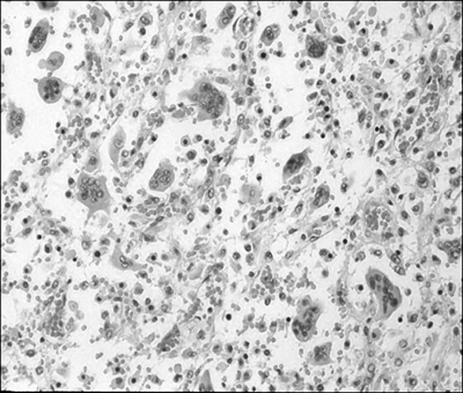
prevent an improper diagnosis or a delay in treatment occurring, open biopsy was performed from the posterior aspect of the sacrum, and histopathological examination revealed numerous multinucleated giant cells within a background of scant stroma consistent with GCT (Fig. 4).

Figure 1

An anteroposterior plain radiograph shows an osteolytic area involving almost the entire sacrum below the S1 vertebral body.

Figure 2

Sagittal T2-weighted magnetic resonance imaging shows an expansile soft tissue mass with a heterogeneous signal density with presacral extension.

Figure 3

A technetium-99m methylene diphosphonate whole-body bone scan reveals a high uptake area in the sacrum.

Figure 4

Hematoxylin and eosin (H&E)-stained biopsy specimen shows multinucleated giant cells within a background of stromal cells (original magnification, ×400).

With the goal of pain relief, tumor resection and prevention of further neurological deterioration, the patient underwent high sacral amputation using a staged anterior and posterior approach. In the first stage, the anterior procedure with a transperitoneal approach was performed. The anterior aspect of the tumor was exposed, and internal iliac and middle sacral vessels were ligated. The anterior osteotomy was performed through the lower border of the S1 vertebral body at the level just below the 
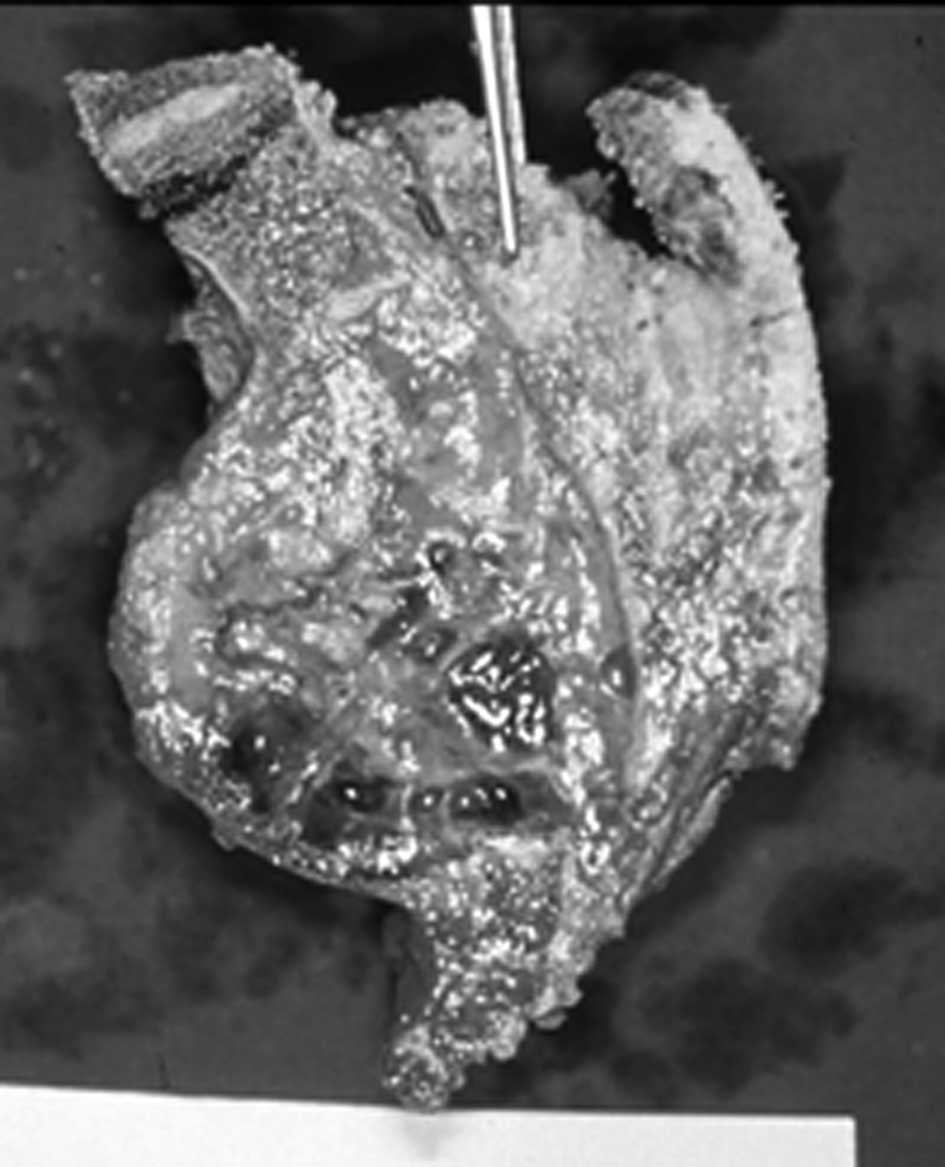
S1 sacral foramina. In the second stage, the posterior approach was performed to transect the dural contents, eventually interconnecting the anterior and posterior osteotomies (Fig. 5). The tumor was resected with a small margin and the biopsy tract was included within the resection margins. The S1 roots were preserved. For adjuvant therapy, the patient underwent radiotherapy consisting of a total dose of 5000 rad in 5 weeks.

Figure 5

Osteotomy was performed through the lower border of the S1 vertebral body at the level just below the S1 sacral foramina.

Postoperatively, the patient was able to ambulate without the use of external supports by 6 months. A certain amount of perineal numbness remained, but the patient did not consider the lower extremity numbness to be particularly disabling. The patient suffered difficulties with urinary retention, requiring intermittent urinary self-catheterization. Self-urination and defecation became possible by a voluntary increase of the abdominal muscle pressure.

After 5 years, there was no evidence of local recurrence on serial imaging. The patient had returned to work without significant complaints and was satisfied with the degree of mobility.

In March 2008, the patient reported increasing pain in the back and mid-buttock area. Physical examination of the buttock area revealed a draining sinus on the previous surgery scar with pus-like discharge. MRI of the lumbar and thoracic spine was performed. T2-weighted MRI of the lumbar spine revealed high signal intensity pus draining with a sinus tract into the buttock area (Fig. 6A). MRI studies of the thoracic spine showed diffuse enhancement on the T11 vertebral body with an associated compression deformity suggestive of a bone tumor. There was no cord compression, nor a signal change within the cord (Fig. 6B). A technetium-99m methylene diphosphonate whole-body bone scan was also performed and showed abnormal uptake only at the T11 vertebral body.

Figure 6

(A) Sagittal T2-weighted magnetic resonance imaging (MRI) shows high signal intensity pus draining with a sinus tract at the posterior aspect of the L5 vertebral body. (B) Sagittal T1-weighted enhanced MRI of the thoracic spine shows diffuse enhancement on the T11 vertebral body with an associated compression deformity.

Considering the patient’s clinical history and findings on MRI, a diagnosis of metastatic GCT was considered, involving anterior zones of the vertebral body according to the Weinstein-Boriani-Biagini (WBB) classification system (5,6). The patient subsequently underwent a wide en block resection of T11 and anterior interbody fusion with autogenous iliac strut bone graft. A complete resection of the vertebral body was achieved and specimens were sent for histopathological analysis. The spinal reconstruction and stabilization were performed by posterolateral fusion with autogenous iliac bone graft and pedicle screw fixation. Additionally, the patient had open irrigation and debridement for the infection on the buttock.

The histopathological examination revealed numerous multinucleated giant cells within a background of scant stroma, consistent with GCT. For adjunctive therapy, the patient again underwent radiotherapy consisting of a total dose of 5000 rads in 5 weeks.

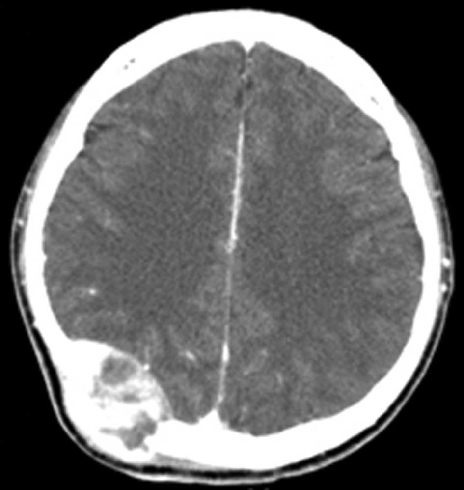
In October 2008, the patient reported newly developed localized swelling on the right occipital area. A diffuse swelling was noted in the right parieto-occipital area. CT scan of the brain revealed a large well-defined hyperdense contrast-enhancing lesion originating from the right parieto-occipital bone (Fig. 7). The patient subsequently underwent a right parieto-occipital craniectomy, followed by cranioplasty. Adjunctive radiotherapy was repeated. The histopathological examination has confirmed the diagnosis of GCT of bone.

Figure 7

An axial computed tomography imaging scan of the brain shows a large well-defined hyperdense contrast enhancing lesion originating from the right parieto-occipital area tract.

Currently, the patient continues to be free of recurrence 26 months following the T11 corpectomy, and 19 months following the craniectomy.

Discussion

The majority of GCTs occur in the ends of the long bones, usually the distal femur, proximal tibia and distal radius (1,2). GCTs rarely occur in the spine, with 2–5% of tumors found in the spine above the sacrum (2–4). Neoplasia of the skull bones are also extremely rare, accounting for only 2.4–2.6% of all primary bone tumors, and when they occur, the sphenoid bone is known to be the most common site, followed by the temporal bone (5,7). Although GCTs exhibit a propensity for aggressive local recurrence, there are few reports of metastases or recurrence from GCT of the spine. Additionally, GCTs are difficult to determine due to the low incidence of these lesions (2,4).

Multifocal GCTs of bone have been addressed in previous reports, mostly occurring in the limbs. On only one occasion has the multifocal GCT of bone been reported in the spine, occurring primarily in the thoracic spine, and two years later, in the sacrum (8). Due to multiple lesions in the axial skeleton in the absence of lung involvement and an open foramen ovale, it is difficult to confirm whether the tumors were actually multifocal in their origin or spread through the vertebral venous complex (9,10). The Batson’s venous plexus, which connects the deep pelvic veins to the internal vertebral venous plexuses, may provide a route for the spread of tumors arising from the pelvis to the vertebral column or brain (10). The primary tumor and metastatic lesions usually have the same histological features; however, histological grades and findings do not have practical value in the prediction of the outcome or the risk of metastatic spread (11,12).

Optimal management of GCT of bone is known to be complete resection of the tumor with wide margins if possible, and according to Mnaymneh et al (13), there was no recurrence when the primary tumor excision was performed, whereas recurrent malignant changes were reported in approximately half or more of groups undergoing curettage and irradiation (3,14). Due to the propensity of GCTs for aggressive local recurrence, adjuvant radiation therapy to the resection site may be adopted. Although it is widely held that radiation is capable of inducing sarcomatous transformation and soft tissue damage, it has been suggested that this risk was possibly associated with the now obsolete ortho-voltage radiation therapy rather than the currently used mega-voltage radiation therapy (15–17). Fujimoto et al (18) report favorable results in three consecutive cases diagnosed with GCT of the spine, which were treated with radiotherapy and bisphosphonate (BP) as a new treatment option.

In the present case, the patient was found to have multifocal GCTs in extremely unusual locations. The first in the sacrum, the second, 15 years later, in the thoracic spine, and the third, 15 years and 6 months later from the onset of the first tumor in the sacrum, in the parieto-occipital area of the skull. The patient had neither lung involvement nor a sign of cardiac septal defect. The tumors in the thoracic spine and skull may be thought of as metastases of the primary lesion in the sacrum with late recurrence. As the patient had an open biopsy for the initial diagnosis, it is possible that the biopsy margins were contaminated in the first resection and the infection of primary site, which occured 15 years later may be related to residual active disease (15,19,20). The 15-year interval between the primary lesion in the sacrum and recurrences in the thoracic spine and skull was longer than the usual interval that has been reported in other studies (19). Thus, we report an extremely rare case of GCT occurring in the axial skeleton, involving the sacrum, thoracic spine and parieto-occipital skull in more than 15 years of follow up.

References

1 

Donthineni R, Boriani L, Ofluoglu O and Bandiera S: Metastatic behaviour of giant cell tumour of the spine. Int Orthop. 33:497–501. 2009. View Article : Google Scholar : PubMed/NCBI

2 

Goldenberg RR, Campbell CJ and Bonfiglio M: Giant-cell tumor of bone. An analysis of two hundred and eighteen cases. J Bone Joint Surg Am. 52:619–664. 1970.PubMed/NCBI

3 

Sanjay BK, Sim FH, Unni KK, McLeod RA and Klassen RA: Giant-cell tumours of the spine. J Bone Joint Surg Br. 75:148–154. 1993.PubMed/NCBI

4 

Kumar R, Guinto FC Jr, Madewell JE, David R and Shirkhoda A: Expansile bone lesions of the vertebra. Radiographics. 8:749–769. 1988. View Article : Google Scholar : PubMed/NCBI

5 

Bitoh S, Takimoto N, Nakagawa H, Namba J, Sakaki S and Gohma T: Giant cell tumor of the skull. Surg Neurol. 9:185–188. 1978.PubMed/NCBI

6 

Boriani S, Weinstein JN and Biagini R: Primary bone tumors of the spine. Terminology and surgical staging Spine (Phila Pa 1976). 22:1036–1044. 1997. View Article : Google Scholar : PubMed/NCBI

7 

Bertoni F, Unni KK, Beabout JW and Ebersold MJ: Giant cell tumor of the skull. Cancer. 70:1124–1132. 1992. View Article : Google Scholar : PubMed/NCBI

8 

Kos CB, Taconis WK, Fidler MW and ten Velden JJ: Multifocal giant cell tumors in the spine. A case report Spine (Phila Pa 1976). 22:821–822. 1997. View Article : Google Scholar : PubMed/NCBI

9 

Onuigbo WI: Batson’s theory of vertebral venous metastasis: a review. Oncology. 32:145–150. 1975.

10 

Batson OV: The function of the vertebral veins and their role in the spread of metastases. Clin Orthop Relat Res. 312:4–9. 1995.PubMed/NCBI

11 

McGough RL, Rutledge J, Lewis VO, Lin PP and Yasko AW: Impact severity of local recurrence in giant cell tumor of bone. Clin Orthop Relat Res. 438:116–122. 2005. View Article : Google Scholar : PubMed/NCBI

12 

Lausten GS, Jensen PK, Schiodt T and Lund B: Local recurrences in giant cell tumour of bone. long-term follow up of 31 cases. Int Orthop. 20:172–176. 1996. View Article : Google Scholar : PubMed/NCBI

13 

Mnaymneh WA, Dudley HR and Mnaymneh LG: Giant-cell tumor of bone. an analysis and follow-up study of the forty-one cases observed at the Massachusetts General Hospital between 1925 and 1960. J Bone Joint Surg Am. 46:63–75. 1964.PubMed/NCBI

14 

Shikata J, Yamamuro T, Kotoura Y, Mikawa Y, Iida H and Maetani S: Total sacrectomy and reconstruction for primary tumors. report of two cases. J Bone Joint Surg Am. 70:122–125. 1988.PubMed/NCBI

15 

Refai D, Dunn GP and Santiago P: Giant cell tumor of the thoracic spine: case report and review of the literature. Surg Neurol. 71:228–233. 2009. View Article : Google Scholar : PubMed/NCBI

16 

Caudell JJ, Ballo MT, Zagars GK, et al: Radiotherapy in the management of giant cell tumor of bone. Int J Radiat Oncol Biol Phys. 57:158–165. 2003. View Article : Google Scholar : PubMed/NCBI

17 

Nair MK and Jyothirmayi R: Radiation therapy in the treatment of giant cell tumor of bone. Int J Radiat Oncol Biol Phys. 43:1065–1069. 1999. View Article : Google Scholar : PubMed/NCBI

18 

Fujimoto N, Nakagawa K, Seichi A, et al: A new bisphosphonate treatment option for giant cell tumors. Oncol Rep. 8:643–647. 2001.PubMed/NCBI

19 

Bergh P, Kindblom LG, Gunterberg B, Remotti F, Ryd W and Meis-Kindblom JM: Prognostic factors in chordoma of the sacrum and mobile spine: a study of 39 patients. Cancer. 88:2122–2134. 2000. View Article : Google Scholar : PubMed/NCBI

20 

Fourney DR, Rhines LD, Hentschel SJ, et al: En bloc resection of primary sacral tumors: classification of surgical approaches and outcome. J Neurosurg Spine. 3:111–122. 2005. View Article : Google Scholar : PubMed/NCBI

Related Articles

  • Abstract
  • View
  • Download
  • Twitter
Copy and paste a formatted citation
Spandidos Publications style
Park Y, Lee J, Baek S and Park C: The rare case of giant cell tumor occuring in the axial skeleton after 15 years of follow-up: Case report. Oncol Lett 2: 1323-1326, 2011.
APA
Park, Y., Lee, J., Baek, S., & Park, C. (2011). The rare case of giant cell tumor occuring in the axial skeleton after 15 years of follow-up: Case report. Oncology Letters, 2, 1323-1326. https://doi.org/10.3892/ol.2011.402
MLA
Park, Y., Lee, J., Baek, S., Park, C."The rare case of giant cell tumor occuring in the axial skeleton after 15 years of follow-up: Case report". Oncology Letters 2.6 (2011): 1323-1326.
Chicago
Park, Y., Lee, J., Baek, S., Park, C."The rare case of giant cell tumor occuring in the axial skeleton after 15 years of follow-up: Case report". Oncology Letters 2, no. 6 (2011): 1323-1326. https://doi.org/10.3892/ol.2011.402
Copy and paste a formatted citation
x
Spandidos Publications style
Park Y, Lee J, Baek S and Park C: The rare case of giant cell tumor occuring in the axial skeleton after 15 years of follow-up: Case report. Oncol Lett 2: 1323-1326, 2011.
APA
Park, Y., Lee, J., Baek, S., & Park, C. (2011). The rare case of giant cell tumor occuring in the axial skeleton after 15 years of follow-up: Case report. Oncology Letters, 2, 1323-1326. https://doi.org/10.3892/ol.2011.402
MLA
Park, Y., Lee, J., Baek, S., Park, C."The rare case of giant cell tumor occuring in the axial skeleton after 15 years of follow-up: Case report". Oncology Letters 2.6 (2011): 1323-1326.
Chicago
Park, Y., Lee, J., Baek, S., Park, C."The rare case of giant cell tumor occuring in the axial skeleton after 15 years of follow-up: Case report". Oncology Letters 2, no. 6 (2011): 1323-1326. https://doi.org/10.3892/ol.2011.402
Follow us
  • Twitter
  • LinkedIn
  • Facebook
About
  • Spandidos Publications
  • Careers
  • Cookie Policy
  • Privacy Policy
How can we help?
  • Help
  • Live Chat
  • Contact
  • Email to our Support Team