Introduction
Uterine sarcomas are rare, accounting for 3–7% of
malignant diseases in the uterine corpus. They have been classified
into three main histologic subgroups: carcinosarcoma (CS),
leiomyosarcoma (LMS) and endometrial stromal sarcoma (ESS). Our
previous study showed that five-year survival rates among 121
patients were 52.1, 61.3 and 68.4% for CS, LMS and ESS,
respectively (1).
The standard therapy for uterine sarcoma is surgery;
however, adjuvant chemotherapy is generally administered in the
advanced stages of the disease. Previous studies have reported that
the response rate for paclitaxel was 18% (2) and 19% for cisplatin (3). The poor response to chemotherapy
reflects the drug resistance of uterine sarcoma.
SXR is expressed in the colon, intestine, lungs and
kidneys, where it plays a vital role in the metabolism of
endogenous substances, such as bile acids, hormones and vitamins
(4,5). While paclitaxel is a key anticancer
drug for CS, as well as for epithelial ovarian cancer, several
studies have demonstrated that SXR agonists such as rifampicin
depress the activity of paclitaxel (6,7) and
induce cellular proliferation in cancers such as ovarian (6), endometrial (8) and breast cancer (9). Further research has confirmed that
downregulation of SXR inhibits endometrial cancer cell growth and
induces apoptosis (10). Cytochrome
P450 3A4 (CYP3A4) has been shown to enhance SXR activity in an SXR
knockout animal model (11,12). Although multiple drug resistance 1
(MDR 1) has not been found to induce the activation of SXR in
ovarian cancer (6), this has been
reported in breast (9) and
endometrial cancer (13). We
previously reported that SXR overexpression is a prognostic factor
in epithelial ovarian cancer and represents a useful marker for
identifying patients at high risk of recurrence or mortality
(14). However, the status of SXR
has not yet been investigated in uterine sarcomas. In this study we
analyzed the status of SXR expression in CS and correlated the
findings with various clinicopathological characteristics. In
addition, we also examined ESS, LMS, benign leiomyoma and normal
endometria for SXR expression and compared the findings with those
in CS. Finally, we evaluated the correlation between SXR expression
and the clinicopathological features of uterine sarcomas.
Generally, it is difficult to histologically
differentiate between LMS and ESS. The expression of CD10 and
α-smooth muscle actin (αSMA) is measured to aid in the diagnosis of
ESS and LMS. Therefore, we investigated the expression of CD10 and
αSMA as well as SXR and evaluated whether SXR expression has the
capacity to be a diagnostic marker for uterine sarcomas.
Materials and methods
Patients and tissue specimens
Forty-seven patients with uterine sarcomas (6 ESS,
17 LMS and 24 CS), 5 patients with uterine leiomyoma and 5 patients
with normal myometrium who underwent surgical treatment between
1993 and 2008 at Tohoku University Hospital (Sendai, Japan), were
included in this study. Data including age, histological subtype,
stage, residual tumor, metastasis, chemotherapy (TJ,
pacilitaxel+carboplatin; IAP, fosfamide+doxorubicin+cisplatin),
recurrence and clinical outcome were collected. Histologic subtypes
were determined according to WHO criteria. This study was approved
by the Ethical Committee of Tohoku University School of Medicine
and informed consent was obtained from the patients.
Disease-free survival and overall survival were
measured from the date of initial surgery to the date of recurrence
and/or mortality, or the date of the last visit. Patients with
recurrence were treated with surgical resection or platinum-based
chemotherapy. For survival estimates, patients who were alive or
lost to follow-up were censored in 2008. The median follow-up
period was 14.3 months (range 1–61 months). All specimens were
fixed in 10% formalin for 24 to 48 h, embedded in paraffin and cut
into 3 μm sections.
Immunohistochemistry
Tissue sections were immunostained by the
streptavidin-biotin method using a Histofine kit
(Nichirei-Biosciences, Tokyo, Japan). The antibodies used in this
study are listed in Table I. The
immunohistochemistry (IHC) method used has been previously
described (9). Briefly, after the
sections were dewaxed and rehydrated, the sections were placed in
target retrieval solution or citric acid buffer (2 μM citric
acid and 9 mM trisodium citrate dehydrate, pH 6.0) and autoclaved
at 120°C for 5 min for antigen retrieval. For αSMA, the slides were
digested with trypsin at 37°C for 30 min. The antigen-antibody
complex was then visualized with 3,3’-diaminobenzidine (1 mM
3,3’-diaminobenzidine, 50 mmol/l Tris.HCL, pH 7.6 and 0.006%
H2O2) and counter-stained with hematoxylin.
Normal small intestine was used as a positive control for SXR. In
order to distinguish between ESS and LMS, IHC for CD10 and αSMA was
also performed.
 | Table ISummary of primary antibodies used in
this study. |
Table I
Summary of primary antibodies used in
this study.
| Antibody | Source | Optimal dilution | Antibody
retrieval |
|---|
| SXR (monoclonal) | Perseus proteromics
(Japan) | 1:400 | Autoclavea |
| ERα (monoclonal) | Invitrogen (UK) | 1:1 | Autoclavea |
| PR (monoclonal) | Chemicon (USA) | 1:50 | Autoclavea |
| Ki-67
(monoclonal) | DAKO (Denmark) | 1:100 | Autoclavea |
| CD10
(monoclonal) | Nichirei (Japan) | 1:1 | Autoclaveb |
| αSMA
(monoclonal) | DAKO (Denmark) | 1:300 | Trypsinc |
Immunohistochemical scoring system
All cases were scored by a semi-quantitative
histological scoring (HSCORE) method. Immunostaining intensity for
each specimen was classified as: 1 (none or weak staining), 2
(moderate staining) and 3 (strong staining). The HSCORE of each
case was obtained by multiplying each intensity level with the
corresponding percentage of positive cells using the following
formula: HSCORE = Σ(I*LI) where I and LI represent the intensity
and labeling index, respectively (9,10). The
final scores ranged from 0 to 300. Specimens with a HSCORE >40
were regarded as SXR-positive, while a HSCORE <40 was regarded
as SXR-negative. The LI was obtained for carcinoma cells as
described by Sasano et al(15). Briefly, two of the authors (X.Y. and
J.A.) independently evaluated at least 500 carcinoma cells
microscopically. Immunostained slides were evaluated using a
double-headed light microscope. Inter-observer differences were
<5%.
Statistical analysis
Student’s t-test was used to analyze the association
of SXR HSCORE with nuclear receptor status and patient
characteristics. Survival was analyzed using the Kaplan-Meier
method. Spearman’s Rho was used for analysis of IHC results with
regard to SXR expression and nuclear receptors or Ki67 antigen
expression. Comparison of positive rates was undertaken using the
χ2 test. Statview 5.0 software (SAS Institute Inc.,
Cary, NC, USA) was used for all statistical analyses. P<0.05 was
considered to indicate a statistically significant difference.
Results
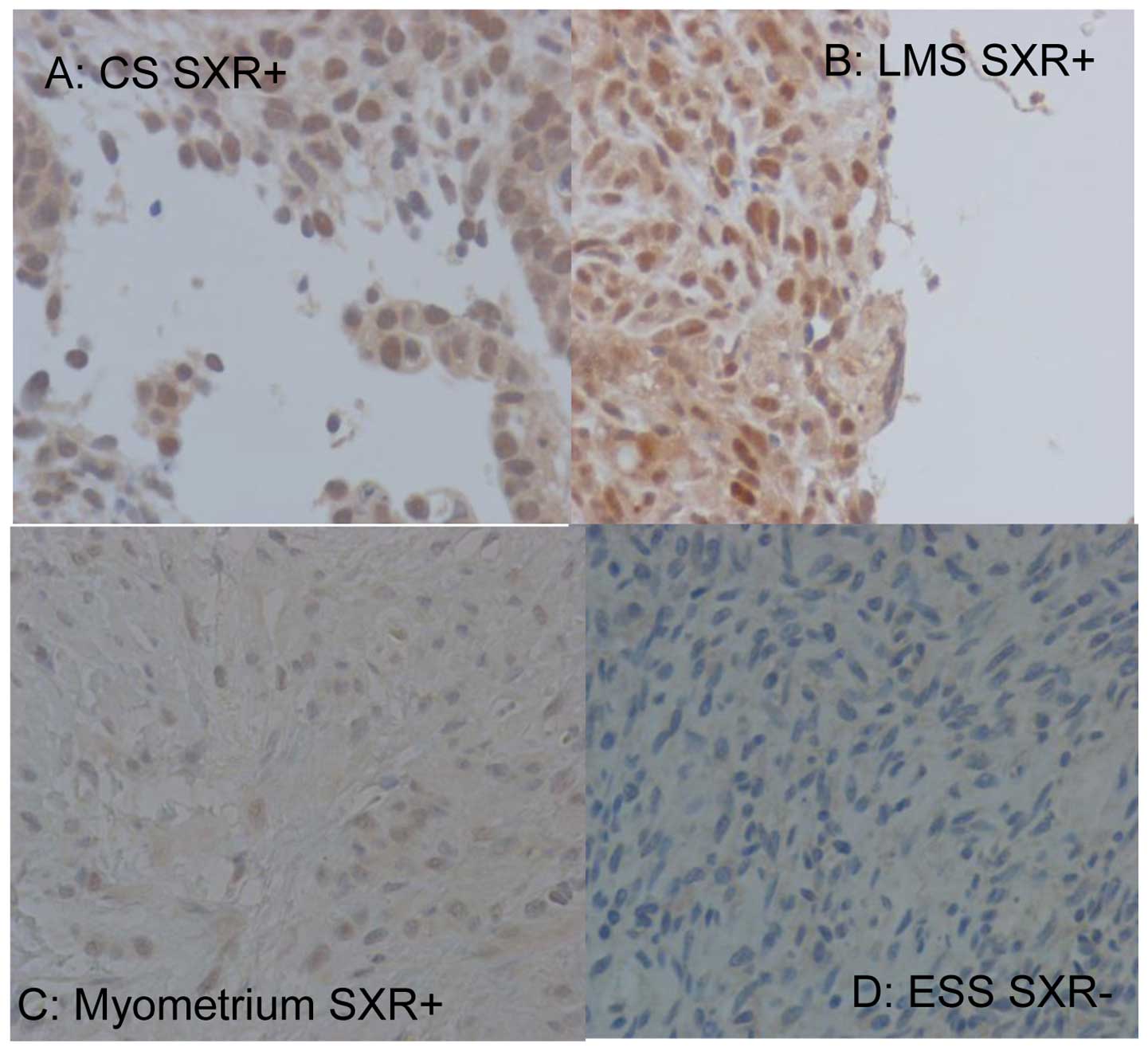
SXR expression was detected in the nuclei of uterine
sarcoma cells by IHC (Fig. 1A and
B). An SXR HSCORE >40 was observed in 4 of 17 (23.5%) LMS
cases and 3 of 24 (12.5%) CS cases. In normal myometrium, positive
expression of SXR was observed in 1 of 5 specimens (Fig. 1C). No significant difference in SXR
expression was observed between uterine sarcoma and normal
myometrium (P=0.764), while no SXR expression was observed in ESS
cases (Fig. 1D) and benign
leiomyomas.
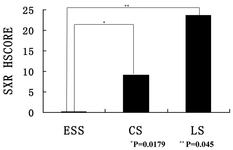
The mean SXR HSCOREs for CS and LMS were 9.13 (range
0–57.2) and 23.6 (range 0–135.6), respectively. The degree of
expression was higher in LMS than in CS. Significant differences in
median SXR HSCOREs were observed between ESS and CS and ESS and LMS
(Fig. 2; P=0.0179 and 0.045,
respectively). The correlations between SXR expression and
clinicopathological features were analyzed (Table II). There were significant
differences between SXR expression and stage, age and Ki67
expression in CS (P<0.05), while no significant differences were
identified in LMS.
| Table IIAssocation of SXR HSCORE and
clinicopathological features in leiomysarcoma and
carcinosarcoma. |
Table II
Assocation of SXR HSCORE and
clinicopathological features in leiomysarcoma and
carcinosarcoma.
| Clinicopathological
features | Carcinosarcoma | Leiomyosarcoma |
|---|
|
|
|---|
| n | SXR HSCORE
(range) | P-value | n | SXR HSCORE
(range) | P-value |
|---|
| Age (years) | | | | | | |
| ≤50 | 3 | 0 (0) | 0.017a | 4 | 33.95 (0–135.8) | 0.3064 |
| >50 | 21 | 10.44 (0–57.2) | | 13 | 20.42 (0–120.2) | |
| Stage | | | | | | |
| I–II | 8 | 0 (0) | 0.0352a | 5 | 8.04 (0–40.2) | 0.3716 |
| III–IV | 16 | 13.7 (0–57.2) | | 12 | 30.08 (0–135.8) | |
| Residual tumor | | | | | | |
| Optimal | 17 | 10.26 (0–57.2) | 0.22 | 12 | 22.12 (0–120.2) | 0.4201 |
| Suboptimal | 7 | 6.4 (0–44.8) | | 5 | 27.16 (1–135.8) | |
| Chemotherapy | | | | | | |
| Yes | 18 | 10.89 (0–57.2) | 0.2034 | 5 | 29.08 (0–120.2) | 0.7559 |
| No | 6 | 3.85 (0–23.1) | | 12 | 21.32 (0–135.8) | |
| Metastasis | | | | | | |
| Yes | 15 | 12.99 (0–57.2) | 0.085 | 13 | 27.77 (0–135.8) | 0.5062 |
| No | 9 | 2.7 (0–24.3) | | 4 | 10.05 (0–40.2) | |
| Ki67 (%) | | | | | | |
| <15 | 3 | 0 (0) | 0.017a | 8 | 13.13 (0–79.8) | 0.3799 |
| ≥15 | 21 | 10.44 (0–57.2) | | 9 | 32.91
(0–135.8) | |
| ERα | | | | | | |
| Negative | 19 | 8.53 (0–46.3) | 0.3746 | 9 | 26.69
(0–120.2) | 0.7734 |
| Positive | 5 | 11.44 (0–57.2) | | 8 | 20.13
(0–135.8) | |
| PR | | | | | | |
| Negative | 16 | 10.13 (0–46.2) | 0.3523 | 8 | 20.13
(0–135.8) | 0.7734 |
| Positive | 8 | 7.15 (0–57.2) | | 9 | 26.69
(0–120.2) | |
| Total | 24 | 9.13 (0–57.2) | | 17 | 23.6 (0–135.8) | |
We analyzed the association between SXR expression
and survival rate or clinical stage in CS. SXR-positive cases were
detected in 3 of 9 (33.3%) advanced-stage patients with CS, whereas
there were only 3 of 17 (17.6%) patients with CS who were
disease-free during the follow-up period. In CS patients who were
SXR-positive, there was no significant correlation with regard to
survival (data not shown). Expression of ERα and PR was not
significantly associated with disease-free survival or overall
survival. Spearman’s Rho analysis showed that there was a
statistically significant correlation between the HSCORE and Ki67
expression levels in CS (r=0.474, P=0.0230). The positive rates for
CD10 were 23.5 and 100% in LMS and ESS, respectively (P=0.0011).
The positive rates for αSMA were 58.8 and 66.7% in LMS and ESS,
respectively, which were not significantly different.
Discussion
Gupta et al found that SXR activation induced
cell proliferation and drug resistance in ovarian cancer cells
(6). In addition, SXR expression
was also detected in the normal endometrium, in the proliferative
and secretory phases (6,16). Other studies have reported SXR
expression in normal and cancer tissues from the liver, breast and
uterus (6,17,18).
In our present study, SXR expression was detected in LMS and CS,
but not in ESS. The percentage of cases with advanced-stage CS with
positive SXR was higher than the percentage observed in the early
stages, and a significant correlation between SXR expression and
stage was found for CS (P<0.05).
It has been reported that TJ chemotherapy is
effective for CS (19). However, we
did not find an association between SXR expression and the efficacy
of chemotherapy in uterine sarcomas. The poor response to
chemotherapy reflects drug resistance in uterine sarcomas. In
endometrial cancer, CYP3A4 and MDR1 activity were induced by the
activation of SXR (8). We
previously reported that SXR is a prognostic factor in epithelial
ovarian cancer and may represent a useful marker for identifying
patients at risk of recurrence or mortality (14). SXR is induced by paclitaxel, which
is a major anti-cancer drug for CS as well as epithelial ovarian
cancer. We investigated the correlation between SXR expression and
CS; however, there was no significant correlation observed between
SXR-positive status and both disease-free survival and overall
survival. It has been reported in mice that SXR has two isoforms,
SXR1 and SXR2 (20). These results
suggest that the expression of SXR isoforms differs in different
organs. The role of SXR isoforms in CS may be different from that
in epithelial ovarian cancer. Further investigation is needed to
clarify the status of SXR isoforms in human uterine sarcomas.
This study showed that there is a significant
correlation between SXR-positive status and stage and metastasis in
uterine sarcomas. In CS, SXR expression was also significantly
related to stage and Ki67 expression. Our results support an
association between SXR expression and malignant behavior.
Overexpression of SXR may aid in identifying patients at an
advanced stage of CS.
The assessment of SXR expression by the HSCORE
method incorporates the intensity of staining and the LI
(percentage of stained cells for each intensity level). Therefore,
in comparison to a standard evaluation of immunohistochemical
results by a 3-tier score (1),
HSCORE provides a more accurate and homogeneous assessment of
protein expression levels in individual cases. The expression
levels using monoclonal nuclear antibodies for ERα, PR and Ki67
were 10–20% of the SXR LI in uterine sarcomas. The HSCORE range was
0–300 and the positive HSCORE range was inferred to be 30–60.
Therefore, this suggests that a HSCORE of 40 is the threshold for
identification of uterine sarcomas.
It is often difficult for pathologists to
distinguish LMS from ESS histologically. The determination of CD10
and αSMA expression is employed to aid in the diagnosis of ESS and
LMS. In this study, the positive rates for CD10 were 23.5 and 100%
in LMS and ESS, respectively (P=0.0011). In addition, the
expression of SXR in LMS was significantly higher than that in ESS,
in which it was completely absent. Therefore, our study suggests
that SXR may be used as a diagnostic marker to identify LMS and
ESS.
This is the first study to evaluate the correlation
between SXR expression and clinical outcomes in uterine sarcomas.
Overexpression of SXR may be employed to identify patients at
advanced stages. Further studies are needed to clarify the role of
SXR in the biology of human uterine sarcomas. Understanding the
mechanisms of SXR may aid in the development of chemotherapeutic
regimens specifically designed against SXR and its target
genes.
Acknowledgements
This study was supported in part by a
Grant-in-Aid for Scientific Research on Priority Areas, a
Grant-in-Aid for Scientific Research (B) and (C), a Grant-in-Aid
for Young Scientists (B), a Grant-in-Aid for Exploratory Research,
from the Ministry of Education, Science, Sports and Culture, Japan,
a Grant-in-Aid from the Ministry of Health, Labor and Welfare,
Japan, the 21st Century COE Program Special Research Grant (Tohoku
University) from the Ministry of Education, Culture, Sports,
Science and Technology, Japan, a Grant-in-Aid from the Kurokawa
Cancer Research Foundation, a Grant-in-Aid from All Japan Coffee
Association, Japan Coffee Association and the Uehara Memorial
Foundation.
References
|
1
|
Akahira J, Tokunaga H, Toyoshima M, et al:
Prognoses and prognostic factors of carcinosarcoma, endometrial
stromal sarcoma and uterine leiomyosarcoma: a comparison with
uterine endometrial adenocarcinoma. Oncology. 71:333–340. 2006.
View Article : Google Scholar
|
|
2
|
Curtin JP, Blessing JA, Soper JT, et al:
Paclitaxel in the treatment of carcinosarcoma of the uterus: a
gynecologic oncology group study. Gynecol Oncol. 83:268–270. 2001.
View Article : Google Scholar : PubMed/NCBI
|
|
3
|
Thigpen T, Blessing JA, Beecham J, et al:
Phase II trial of cisplatin as first-line chemotherapy in patients
with advanced or recurrent uterine sarcomas: a gynecologic oncology
group study. J Clin Oncol. 9:1962–1966. 1991.PubMed/NCBI
|
|
4
|
Takara K, Takagi K, Tsujimoto M, et al:
Digoxin up-regulates multidrug resistance transporter (MDR1) mRNA
and simultaneously down-regulates steroid xenobiotic receptor mRNA.
Biochem Biophys Res Commun. 306:116–120. 2003. View Article : Google Scholar : PubMed/NCBI
|
|
5
|
Miki Y, Suzuki T, Tazawa C, et al: Steroid
and xenobiotic receptor (SXR), cytochrome P450 3A4 and multidrug
resistance gene 1 in human adult and fetal tissues. Mol Cell
Endocrinol. 231:75–85. 2005. View Article : Google Scholar : PubMed/NCBI
|
|
6
|
Gupta D, Venkatesh M, Wang H, et al:
Expanding the roles for pregnane X receptor in cancer:
proliferation and drug resistance in ovarian cancer. Clin Cancer
Res. 14:5332–5340. 2008. View Article : Google Scholar : PubMed/NCBI
|
|
7
|
Wang H, Huang H, Li H, et al: Activated
pregnenolone X-receptor is a target for ketoconazole and its
analogs. Clin Cancer Res. 13:2488–2495. 2007. View Article : Google Scholar : PubMed/NCBI
|
|
8
|
Masuyama H, Hiramatsu Y, Kodama J, et al:
Expression and potential roles of pregnane X receptor in
endometrial cancer. J Clin Endocrinol Metab. 88:4446–4454. 2003.
View Article : Google Scholar : PubMed/NCBI
|
|
9
|
Miki Y, Suzuki T, Kitada K, et al:
Expression of the steroid and xenobiotic receptor and its possible
target gene, organic anion transporting polypeptide-A in human
breast carcinoma. Cancer Res. 66:535–542. 2006. View Article : Google Scholar : PubMed/NCBI
|
|
10
|
Masuyama H, Nakatsukasa H, Takamoto N, et
al: Down-regulation of pregnane X receptor contributes to cell
grown inhibitor and apoptosis by anticancer agents in endometrial
cancer cells. Mol Pharmacol. 72:1045–1053. 2007. View Article : Google Scholar : PubMed/NCBI
|
|
11
|
Staudinger JL, Goodwin B, Jones SA, et al:
The nuclear receptor PXR is a lithocholic acid sensor that protects
against liver toxicity. Proc Natl Acad Sci USA. 98:3369–3374. 2001.
View Article : Google Scholar
|
|
12
|
Xie W, Barwick JL, Downes M, et al:
Humanized xenobiotic response in mice expressing nuclear receptor
SXR. Nature. 406:435–439. 2000. View
Article : Google Scholar : PubMed/NCBI
|
|
13
|
Masuyama H, Suwaki N, Tateishi Y, et al:
The pregnane X receptor regulates gene expression in a ligand- and
promoter-selective fashion. Mol Endocrinol. 19:1170–1180. 2005.
View Article : Google Scholar : PubMed/NCBI
|
|
14
|
Yue X, Akahira J, Utsunomiya H, et al:
Steroid and xenobiotic receptor (SXR) as a possible prognostic
marker in epithelial ovarian cancer. Pathl Int. 60:400–406. 2010.
View Article : Google Scholar : PubMed/NCBI
|
|
15
|
Sasano H, Frost AR, Saitoh R, et al:
Aromatase and 17β-hydroxysteroid dehydrogense type 1 in human
breast carcinoma. J Clin Endocrinol Metab. 81:4042–4046. 1996.
|
|
16
|
Masuyama H, Hiramatsu Y, Mizutani H, et
al: The expression of pregnane X receptor and its target gene,
cytochrome P450 3A1 in perinatal mouse. Mol Cell Endocrinol.
172:47–56. 2001. View Article : Google Scholar : PubMed/NCBI
|
|
17
|
Blumberg B, Sabbagh W Jr, Juguilion H, et
al: SXR, a novel and xenobiotic-sensing nuclear receptor. Genes
Dev. 12:3195–3205. 1998. View Article : Google Scholar : PubMed/NCBI
|
|
18
|
Suzuki T, Moriya T, Sugawara A, et al:
Retinoid receptor in human breast carcinoma: possible modulators of
in situ estrogen metabolism. Breast Cancer Res Treat. 65:31–40.
2001. View Article : Google Scholar : PubMed/NCBI
|
|
19
|
Toyoshima M, Akahira J, Matsunaga G, et
al: Clinical experience with combination paclitaxel and carboplatin
therapy for advanced or recurrent carcinosarcoma of the uterus.
Gynecol Oncol. 94:774–778. 2004. View Article : Google Scholar : PubMed/NCBI
|
|
20
|
Dotzlaw H, Leygue E, Watson P, et al: The
human orphan receptor PXR messenger RNA is expressed in both normal
and neoplastic breast tissue. Clin Cancer Res. 5:2103–2107.
1999.PubMed/NCBI
|