Introduction
Despite progress in early detection, radical
surgical treatments and multimodal therapeutic approaches, the
prognosis of lung cancer remains poor. The prognosis of patients
with non-small cell lung cancer (NSCLC) is determined by the
disease stage as defined by the International Association for the
Study of Lung Cancer. However, this classification is not a precise
prediction tool compared to those used for other primary cancers.
For example, patients with completely excised stage I NSCLC develop
local relapses in up to 30% of cases and have a 5-year survival
rate of 58–73% (1). This
distinctive NSCLC behavior has encouraged the assessment of a
number of pathological and laboratory markers which may offer
supplementary prognostic information, including epidermal growth
factor receptors, angiogenic growth factors, the K-ras oncogene and
disseminated tumor cells. Certain studies have suggested that
micrometastases, which have not yet been detected by conventional
lymph node histopathological examination, may offer additional
prognostic information (2–4). A number of molecular methods,
including the polymerase chain reaction (PCR) for p53 and K-ras and
the reverse transcriptase-polymerase chain reaction (RT-PCR) for
carcinoembryonic antigen (CEA), cytokeratin 7 (CK7), cytokeratin 19
(CK19) and mucin 1 (MUC1) have been used for the detection of
micrometastases in lymph nodes. The prognostic role of these
markers remains controversial and there is no agreement as to
whether molecular technologies should be used during routine
pathological evaluation (5).
The purpose of this prospective study was to
determine the impact of lymph node micrometastases, detected with
quantitative real-time RT-PCR assays for CEA mRNA, on 5-year
survival in patients undergoing complete resection of clinical
stage I NSCLC. This study follows a preliminary study published in
2005 which focused on the disease-free interval (6).
Materials and methods
Patients
Mediastinal and hilar lymph nodes from clinical
stage I NSCLC patients treated with pulmonary lobectomy at the Unit
of Thoracic Surgery and Lung Transplantation (Ca' Granda General
Hospital, Milan, Italy) between October 2000 and February 2004 were
studied following Ethics Committee approval. All patients had been
staged clinically by means of chest, abdominal and brain computed
tomographic scans, positron emission tomography scans, bronchoscopy
and mediastinoscopy (if mediastinal lymph node enlargement or
positivity for PET were detected). Patients with a history of
previous malignancy or a forced expiratory volume in 1 sec
(FEV1) <80% of the predicted value, were excluded.
Consent was obtained from each patient. The patient population
included a cluster of NSCLC cases previously reported and a set of
additional patients that had been studied prospectively following
the original publication (3).
Study protocol
The study protocol was extensively reported in our
previous study (6) and is
summarized as follows. Systematic lymphadenectomy was performed
immediately after thoracic incision and was followed by pulmonary
lobectomy. Each lymph node was divided into two sections; one
section was frozen at −80°C and the second section was
formalin-fixed. A fragment of the primary tumor was frozen. The
standard curve was constructed from a mix of lymphocytes from a
healthy donor and CEA-expressing MCF-7 cells (American Type
Collection, Rockville, MD, USA). Specimens from cell lines, control
lymph nodes, lymph nodes from cancer patients and primary tumors
were homogenized (TRIzol solution; Invitrogen, Carlsbad, CA, USA)
and spectrophotometry determined the purity and quantity of the
mRNA extracted. Total mRNA was reverse-transcribed in DNA with
reagents from Applied Biosystems (Foster City, CA, USA). The ABI
Prism 7700 Sequence Detection System (Applied Biosystems) was used
for the quantitative assessment of CEA. During the DNA
polymerization, the TaqMan (Applied Biosystems) probe was
hydrolyzed and fluorescence was emitted. The charge-coupled device
camera on the Prism 7700 device continuously collected the
fluorescence emissions during the amplification cycles. The
threshold cycle (Ct) was noted when the fluorescence signal reached
10 times the standard deviation of the background. The linear
regression of the standard curve determined the number of
CEA-positive cells. All PCRs were performed in duplicate (Universal
TaqMan 2X PCR Mastermix; Applied Biosystems) in a volume containing
0.1 μmol/l of TaqMan probe and 0.3 μmol/l of each
primer. The thermal profile included 2 min at 50°C, 10 min at 95°C
followed by 40 cycles (50 for lymph nodes and standard curve
points) at 95°C for 15 sec and 1 min at 60°C. The following
sequences of primers were used: forward 5′-ATT CCA TAG TCA AGA GCA
TCA CA-3′, reverse 5′-GCA AAT GCT TTA AGG AAG AAG-3′ and TaqMan
probe 5′-(6-FAM) TGA AAT GAA GAA ACT ACA CCA GGG CTG CTA TAT
(TAMRA)-3′ for CEA (National Center for Biotechnology Information
accession number NM_004363) and forward 5′-TCC TTC CTG GGC ATG
GAG-3′, reverse 5′-AGG AGG AGC AAT GAT CTT GAT CTT-3′ and TaqMan
probe 5′-(6-FAM) CCT GTG GCA TCC ACG AAA CTA CCT TC-(TAMRA)-3′ for
ACTB (National Center for Biotechnology Information accession
number NM_001101). Threshold cycle values <37.42 were considered
significant for the presence of cancer cells in the lymph nodes.
The formalin-fixed specimens underwent routine hematoxylin and
eosin (H&E) staining. Following surgery, the patients were
examined every 2 months in the first year and every 3 months
thereafter. If recurrence was suspected, the appropriate
investigations were undertaken. Adjuvant chemotherapy and/or
radiotherapy were administered following confirmation of
recurrence. The survival rate and disease-free interval were
recorded. The follow-up was censored in the case of a second
malignancy. All the patients were reclassified according to the
seventh edition of the TNM classification for lung cancer (1).
Statistical analysis
Survival functions were estimated using the
Kaplan-Meier method and the differences were evaluated by the
log-rank or the Wilcoxon test, as appropriate. The Cox proportional
hazards test was used to identify the factors that influenced the
survival rate and disease-free interval. The regression analysis
was applied to standard curves and the analysis of variance was
used where indicated.
Results
Three-hundred and thirty-one consecutive NSCLC stage
I patients were evaluated. Sixty-one patients with previous
malignancy, 95 with FEV1 <80%, 24 requiring
pneumonectomy and 13 patients requiring sleeve-lobectomy were
excluded and 27 patients did not provide consent. A set of 15
patients with a final diagnosis of benign disease, 22 patients with
a diagnosis of pulmonary carcinoid tumor and 2 diagnosed with
small-cell lung cancer were also excluded. Four patients had
specimens not adequately frozen, 4 patients were upstaged to T4 and
9 patients were found to have unexpected N2 disease at routine
staining, causing their removal from the trial. The remaining 55
patients constituted the study group.
The 55 patients included 42 males and 13 females.
The mean age was 65.1 years (range, 36–80 years). The NSCLC tumors
included 32 (58.1%) adenocarcinomas, 17 (30.9%) squamous cell
carcinomas, 4 large cell carcinomas (7.3%) and 2 (3.7%)
adenosquamous carcinomas. Patients' characteristics are shown in
Table I.
 | Table IPatient characteristics. |
Table I
Patient characteristics.
| Variable | Data |
|---|
| No. of
patients | 55 |
| Male:female
ratio | 42:13 |
| Average age
(range), years | 65.1 (36–80) |
| Tumor
histotype | |
|
Adenocarcinomas | 32 |
| Squamous cell
carcinoma | 17 |
| Large cell
carcimoma | 4 |
| Adenosquamous
carcinoma | 2 |
| Median follow-up,
months | 45.8 |
| Recurrences | 20 |
| Cancer-related
mortalities | 15 |
A total of 609 lymph nodes were examined with
H&E staining and quantitative real-time RT-PCR for CEA mRNA.
The lymph nodes from the 55 patients were pathologically staged by
routine histopathological examination as pN0=44 and pN1=11. RT-PCR
for CEA mRNA revealed the presence of cancer cells in 20 patients
(36.3%). Table II details the N
status according to morphologic and molecular procedures. CEA
transcript levels were detected in all the primary tumor specimens,
with values ranging from 0.044 to 1782.88.
| Table IIN factor according to hematoxylin and
eosin staining and quantitative real-time RT-PCR for CEA mRNA. |
Table II
N factor according to hematoxylin and
eosin staining and quantitative real-time RT-PCR for CEA mRNA.
| pN factor | Hematoxylin and
eosin staining | Real-time RT-PCR
for CEA mRNA |
|---|
| N0 | 44 | 35 |
| N1 | 11 | 5 |
| N2 | 0 | 15 |
Contingency analyses did not reveal any correlation
between the presence of micrometastases and the tumor mass
characteristics (pathological stage T1 or T2), histotype, tumor
grade, age, gender and levels of CEA mRNA in the primary
cancer.
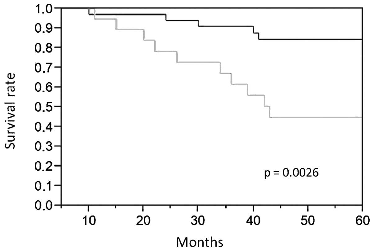
The mean follow-up time of the 55 enrolled patients
was 45.8 months. There were no patients lost to follow-up, cancer
recurrence occurred in 20 patients, and 15 patients succumbed to
their cancer. Statistically significant survival differences were
observed between patients with and without lymph node
micrometastases (P=0.0026; Fig. 1).
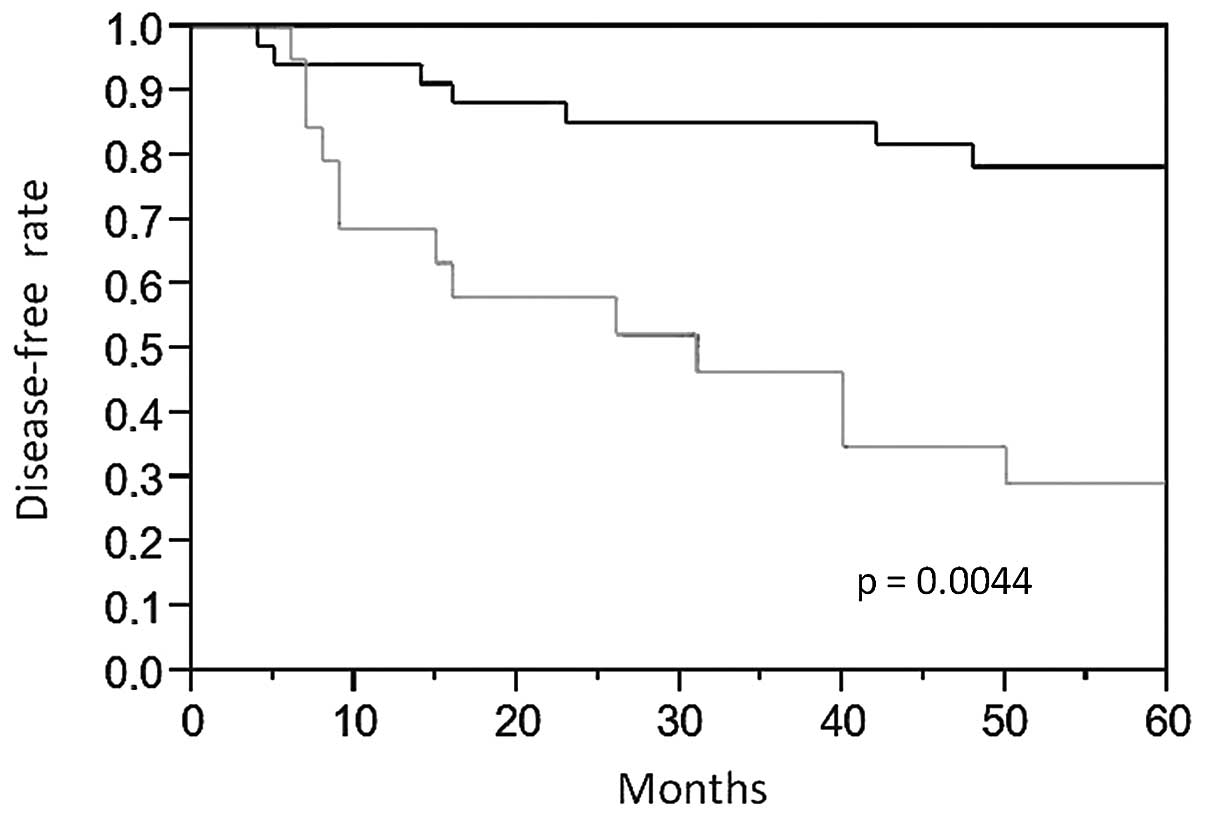
There were also statistically significant differences in the
disease-free intervals between patients with and without
micrometastases (P=0.0044; Fig. 2).
Cox regression multivariate analyses of age, gender, T status,
histology and grading revealed that the presence of micrometastases
was an independent predictor for worse prognosis (P=0.0098) and
shorter disease-free interval (P=0.0137; Table III).
| Table IIISurvival and disease-free interval
multivariate analysis. |
Table III
Survival and disease-free interval
multivariate analysis.
| Absence of
micrometastases | RR | Standard error | 95% CI | P-value |
|---|
| Survival | −1.021 | 0.416 | −1.916 to
−0.242 | 0.009 |
| Disease-free
interval | −0.804 | 0.339 | −1.526 to
−0.163 | 0.013 |
Discussion
Lymph node metastases are indicative of a poor
prognosis in NSCLC, as well as in other solid tumors, as they
provide clear evidence of systemic dissemination, a factor that
currently influences the prognosis and therapy. The standard
procedure to evaluate lymph node metastases from solid tumors is
histopathological analysis, although a more accurate process is
advisable. In 1948, Saphir and Amromin reported that a limited
number of sections taken from axillary lymph nodes following breast
cancer surgery, were not sufficient enough to establish whether
metastases were present or not (7).
The study reviewed lymph nodes which demonstrated no tumors in
standard examinations through serial sectioning, hence the
metastases that were revealed were named ‘obscure’ axillary lymph
node metastases. Since then, the presence of tumor cells in
axillary lymph nodes, which were initially assessed as negative on
standard examinations, have been reported; however, their
prognostic significance remains unclear. The origin of such
indeterminate significance may be related to the distinctive
behavior of the isolated tumor cells (or micrometastases), which
remains to be investigated. Animal experiments have demonstrated
that micrometastases are suppressed by apoptosis and that primary
tumor removal induces angiogenesis, leading to metastatic
development (8). Research on
colorectal cancer has revealed that, in the absence of the primary
tumor, both vascular density and metabolic activity in the
metastases are increased, while the level of apoptosis is decreased
(9).
Several studies have evaluated the presence of
micrometastases within intra-thoracic lymph nodes by
immunohistochemistry using various antibodies. A recent study
reviewed 13 trials correlating micrometastases and survival, in
addition to the authors' personal experience (10). This meta-analysis of 835 NSCLC cases
revealed no significant correlation between lymph node
micrometastases and survival. The result is not surprising in our
opinion: the analyzed studies utilized a mosaic of different
antibodies, each with specific and different points of strength and
weakness. A major problem is the use of immunohistochemistry; such
a procedure requires a meticulous and expert pathologist reading
thousands of slides, therefore personal interpretation is often a
problem. This is the same problem that we encountered in a trial
correlating bone marrow micrometastases and survival, the results
of which were negative (11).
Efforts have been made to detect lymph node
micrometastases at molecular levels and quantitative real-time
RT-PCR has been shown to be more sensitive than
immunohistochemistry for the detection of micrometastases in
patients with solid tumors (12,13).
In 2002, D'Cunha et al revealed the effectiveness of
quantitative real-time RT-PCR using CEA mRNA in detecting tumor
cells in lymph nodes of patients with NSCLC (14). Maeda et al, following the
analysis of several molecular markers, evaluated the sensitivity of
CEA mRNA for nodal metastases (14). The study, published in 2006,
revealed that the slices without tumor cells and lymph nodes
obtained from thymoma patients showed no amplification. By
contrast, CEA mRNA was detected by RT-PCR in all primary NSCLC and
positive lymph nodes as well as in 25% of the H&E-negative
lymph nodes (15). This study
supports our previous paper published in 2005. Following the
determination of a threshold cycle in the 14 control lymph nodes,
we also detected CEA mRNA in all the primary NSCLC samples, all the
positive lymph nodes and in 36% of patients with negative lymph
nodes in routine staining (6). Our
preliminary study reported a positive correlation between early
cancer recurrence and CEA mRNA detection in the intra-thoracic
lymph nodes (P=0.021). The present study contained an additional
set of patients who completed the 5-year follow-up. The results
were encouraging and congruent with the preliminary report.
Survival rates, as well as the disease-free intervals, were clearly
affected by CEA mRNA detection in the intrathoracic lymph nodes.
Moreover, the multivariate analysis identified the micrometastases
to be a negative predictor for survival and disease-free interval.
Such results may have an essential clinical relevance, for example,
molecular diagnosis of node involvement may be useful for selecting
appropriate candidates from among pN0-1 patients to receive
postoperative chemotherapy.
The present study has several points of strength.
The marker CEA is effective in the identification of NSCLC cells in
the intra-thoracic lymph nodes and false-negative results did not
occur in our results but were observed in other studies. The RT-PCR
technology is relatively simple and above all an automated
procedure, avoiding any problems relating to personal
interpretation. The clinical controls were also meticulous, no
patients were lost to the follow-up and survival was unaffected by
poor respiratory function.
The weakest point of this study was the number of
patients, which prevented any significant comparisons between TNM
and molecular staging. A large multicentre trial should further
investigate these findings, considering that the data from the
surviving patients were encouraging.
In our opinion, the molecular intra-thoracic lymph
node staging is a promising new frontier in NSCLC classification
which may identify patients requiring specific treatment. In this
setting, the development of the loop-mediated isothermal
amplification method (which was recently introduced as a fast
diagnostic procedure for infectious disorders) may prove
significant as it has been successfully tested for detecting CEA
mRNA in intra-thoracic lymph nodes. Such new rapid procedures may
be proposed during mediastinoscopy or transbronchial preoperative
lymph node staging by selecting patients who require neoadjuvant
therapy (16).
In conclusion, the present prospective study
provided evidence for a strong correlation between the molecular
detection of intra-thoracic lymph node micrometastases and 5-year
cancer specific survival as well as the disease-free interval in
patients who underwent pulmonary lobectomy for early-stage
NSCLC.
Acknowledgements
The authors wish to acknowledge
Professor Maria Martellini, Mr. Francesco Caridei and Mrs. Maria
Grazia Vitali for their support.
References
|
1.
|
R Rami-PortaJJ CrowleyP GoldstrawThe
revised TNM staging system for lung cancerAnn Thorac Cardiovasc
Surg1549200919262443
|
|
2.
|
CT SalernoS FrizelleGA NiehansSB HOM
JakkulaRA KratzkeMA MaddausDetection of occult micro-metastases in
non-small cell lung carcinoma by reverse transcriptase-polymerase
chain
reactionChest11315261532199810.1378/chest.113.6.15269631789
|
|
3.
|
L BonavinaD SoligoN QuiriciP BossolascoB
CesanaG Lambertenghi DeliliersA PeracchiaBone marrow-disseminated
tumor cells in patients with carcinoma of the esophagus or
cardiaSurgery1291522200110.1067/msy.2001.10950311150029
|
|
4.
|
A RuffatoS MattioliS PileriN DaddiF
D'OvidioV PilottiP TazzariDo bone marrow isolated tumor cells
influence long-term survival of non-small cell lung cancer?Eur J
Cardiothorac
Surg35463468200910.1016/j.ejcts.2008.11.01719150243
|
|
5.
|
AM MarchevskyJH QiaoS KrajisnikJM
MirochaRJ McKennaThe prognostic significance of intranodal isolated
tumor cells and micrometastases in patients with non-small cell
carcinoma of the lungJ Thorac Cardiovasc
Surg126551557200310.1016/S0022-5223(03)00123-512928657
|
|
6.
|
M NosottiM FalleniA PalleschiC PellegriniF
AlessiS BosariL SantambrogioQuantitative real-time polymerase chain
reaction detection of lymph node lung cancer micrometastasis using
carcinoembryonic antigen
markerChest12815391544200510.1378/chest.128.3.1539
|
|
7.
|
O SaphirGD AmrominObscure axillary
lymph-node metastasis in carcinoma of the
breastCancer1238241194810.1002/1097-0142(194807)1:2%3C238::AID-CNCR2820010208%3E3.0.CO;2-U18875036
|
|
8.
|
MS O’ReillyL HolmgrenC ChenJ
FolkmanAngiostatin induces and sustains dormancy of human primary
tumors in miceNat Med268969219968640562
|
|
9.
|
CF PeetersRM de WaalT WobbesTJ
RuersMetastatic dormancy imposed by the primary tumor: does it
exist in humans?Ann Surg
Oncol1533083315200810.1245/s10434-008-0029-518685897
|
|
10.
|
AM MarchevskyR GuptaD KusuancoJ MirochaRJ
McKenna JrThe presence of isolated tumor cells and micro-metastases
in the intrathoracic lymph nodes of patients with lung cancer is
not associated with decreased survivalHum
Pathol4115361543201010.1016/j.humpath.2010.04.00620656322
|
|
11.
|
M NosottiD TosiA PalleschiL RossoP
MendogniL SantambrogioImmunocytochemical detection of occult tumor
cells in the bone marrow: prognostic impact on early stages of lung
cancerEur Surg Res41267271200810.1159/00014196118594145
|
|
12.
|
S NoguchiT AiharaS NakamoriK MotomuraH
InajiS ImaokaH KoyamaThe detection of breast carcinoma
micrometastases in axillary lymph nodes by means of reverse
transcriptase-polymerase chain
reactionCancer7415951600199410.1002/1097-0142(19940901)74:5%3C1595::AID-CNCR2820740516%3E3.0.CO;2-L
|
|
13.
|
K KubotaH NakanishiN HikiQuantitative
detection of micrometastases in the lymph nodes of gastric cancer
patients with real-time RT-PCR: a comparative study with
immunohistochemistryInt J
Cancer105136143200310.1002/ijc.1103112672044
|
|
14.
|
J D'CunhaAL CorfitsJE Herndon IIMolecular
staging of lung cancer: real-time polymerase chain reaction
estimation of lymph node micrometastatic tumor cell burden in stage
I non-small cell lung cancer - preliminary results of Cancer and
Leukemia Group B Trial 9761J Thorac Cardiovasc
Surg1234844912002
|
|
15.
|
J MaedaM InoueM OkumuraDetection of occult
tumor cells in lymph nodes from non-small cell lung cancer patients
using reverse transcription-polymerase chain reaction for
carcinoembryonic antigen mRNA with the evaluation of its
sensitivityLung Cancer52235240200610.1016/j.lungcan.2005.12.008
|
|
16.
|
J MaedaM InoueK NakabayashiRapid diagnosis
of lymph node metastasis in lung cancer with loop-mediated
isothermal amplification assay using carcinoembryonic
antigen-mRNALung
Cancer65324327200910.1016/j.lungcan.2008.12.00319144442
|