Introduction
Desmoplastic small round cell tumor (DSRCT) is a
rare and aggressive neoplasm that was first described by Gerald and
Rosai (1). Although not unknown in
females, DSRCT mainly affects young males and generally presents as
a widely disseminated tumor within the peritoneal cavity. Other
primary sites, including the paratesticular area, ovary, thorax,
lung, and intracranial or head and neck areas, have also been
documented (2–8). The majority of the literature
regarding this particular type of tumor is comprised of case
reports (9).
Clinically, DSRCT is usually diagnosed at an
advanced stage, is highly aggressive and spreads along the
peritoneum and mesothelial lined surfaces. Upon diagnosis, the
tumor typically consists of a single large mass (occasionally as
large as 40 cm in diameter) and multiple smaller masses scattered
throughout the peritoneum, although other areas of origin have been
noted. Common symptoms upon presentation include abdominal pain,
hepatomegaly, ascites and hydronephrosis, and are non-specific and
non-diagnostic (9). A definitive
diagnosis is based on the identification of a reciprocal chromosome
translocation, t(11;22)(p13;q12) (10). Due to the rarity of the tumor, its
younger and overall healthier patient population (compared with
other tumor types) and the fact that it lacks definitive
histological and immunohistological features (9), DSRCT diagnosis may frequently be
delayed or the tumor may be entirely misdiagnosed as a different
type of abdominal sarcoma.
The scarcity of studies that are specific to DSRCT,
as well as the non-specific presentation of the tumor, has resulted
in difficulties regarding the treatment of the disease; according
to the literature, no curative outcome has been achieved thus far
(11). This is partly due to the
fact that there are no available models that simulate the behavior
of DSRCT outside the patient. The present study aimed to rectify
this through the development of several DSRCT tissue cultures and
xenograft lines.
Materials and methods
Patients and tissues
Given the low incidence of DSRCT, a limited number
of patients were available for enrollment in the present study.
Samples of tissues that had been removed during surgeries and
biopsies, and that the local pathologists had confirmed as DSRCT,
were received from surgical centers worldwide. Written informed
consent was obtained from the patients for the use of their tissues
in this study. The tissues were minced into small fragments (∼3
mm3) at the site of the surgery, and placed in sterile
tubes containing RPMI-1640 medium supplemented with 10% fetal calf
serum (FCS) and antibiotics (penicillin 50 ng/ml, streptomycin 50
ng/ml and neomycin 100 ng/ml). The tissue samples were shipped
overnight on wet ice, although certain samples that were shipped
from overseas took considerably longer than this to arrive. Upon
arrival, the tumor samples were immediately processed for the
development of xenograft lines. Following the establishment of the
xenograft lines, several were transferred for use in cell
cultures.
All portions of this study involving the use of
laboratory animals were approved by the Institutional Animal Care
and Use Committee (IACUC) of the CHRISTUS Stehlin Foundation for
Cancer Research in Houston, Texas, USA. This committee operates
under full compliance with OLAW (Office of laboratory animal
welfare) regulations. All human tissues were obtained with full and
proper consent from patients or their legal guardians.
Xenograft establishment
The tissues were transferred to fresh, sterile
phosphate-buffered saline (PBS) solution and rinsed. They were then
further minced to a final fragment size of <1 mm3
using crossed scalpels or sharp iris scissors. The fragments were
then transferred using a wide-tip pipette to a 15- or 50-ml sterile
centrifuge tube or a universal container, and centrifuged gently
for 2 min. The supernatant was removed and replaced with RPMI-1640
medium to a volume equal to that of the tumor. Following mixing,
the suspended tumor fragments were injected subcutaneously with a
16-gauge needle into the dorsal flanks of non-inbred Swiss nude
mice (nu−/nu−), and allowed to grow. The mice
were inspected daily for a minimum of six months. When growth was
detected, the volume of the tumor was determined by measurement of
the three main diameters with calipers. The tumors were harvested
and passaged once their volume reached ∼4,000 mm3.
Following every passage, each tumor was studied by protein gel
electrophoresis for allozymic differences at two enzyme loci. The
xenografts were also examined histologically to confirm that the
morphological characteristics of DSRCT had been retained through
the establishment process. Once established, each xenograft or cell
culture tumor line was assigned a unique three letter code used for
identification, e.g., BER or ZUC.
Cell culture
There was no single procedure utilized for all
specimens. An initial gross examination of the tumor specimen
assisted the determination of the appropriate following steps. A
general procedure involved mincing the tumor fragments as finely as
possible using sharp, sterile iris scissors. The tumor specimen was
then washed extensively in PBS solution and transferred to a flask
that had been pretreated with collagen. Fresh DMEM/F12 medium
supplemented with 10% FCS was added to the flask and attachments to
the flask surface were facilitated for 18–24 h at 37°C. The medium
was replaced no more than 24 h after the cells were first placed in
the flask. This procedure removed any unattached or necrotic tissue
that may have disrupted the growth of the attached fragments. The
growth medium was replaced regularly (at least weekly, but more
often if necessary) in the first flask until the outgrowth had
spread to cover 50% of the growth surface, at which point the cells
were passaged. Fibroblastic growth was culled from the cultures
slowly over the course of the experiment (≥6 months) by careful,
selective trypsinization.
Results
Morphology
Upon surgical removal and macroscopic observation,
the DSRCT xenografts were soft to the touch and appeared to be
hypervascularized and flushed with blood. Microscopically, the
xenografts were similar in appearance to metastatic tumors isolated
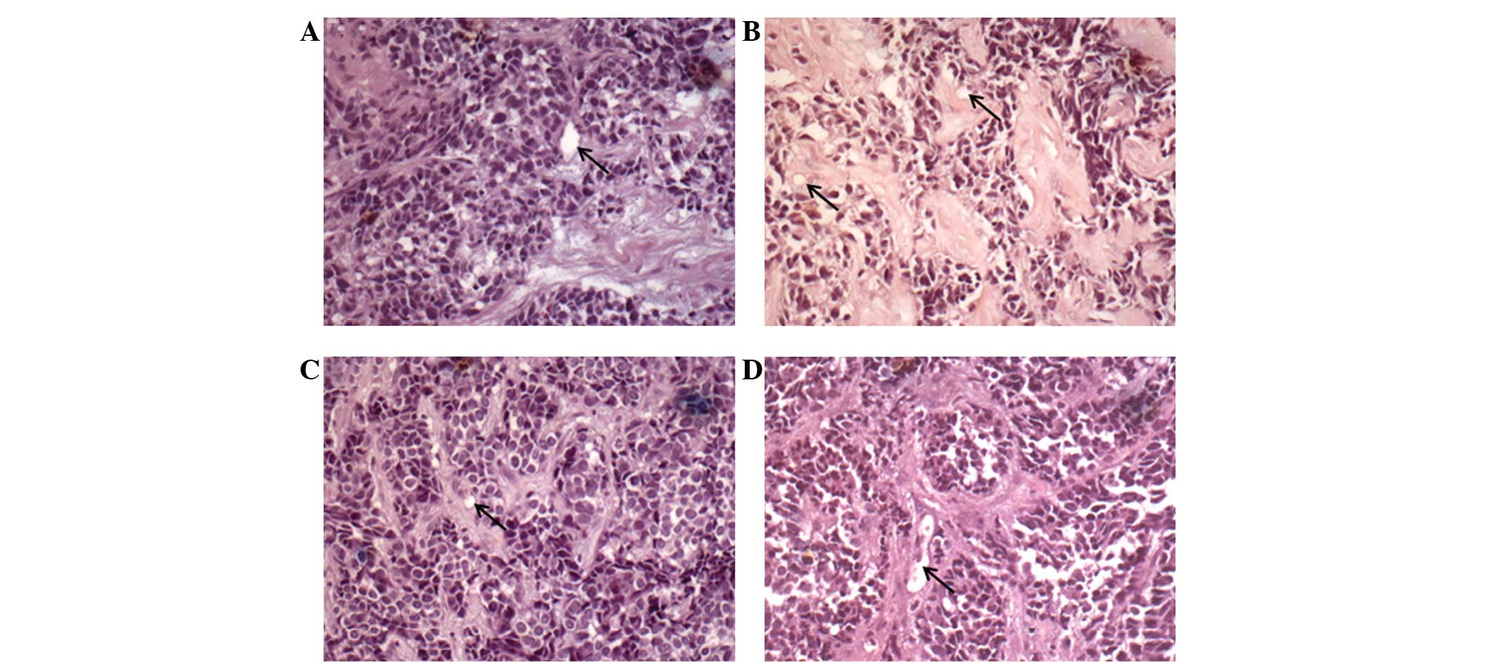
from patients. As demonstrated in Fig.
1, the tumors consisted of small compact cells, arranged in
clusters of varying sizes and surrounded by desmoplastic stromal
tissues, which were primarily fibroblastic in nature. The quantity
of stromal tissue varied from field to field, even within the same
xenograft passage. The tumor cells were small with large, round,
hyperchromatic nuclei. Little cytosol was observed and the cell
borders were typically indistinct. Heavy vascularization was
evident in the stromal tissue, while the majority of the core small
cell sections of the tumor were lacking in any significant vascular
recruitment.
In several instances, the tissue received from the
patient sites was either dry, frozen solid or unrefrigerated. No
growth was predicted or observed in these samples. However, even
under these circumstances, the tissues were implanted into the mice
and treated in the same way as the remaining tissues that had been
received, in case any cells had survived.
Of the 14 human DSRCT specimens that were received
in good condition and implanted into the nude mice, nine (64%) were
considered to be positive for growth. These tumor lines were
assigned the code names; ZUC, BER, VOS, MYE, BOD, UEK, ORA, DYC and
CAR. In all positive cases, the site of inoculation grew a tumor
that was histologically compatible (Fig. 1) with the original tissue, and this
tumor was able to be retransplanted into other nude mice for serial
passaging. For the nine established xenograft lines, the mean time
to the first passage (nude passage 1, NP1) was 192 days (Table I). The time to the first passage is
an effective measure of tumor malignancy, i.e., how well the tumor
adapts to an environment other than its tissue of origin.
 | Table IGrowth of primary DSRCT samples as
xenografts in nude mice. |
Table I
Growth of primary DSRCT samples as
xenografts in nude mice.
| Tumor | Date received | Time to NP1
(days) |
|---|
| ZUC | 01/28/2005 | 145 |
| BER | 02/11/2005 | 75 |
| VOS | 04/15/2005 | 271 |
| MYE | 10/04/2005 | 113 |
| BOD | 11/18/2006 | 186 |
| UEK | 01/11/2007 | 209 |
| ORA | 05/23/2008 | 306 |
| DYC | 02/13/2009 | 250 |
| CAR | 05/18/2010 | 177 |
Attempts to inject the tumor fragments
intraperitoneally were conducted in order to simulate orthotopic
inoculation; however, for reasons that are presently unclear, these
attempts were universally unsuccessful at generating tumors in the
nude mice.
When cell lines become established, either as
xenografts or in tissue culture, their growth characteristics may
markedly change compared with those of primary inoculations of
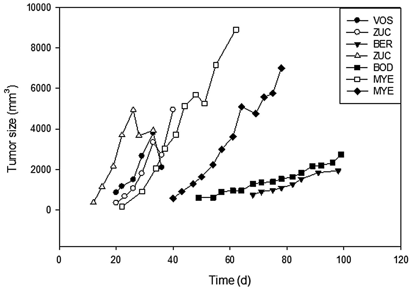
patient tissues in nude mice. The growth rates of the established
xenograft lines were separated into two categories: a rapid growth
group and an indolent growth group (Fig. 2). The mean time from tumor injection
to palpable growth (200–300 mm3) was 22.4 days for the
rapid growth group and 58 days for the indolent growth group.
Of the few samples that yielded sufficient tissue to
split between the xenograft lines and tissue cultures, none yielded
primary lines in tissue culture. However, several lines were able
to be established as nude transfer (NUT) lines, which were
xenografts that were surgically excised from nude mice and
subsequently grown in culture. The stromal tissue was culled slowly
from these cultures by selective trypsinization over the course of
the experiment (≥6 months).
Discussion
Histologically, the DSRCT xenografts closely
resembled the tumors from which they originated; they appeared to
be small compact cells, with large nuclei and little cytoplasm,
arranged in clusters threaded with desmoplastic tissue. The
simplicity of the cell structure, combined with the multifocal
nature of the disease in patients, suggested the possibility that
an unusually large proportion of the tumor cells may be cancer stem
cells.
When excised from the mice, the xenografts were
observed to be soft, well-vascularized and flushed with blood. The
majority of other dorsally grown subcutaneous xenografts exhibit a
necrotic core that has been starved of nutrients by the lack of
sufficient vascularization. In numerous types of solid tumor,
particularly those of the breast, desmoplasia is initiated by the
secretion of platelet-derived growth factor (PDGF), which acts in a
paracrine manner on stromal fibroblasts (12). The same mechanism is likely to be
involved in DSRCT, while the dense vascularization of the stromal
tissues closely resembles the situation in a number of types of
colon carcinoma, where the recruited fibroblasts contribute to
vascular maturation (13).
While the present data indicate two distinct
populations of DSRCT as far as growth rate is concerned, the
underlying mechansims for these differences are unclear.
Traditional cytotoxic agents are more effective against
fast-growing tumors, and slower-growing tumors are less responsive
to cell cycle phase-specific cytotoxic agents (14). Therefore, we predicted that the
DSRCT lines that exhibited more indolent growth would be less
susceptible to cell cycle-specific chemotherapy, such as with
camptothecins or methotrexate. Limited testing in the present study
suggested that this, in fact, was not the case.
DSRCT belongs to a larger family of tumors known as
small round cell sarcomas. Neoplasms, including Wilms’ tumor and
Ewing’s sarcoma, also belong to this category. DSRCT is
significantly less common than the remaining tumors of the family,
but have the worst prognosis. DSRCTs have been demonstrated to be
refractory to chemotherapy and almost uniformly fatal (9), however, the tumors may respond well to
camptothecin compounds, which have been identified to have a
certain effectiveness against indolent tumors (15,16).
Abbreviations:
|
DSRCT
|
desmoplastic small round cell
tumor
|
|
FCS
|
fetal calf serum
|
|
PDGF
|
platelet derived growth factor
|
Acknowledgements
This study was supported by the
Scranton Family (Houston, TX, USA), and by numerous smaller,
individual donations to the Christus Stehlin Foundation for Cancer
Research (Houston, TX, USA) and the Friends of the Stehlin
Foundation (Houston, TX, USA). The authors would like to thank Ms.
Gabrielle Patout for assistance in the preparation of this
manuscript.
References
|
1
|
Gerald WL and Rosai J: Case 2.
Desmoplastic small cell tumor with divergent differentiation.
Pediatr Pathol. 9:177–183. 1989. View Article : Google Scholar : PubMed/NCBI
|
|
2
|
Cummings OW, Ulbright TM, Young RH, Dei
Tos AP, Fletcher CD and Hull MT: Desmoplastic small round cell
tumors of the paratesticular region. A report of six cases. Am J
Surg Pathol. 21:219–225. 1997. View Article : Google Scholar : PubMed/NCBI
|
|
3
|
Parkash V, Gerald WL, Parma A, Miettinen M
and Rosai J: Desmoplastic small round cell tumor of the pleura. Am
J Surg Pathol. 19:659–665. 1995. View Article : Google Scholar : PubMed/NCBI
|
|
4
|
Tison V, Cerasoli S, Morigi F, Ladanyi M,
Gerald WL and Rosai J: Intracranial desmoplastic small-cell tumor.
Report of a case. Am J Surg Pathol. 20:112–117. 1996. View Article : Google Scholar : PubMed/NCBI
|
|
5
|
Adsay V, Cheng J, Athanasian E, Gerald W
and Rosai J: Primary desmoplastic small cell tumor of soft tissues
and bone of the hand. Am J Surg Pathol. 23:1408–1413. 1999.
View Article : Google Scholar : PubMed/NCBI
|
|
6
|
Young RH, Eichhorn JH, Dickersin GR and
Scully RE: Ovarian involvement by the intra-abdominal desmoplastic
small round cell tumor with divergent differentiation: a report of
three cases. Hum Pathol. 23:454–464. 1992. View Article : Google Scholar : PubMed/NCBI
|
|
7
|
Wolf AN, Ladanyi M, Paull G, Blaugrund JE
and Westra WH: The expanding clinical spectrum of desmoplastic
small round-cell tumor: a report of two cases with molecular
confirmation. Hum Pathol. 30:430–435. 1999. View Article : Google Scholar : PubMed/NCBI
|
|
8
|
Syed S, Haque AK, Hawkins HK, Sorensen PH
and Cowan DF: Desmoplastic small round cell tumor of the lung. Arch
Pathol Lab Med. 126:1226–1228. 2002.PubMed/NCBI
|
|
9
|
Chang F: Desmoplastic small round cell
tumors: cytologic, histologic, and immunohistochemical features.
Arch Pathol Lab Med. 130:728–732. 2006.PubMed/NCBI
|
|
10
|
Sawyer JR, Tryka AF and Lewis JM: A novel
reciprocal chromosome translocation t(11;22)(p13;q12) in an
intraabdominal desmoplastic small round-cell tumor. Am J Surg
Pathol. 16:411–416. 1992. View Article : Google Scholar : PubMed/NCBI
|
|
11
|
Mrabti H, Kaikani W, Ahbeddou N, Abahssain
H, El Khannoussi B, Amrani M and Errihani H: Metastatic
desmoplastic small round cell tumor controlled by an
anthracycline-based regimen: review of the role of chemotherapy. J
Gastrointest Cancer. 43:103–109. 2012. View Article : Google Scholar : PubMed/NCBI
|
|
12
|
Shao ZM, Nguyen M and Barsky SH: Human
breast carcinoma desmoplasia is PDGF initiated. Oncogene.
19:4337–4345. 2000. View Article : Google Scholar : PubMed/NCBI
|
|
13
|
Schmid SA, Dietrich A, Schulte S, Gaumann
A and Kunz-Schughart LA: Fibroblastic reaction and vascular
maturation in human colon cancers. Int J Radiat Biol. 85:1013–1025.
2009. View Article : Google Scholar : PubMed/NCBI
|
|
14
|
Schwartz RN, Blanke CD and Pesko LJ:
Targeted therapies in the treatment of colorectal cancer: what
managed care needs to know. J Manag Care Pharm. 10(5 Suppl B):
S2–S13. 2004.PubMed/NCBI
|
|
15
|
Bartlett NL, Johnson JL, Wagner-Johnston
N, Ratain MJ and Peterson BA; Cancer and Leukemia Group B: Phase II
study of 9-aminocamptothecin in previously treated lymphomas:
results of Cancer and Leukemia Group B 9551. Cancer Chemother
Pharmacol. 63:793–798. 2009. View Article : Google Scholar : PubMed/NCBI
|
|
16
|
Sarris AH, Phan A, Goy A, Romaguera J,
Hagemeister FB, Rodriguez MA, McLaughlin P, Pro B, Medeiros LJ,
Samuels B, Mesina O, Bleyer AW and Cabanillas F: Irinotecan in
relapsed or refractory non-Hodgkin’s lymphomas. Indications of
activity in a phase II trial. Oncology (Williston Park). 16(8 Suppl
7): 27–31. 2002.
|