Introduction
The fruit of Cornus officinalis is effective
for treating liver and lung disease in traditional Chinese medicine
(1). In recent decades, the
pharmacology and phytochemistry of the fruit of Cornus
officinalis have been extensively investigated. Clinical
studies have demonstrated that this fruit has anticancer and
antioxidative roles (2,3). Based on various clinical roles,
several effective water-soluble components of the fruit of
Cornus officinalis have been identified. For instance,
gallic acid has antitumor properties (4) and oleanolic acid has antiulcer
properties and markedly reduces blood pressure and blood glucose
levels (5). Morroniside is used to
suppress the generation of superoxide anions in a
concentration-dependent manner, and protects nerve cells from
H2O2-induced oxidation injury (6,7). At
the molecular level, morroniside can enhance the activity of
alkaline phosphatase and decrease caspase-3 mRNA levels (8). The protection of cell growth by
morroniside is hypothesized to be involved in regulation of cell
proliferation and apoptosis.
The retinoblastoma (Rb)/pocket protein family is
made up of tumor suppressor proteins involved in cell
proliferation, apoptosis and cell differentiation (9,10). The
Rb protein is one of the master regulators of the eukaryotic cell
cycle, regulating the G1/G0 phase and also the S and G2 phases
(11). Rb protein activity is
regulated by phosphorylation. In normal cells during the M-to-G1
transition, the Rb protein is progressively dephosphorylated by
protein phosphatase 1, enabling it to bind E2F, blocking
transcription of multiple genes involved in cell cycle progression
and elongating the G1/G0 phases. Following mitogenic stimulation of
the cell, the Rb protein is phosphorylated by cyclin-dependent
kinase (CDK) 4/6-cyclin D complexes. Phosphorylation inactivates
the Rb protein, resulting in release of E2F, enabling E2F to
transcriptionally activate genes that facilitate the G1/S
transition and S phase. The Rb protein remains
phosphorylated/inactive throughout the S, G2 and M phases through
hyperphosphorylation by cyclin E/CDK2 complexes (12–15).
The present study showed that morroniside decreases
cell apoptosis induced by H2O2 in HELF cells
but not in A549 cells. Additionally, the S phase of the cell cycle
was returned to normal function and Rb protein levels were
downregulated by morroniside. These results indicate that the
effects of morroniside on HELF and A549 cells may correlate with
the regulation of Rb protein levels and S phase length.
Materials and methods
Cells and reagents
HELF and A549 cells were purchased from Shanghai
Institute of Biochemistry and Cell Biology (Shanghai, China).
Morroniside was provided by Jiangsu ZhongKang New Drug Fingerprint
R&D Co., Ltd (Nanjing, China) at a purity of >98%; MTT,
acridine orange (AO), ethidium bromide (EB), RNase A, propidium
iodide (PI) and trypsin were purchased from Sigma (St. Louis, MO,
USA). RPMI 1640 medium was obtained from Gibco-BRL (Carlsbad, CA,
USA) and fetal bovine serum (FBS) was from Sijiqing Biotechnology
(Hangzhou, China). Cell culture and drug incorporation. HELF and
A549 cells were cultured in RPMI 1640 medium with 10% (v/v) FBS,
100 U/ml streptomycin and 100 U/ml penicillin, at 37°C with 5%
CO2. The cells digested with 0.25% trypsin were cultured
in 96-well plates (for proliferation analysis), 6-well plates (for
morphology observation) or flasks (for cell cycle analysis). In all
experiments, HELF/A549 cells were cultured without treatment
(normal control), with 300 μM H2O2/0.2 nM
staurosporine (model) or with combined treatment of
H2O2/staurosporine and morroniside at various
doses (12.5, 25, 50, 100 and 200 μg/ml).
Cell proliferation analysis
Cell proliferation was determined by MTT assay. HELF
cells in exponential growth were seeded at a final concentration of
6×10⁴ cells/ml in a 96-well plate. At 50% confluency,
the cells were pretreated with morroniside at various
concentrations for 2 h, exposed to H2O2 or
staurosporine (0.2 nM) and incubated for 48 h. Next, 50 ml MTT (1
mg/ml) was added to each well, followed by incubation for 4 h at
37°C. The supernatants were discarded and the formazan product was
dissolved in 100 μl dimethyl sulfoxide. The 96-well plate was
agitated on a micro-vibrator for an additional 10 min. The optical
density of each well was measured at λ490 nm using an
enzyme-immunoassay instrument.
Morphological examination
Cell groups were subjected to various designated
treatments, as discussed. The cells were observed under a
fluorescence microscope and photographed. Cells were seeded at a
concentration of 1×10⁵/well in a 6-well culture plate. A
cover slip was placed on the bottom of each well to allow cells to
grow on the surface as a monolayer. At 40–60% confluency, the cells
were divided into three groups: Normal control group;
H2O2-treated group, treated with 300 μM
H2O2 and incubated for 48 h; morroniside plus
H2O2-treated group, pretreated with 100 μg/ml
morroniside for 2 h and exposed to 300 μM
H2O2. After a 48-h incubation period, the
cell culture was washed with phosphate-buffered saline (PBS),
stained with 10 μg/ml AO/EB for 2 min and observed under a
fluorescence microscope (Olympus BX60; Olympus, Tokyo, Japan).
Flow cytometry
Cells were treated as discussed. After 24-h culture,
cells were washed twice with ice-cold PBS buffer (pH 7.4) prior to
fixing with 75% ice-cold ethanol and staining with PI (1 mg/ml) in
the presence of 1% RNAase A for 30 min. The samples were
subsequently analyzed by flow cytometry (BD Biosciences, Franklin
Lakes, NJ, USA). The Cell Quest software (BD Biosciences) was
applied to analyze apoptosis rates and the cell cycle.
Western blotting
HELF cells were lysed in cold lysis buffer [40 mM
HEPES (pH 7.5), 150 mM NaCl, 1.5 mM MgCl2, 1 mM EDTA, 1
mM dithiothreitol and 1 mM fresh phenylmethylsulfonyl fluoride].
Lysates were centrifuged at 12,000 × g for 15 min. The supernatant
was collected and the protein concentration was determined by Lowry
protein assay. Next, 20 μg protein was loaded in each well,
electrophoresed by SDS-PAGE and transferred onto nitrocellulose
membranes. The membranes were blocked with 5% non-fat dried milk in
PBS with 0.5 ml/l Tween-20 for 2 h at room temperature, and
incubated overnight at 4°C with primary antibodies against human
P16, P27, P53 or Rb proteins, purchased from Beyotime (Nanjing,
China). This was followed by incubation with a horseradish
peroxidase-conjugated secondary antibody (Bioworld Technology, St.
Louis Park, MN, USA) at room temperature for 1 h. Enhanced
chemiluminescence was used to detect the results.
Statistical analysis
The statistical analysis among various groups was
performed using one-way analysis of variance with Scheffé’s test.
All data were processed with SPSS software (SPSS, Inc., Chicago,
IL, USA). The results were expressed as the mean ± SD. P<0.05
and P<0.01 were considered to indicate a statistically
significant and highly statistically significant difference,
respectively.
Results
Morroniside reverses inhibition of cell
growth in human embryonic lung fibroblasts cells induced by
H2O2
Preliminary data indicated that the crude extract of
morroniside protected the lung in aging and immune-defective mice.
In order to understand whether morroniside regulates the
proliferation of lung cells, the growth of HELF cells treated with
morroniside (98% purity) was examined. MTT was applied to analyze
HELF growth under H2O2 treatment and with the
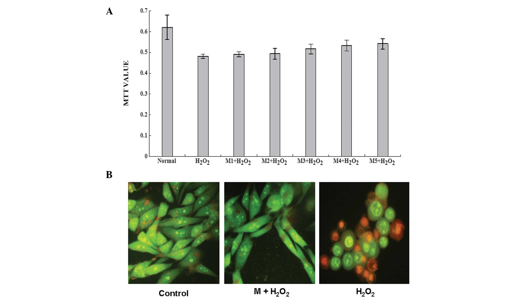
addition of morroniside at various concentrations (Fig. 1A). Results showed that 300 μM
H2O2 treatment decreased HELF growth, as
expected, while 100 μg/ml morroniside markedly increased HELF cell
growth and decreased the negative effect on growth under
H2O2 treatment. This indicates that
morroniside protects HELF proliferation against
H2O2 treatment.
Additionally, under a fluorescence microscope,
staining with mixed dye (AO/EB) showed that, compared with the
normal control group, the amount of cells was lower in the model
group but increased with the treatment of 100 μg/ml morroniside.
The control group was morphologically normal, with evenly-stained
bright green nuclei. Under H2O2 treatment,
however, the cells were wrinkled and chromatin was condensed and
orange in color. The addition of morroniside morphologically
restored the cells back to the morphological appearance of the
control group (Fig. 1B). These
results indicate that morroniside blocks the negative effect of
H2O2 treatment on HELF cells.
Morroniside specifically antagonizes
H2O2-induced HELF apoptosis
H2O2 treatment can result in
various consequences in terms of cell growth, including apoptosis,
autophagy and necrosis (16). To
understand whether morroniside treatment is involved in the
regulation of apoptosis, flow cytometry was used to analyze the
cell cycle and rate of apoptosis in HELF cells under various
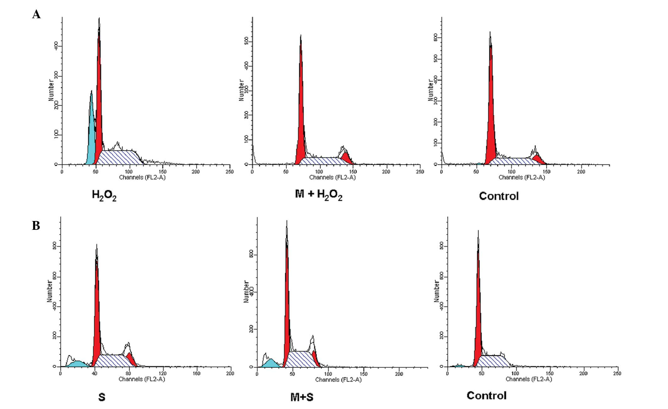
treatments (Fig. 2A). Results
demonstrated that 300 μM H2O2 induced a 41%
rate of apoptosis in HELF cells (control group apoptosis rate,
0.48%), however, the addition of morroniside (100 μg/ml) 2 h prior
to H2O2 treatment maintained the apoptotic
rate at the normal control level, indicating that morroniside
protected HELF cells from the H2O2-induced
apoptosis. Furthermore, H2O2 treatment
elongated the S phase of the cell cycle, while morroniside markedly
decreased the duration of the S phase to that of normal cells,
suggesting that morroniside repairs the dysfunctional regulation of
the S phase induced by H2O2. To understand
whether morroniside regulates the common signaling pathways of
apoptosis, staurosporine was used as an apoptotic model to examine
the consequences of morroniside treatment on staurosporine-induced
apoptosis (Fig. 2B). Flow cytometry
results showed that 0.2 nM staurosporine caused an 8.1% apoptotic
rate in HELF cells and the combination of morroniside and
staurosporine resulted in a similar apoptotic rate of 8.3%. This
suggests that morroniside may not reverse apoptosis induced by
staurosporine. Therefore, morronoside is involved in
H2O2 signaling and likely to regulate the
upstream apoptotic signaling pathways.
Morroniside does not reverse apoptosis of
lung cancer A549 cells
As morroniside restored HELF apoptosis to normal
levels, the apoptotic effects of morroniside were examined in
cancer cells. The A549 cell line was used for the same experiments
and 100 μg/ml morroniside was applied 2 h prior to
H2O2 treatment. Flow cytometry showed that
100 μM H2O2 induced HELF apoptosis at rate of
8.1%. Notably, the combination of morroniside and
H2O2 caused a 7.8% rate of apoptosis
(Fig. 3A) and MTT results showed
that morroniside markedly increased A549 proliferation but did not
reduce the H2O2-induced inhibition of A549
cell growth. These primary results indicate that morroniside has no
apparent effect on A549 growth under H2O2
treatment (Fig. 3B).
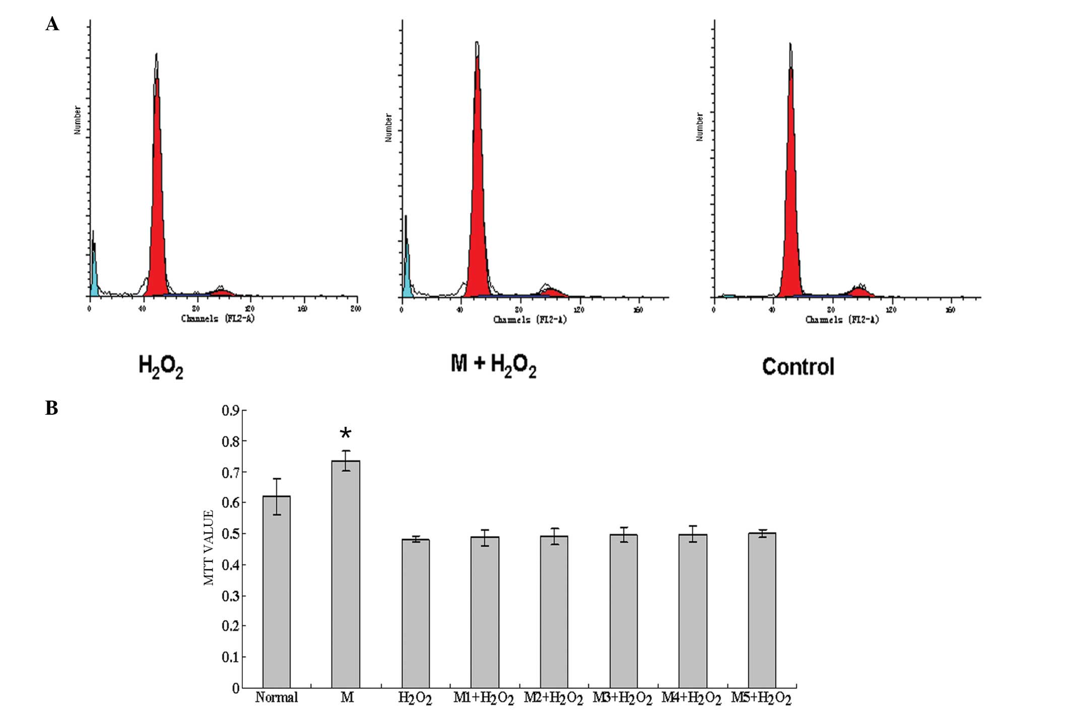 | Figure 3Morroniside does not inhibit the
apoptosis of A549 cells induced by H2O2. (A)
A549 apoptosis under various treatments. Treatment with 300 μM
H2O2 induced an 8.1% rate of apoptosis in
A549 cells. The addition of 100 μg/ml morroniside 2 h prior to
H2O2 treatment showed a similar apoptotic
rate of 7.8%. (B) A549 cell growth was measured by MTT. Compared
with the control group, morroniside significantly increased A549
growth, H2O2 treatment decreased cell growth
and combined treatment of morroniside and
H2O2 did not improve cell growth (P<0.01).
M, morroniside; M1-5, 12.5, 25, 50, 100 and 200 μg/ml morroniside,
respectively. *Compared with the H2O2 group
the addition of morroniside showed no significant difference to
cell growth. Blue, apoptotic cells; red, normal cells. |
Morroniside regulates the S phase of the
cell cycle and negatively regulates Rb protein levels
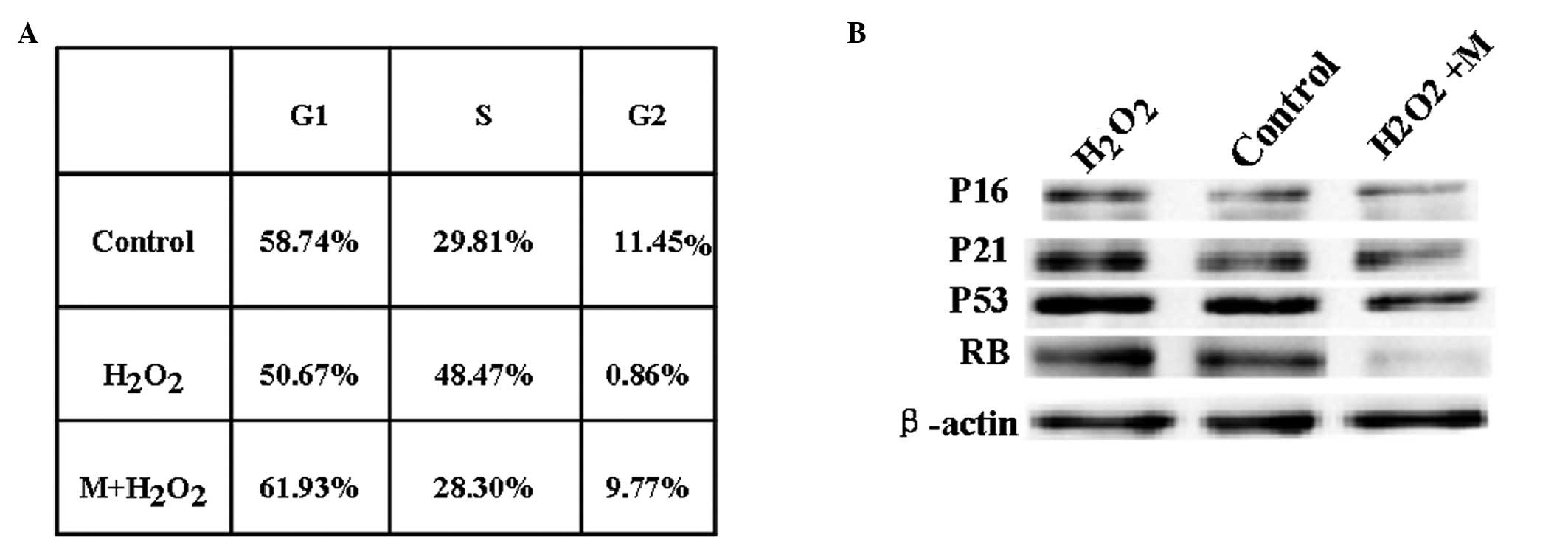
In normal HELF cells, flow cytometry showed that the
proportion of the cell cycle in the S phase was 28.8% and that
H2O2 increased this to 48.5%. By contrast,
morroniside reduced this to 28.3%, indicating that morroniside
regulates the cell cycle during the S phase (Fig. 4A). To elucidate the potential
signaling pathways regulated by morroniside, the levels of P16,
P27, P53 and Rb proteins, all critical factors in the S phase and
the regulation of cell growth and apoptosis, were examined
(17). Western blotting showed that
Rb and P53 proteins were markedly reduced under the combined
treatment of morroniside and H2O2. However,
H2O2 treatment slightly increased P53 protein
levels but did not affect Rb protein levels, suggesting that Rb
protein levels are not regulated by H2O2 but
may mediate H2O2-induced apoptosis (Fig. 4B). The loss of Rb protein positively
correlated with cell proliferation. These results suggest that the
downregulation of Rb protein expression by morroniside may be
relevant to the mediation of S phase length.
Discussion
Previous preliminary experiments (data not shown)
have shown that morroniside plays a specific role in the protection
of lung cell proliferation, suggesting a potential medicinal use of
morroniside to protect normal cells from the side effects of
anticancer medication. The present study showed that morroniside
exhibits differential effects on apoptosis in HELF and A549 cells
under H2O2 treatment. Notably, morroniside
significantly reversed the negative effects of
H2O2 on HELF cell growth but not on A549
cells, indicating that morroniside-regulated signaling pathways are
relevant for partially differentiating between the signaling
pathways in HELF and A549 cells. Furthermore, morroniside protected
the S phase and downregulated Rb protein, suggesting that the
molecular mechanisms of morroniside mediating the cell cycle (S
phase) may be relevant to Rb protein signaling.
Results of the present study suggest that the
different signaling pathways of HELF and A549 cell lines interact
with H2O2 signaling and are specific to the
regulation of S phase. H2O2 treatment was
applied to establish a model of hepatocellular oxidative stress, as
H2O2 acts as a harmful factor in these cells,
resulting in accumulation of reactive oxygen species (ROS) and an
imbalance between the cell oxidation and antioxidant system
(18). Results showed that
H2O2 did not increase Rb protein levels,
suggesting that morroniside and H2O2
signaling may interact downstream of the Rb protein. The
interaction of signaling pathways regulated by ROS and morroniside
requires further investigation. Notably, results also showed that
morroniside did not reverse staurosporine-induced apoptosis,
indicating that morroniside may not directly target the apoptosis
signaling pathways, but acts on the upstream molecules beyond the
apoptotic signaling pathways. Furthermore, staurosporine, an
inhibitor of protein kinase C, was used to establish an apoptosis
model through elongation of G2 phase (19). Morroniside was found to restore the
H2O2-induced imbalance of the S phase only
but not staurosporine-induced G2 phase imbalance, suggesting that
morroniside may be an S phase-specific regulator.
Results of the present study imply that the
downregulation of Rb protein expression may be relevant to S phase
regulation. It is well known that Rb protein plays roles throughout
the cell cycle. The phosphorylation and dephosphorylation of Rb
play key roles in the cell cycle (20). Usually, Rb protein levels undergo no
significant change through the cell cycle (21). However, results of the present study
showed that Rb protein was markedly downregulated by morroniside.
Morronisde also caused the rebalance of the S phase length in HELF
cells, which was previously increased by H2O2
treatment. It remains unclear how Rb protein levels are involved in
S phase regulation. Identification of the signaling pathways
regulated by morroniside and the molecular mechanisms regulating Rb
protein stability require further in vitro and in
vivo study.
In conclusion, morroniside was found to inhibit
apoptosis in normal HELF cells but not in the A549 cancer cell
line. These effects included the protection of cell proliferation
and normal cell morphology, and the restoration of the S phase to
normal levels. Furthermore, the interaction between morroniside and
H2O2 signaling was found to be involved in
HELF proliferation and apoptosis. Thus, morroniside is a potential
compound for clinical amelioration of the side effects of
anticancer treatments.
Acknowledgements
This study was supported by grants from the National
Nature Science Foundation of China (no. 30772851), the Priority
Academic Program Development of Jiangsu Higher Education
Institutions and the National Science and Technology Research
Supporting Program in Chinese Medicine in ‘The 11th Five-Year Plan’
(no. 2006BAI11B08-01).
References
|
1
|
Liu H and Xu H: Advancement in research of
Fructus Corni Officinalis and its main component. Nanjing
Zhongyiyao Daxue Xuebao. 19:254–256. 2003.(In Chinese).
|
|
2
|
Zhang L, Yuan Z, Du Y and Wang C: Recent
development and prospect of Cornus officinalis. Zhongguo
Zhongyao Zazhi. 35:952–956. 2004.(In Chinese).
|
|
3
|
Song Q and Zhou Y: Recent Research on the
pharmacological action of Cornus officinalis. Zhongyiyao
Xinxi. 23:24–25. 2006.(In Chinese).
|
|
4
|
Li X, Cui L and Zhu D: Research progress
on biological effects of gallic acid. Zhongguo Yaoshi. 7:767–769.
2004.(In Chinese).
|
|
5
|
Zhou Z and Yuan D: Pharmacological
research progress of oleanolic acid. Zhongguo Yiyuan Yaoxue Zazhi.
28:2031–2033. 2008.(In Chinese).
|
|
6
|
Wei S, Chi H, Kodama H and Chen G:
Anti-inflammatory effect of three iridoids in human neutrophils.
Nat Prod Res. 27:911–915. 2013. View Article : Google Scholar : PubMed/NCBI
|
|
7
|
Ai HX, Wang W, Sun FL, Huang WT, An Y and
Li L: Morroniside inhibits H2O2-induced
apoptosis in cultured nerve cells. Zhongguo Zhong Yao Za Zhi.
33:2109–2112. 2008.(In Chinese).
|
|
8
|
Li M, Wang W, Wang P, Yang K, Sun H and
Wang X: The pharmacological effects of morroniside and loganin
isolated from Liuweidihuang Wan, on MC3T3-E1 cells. Molecules.
15:7403–7414. 2010. View Article : Google Scholar : PubMed/NCBI
|
|
9
|
Gordon GM and Du W: Conserved RB functions
in development and tumor suppression. Protein Cell. 2:864–878.
2011. View Article : Google Scholar : PubMed/NCBI
|
|
10
|
Poznic M: Retinoblastoma protein: a
central processing unit. J Biosci. 34:305–312. 2009. View Article : Google Scholar
|
|
11
|
Wikman H and Kettunen E: Regulation of the
G1/S phase of the cell cycle and alterations in the RB pathway in
human lung cancer. Expert Rev Anticancer Ther. 6:515–530. 2006.
View Article : Google Scholar : PubMed/NCBI
|
|
12
|
Kolupaeva V and Basilico C: Overexpression
of cyclin E/CDK2 complexes overcomes FGF-induced cell cycle arrest
in the presence of hypophosphorylated Rb proteins. Cell Cycle.
11:2557–2566. 2012. View
Article : Google Scholar : PubMed/NCBI
|
|
13
|
Kolupaeva V and Janssens V: PP1 and PP2A
phosphatases - cooperating partners in modulating retinoblastoma
protein activation. FEBS J. 280:627–643. 2013. View Article : Google Scholar
|
|
14
|
Aksoy O, Chicas A, Zeng T, Zhao Z,
McCurrach M, Wang X and Lowe SW: The atypical E2F family member
E2F7 couples the p53 and RB pathways during cellular senescence.
Genes Dev. 26:1546–1557. 2012. View Article : Google Scholar : PubMed/NCBI
|
|
15
|
Naqsh e Zahra S, Khattak NA and Mir A:
Comparative modeling and docking studies of p16ink4/cyclin D1/Rb
pathway genes in lung cancer revealed functionally interactive
residue of RB1 and its functional partner E2F1. Theor Biol Med
Model. 10:12013.
|
|
16
|
Aiken RJ and Roman SD: Antioxidant systems
and oxidative stress in the testes. Oxid Med Cell Longev. 1:15–24.
2008. View Article : Google Scholar
|
|
17
|
Ye Y, Wang D, Su C, Rong T and Guo A:
Combined detection of p53, p16, Rb, and EGFR mutations in lung
cancer by suspension microarray. Genet Mol Res. 8:1509–1518. 2009.
View Article : Google Scholar : PubMed/NCBI
|
|
18
|
Kohen R and Nyska A: Oxidation of
biological systems: oxidative stress phenomena, antioxidants, redox
reactions, and methods for their quantification. Toxicol Pathol.
30:620–650. 2002. View Article : Google Scholar
|
|
19
|
Antonsson A and Persson JL: Induction of
apoptosis by staurosporine involves the inhibition of expression of
the major cell cycle proteins at the G(2)/m checkpoint accompanied
by alterations in Erk and Akt kinase activities. Anticancer Res.
29:2893–2898. 2009.
|
|
20
|
Rubin SM: Deciphering the retinoblastoma
protein phosphorylation code. Trends Biochem Sci. 38:12–19. 2013.
View Article : Google Scholar : PubMed/NCBI
|
|
21
|
Burkhart DL, Ngai LK, Roake CM, Viatour P,
Thangavel C, Ho VM, Knudsen ES and Sage J: Regulation of RB
transcription in vivo by RB family members. Mol Cell Biol.
30:1729–1745. 2010. View Article : Google Scholar : PubMed/NCBI
|