Spandidos Publications Logo
  • About
    • About Spandidos
    • Aims and Scopes
    • Abstracting and Indexing
    • Editorial Policies
    • Reprints and Permissions
    • Job Opportunities
    • Terms and Conditions
    • Contact
  • Journals
    • All Journals
    • Oncology Letters
      • Oncology Letters
      • Information for Authors
      • Editorial Policies
      • Editorial Board
      • Aims and Scope
      • Abstracting and Indexing
      • Bibliographic Information
      • Archive
    • International Journal of Oncology
      • International Journal of Oncology
      • Information for Authors
      • Editorial Policies
      • Editorial Board
      • Aims and Scope
      • Abstracting and Indexing
      • Bibliographic Information
      • Archive
    • Molecular and Clinical Oncology
      • Molecular and Clinical Oncology
      • Information for Authors
      • Editorial Policies
      • Editorial Board
      • Aims and Scope
      • Abstracting and Indexing
      • Bibliographic Information
      • Archive
    • Experimental and Therapeutic Medicine
      • Experimental and Therapeutic Medicine
      • Information for Authors
      • Editorial Policies
      • Editorial Board
      • Aims and Scope
      • Abstracting and Indexing
      • Bibliographic Information
      • Archive
    • International Journal of Molecular Medicine
      • International Journal of Molecular Medicine
      • Information for Authors
      • Editorial Policies
      • Editorial Board
      • Aims and Scope
      • Abstracting and Indexing
      • Bibliographic Information
      • Archive
    • Biomedical Reports
      • Biomedical Reports
      • Information for Authors
      • Editorial Policies
      • Editorial Board
      • Aims and Scope
      • Abstracting and Indexing
      • Bibliographic Information
      • Archive
    • Oncology Reports
      • Oncology Reports
      • Information for Authors
      • Editorial Policies
      • Editorial Board
      • Aims and Scope
      • Abstracting and Indexing
      • Bibliographic Information
      • Archive
    • Molecular Medicine Reports
      • Molecular Medicine Reports
      • Information for Authors
      • Editorial Policies
      • Editorial Board
      • Aims and Scope
      • Abstracting and Indexing
      • Bibliographic Information
      • Archive
    • World Academy of Sciences Journal
      • World Academy of Sciences Journal
      • Information for Authors
      • Editorial Policies
      • Editorial Board
      • Aims and Scope
      • Abstracting and Indexing
      • Bibliographic Information
      • Archive
    • International Journal of Functional Nutrition
      • International Journal of Functional Nutrition
      • Information for Authors
      • Editorial Policies
      • Editorial Board
      • Aims and Scope
      • Abstracting and Indexing
      • Bibliographic Information
      • Archive
    • International Journal of Epigenetics
      • International Journal of Epigenetics
      • Information for Authors
      • Editorial Policies
      • Editorial Board
      • Aims and Scope
      • Abstracting and Indexing
      • Bibliographic Information
      • Archive
    • Medicine International
      • Medicine International
      • Information for Authors
      • Editorial Policies
      • Editorial Board
      • Aims and Scope
      • Abstracting and Indexing
      • Bibliographic Information
      • Archive
  • Articles
  • Information
    • Information for Authors
    • Information for Reviewers
    • Information for Librarians
    • Information for Advertisers
    • Conferences
  • Language Editing
Spandidos Publications Logo
  • About
    • About Spandidos
    • Aims and Scopes
    • Abstracting and Indexing
    • Editorial Policies
    • Reprints and Permissions
    • Job Opportunities
    • Terms and Conditions
    • Contact
  • Journals
    • All Journals
    • Biomedical Reports
      • Information for Authors
      • Editorial Policies
      • Editorial Board
      • Aims and Scope
      • Abstracting and Indexing
      • Bibliographic Information
      • Archive
    • Experimental and Therapeutic Medicine
      • Information for Authors
      • Editorial Policies
      • Editorial Board
      • Aims and Scope
      • Abstracting and Indexing
      • Bibliographic Information
      • Archive
    • International Journal of Epigenetics
      • Information for Authors
      • Editorial Policies
      • Editorial Board
      • Aims and Scope
      • Abstracting and Indexing
      • Bibliographic Information
      • Archive
    • International Journal of Functional Nutrition
      • Information for Authors
      • Editorial Policies
      • Editorial Board
      • Aims and Scope
      • Abstracting and Indexing
      • Bibliographic Information
      • Archive
    • International Journal of Molecular Medicine
      • Information for Authors
      • Editorial Policies
      • Editorial Board
      • Aims and Scope
      • Abstracting and Indexing
      • Bibliographic Information
      • Archive
    • International Journal of Oncology
      • Information for Authors
      • Editorial Policies
      • Editorial Board
      • Aims and Scope
      • Abstracting and Indexing
      • Bibliographic Information
      • Archive
    • Medicine International
      • Information for Authors
      • Editorial Policies
      • Editorial Board
      • Aims and Scope
      • Abstracting and Indexing
      • Bibliographic Information
      • Archive
    • Molecular and Clinical Oncology
      • Information for Authors
      • Editorial Policies
      • Editorial Board
      • Aims and Scope
      • Abstracting and Indexing
      • Bibliographic Information
      • Archive
    • Molecular Medicine Reports
      • Information for Authors
      • Editorial Policies
      • Editorial Board
      • Aims and Scope
      • Abstracting and Indexing
      • Bibliographic Information
      • Archive
    • Oncology Letters
      • Information for Authors
      • Editorial Policies
      • Editorial Board
      • Aims and Scope
      • Abstracting and Indexing
      • Bibliographic Information
      • Archive
    • Oncology Reports
      • Information for Authors
      • Editorial Policies
      • Editorial Board
      • Aims and Scope
      • Abstracting and Indexing
      • Bibliographic Information
      • Archive
    • World Academy of Sciences Journal
      • Information for Authors
      • Editorial Policies
      • Editorial Board
      • Aims and Scope
      • Abstracting and Indexing
      • Bibliographic Information
      • Archive
  • Articles
  • Information
    • For Authors
    • For Reviewers
    • For Librarians
    • For Advertisers
    • Conferences
  • Language Editing
Login Register Submit
  • This site uses cookies
  • You can change your cookie settings at any time by following the instructions in our Cookie Policy. To find out more, you may read our Privacy Policy.

    I agree
Search articles by DOI, keyword, author or affiliation
Search
Advanced Search
presentation
Oncology Letters
Join Editorial Board Propose a Special Issue
Print ISSN: 1792-1074 Online ISSN: 1792-1082
Journal Cover
July-2015 Volume 10 Issue 1

Full Size Image

Sign up for eToc alerts
Recommend to Library

Journals

International Journal of Molecular Medicine

International Journal of Molecular Medicine

International Journal of Molecular Medicine is an international journal devoted to molecular mechanisms of human disease.

International Journal of Oncology

International Journal of Oncology

International Journal of Oncology is an international journal devoted to oncology research and cancer treatment.

Molecular Medicine Reports

Molecular Medicine Reports

Covers molecular medicine topics such as pharmacology, pathology, genetics, neuroscience, infectious diseases, molecular cardiology, and molecular surgery.

Oncology Reports

Oncology Reports

Oncology Reports is an international journal devoted to fundamental and applied research in Oncology.

Experimental and Therapeutic Medicine

Experimental and Therapeutic Medicine

Experimental and Therapeutic Medicine is an international journal devoted to laboratory and clinical medicine.

Oncology Letters

Oncology Letters

Oncology Letters is an international journal devoted to Experimental and Clinical Oncology.

Biomedical Reports

Biomedical Reports

Explores a wide range of biological and medical fields, including pharmacology, genetics, microbiology, neuroscience, and molecular cardiology.

Molecular and Clinical Oncology

Molecular and Clinical Oncology

International journal addressing all aspects of oncology research, from tumorigenesis and oncogenes to chemotherapy and metastasis.

World Academy of Sciences Journal

World Academy of Sciences Journal

Multidisciplinary open-access journal spanning biochemistry, genetics, neuroscience, environmental health, and synthetic biology.

International Journal of Functional Nutrition

International Journal of Functional Nutrition

Open-access journal combining biochemistry, pharmacology, immunology, and genetics to advance health through functional nutrition.

International Journal of Epigenetics

International Journal of Epigenetics

Publishes open-access research on using epigenetics to advance understanding and treatment of human disease.

Medicine International

Medicine International

An International Open Access Journal Devoted to General Medicine.

Journal Cover
July-2015 Volume 10 Issue 1

Full Size Image

Sign up for eToc alerts
Recommend to Library

  • Article
  • Citations
    • Cite This Article
    • Download Citation
    • Create Citation Alert
    • Remove Citation Alert
    • Cited By
  • Similar Articles
    • Related Articles (in Spandidos Publications)
    • Similar Articles (Google Scholar)
    • Similar Articles (PubMed)
  • Download PDF
  • Download XML
  • View XML
Case Report

Primary carcinoid tumor of the kidney with estrogen and progesterone receptor expression. A case report

  • Authors:
    • Chunhua Lin
    • Jitao Wu
    • Zhenli Gao
    • Guimei Qu
    • Wei Wang
    • Guohua Yu
  • View Affiliations / Copyright

    Affiliations: Department of Surgery, Shandong University School of Medicine, Jinan, Shandong 250012, P.R. China, Department of Urological Surgery, Affiliated Yantai Yuhuangding Hospital, Medical College of Qingdao University, Yantai, Shandong 264000, P.R. China, Department of Pathology, Affiliated Yantai Yuhuangding Hospital, Medical College of Qingdao University, Yantai, Shandong 264000, P.R. China
  • Pages: 449-452
    |
    Published online on: April 30, 2015
       https://doi.org/10.3892/ol.2015.3167
  • Expand metrics +
Metrics: Total Views: 0 (Spandidos Publications: | PMC Statistics: )
Metrics: Total PDF Downloads: 0 (Spandidos Publications: | PMC Statistics: )
Cited By (CrossRef): 0 citations Loading Articles...

This article is mentioned in:


Abstract

Primary carcinoid tumors are uncommon neoplasms in the kidney. The current study presents a case of primary carcinoid tumor of the kidney in a 49-year‑old female who suffered from painless gross hematuria for half a month. Left hydronephrosis, a horseshoe kidney and a space‑occupying lesion of the left ureter were found by abdominal computed tomography scans and ultrasonic testing. Surgery was performed and an oval tumor was found under the left ureter; the tumor and left kidney were excised completely. The neoplasm was composed of solid nests of cells, trabeculae, adenoid structures and anastomosing cords in a loose and myxoid background. The tumor cells, which were consistent in volume, exhibited centrally oval nuclei with inconspicuous nucleoli, and eosinophilic finely granular cytoplasm. Upon immunohistochemical staining, the neoplastic cells were positive for AE1/AE3, vimentin, synaptophysin, chromogranin A, estrogen receptor and progesterone receptor, while being negative for epithelial membrane antigen, inhibin A, cluster of differentiation (CD)99, S‑100 and CD10. Based on the histological characteristics, a diagnosis of primary carcinoid tumor of the left kidney was formed. The patient did not receive further treatment. The total follow‑up period was 18 months after the surgery and repeated imaging examinations every 6 months revealed no recurrence.

Introduction

Carcinoid tumors are a group of neuroendocrine tumors that occur most frequently in the gastrointestinal tract as well as in other organs comprising argyrophil cells; ~85% these tumors occur in the digestive tract, with 10% occurring in the lung and 5% occurring in other organs such as the testis (1) and bladder (2). The clinical manifestations of carcinoid tumors may not be apparent, or limited to local symptoms. However, carcinoid syndromes often have obvious systemic symptoms, such as unexplained intermittent diarrhea, flushing, facial telangiectasia, paroxysmal asthma or psychiatric symptoms (3). The diagnosis of carcinoid tumor is dependent on biopsy, and surgical excision is the first treatment. Patients without carcinoid syndrome have a better prognosis (4).

Primary carcinoid tumor in the kidney is uncommon and the expression of the estrogen receptor (ER) and the progesterone receptor (PR) has not been reported. Due to the rarity of this tumor, its clinicopathological characteristics, prognosis and histogenesis have not been fully characterized. The current study reports an unusual case of a carcinoid tumor of the kidney with ER and PR expression in a 49-year-old female who presented with a 2-year history of hypertension. Abdominal computed tomography scans revealed a left horseshoe kidney. Clinicopathological characteristics of primary carcinoid tumors are also discussed by reviewing the literature. Written informed consent was obtained from the patient for publication of this case report and any accompanying images.

Case report

Clinical data

A 49-year-old female with a history of hypertension for two years presented with a half-month history of painless gross hematuria. The patient was admitted to the Department of Urological Surgery in the Affiliated Yantai Yuhuangding Hospital (Medical College of Qingdao University, Yantai, Shandong, China). No abnormal physical signs were found during a physical examination. 
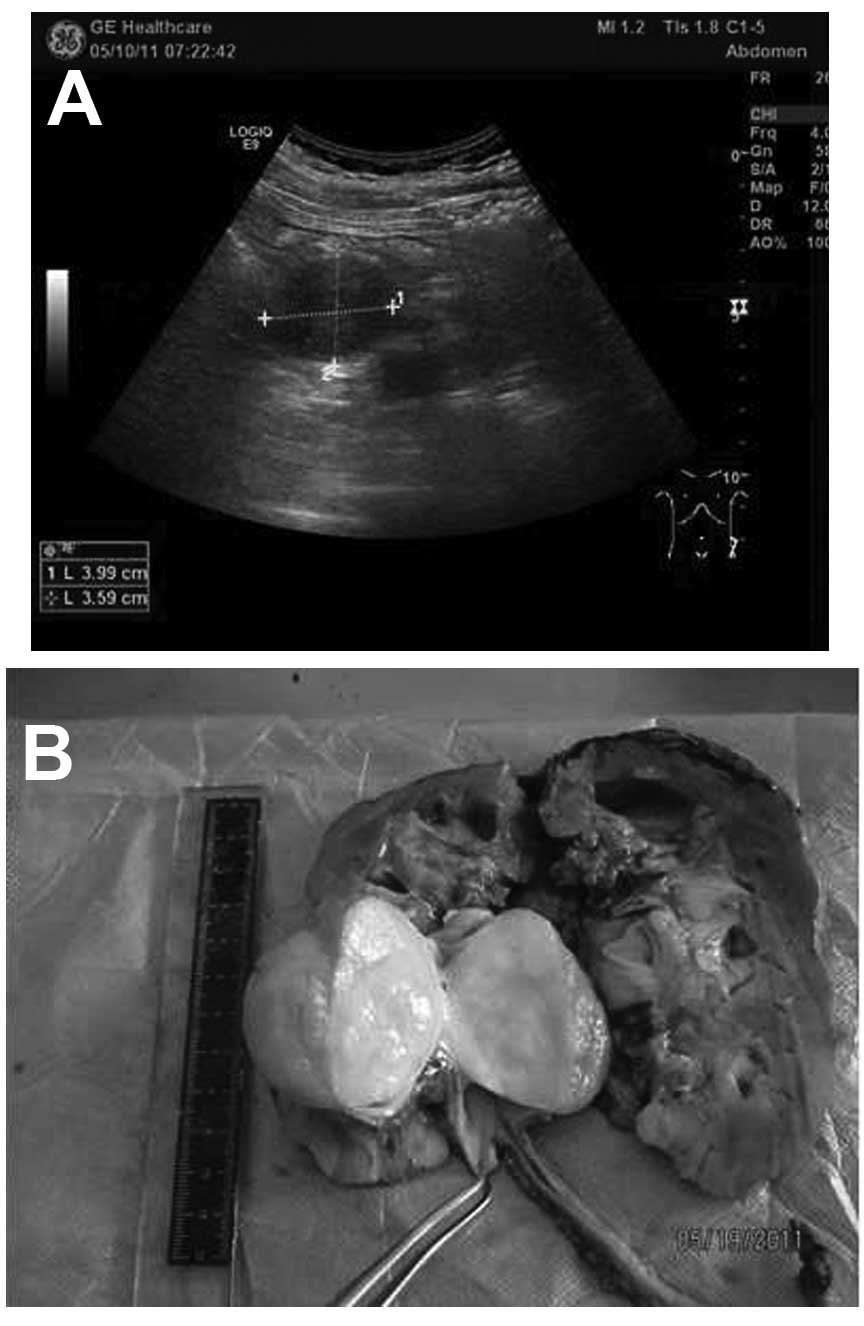
Abdominal computed tomography (CT) scans and ultrasonic testing revealed left hydronephrosis, a horseshoe kidney and a space-occupying lesion in the left ureter, while the right kidney and other visceral organs were normal (Fig. 1A). The hematological findings, endocrine index, erythrocyte sedimentation rate, blood biochemistry, electrolytes, blood urea nitrogen and serum creatinine levels were within the normal ranges. The surgery was performed under general anesthesia and in a right lateral decubitus position at 70°. The left kidney, which presented with black coloration and a horseshoe appearance, was exposed and dissociated. An oval tumor, ~5×4×3.6 cm, was observed under the ureter. The boundaries of the mass were clear. The cross-section of the tumor was faint yellow, homogeneous and pliable in quality (Fig. 1B).

Figure 1.

(A) Ultrasonic examination revealing left hydronephrosis and a space-occupying lesion in the left ureter. (B) Tumor positioned under the uterer, the with a volume of ~5×4×3.6 cm. The boundaries of the mass were clear and the section was faintly yellow.

Pathological findings

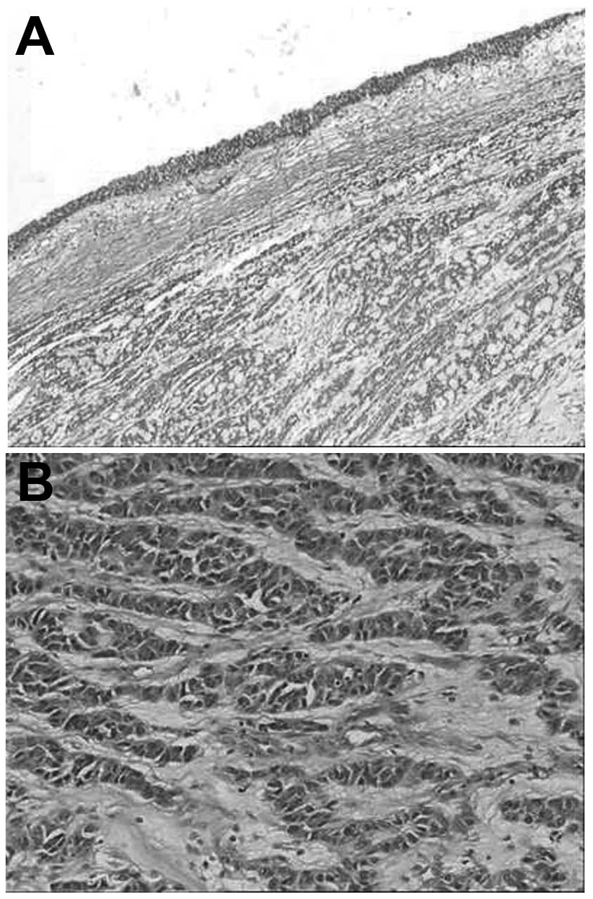
Microscopically, the tumor was located under the mucosa of the pelvis and showed aggressive growth. The neoplasm was composed of solid nests of cells, trabeculae, adenoid structures and anastomosing cords in a loose and myxoid background (Fig. 2A). The tumor cells, which were consistent in volume, exhibited centrally placed oval nuclei with inconspicuous nucleoli, and eosinophilic finely granular cytoplasm. The mitotic rate was <1 per 10 high-power fields (Fig. 2B).

Figure 2.

(A) Tumor was located under the mucosa of the pelvis and the neoplasm was composed of solid nests of cells, trabeculae, adenoid structures and anastomosing cords in a loose and myxoid background (hematoxylin and eosin staining; magnification, x100). (B) Neoplastic cells were similar in size and exhibited centrally placed oval nuclei with eosinophilic finely granular cytoplasm, and a low mitotic rate (hematoxylin and eosin staining; magnification, x200).

Immunohistochemical findings

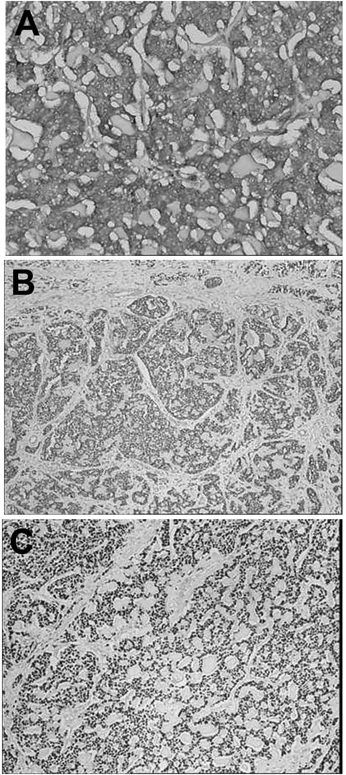
Immunohistochemical staining suggested that the neoplastic cells were positive for AE1/AE3, vimentin, synaptophysin (Syn; Fig. 3A), chromogranin A (CgA), ER (Fig. 3B) and PR (Fig. 3C), while being negative for epithelial membrane antigen, inhibin A, cluster of differentiation (CD)99, S-100 and CD10. The cells that were positive for Ki-67 were dispersive. All the primary antibodies used are listed in Table I. On the basis of histomorphology in light microscopy and the presence of immunohistochemical staining, a diagnosis of primary carcinoid tumor of the left kidney was made. The patient did not receive further treatment. The total follow-up period was 18 months after the surgery. Repeated CT scans and abdominal ultrasonography every 6 months revealed no recurrence or residual lesion.

Figure 3.

Immunostaining of the tumor. The tumor cells were positive for (A) synaptophysin (magnification, x200), (B) estrogen receptor (magnification, x100) and (C) progesterone receptor (magnification, x100).

Table I.

Antibodies employed in the immunohistochemistry applied for the case.

Table I.

Antibodies employed in the immunohistochemistry applied for the case.

AntibodyCloneSourceDilution
Anti-S1004C49Lab vision1:150
Anti-cytokeratinAE1/AE3Dako1:100
Anti-vimentinSP20Lab vision1:100
Anti-Ki-67MIB-1Lab vision1:100
Anti-CD99O13Dako1:100
Anti-CD1056C6Lab vision1:100
Anti-inhibin AR11Dako1:100
Anti-EMAE19Dako1:100
Anti-ER1D5Dako1:100
Anti-PREP2Dako1:100
Anti-SynSP11Dako1:100
Anti-CgAEP38Dako1:100

[i] Dako, Glostrup, Denmark. Lab vision corporation, Fremont, CA, USA. CD, cluster of differentiation; EMA, epithelial membrane antigen; ER, estrogen receptor; PR, progesterone receptor; Syn, synaptophysin; CgA, chromogranin A.

Discussion

Carcinoid tumors of the kidney are rare. The pathogenesis of renal carcinoid tumors remains under debate (5–9). Certain previous studies have attributed the genesis of this tumor to congenital renal abnormalities, while other studies have suggested that the tumor cells are derived from multipotential primitive stem cells, which are induced to neuroendocrine differentiation (10–12). Notably, in the present case, the ER and PR sex hormone receptors were strongly expressed in the tumor cells. ER and PR expression has previously been detected not only in hormone target organs, such as the breast, ovaries and endometrium, but also in the digestive tract and pancreas (13,14). However, the expression of ER and PR in carcinoid tumors of the kidney has not been reported. The present results may not be of use in inferring the pathogenesis of the disease, as no relative literature has been found by review, however, we speculate that the ER and PR may be a potential therapeutic target in patients with this condition (13). Further study is required to investigate this association in the future.

Clinically, Hansel et al reported that 50% of 21 carcinoid tumor patients studied were younger than 50-years-old, and that the tumor showed no tendencies in gender and location. Bloody urine and pain in the lower back proved to be the most common clinical manifestations. Horseshoe kidneys was frequently present, while no other characteristics were known to distinguish renal cell carcinoma on imaging. Endocrine syndrome was not found in the blood examination (15). The current study reports the case of a 49-year-old female who presented with painless and gross hematuria. A horseshoe kidney on imaging was a contributory factor for the diagnosis.

Pathological examination remains the gold standard of diagnosis for renal carcinoid tumors (15,16). Under the microscope, the classic histological characteristics of renal carcinoid tumors are a band or ribbon-like pattern accompanied by solid nests of cells and adenoid structures within a loose stroma. Rosette structures are characteristic of these lesions, however, this was not found in the present case. Tumor cells are similar in size, with a granular eosinophilic cytoplasm. Round to oval nuclei are uniform, with rare mitotic events. Used together with histological features, neuroendocrine immunostaining would aid in distinguishing carcinoid tumors from other diagnoses, such as small cell carcinoma, pheochromocytoma and primitive neuroectodermal tumor. With regard to the present case, a diagnosis of renal carcinoid tumor may have been regarded as presumptive based on the observation of typical morphological characteristics by light microscopy, as this is subject to validation by positive immunohistochemical staining of cytokeratin, vimentin and endocrine markers (CgA, Syn or CD56 protein).

No standard therapy focusing on renal carcinoid tumors has yet been proposed and surgical resection is currently the only treatment (17). Hormonotherapy may be a novel method aimed at patients with positive ER and PR expression. Necrosis, metastasis, an age of >50 years, a tumor diameter >4 cm and more karyokinesis are presumed to potentially indicate an ominous prognosis. However, no clear prognosis factors are generally accepted and further case accumulation is required (18,19).

In conclusion, the current study presents the clinical manifestations and pathological features of a case of renal carcinoid tumor expressing ER and PR. The expression of ER and PR in carcinoid tumors of the kidney is unusual, and these molecules may be a potential therapeutic targets in such patients.

Acknowledgements

This study was supported by the Project of Administration of Traditional Chinese Medicine of Shandong Province (grant no. 2009222).

References

1 

Han X, Yu L, Yang S and Zheng J: Primary neuroendocrine tumor of the testis: A study of clinicopathological features. Int J Clin Exp Pathol. 7:1771–1776. 2014.PubMed/NCBI

2 

Kaplan AL, Margolis DJ, Said J and Chin AI: Primary carcinoid tumor of urinary bladder discovered on pelvic magnetic resonance imaging. Urology. 80:e55–57. 2012. View Article : Google Scholar : PubMed/NCBI

3 

Pavel M, Hörsch D, Caplin M, et al: Telotristat etiprate for carcinoid syndrome: A single-arm, multicenter trial. J Clin Endocrinol Metab. 100:1511–1519. 2015. View Article : Google Scholar : PubMed/NCBI

4 

Chi Y, Du F, Zhao H, Wang JW and Cai JQ: Characteristics and long-term prognosis of patients with rectal neuroendocrine tumors. World J Gastroenterol. 20:16252–16257. 2014.PubMed/NCBI

5 

Roy S, Hooda S, Huang GJ, Pantanowitz L and Parwani AV: A novel case of concurrent renal tumors: chromophobe renal cell carcinoma and carcinoid tumor of the kidney with brief review of renal neuroendocrine tumors. Int J Surg Pathol. 20:531–535. 2012. View Article : Google Scholar : PubMed/NCBI

6 

Kato Y, Nakamura K, Yamada Y, et al: A rare case of metastatic renal carcinoid. BMC Urol. 10:222010. View Article : Google Scholar : PubMed/NCBI

7 

Litwinowicz R, Szpor J, Januś G, et al: Primary carcinoid tumour in horseshoe kidney. Pol J Pathol. 62:72–74. 2011.PubMed/NCBI

8 

Eble JN, Sauter G, Epstein JI and Sesterhenn IA: Renal carcinoid tumourWorld Health Organization Classification of Tumors. Pathology and Genetics of Tumors of the Urinary System and Male Genital Organs. 3rd. IARC Press; Lyon, France: pp. 812004

9 

Raslan WF, Ro JY, Ordonez NG, et al: Primary carcinoid of the kidney. Immunohistochemical and ultrastructural studies of five patients. Cancer. 72:2660–2666. 1993. View Article : Google Scholar : PubMed/NCBI

10 

Takeshima Y, Inai K and Yoneda K: Primary carcinoid tumor of the kidney with special reference to its histogenesis. Pathol Int. 46:894–900. 1996. View Article : Google Scholar : PubMed/NCBI

11 

Lodding P, Hugosson J and Hansson G: Primary carcinoid tumour with ossification masquerading as calyx stone in a horseshoe kidney. Scand J Urol Nephrol. 31:575–578. 1997. View Article : Google Scholar : PubMed/NCBI

12 

Kuroda N, Tanaka A, Ohe C, et al: Review of renal carcinoid tumor with focus on clinical and pathobiological aspects. Histol Histopathol. 28:15–21. 2013.PubMed/NCBI

13 

Liang JJ, Alrawi S, Fuller GN and Tan D: Carcinoid tumors arising in tailgut cysts may be associated with estrogen receptor status: case report and review of the literature. Int J Clin Exp Pathol. 1:539–543. 2008.PubMed/NCBI

14 

Viale G, Doglioni C, Gambacorta M, Zamboni G, Coggi G and Bordi C: Progesterone receptor immunoreactivity in pancreatic endocrine tumors. An immunocytochemical study of 156 neuroendocrine tumors of the pancreas, gastrointestinal and respiratory tracts, and skin. Cancer. 70:2268–2277. 1992. View Article : Google Scholar : PubMed/NCBI

15 

Hansel DE, Epstein JI, Berbescu E, Fine SW, Young RH and Cheville JC: Renal carcinoid tumor: A clinicopathologic study of 21 cases. Am J Surg Pathol. 31:1539–1544. 2007. View Article : Google Scholar : PubMed/NCBI

16 

Canacci AM and MacLennan GT: Carcinoid tumor of the kidney. J Urol. 180:21932008. View Article : Google Scholar : PubMed/NCBI

17 

Kawajiri H, Onoda N, Ohira M, et al: Carcinoid tumor of the kidney presenting as a large abdominal mass: Report of a case. Surg Today. 34:86–89. 2004. View Article : Google Scholar : PubMed/NCBI

18 

McCaffrey JA, Reuter VV, Herr HW, Macapinlac HA, Russo P and Motzer RJ: Carcinoid tumor of the kidney. The use of somatostatin receptor scintigraphy in diagnosis and management. Urol Oncol. 5:108–111. 2000. View Article : Google Scholar : PubMed/NCBI

19 

Romero FR, Rais-Bahrami S, Permpongkosol S, Fine SW, Kohanim S and Jarrett TW: Primary carcinoid tumors of the kidney. J Urol. 176:2359–2366. 2006. View Article : Google Scholar : PubMed/NCBI

Related Articles

  • Abstract
  • View
  • Download
  • Twitter
Copy and paste a formatted citation
Spandidos Publications style
Lin C, Wu J, Gao Z, Qu G, Wang W and Yu G: Primary carcinoid tumor of the kidney with estrogen and progesterone receptor expression. A case report. Oncol Lett 10: 449-452, 2015.
APA
Lin, C., Wu, J., Gao, Z., Qu, G., Wang, W., & Yu, G. (2015). Primary carcinoid tumor of the kidney with estrogen and progesterone receptor expression. A case report. Oncology Letters, 10, 449-452. https://doi.org/10.3892/ol.2015.3167
MLA
Lin, C., Wu, J., Gao, Z., Qu, G., Wang, W., Yu, G."Primary carcinoid tumor of the kidney with estrogen and progesterone receptor expression. A case report". Oncology Letters 10.1 (2015): 449-452.
Chicago
Lin, C., Wu, J., Gao, Z., Qu, G., Wang, W., Yu, G."Primary carcinoid tumor of the kidney with estrogen and progesterone receptor expression. A case report". Oncology Letters 10, no. 1 (2015): 449-452. https://doi.org/10.3892/ol.2015.3167
Copy and paste a formatted citation
x
Spandidos Publications style
Lin C, Wu J, Gao Z, Qu G, Wang W and Yu G: Primary carcinoid tumor of the kidney with estrogen and progesterone receptor expression. A case report. Oncol Lett 10: 449-452, 2015.
APA
Lin, C., Wu, J., Gao, Z., Qu, G., Wang, W., & Yu, G. (2015). Primary carcinoid tumor of the kidney with estrogen and progesterone receptor expression. A case report. Oncology Letters, 10, 449-452. https://doi.org/10.3892/ol.2015.3167
MLA
Lin, C., Wu, J., Gao, Z., Qu, G., Wang, W., Yu, G."Primary carcinoid tumor of the kidney with estrogen and progesterone receptor expression. A case report". Oncology Letters 10.1 (2015): 449-452.
Chicago
Lin, C., Wu, J., Gao, Z., Qu, G., Wang, W., Yu, G."Primary carcinoid tumor of the kidney with estrogen and progesterone receptor expression. A case report". Oncology Letters 10, no. 1 (2015): 449-452. https://doi.org/10.3892/ol.2015.3167
Follow us
  • Twitter
  • LinkedIn
  • Facebook
About
  • Spandidos Publications
  • Careers
  • Cookie Policy
  • Privacy Policy
How can we help?
  • Help
  • Live Chat
  • Contact
  • Email to our Support Team