Case Report
Retroperitoneal cystic immature teratoma: A case report
- Authors:
- Junxia Li
- Peiyou Gong
- Fengli Liu
- Ping Sun
- Chengrong Wu
-
View Affiliations / Copyright
Affiliations:
Department of Oncology, Yantai Yuhuangding Hospital, Yantai, Shandong 264000, P.R. China, Department of Radiology, Yantai Yuhuangding Hospital, Yantai, Shandong 264000, P.R. China
-
Pages:
1023-1025
|
Published online on:
May 22, 2015
https://doi.org/10.3892/ol.2015.3256
- Expand metrics +
Metrics:
Total
Views: 0
(Spandidos Publications: | PMC Statistics:
)
Metrics:
Total PDF Downloads: 0
(Spandidos Publications: | PMC Statistics:
)
This article is mentioned in:
Abstract
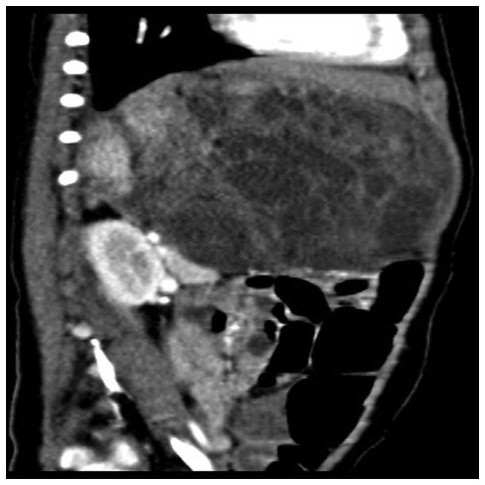
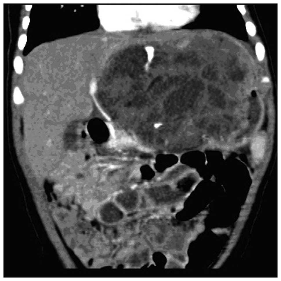
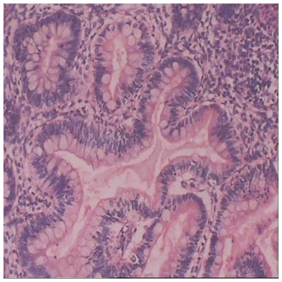
Retroperitoneal cystic immature teratoma (RCIT) is a rare disease. RCITs manifest as solid and cystic masses. In pathological sections, cysts of various sizes, with internal hemorrhage and necrosis, are observed. Components of all germ layer tissue are also observed, the majority of which is located within the endoderm. As the tumor contains undifferentiated immature tissue components, RCITs are also termed malignant teratomas. Immature teratomas grow rapidly, often invading adjacent tissue to cause serious symptoms, and transfer through the blood and lymph vessels, often resulting in glandular cancer. The present study reports the case of an infant with RCIT. The female patient, aged six months and six days, was hospitalized due to an abdominal mass. Physical examination revealed a large mass (10x8 cm) below the xiphoid in the epigastrium. The mass, which ranged from the xiphoid to the umbilical region, was friable, and possessed a smooth surface, clear boundaries and poor activity, without tenderness. Upper abdominal computed tomography (CT) revealed a large, solid, cystic mass in the left, middle and lower retroperitoneum. The patient was admitted to the Yantai Yuhuangding Hospital for surgery. The pre-operative examination was improved following admission by documenting parameters that included the results from routine blood tests, bleeding and clotting times and cardiography. Retroperitoneal tumor resection was then performed. During resection, the tumor was found to originate from the retroperitoneum. As the tumor involved the gastric wall, a section of the gastric wall was resected, in addition to the tumor. The resection surface was yellow and friable. Pathological examination of tumor tissue sections revealed the involvement of immature nerves and mesenchymal components, confirming the diagnosis of a grade II immature teratoma. Subsequent to six months of outpatient follow-up, the patient had recovered well, without recurrence. RCIT is a clinically rare disease, and the present study adds to the current understanding of this rare condition in infants.
View References
|
1
|
Aggarwal SK, Keshri A and Agarwal P:
Immature teratoma of the nose and paranasal sinuses masquerading as
bilateral nasal polyposis: a unique presentation. J Postgrad Med.
59:138–141. 2013. View Article : Google Scholar : PubMed/NCBI
|
|
2
|
Ishida M, Hotta M, Ohta M, Taga T, Ohta S,
Takeuchi Y and Okabe H: A case of retroperitoneal immature teratoma
with nephroblastic components. J Pediatr Hematol Oncol. 34:e22–e25.
2012. View Article : Google Scholar : PubMed/NCBI
|
|
3
|
Yanai H, Matsuura H, Kawasaki M, Takada Y,
Tabuchi Y and Yoshino Tadashi: Immature teratoma of the ovary with
a minor rhabdomyosarcomatous component and fatal
rhabdomyosarcomatous metastases: The first case in a child. Int J
Gynecol Pathol. 21:82–85. 2002. View Article : Google Scholar : PubMed/NCBI
|
|
4
|
Grammatikopoulou I, Kontomanolis EN,
Chatzaki E, Chouridou E, Pavlidis P, Papadopoulos EM and
Lambropouloux M: Immature malignant sacrococcygeal teratoma: case
report and review of the literature. Clin Exp Obstet Gynecol.
40:437–439. 2013.PubMed/NCBI
|
|
5
|
Hasiotou M, Vakaki M, Pitsoulakis G,
Zarifi M, Sammouti H, Konstadinidou CV and Koudoumnakis E:
Congenital cervical teratomas. Int J Pediatr Otorhinolaryngol.
68:1133–1139. 2004. View Article : Google Scholar : PubMed/NCBI
|
|
6
|
Marina NM, Cushing B, Giller R, Cohen L,
Lauer SJ, Ablin A, Weetman R, Cullen J, Rogers P, Vinocur C, et al:
Complete surgical excision is effective treatment for children with
immature teratomas with or without malignant elements: A Pediatric
Oncology Group/Children's Cancer Group Intergroup Study. J Clin
Oncol. 17:2137–2143. 1999.PubMed/NCBI
|
|
7
|
Bogner G, Wolfrum-Ristau P, Schneider W,
Fischer T and Jacobs VR: Local foreign body reaction of peritoneum
after rupture of cystic partially immature teratoma. J Minim
Invasive Gynecol. 21:959–962. 2014. View Article : Google Scholar : PubMed/NCBI
|
|
8
|
Matsushita H and Tani H: Successful
infertility treatment following fertility-sparing surgery and
chemotherapy for ovarian immature teratoma: A case report and a
literature review. Reprod Med Biol. 10:193–198. 2011. View Article : Google Scholar
|
|
9
|
Lee DH, Yoon TM, Lim SC and Lee JK:
Immature teratoma of the parapharyngeal space presenting with
airway obstruction in an infant. B-ENT. 10:71–73. 2014.PubMed/NCBI
|
|
10
|
Anilkumar MG, Jagadishkumar K, Girish GN
and Sunila: Immature gastric teratoma in an infant. Indian J Surg.
75 (Suppl 1):453–455. 2013. View Article : Google Scholar : PubMed/NCBI
|