Spandidos Publications Logo
  • About
    • About Spandidos
    • Aims and Scopes
    • Abstracting and Indexing
    • Editorial Policies
    • Reprints and Permissions
    • Job Opportunities
    • Terms and Conditions
    • Contact
  • Journals
    • All Journals
    • Oncology Letters
      • Oncology Letters
      • Information for Authors
      • Editorial Policies
      • Editorial Board
      • Aims and Scope
      • Abstracting and Indexing
      • Bibliographic Information
      • Archive
    • International Journal of Oncology
      • International Journal of Oncology
      • Information for Authors
      • Editorial Policies
      • Editorial Board
      • Aims and Scope
      • Abstracting and Indexing
      • Bibliographic Information
      • Archive
    • Molecular and Clinical Oncology
      • Molecular and Clinical Oncology
      • Information for Authors
      • Editorial Policies
      • Editorial Board
      • Aims and Scope
      • Abstracting and Indexing
      • Bibliographic Information
      • Archive
    • Experimental and Therapeutic Medicine
      • Experimental and Therapeutic Medicine
      • Information for Authors
      • Editorial Policies
      • Editorial Board
      • Aims and Scope
      • Abstracting and Indexing
      • Bibliographic Information
      • Archive
    • International Journal of Molecular Medicine
      • International Journal of Molecular Medicine
      • Information for Authors
      • Editorial Policies
      • Editorial Board
      • Aims and Scope
      • Abstracting and Indexing
      • Bibliographic Information
      • Archive
    • Biomedical Reports
      • Biomedical Reports
      • Information for Authors
      • Editorial Policies
      • Editorial Board
      • Aims and Scope
      • Abstracting and Indexing
      • Bibliographic Information
      • Archive
    • Oncology Reports
      • Oncology Reports
      • Information for Authors
      • Editorial Policies
      • Editorial Board
      • Aims and Scope
      • Abstracting and Indexing
      • Bibliographic Information
      • Archive
    • Molecular Medicine Reports
      • Molecular Medicine Reports
      • Information for Authors
      • Editorial Policies
      • Editorial Board
      • Aims and Scope
      • Abstracting and Indexing
      • Bibliographic Information
      • Archive
    • World Academy of Sciences Journal
      • World Academy of Sciences Journal
      • Information for Authors
      • Editorial Policies
      • Editorial Board
      • Aims and Scope
      • Abstracting and Indexing
      • Bibliographic Information
      • Archive
    • International Journal of Functional Nutrition
      • International Journal of Functional Nutrition
      • Information for Authors
      • Editorial Policies
      • Editorial Board
      • Aims and Scope
      • Abstracting and Indexing
      • Bibliographic Information
      • Archive
    • International Journal of Epigenetics
      • International Journal of Epigenetics
      • Information for Authors
      • Editorial Policies
      • Editorial Board
      • Aims and Scope
      • Abstracting and Indexing
      • Bibliographic Information
      • Archive
    • Medicine International
      • Medicine International
      • Information for Authors
      • Editorial Policies
      • Editorial Board
      • Aims and Scope
      • Abstracting and Indexing
      • Bibliographic Information
      • Archive
  • Articles
  • Information
    • Information for Authors
    • Information for Reviewers
    • Information for Librarians
    • Information for Advertisers
    • Conferences
  • Language Editing
Spandidos Publications Logo
  • About
    • About Spandidos
    • Aims and Scopes
    • Abstracting and Indexing
    • Editorial Policies
    • Reprints and Permissions
    • Job Opportunities
    • Terms and Conditions
    • Contact
  • Journals
    • All Journals
    • Biomedical Reports
      • Information for Authors
      • Editorial Policies
      • Editorial Board
      • Aims and Scope
      • Abstracting and Indexing
      • Bibliographic Information
      • Archive
    • Experimental and Therapeutic Medicine
      • Information for Authors
      • Editorial Policies
      • Editorial Board
      • Aims and Scope
      • Abstracting and Indexing
      • Bibliographic Information
      • Archive
    • International Journal of Epigenetics
      • Information for Authors
      • Editorial Policies
      • Editorial Board
      • Aims and Scope
      • Abstracting and Indexing
      • Bibliographic Information
      • Archive
    • International Journal of Functional Nutrition
      • Information for Authors
      • Editorial Policies
      • Editorial Board
      • Aims and Scope
      • Abstracting and Indexing
      • Bibliographic Information
      • Archive
    • International Journal of Molecular Medicine
      • Information for Authors
      • Editorial Policies
      • Editorial Board
      • Aims and Scope
      • Abstracting and Indexing
      • Bibliographic Information
      • Archive
    • International Journal of Oncology
      • Information for Authors
      • Editorial Policies
      • Editorial Board
      • Aims and Scope
      • Abstracting and Indexing
      • Bibliographic Information
      • Archive
    • Medicine International
      • Information for Authors
      • Editorial Policies
      • Editorial Board
      • Aims and Scope
      • Abstracting and Indexing
      • Bibliographic Information
      • Archive
    • Molecular and Clinical Oncology
      • Information for Authors
      • Editorial Policies
      • Editorial Board
      • Aims and Scope
      • Abstracting and Indexing
      • Bibliographic Information
      • Archive
    • Molecular Medicine Reports
      • Information for Authors
      • Editorial Policies
      • Editorial Board
      • Aims and Scope
      • Abstracting and Indexing
      • Bibliographic Information
      • Archive
    • Oncology Letters
      • Information for Authors
      • Editorial Policies
      • Editorial Board
      • Aims and Scope
      • Abstracting and Indexing
      • Bibliographic Information
      • Archive
    • Oncology Reports
      • Information for Authors
      • Editorial Policies
      • Editorial Board
      • Aims and Scope
      • Abstracting and Indexing
      • Bibliographic Information
      • Archive
    • World Academy of Sciences Journal
      • Information for Authors
      • Editorial Policies
      • Editorial Board
      • Aims and Scope
      • Abstracting and Indexing
      • Bibliographic Information
      • Archive
  • Articles
  • Information
    • For Authors
    • For Reviewers
    • For Librarians
    • For Advertisers
    • Conferences
  • Language Editing
Login Register Submit
  • This site uses cookies
  • You can change your cookie settings at any time by following the instructions in our Cookie Policy. To find out more, you may read our Privacy Policy.

    I agree
Search articles by DOI, keyword, author or affiliation
Search
Advanced Search
presentation
Oncology Letters
Join Editorial Board Propose a Special Issue
Print ISSN: 1792-1074 Online ISSN: 1792-1082
Journal Cover
August-2015 Volume 10 Issue 2

Full Size Image

Sign up for eToc alerts
Recommend to Library

Journals

International Journal of Molecular Medicine

International Journal of Molecular Medicine

International Journal of Molecular Medicine is an international journal devoted to molecular mechanisms of human disease.

International Journal of Oncology

International Journal of Oncology

International Journal of Oncology is an international journal devoted to oncology research and cancer treatment.

Molecular Medicine Reports

Molecular Medicine Reports

Covers molecular medicine topics such as pharmacology, pathology, genetics, neuroscience, infectious diseases, molecular cardiology, and molecular surgery.

Oncology Reports

Oncology Reports

Oncology Reports is an international journal devoted to fundamental and applied research in Oncology.

Experimental and Therapeutic Medicine

Experimental and Therapeutic Medicine

Experimental and Therapeutic Medicine is an international journal devoted to laboratory and clinical medicine.

Oncology Letters

Oncology Letters

Oncology Letters is an international journal devoted to Experimental and Clinical Oncology.

Biomedical Reports

Biomedical Reports

Explores a wide range of biological and medical fields, including pharmacology, genetics, microbiology, neuroscience, and molecular cardiology.

Molecular and Clinical Oncology

Molecular and Clinical Oncology

International journal addressing all aspects of oncology research, from tumorigenesis and oncogenes to chemotherapy and metastasis.

World Academy of Sciences Journal

World Academy of Sciences Journal

Multidisciplinary open-access journal spanning biochemistry, genetics, neuroscience, environmental health, and synthetic biology.

International Journal of Functional Nutrition

International Journal of Functional Nutrition

Open-access journal combining biochemistry, pharmacology, immunology, and genetics to advance health through functional nutrition.

International Journal of Epigenetics

International Journal of Epigenetics

Publishes open-access research on using epigenetics to advance understanding and treatment of human disease.

Medicine International

Medicine International

An International Open Access Journal Devoted to General Medicine.

Journal Cover
August-2015 Volume 10 Issue 2

Full Size Image

Sign up for eToc alerts
Recommend to Library

  • Article
  • Citations
    • Cite This Article
    • Download Citation
    • Create Citation Alert
    • Remove Citation Alert
    • Cited By
  • Similar Articles
    • Related Articles (in Spandidos Publications)
    • Similar Articles (Google Scholar)
    • Similar Articles (PubMed)
  • Download PDF
  • Download XML
  • View XML
Case Report

Retroperitoneal cystic immature teratoma: A case report

  • Authors:
    • Junxia Li
    • Peiyou Gong
    • Fengli Liu
    • Ping Sun
    • Chengrong Wu
  • View Affiliations / Copyright

    Affiliations: Department of Oncology, Yantai Yuhuangding Hospital, Yantai, Shandong 264000, P.R. China, Department of Radiology, Yantai Yuhuangding Hospital, Yantai, Shandong 264000, P.R. China
  • Pages: 1023-1025
    |
    Published online on: May 22, 2015
       https://doi.org/10.3892/ol.2015.3256
  • Expand metrics +
Metrics: Total Views: 0 (Spandidos Publications: | PMC Statistics: )
Metrics: Total PDF Downloads: 0 (Spandidos Publications: | PMC Statistics: )
Cited By (CrossRef): 0 citations Loading Articles...

This article is mentioned in:


Abstract

Retroperitoneal cystic immature teratoma (RCIT) is a rare disease. RCITs manifest as solid and cystic masses. In pathological sections, cysts of various sizes, with internal hemorrhage and necrosis, are observed. Components of all germ layer tissue are also observed, the majority of which is located within the endoderm. As the tumor contains undifferentiated immature tissue components, RCITs are also termed malignant teratomas. Immature teratomas grow rapidly, often invading adjacent tissue to cause serious symptoms, and transfer through the blood and lymph vessels, often resulting in glandular cancer. The present study reports the case of an infant with RCIT. The female patient, aged six months and six days, was hospitalized due to an abdominal mass. Physical examination revealed a large mass (10x8 cm) below the xiphoid in the epigastrium. The mass, which ranged from the xiphoid to the umbilical region, was friable, and possessed a smooth surface, clear boundaries and poor activity, without tenderness. Upper abdominal computed tomography (CT) revealed a large, solid, cystic mass in the left, middle and lower retroperitoneum. The patient was admitted to the Yantai Yuhuangding Hospital for surgery. The pre-operative examination was improved following admission by documenting parameters that included the results from routine blood tests, bleeding and clotting times and cardiography. Retroperitoneal tumor resection was then performed. During resection, the tumor was found to originate from the retroperitoneum. As the tumor involved the gastric wall, a section of the gastric wall was resected, in addition to the tumor. The resection surface was yellow and friable. Pathological examination of tumor tissue sections revealed the involvement of immature nerves and mesenchymal components, confirming the diagnosis of a grade II immature teratoma. Subsequent to six months of outpatient follow-up, the patient had recovered well, without recurrence. RCIT is a clinically rare disease, and the present study adds to the current understanding of this rare condition in infants.

Introduction

Retroperitoneal cystic immature teratoma (RCIT), also termed malignant teratoma, is composed of immature tissue undergoing embryogenesis, the majority of which is constituted by neuroglia and neural tube-like structures. RCIT demonstrates pathological manifestations such as undifferentiated cells and increased cell mitosis (1). RCIT rarely occurs in children, accounting for <1% of all childhood tumors (2). The tumor has malignant potential and recurrence is common. As RCIT exhibits a high-risk of developing into malignant teratoma, the treatment modalities for RCIT are similiar to those for malignant teratoma, which include surgical resection and combined treatment with chemotherapy and radiotherapy (3).

Malignant germinomas, including seminoma, dysgerminoma, embryonic carcinoma and endodermal sinus tumors, were historically termed malignant teratomas. However, these tumors are the result of cellular de-differentiation in various regions during the cellular movement from the yolk sac to gonads during embryonic development. Malignant germinomas are not usually classified into teratomas, as they possess no pathologically observable triploblastic structure (4). Imaging techniques, including ultrasonography, computed tomography (CT) and magnetic resonance imaging (MRI), are required for the diagnosis and surgical management of teratomas (5). On MRI, RCIT exhibits a pattern of mixed signal intensity and enhancement, which presents as uniformed hypodensity on T1 weighted imaging (WI) and hyperintensity on T2WI, with enhancement of the tumor capsule. On CT imaging, RCIT often exhibits heterogeneous density due to the presence of various well-differentiated components. Subsequent to confirmation of the diagnosis of RCIT, an early resection procedure must be performed. The tumor requires thorough resection to prevent the recurrence of the tumor from any remaining pluripotent cells.

Case report

The study was approved by the ethics committee of Yantai Yuhuangding Hospital (Yantai, China). A female aged six months and six days was admitted to the Yantai Yuhuangding Hospital (Yantai, Shandong, China) for 7 days in April 2014, due to an abdominal mass. A 12-cm mass was identified in the abdomen of the infant one week prior to hospitalization. The infant demonstrated no abdominal distension, abdominal pain, diarrhea or fever. The patient was in high spirits, slept well and had a good appetite. No evident abnormalities or significant weight loss were observed.

The results of the physical examination revealed the presence of abdominal distention and clear abdominal veins. No evident dilation or bowel obstruction was identified. A large mass, 10×8 cm in size, was found below the xiphoid in the epigastrium. The mass ranged between the xiphoid and umbilical region. The lesion was friable, and possessed a smooth surface, clear boundaries and poor activity, without tenderness or rebound tenderness. The liver, spleen and lower ribs were not examined. Bowel sounds were identified, without hyperthyroidism and gurgling.

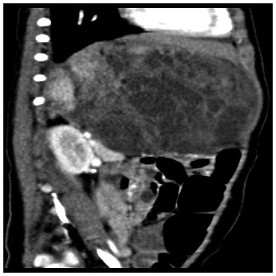
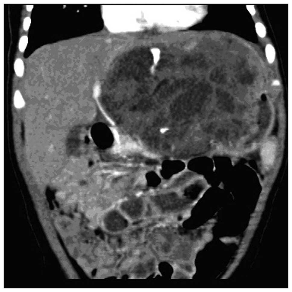
Dynamic enhanced CT of the upper abdomen revealed a large solid cystic mass in the fat space of the left, middle and lower retroperitoneum. The internal diameter of the mass was indicated to measure 66×100 mm by CT, and the mass contained plaque calcification. Following enhanced scanning, the solid region of the tumor demonstrated evident enhancement and was shifted upon compression near the intestine, pancreas and left lobe of the liver (Figs. 1–3). The disease was likely to be diagnosed as a left, middle and lower retroperitoneal malignant tumor based on the imaging data. Retroperitoneal tumor resection was subsequently performed. During resection, the tumor was found to originate from the retroperitoneal space. As the tumor involved the gastric wall and grew into the gastric cavity, a section of the gastric wall was 
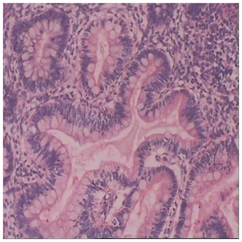
resected in addition to the tumor. The pathologically examined tumor tissue indicated the presence of nodosities. The resection surface was yellow and friable, with a portion that appeared similar to rotten meat and contained liquids and mucous. The tumor demonstrated infiltrative growth into the gastric wall, and did not involve the resection margins. A moderate quantity of immature nerves and mesenchymal components were evident in the microscopic examination of the tumor, which was diagnosed by pathology as grade II immature teratoma (Fig. 4). Except the routine prevention of infection and nutritional support treatment, no further treatment following surgery was required. Subsequent to six months of outpatient follow-ups, the patient had recovered well, without recurrence.

Figure 1.

Computed tomography revealing low-density shadows that were observed in the retroperitoneal cystic mass, which was surrounded by enhanced separations.

Figure 3.

Coronal view of computed tomography, revealing plaque calcification.

Figure 4.

Tumor tissue was fixed with 4% neutral formaldehyde solution, dehydrated and embedded in paraffin. Tissue sections were sliced into 4-µm thick microscopic sections. Morphological features of the tumor were assessed by hematoxylin and eosin staining. The pathological analysis of the tumor tissue revealed a small-to-moderate proportion of immature neural and mesenchymal components, harboring characteristics of a grade II immature teratoma (magnification, x100), and the cells formed a structure that simulated a Homer-Wright rosette.

Discussion

Immature teratomas contain varying quantities of neuroectodermal or blastemal tissues and may be graded by the quantity of immature neuroglial tissue (6). Immature teratomas have been mostly reported in the ovaries of young females, and the diagnosis of these tumors is based mainly on the pathological evaluation of the tumor tissue. A previous immunohistological study investigating immature teratoma tissue reported the presence of partial neuroendocrine differentiation of immature origin (7). Complete resection, along with chemotherapy, is the main treatment method for immature teratomas (8).

Lee et al (9) reported immature teratoma of the parapharyngeal space, and reported that germ cell tumors often occur in infants, with the sacrococcygeal region, gonads and mediastinum as the most common sites. According to Anilkumar et al (10), gastric teratomas are extremely rare, accounting for <1% of all teratomas in infants and children. In total, >100 cases of gastric teratomas have been reported in the literature, and extremely few of these are of the immature variety.

The present study reports the case of an infant that developed RCIT. The tumor involved the gastric wall, and was large, ~12 cm, in size. The clinical feature of the lesion was a progressively increasing painless mass. Imaging features included the identification of a cystoid mass surrounded by separations. Enhancement of the separations was evident in the mass, with calcification also observed. No clear fat components were identified in the lesion. The patient was diagnosed by post-operative pathology and immunohistochemical analysis. Immature neural tissues of various proportions, including primitive neural canal and neural epithelial components, and other immature components, including immature cartilage, primary small cells and mesenchymal tissues, were observed.

In summary, retroperitoneal immature teratoma is rare in clinical practice, and is challenging to diagnose. It is also difficult to differentiate retroperitoneal immature teratoma from retroperitoneal neurogenic and yolk sac tumors, which occurs mostly in the axis of the body. The present study provides additional information on the occurrence of this rare tumor in infants.

Acknowledgements

The authors thank the patient in the present study for their participation and colleagues in the Department of Pediatric Surgery of Yantai Yuhuangding Hospital for their assistance.

References

1 

Aggarwal SK, Keshri A and Agarwal P: Immature teratoma of the nose and paranasal sinuses masquerading as bilateral nasal polyposis: a unique presentation. J Postgrad Med. 59:138–141. 2013. View Article : Google Scholar : PubMed/NCBI

2 

Ishida M, Hotta M, Ohta M, Taga T, Ohta S, Takeuchi Y and Okabe H: A case of retroperitoneal immature teratoma with nephroblastic components. J Pediatr Hematol Oncol. 34:e22–e25. 2012. View Article : Google Scholar : PubMed/NCBI

3 

Yanai H, Matsuura H, Kawasaki M, Takada Y, Tabuchi Y and Yoshino Tadashi: Immature teratoma of the ovary with a minor rhabdomyosarcomatous component and fatal rhabdomyosarcomatous metastases: The first case in a child. Int J Gynecol Pathol. 21:82–85. 2002. View Article : Google Scholar : PubMed/NCBI

4 

Grammatikopoulou I, Kontomanolis EN, Chatzaki E, Chouridou E, Pavlidis P, Papadopoulos EM and Lambropouloux M: Immature malignant sacrococcygeal teratoma: case report and review of the literature. Clin Exp Obstet Gynecol. 40:437–439. 2013.PubMed/NCBI

5 

Hasiotou M, Vakaki M, Pitsoulakis G, Zarifi M, Sammouti H, Konstadinidou CV and Koudoumnakis E: Congenital cervical teratomas. Int J Pediatr Otorhinolaryngol. 68:1133–1139. 2004. View Article : Google Scholar : PubMed/NCBI

6 

Marina NM, Cushing B, Giller R, Cohen L, Lauer SJ, Ablin A, Weetman R, Cullen J, Rogers P, Vinocur C, et al: Complete surgical excision is effective treatment for children with immature teratomas with or without malignant elements: A Pediatric Oncology Group/Children's Cancer Group Intergroup Study. J Clin Oncol. 17:2137–2143. 1999.PubMed/NCBI

7 

Bogner G, Wolfrum-Ristau P, Schneider W, Fischer T and Jacobs VR: Local foreign body reaction of peritoneum after rupture of cystic partially immature teratoma. J Minim Invasive Gynecol. 21:959–962. 2014. View Article : Google Scholar : PubMed/NCBI

8 

Matsushita H and Tani H: Successful infertility treatment following fertility-sparing surgery and chemotherapy for ovarian immature teratoma: A case report and a literature review. Reprod Med Biol. 10:193–198. 2011. View Article : Google Scholar

9 

Lee DH, Yoon TM, Lim SC and Lee JK: Immature teratoma of the parapharyngeal space presenting with airway obstruction in an infant. B-ENT. 10:71–73. 2014.PubMed/NCBI

10 

Anilkumar MG, Jagadishkumar K, Girish GN and Sunila: Immature gastric teratoma in an infant. Indian J Surg. 75 (Suppl 1):453–455. 2013. View Article : Google Scholar : PubMed/NCBI

Related Articles

  • Abstract
  • View
  • Download
  • Twitter
Copy and paste a formatted citation
Spandidos Publications style
Li J, Gong P, Liu F, Sun P and Wu C: Retroperitoneal cystic immature teratoma: A case report. Oncol Lett 10: 1023-1025, 2015.
APA
Li, J., Gong, P., Liu, F., Sun, P., & Wu, C. (2015). Retroperitoneal cystic immature teratoma: A case report. Oncology Letters, 10, 1023-1025. https://doi.org/10.3892/ol.2015.3256
MLA
Li, J., Gong, P., Liu, F., Sun, P., Wu, C."Retroperitoneal cystic immature teratoma: A case report". Oncology Letters 10.2 (2015): 1023-1025.
Chicago
Li, J., Gong, P., Liu, F., Sun, P., Wu, C."Retroperitoneal cystic immature teratoma: A case report". Oncology Letters 10, no. 2 (2015): 1023-1025. https://doi.org/10.3892/ol.2015.3256
Copy and paste a formatted citation
x
Spandidos Publications style
Li J, Gong P, Liu F, Sun P and Wu C: Retroperitoneal cystic immature teratoma: A case report. Oncol Lett 10: 1023-1025, 2015.
APA
Li, J., Gong, P., Liu, F., Sun, P., & Wu, C. (2015). Retroperitoneal cystic immature teratoma: A case report. Oncology Letters, 10, 1023-1025. https://doi.org/10.3892/ol.2015.3256
MLA
Li, J., Gong, P., Liu, F., Sun, P., Wu, C."Retroperitoneal cystic immature teratoma: A case report". Oncology Letters 10.2 (2015): 1023-1025.
Chicago
Li, J., Gong, P., Liu, F., Sun, P., Wu, C."Retroperitoneal cystic immature teratoma: A case report". Oncology Letters 10, no. 2 (2015): 1023-1025. https://doi.org/10.3892/ol.2015.3256
Follow us
  • Twitter
  • LinkedIn
  • Facebook
About
  • Spandidos Publications
  • Careers
  • Cookie Policy
  • Privacy Policy
How can we help?
  • Help
  • Live Chat
  • Contact
  • Email to our Support Team