Spandidos Publications Logo
  • About
    • About Spandidos
    • Aims and Scopes
    • Abstracting and Indexing
    • Editorial Policies
    • Reprints and Permissions
    • Job Opportunities
    • Terms and Conditions
    • Contact
  • Journals
    • All Journals
    • Oncology Letters
      • Oncology Letters
      • Information for Authors
      • Editorial Policies
      • Editorial Board
      • Aims and Scope
      • Abstracting and Indexing
      • Bibliographic Information
      • Archive
    • International Journal of Oncology
      • International Journal of Oncology
      • Information for Authors
      • Editorial Policies
      • Editorial Board
      • Aims and Scope
      • Abstracting and Indexing
      • Bibliographic Information
      • Archive
    • Molecular and Clinical Oncology
      • Molecular and Clinical Oncology
      • Information for Authors
      • Editorial Policies
      • Editorial Board
      • Aims and Scope
      • Abstracting and Indexing
      • Bibliographic Information
      • Archive
    • Experimental and Therapeutic Medicine
      • Experimental and Therapeutic Medicine
      • Information for Authors
      • Editorial Policies
      • Editorial Board
      • Aims and Scope
      • Abstracting and Indexing
      • Bibliographic Information
      • Archive
    • International Journal of Molecular Medicine
      • International Journal of Molecular Medicine
      • Information for Authors
      • Editorial Policies
      • Editorial Board
      • Aims and Scope
      • Abstracting and Indexing
      • Bibliographic Information
      • Archive
    • Biomedical Reports
      • Biomedical Reports
      • Information for Authors
      • Editorial Policies
      • Editorial Board
      • Aims and Scope
      • Abstracting and Indexing
      • Bibliographic Information
      • Archive
    • Oncology Reports
      • Oncology Reports
      • Information for Authors
      • Editorial Policies
      • Editorial Board
      • Aims and Scope
      • Abstracting and Indexing
      • Bibliographic Information
      • Archive
    • Molecular Medicine Reports
      • Molecular Medicine Reports
      • Information for Authors
      • Editorial Policies
      • Editorial Board
      • Aims and Scope
      • Abstracting and Indexing
      • Bibliographic Information
      • Archive
    • World Academy of Sciences Journal
      • World Academy of Sciences Journal
      • Information for Authors
      • Editorial Policies
      • Editorial Board
      • Aims and Scope
      • Abstracting and Indexing
      • Bibliographic Information
      • Archive
    • International Journal of Functional Nutrition
      • International Journal of Functional Nutrition
      • Information for Authors
      • Editorial Policies
      • Editorial Board
      • Aims and Scope
      • Abstracting and Indexing
      • Bibliographic Information
      • Archive
    • International Journal of Epigenetics
      • International Journal of Epigenetics
      • Information for Authors
      • Editorial Policies
      • Editorial Board
      • Aims and Scope
      • Abstracting and Indexing
      • Bibliographic Information
      • Archive
    • Medicine International
      • Medicine International
      • Information for Authors
      • Editorial Policies
      • Editorial Board
      • Aims and Scope
      • Abstracting and Indexing
      • Bibliographic Information
      • Archive
  • Articles
  • Information
    • Information for Authors
    • Information for Reviewers
    • Information for Librarians
    • Information for Advertisers
    • Conferences
  • Language Editing
Spandidos Publications Logo
  • About
    • About Spandidos
    • Aims and Scopes
    • Abstracting and Indexing
    • Editorial Policies
    • Reprints and Permissions
    • Job Opportunities
    • Terms and Conditions
    • Contact
  • Journals
    • All Journals
    • Biomedical Reports
      • Information for Authors
      • Editorial Policies
      • Editorial Board
      • Aims and Scope
      • Abstracting and Indexing
      • Bibliographic Information
      • Archive
    • Experimental and Therapeutic Medicine
      • Information for Authors
      • Editorial Policies
      • Editorial Board
      • Aims and Scope
      • Abstracting and Indexing
      • Bibliographic Information
      • Archive
    • International Journal of Epigenetics
      • Information for Authors
      • Editorial Policies
      • Editorial Board
      • Aims and Scope
      • Abstracting and Indexing
      • Bibliographic Information
      • Archive
    • International Journal of Functional Nutrition
      • Information for Authors
      • Editorial Policies
      • Editorial Board
      • Aims and Scope
      • Abstracting and Indexing
      • Bibliographic Information
      • Archive
    • International Journal of Molecular Medicine
      • Information for Authors
      • Editorial Policies
      • Editorial Board
      • Aims and Scope
      • Abstracting and Indexing
      • Bibliographic Information
      • Archive
    • International Journal of Oncology
      • Information for Authors
      • Editorial Policies
      • Editorial Board
      • Aims and Scope
      • Abstracting and Indexing
      • Bibliographic Information
      • Archive
    • Medicine International
      • Information for Authors
      • Editorial Policies
      • Editorial Board
      • Aims and Scope
      • Abstracting and Indexing
      • Bibliographic Information
      • Archive
    • Molecular and Clinical Oncology
      • Information for Authors
      • Editorial Policies
      • Editorial Board
      • Aims and Scope
      • Abstracting and Indexing
      • Bibliographic Information
      • Archive
    • Molecular Medicine Reports
      • Information for Authors
      • Editorial Policies
      • Editorial Board
      • Aims and Scope
      • Abstracting and Indexing
      • Bibliographic Information
      • Archive
    • Oncology Letters
      • Information for Authors
      • Editorial Policies
      • Editorial Board
      • Aims and Scope
      • Abstracting and Indexing
      • Bibliographic Information
      • Archive
    • Oncology Reports
      • Information for Authors
      • Editorial Policies
      • Editorial Board
      • Aims and Scope
      • Abstracting and Indexing
      • Bibliographic Information
      • Archive
    • World Academy of Sciences Journal
      • Information for Authors
      • Editorial Policies
      • Editorial Board
      • Aims and Scope
      • Abstracting and Indexing
      • Bibliographic Information
      • Archive
  • Articles
  • Information
    • For Authors
    • For Reviewers
    • For Librarians
    • For Advertisers
    • Conferences
  • Language Editing
Login Register Submit
  • This site uses cookies
  • You can change your cookie settings at any time by following the instructions in our Cookie Policy. To find out more, you may read our Privacy Policy.

    I agree
Search articles by DOI, keyword, author or affiliation
Search
Advanced Search
presentation
Oncology Letters
Join Editorial Board Propose a Special Issue
Print ISSN: 1792-1074 Online ISSN: 1792-1082
Journal Cover
August-2015 Volume 10 Issue 2

Full Size Image

Sign up for eToc alerts
Recommend to Library

Journals

International Journal of Molecular Medicine

International Journal of Molecular Medicine

International Journal of Molecular Medicine is an international journal devoted to molecular mechanisms of human disease.

International Journal of Oncology

International Journal of Oncology

International Journal of Oncology is an international journal devoted to oncology research and cancer treatment.

Molecular Medicine Reports

Molecular Medicine Reports

Covers molecular medicine topics such as pharmacology, pathology, genetics, neuroscience, infectious diseases, molecular cardiology, and molecular surgery.

Oncology Reports

Oncology Reports

Oncology Reports is an international journal devoted to fundamental and applied research in Oncology.

Experimental and Therapeutic Medicine

Experimental and Therapeutic Medicine

Experimental and Therapeutic Medicine is an international journal devoted to laboratory and clinical medicine.

Oncology Letters

Oncology Letters

Oncology Letters is an international journal devoted to Experimental and Clinical Oncology.

Biomedical Reports

Biomedical Reports

Explores a wide range of biological and medical fields, including pharmacology, genetics, microbiology, neuroscience, and molecular cardiology.

Molecular and Clinical Oncology

Molecular and Clinical Oncology

International journal addressing all aspects of oncology research, from tumorigenesis and oncogenes to chemotherapy and metastasis.

World Academy of Sciences Journal

World Academy of Sciences Journal

Multidisciplinary open-access journal spanning biochemistry, genetics, neuroscience, environmental health, and synthetic biology.

International Journal of Functional Nutrition

International Journal of Functional Nutrition

Open-access journal combining biochemistry, pharmacology, immunology, and genetics to advance health through functional nutrition.

International Journal of Epigenetics

International Journal of Epigenetics

Publishes open-access research on using epigenetics to advance understanding and treatment of human disease.

Medicine International

Medicine International

An International Open Access Journal Devoted to General Medicine.

Journal Cover
August-2015 Volume 10 Issue 2

Full Size Image

Sign up for eToc alerts
Recommend to Library

  • Article
  • Citations
    • Cite This Article
    • Download Citation
    • Create Citation Alert
    • Remove Citation Alert
    • Cited By
  • Similar Articles
    • Related Articles (in Spandidos Publications)
    • Similar Articles (Google Scholar)
    • Similar Articles (PubMed)
  • Download PDF
  • Download XML
  • View XML
Article

Radionuclide imaging in the diagnosis of osteoid osteoma

  • Authors:
    • Jong Hoon Park
    • Kisoo Pahk
    • Sungeun Kim
    • Soon Hyuck Lee
    • Sang Heon Song
    • Jae Gol Choe
  • View Affiliations / Copyright

    Affiliations: Department of Orthopedic Surgery, Korea University Anam Hospital, Seoul 136‑705, Republic of Korea, Department of Nuclear Medicine, Korea University Anam Hospital, Seoul 136‑705, Republic of Korea, Department of Orthopedic Surgery, Soonchunhyang University Bucheon Hospital, Bucheon 420-767, Republic of Korea
  • Pages: 1131-1134
    |
    Published online on: May 25, 2015
       https://doi.org/10.3892/ol.2015.3258
  • Expand metrics +
Metrics: Total Views: 0 (Spandidos Publications: | PMC Statistics: )
Metrics: Total PDF Downloads: 0 (Spandidos Publications: | PMC Statistics: )
Cited By (CrossRef): 0 citations Loading Articles...

This article is mentioned in:


Abstract

Oseteoid osteoma is a well-known type of benign bone-forming tumor, which has previously been diagnosed using plain radiograph imaging. However, diagnosis of osteoid osteoma may be delayed due to ambiguities on plain radiograph images; despite the increasing use of magnetic resonance imaging (MRI), this type of misdiagnosis is not uncommon. The aim of the present study was to evaluate the effectiveness of radionuclide imaging scans for the diagnosis of osteoid osteoma, as this form of imaging was proposed to be a more sensitive test. The characteristics of 18 cases of osteoid osteoma were analyzed based on diagnostic imaging and the time from initial recognition of symptoms by the patient to diagnosis. Diagnostic modalities included plain radiograph, computed tomography (CT), MRI and radionuclide imaging. Among the 18 patients, 14 patients had unique positive findings in plain radiographs. The mean duration between initial cognition of symptoms to the diagnosis for these patients was 5.2 months (range, 3.8‑9.3 months). A total of 4 patients exhibited no radiographic abnormalities in the initial plain radiographs and were diagnosed a mean of 18.5 months (range, 17‑20 months) following the onset of symptoms. Overall, radionuclide imaging was performed on 16 patients and all of the cases demonstrated positive findings. In these cases, 28.6% of osteoid osteoma patients with clinical indications revealed no abnormal findings in plain radiographs. Therefore, in situations such as these, radionuclide imaging may be a useful indicator for diagnosis, as these results have demonstrated that it positively identified all cases of osteoid osteoma. In addition, the results of the present study indicated that if the radionuclide imaging was positive, CT scan was a more valuable diagnostic tool, whereas if the radionuclide imaging was negative, MRI should be recommended for the diagnosis of other undiscovered disease entities.

Introduction

Osteoid osteoma is a type of benign bone-forming tumor, which is characterized as a well-demarcated osteoblastic mass, called a nidus, surrounded by a distinct zone of reactive bone sclerosis; these tumors have limited growth potential and exhibit disproportionate pain (1). In the majority of osteoid osteoma cases, typical radiographic features demonstrate a sclerotic cortical lesion and contain a small lucency that represents a nidus (2,3). However, contrary to the expected presentation of osteoid osteoma, different radiographic findings may be encountered that provide a diagnostic dilemma for the physicians concerned. Prior to the introduction of magnetic resonance imaging (MRI), diagnoses were made using plain radiograph images, bone scans and computed tomography (CT) scans (2–4). However, MRI evaluation is now commonly preferred, particularly when the lesions are located close to a joint or spinal area, and may not be easily detected on plain radiographs. However, it was suggested that there may be a high possibility of false positive results in the diagnosis of osteoid osteoma using MRI (5); therefore, delayed diagnosis of osteoid osteoma is a common issue. The present study aimed to investigate the potential role of radionuclide imaging for the diagnosis of osteoid osteoma, as radionuclide imaging has been reported to be a more sensitive diagnostic modality in osteoid osteoma.

Patients and methods

In the present study, 18 patients with surgically and histologically proven osteoid osteoma were retrospectively enrolled and reviewed at Korea University Anam Hospital (Seoul, Korea) between January 2006 and December 2013. The study was approved by the ethics committee of Korea University Anam Hospital. The ratio of males to females was 10:8 and mean patient age was 18.2 years (range, 4–50 years). The characteristics of the 18 cases of osteoid osteoma were analyzed based on diagnostic imaging and the time from initial recognition of symptoms by the patient to diagnosis. Diagnostic modalities included plain radiograph imaging, CT, MRI and radionuclide imaging.

Case report

Two cases of osteoid osteoma are presented. In these patients, plain radiographs and MRI were unable to provide sufficient findings for diagnosis of osteoid osteoma. However, radionuclide imaging and CT revealed the characteristics of osteoid osteoma.

Case I

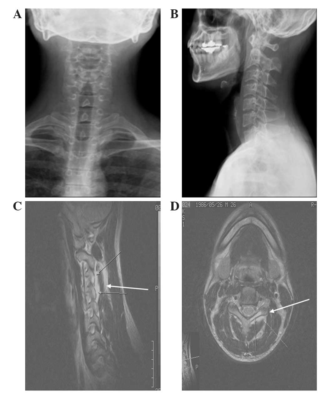
A 26-year-old male presented with severe upper neck pain that had been ongoing for one year. The patient had previously been treated using non-steroidal anti-inflammatory drugs (Naproxen; 200 mg, twice daily), analgesic drugs and physiotherapy in another hospital. However, his symptom had not been relieved and so an MRI study was recommended by the patient's physician, the results of which revealed a non-specific inflammatory lesion on the left third cervical spine (C3) (Fig. 1). The laboratory findings of the patient's blood chemistry revealed an elevated erythrocyte sedimentation rate and supported the MRI 
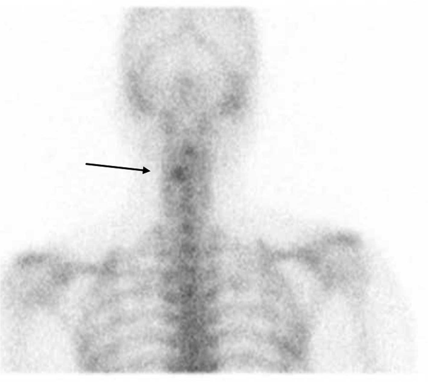
results. When the patient was admitted to Korea University Anam Hospital, a bone scintigraphy was performed using 99mTc-methylene diphosphonate (MDP) and the bone scan image revealed increased focal uptake in the cervical spine 
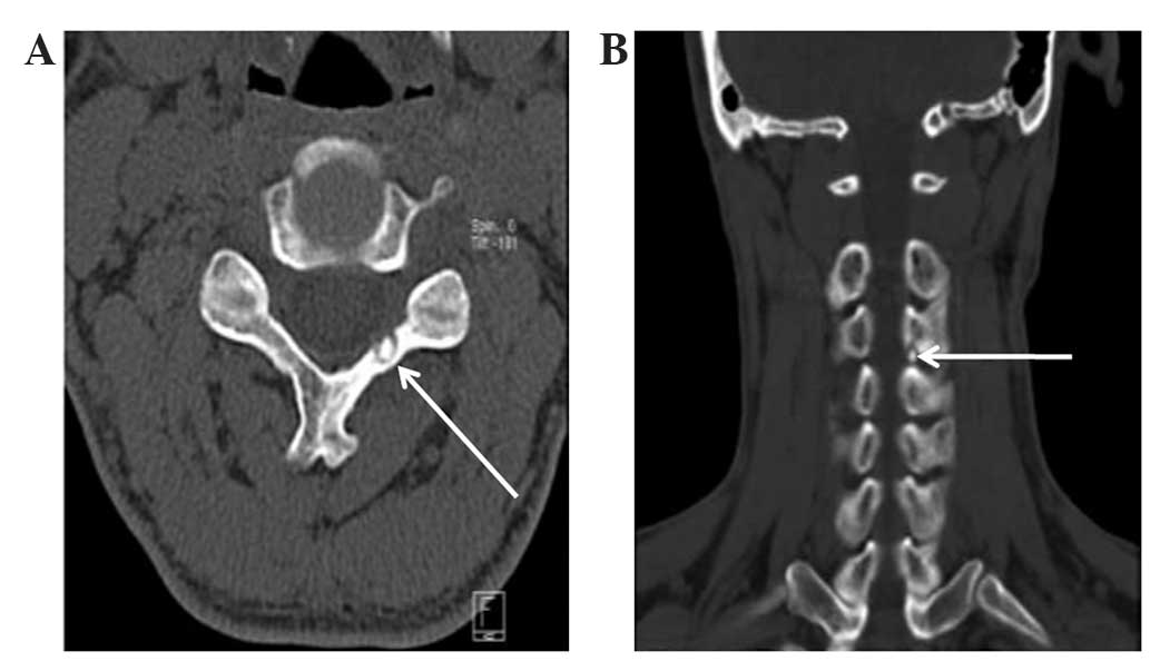
(Fig. 2). Therefore, a CT scan was performed (Fig. 3) and a diagnosis of an osteoid osteoma in the left C3 lamina was confirmed. The duration from first onset of symptoms to diagnosis of osteoid osteoma was 18 months.

Figure 1.

Plain radiographs and MRI scans from Case I. Plain radiographs of cervical spinal (A) anterioposterior and (B) lateral view revealed no specific bony abnormalities. MRI scans of (C) sagittal and (D) axial view revealed a non-specific inflammatory lesion with edematous changes (arrows) at the left third cervical spine lamina. MRI, magnetic resonance imaging.

Figure 2.

99mTc-methylene diphosphonate bone scintigraph image from Case I revealed increased focal uptake (arrow) in the cervical spine.

Figure 3.

CT scans in Case I. CT scans of (A) axial and (B) coronal view revealed a small nidus lesion (white arrows) on left third cervical spine lamina, leading to a diagnosis of osteoid osteoma. CT, computed tomography.

Case II

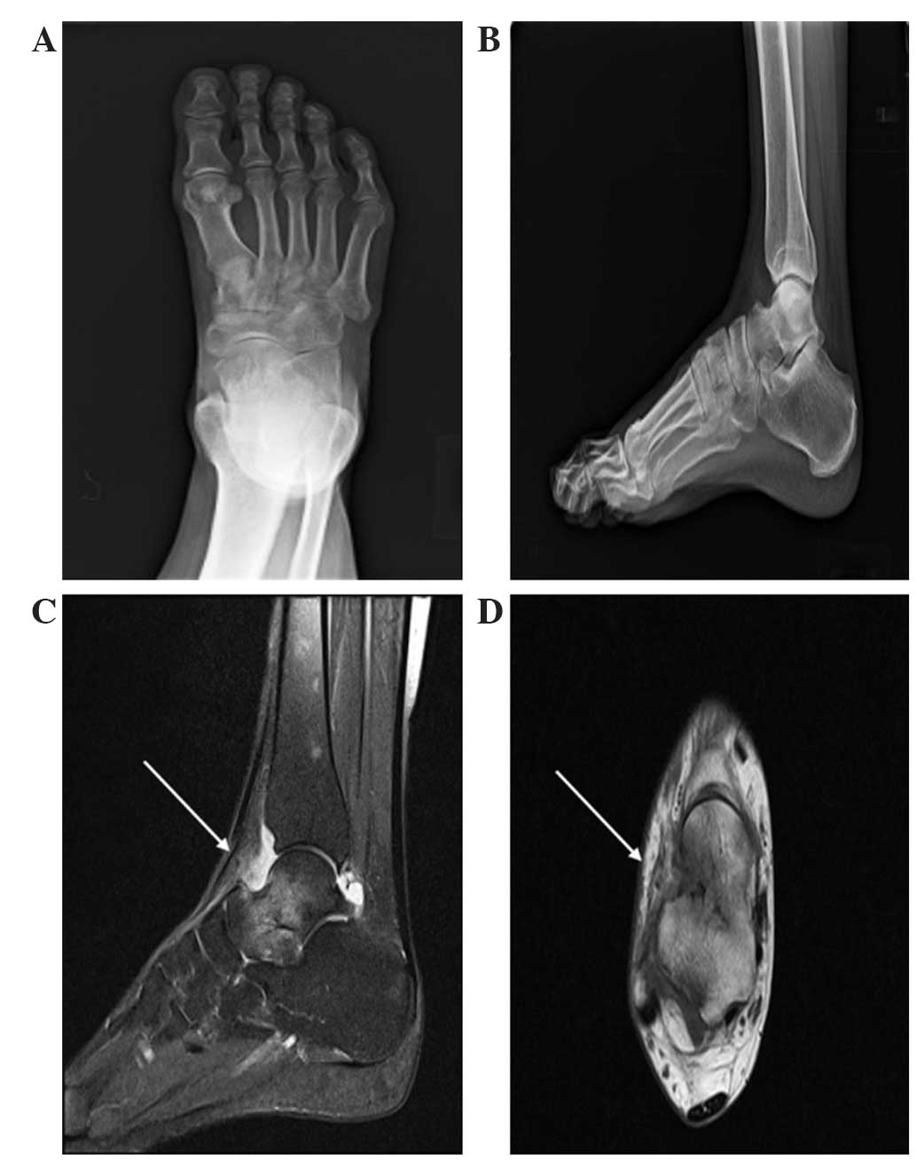
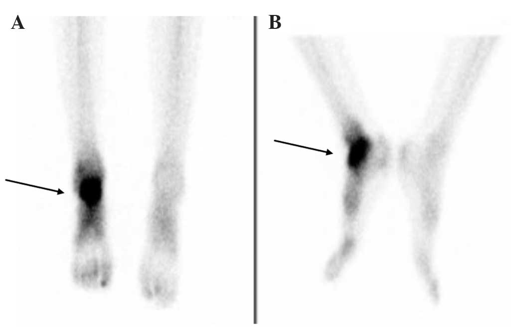
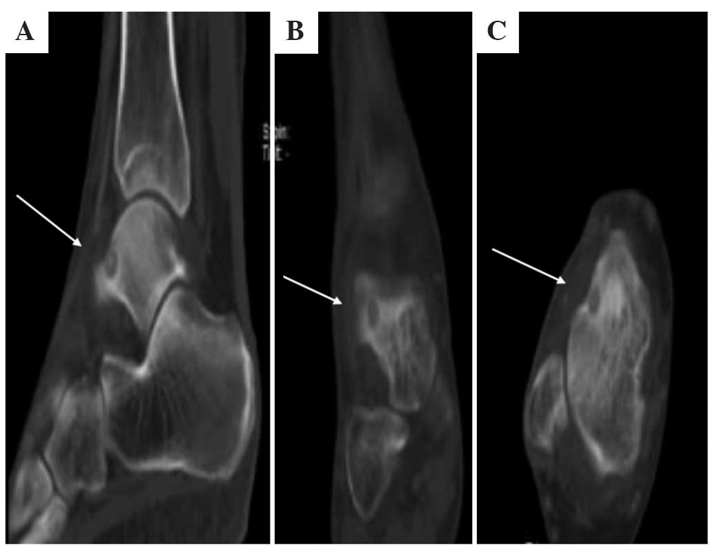
A 46-year-old female presented with right ankle pain, which worsened at night. The patient's symptoms had lasted for one year and were treated as an ankle sprain at another hospital based on the results of plain radiographs, which revealed no specific bony abnormalities (Fig. 4A and B). The patient then underwent an MRI examination at another hospital and was diagnosed with a tumorous lesion on the talus, with inflammatory changes (Fig. 4C and D). The patient was recommended to visit a specialist institute for bone tumors. The patient was admitted to Korea University Anam Hospital, where bone scintigraphy (Fig. 5) and CT (Fig. 6) scans were performed. These scans confirmed the diagnosis of an osteoid osteoma. The duration from first onset of symptoms to diagnosis of osteoid osteoma was 20 months.

Figure 4.

Plain radiographs and MRI scans in Case II. Plain radiographs of the (A) anterioposterior foot and (B) lateral ankle revealed no specific bony abnormalities. MRI demonstrated inflammatory signal changes on the patients talar neck area with reactive edematous changes (white arrows) on ankle joint of the (C) sagittal and (D) axial scans. MRI, magnetic resonance imaging.

Figure 5.

Bone scans in Case II. 99mTc-methylene diphosphonate bone scintigraph image of (A) anterior and (B) medial scans revealed increased focal uptake in the patients talus.

Figure 6.

CT scans in Case II. CT scans of the patients ankle in (A) sagittal, (B) coronal and (C) axial view revealed a small nidus lesion on the talus anteromedial aspect (white arrows), which was diagnosed as osteoid osteoma. CT, computed tomography.

Results

A total of 18 cases of osteoid osteoma were retrospectively reviewed in the present study. Among these 18 patients, 14 patients exhibited unique positive findings suggestive of osteoid osteoma on plain radiographs, with a mean time between initial recognition of symptoms by the patient and diagnosis of 5.2 months (range, 3.8–9.3 months). Of these patients, 5 cases were identified in the femur, 5 cases were located in the tibia, 2 cases were in the humerus and the remaining cases were in the calcaneus and T12 spine. All of these 14 cases underwent MRI and CT scans. However, 2 patients did not undergo radionuclide imaging as it was not required. Out of the total 18 patients, 4 patients exhibited no radiographic abnormalities in the initial plain radiographs: 2 cases were located in the spine (1 cervical and 1 lumbar), another in the talus and another in the capitate. These 4 patients were diagnosed a mean of 18.5 months (range, 17–20 months) following the onset of symptoms. In addition, radionuclide imaging or CT scans were not performed on these patients during the early stage of symptoms; initially, they were treated as an ankle sprain, back and neck sprains and a wrist sprain in each case. Following long term medical treatment and physiotherapy, these patients underwent an MRI study, following which 4 cases were diagnosed as an inflammatory disease and required further CT and bone scans. Radionuclide imaging was later performed in these cases and the results were strongly positive for osteoid osteoma. Overall, radionuclide imaging was performed on 16 patients and all of the cases were positively identified as osteoid osteoma.

Discussion

Osteoid osteoma is a type of benign bone-forming tumor, which accounts for 10–12% of all benign bone tumors (6). Osteoid osteoma is characterized by a well-demarcated osteoblastic mass, called a nidus, surrounded by a distinct zone of reactive bone sclerosis (7). It was reported that >50% of osteoid osteomas occur in the long bones of the lower extremities; in addition, they are often present in the small bones of the hand and feet. However, these tumors rarely occur on the axial skeleton (8). Osteoid osteoma has unique and quite often diagnostic symptoms; the typical clinical symptom is long term pain of increasing severity (9). This pain is often referred to the nearest joint when the tumor is located in the proximity of a joint, which physicians may confuse with arthritic pain (10). The well-known radiographic features of osteoid osteoma are characteristic and diagnostic (7); however, due to localized bone and joint pain without significant abnormality on plain radiographs, patients may be initially referred to a rheumatologist for evaluation. In this situation, the diagnosis is not readily apparent and inflammatory arthritis, degenerative arthritis, gouty arthritis and even septic arthritis may be considered as the diagnosis rather than osteoid osteoma (11).

The typical radiographic and clinical features of osteoid osteoma are not always distinguishable. Intracortical lesions of long bones produce extensive fusiform thickening of the cortex with dense radiopacity, which may obscure the nidus of osteoid osteoma (12). In cases of osteoid osteoma in small bones and the spine, the nidus may not be visible on plain radiographs; therefore, additional imaging studies, including CT, MRI and radionuclide imaging, may be required for the confirmation of diagnosis (13). In general, physicians may prefer to use MRI rather than CT scans, as MRI exaggerates the inflammatory changes around osteoid osteomas (14). However, according to the way images are captured during testing, small surroundings of the nidus may be excluded from examined area, resulting in a misdiagnosis. The present study reported examples of cases of the talus and cervical spine, which demonstrated how misdiagnosis resulted from the exaggerative tendency of MRI. In Case I, a 26-year-old male suffered from severe neck pain ongoing for one year. An MRI of the cervical spine revealed edematous signal change in the left C3 lamina, which was diagnosed as a non-specific inflammatory lesion in C3 lamina and the patient was treated using anti-inflammatory agents. However, when the patient was admitted to Korea University Anam Hospital, bone scintigraphy using 99mTc-MDP was performed, which revealed small increased focal uptake on the C3 lamina; therefore, an accurate diagnosis was confirmed using CT. According to Swee et al (4), plain radiograph images and clinical history were sufficient for the accurate diagnosis of osteoid osteoma in 75% of cases. In such circumstances, no further work-up is necessary, though CT scans may assist the localization of the tumors nidus. It was therefore advised that if the plain radiograph was equivocal, tomography should be directed to the area in question, whereas if the plain radiograph was normal with high index of suspicion, radionuclide imaging should be performed (4). However, at the time of the study by Swee et al, MRI was not popular as a diagnostic modality and so only plain x-ray, tomography and radionuclide imaging were considered as diagnostic modalities. Following the introduction of MRI as a diagnostic tool, physicians may tend to consider MRI as the first choice technique, rather than CT, in cases of ambiguous pain patients (15). However, CT is an effective modality for the diagnosis of osteoid osteoma; although if the plain radiograph is normal, a selection of CT scans as a first choice of radiologic examination is not often obvious.

In conclusion, the results of the present study demonstrated that 28.6% of osteoid osteoma cases with clinical indications revealed no abnormal findings in plain radiographs. However, all radionuclide imaging results in the present study accurately identified positive cases of osteoid osteoma. Therefore, in such situations where plain radiographs are not conclusive, radionuclide imaging may provide a useful tool for diagnosis. In addition, these results suggested that if the radionuclide imaging is positive, CT scans may be more valuable for diagnosis of osteoid osteoma compared with MRI; however, if the radionuclide imaging is negative, MRI should be recommended for the diagnosis of other undiscovered disease entities.

References

1 

Klein MJ, Parisien MV and Schneider-Stock R: Osteoid osteomaWorld Health Organization Classification of Tumours, International Agency for Research on Cancer (IARC), Pathology and Genetics of Tumours of Soft Tissue and Bone. Fletcher CDM, Unni KK and Mertens F: IARC Press; Lyon: pp. 260–261. 2002

2 

Schajowicz F and Lemos C: Osteoid osteoma and osteoblastoma. Closely related entities of osteoblastic derivation. Acta Orthop Scand. 41:272–291. 1970. View Article : Google Scholar : PubMed/NCBI

3 

Sim FH, Dahlin CD and Beabout JW: Osteoid-osteoma: Diagnostic problems. J Bone Joint Surg Am. 57:154–159. 1975.PubMed/NCBI

4 

Swee RG, McLeod RA and Beabout JW: Osteoid osteoma. Detection, diagnosis and localization. Radiology. 130:117–123. 1979. View Article : Google Scholar : PubMed/NCBI

5 

Hosalkar HS, Garg S, Moroz L, Pollack A and Dormans JP: The diagnostic accuracy of MRI versus CT imaging for osteoid osteoma in children. Clin Orthop Relat Res. 433:171–177. 2005. View Article : Google Scholar : PubMed/NCBI

6 

Kransdorf MJ, Stull MA, Gilkey FW and Moser RP Jr: Osteoid osteoma. Radiographics. 11:671–696. 1991. View Article : Google Scholar : PubMed/NCBI

7 

Greenspan A: Benign bone-forming lesions: Osteoma, osteoid osteoma and osteoblastoma. Clinical, imaging, pathologic and differential considerations. Skeletal Radiol. 22:485–500. 1993. View Article : Google Scholar : PubMed/NCBI

8 

Sabanas AO, Bickel WH and Moe JH: Natural history of osteoid osteoma of the spine; review of the literature and report of three cases. Am J Surg. 91:880–889. 1956. View Article : Google Scholar : PubMed/NCBI

9 

Ebrahimzadeh MH, Ahmadzadeh-Chabock H and Ebrahimzadeh AR: Osteoid osteoma: A diagnosis for radicular pain of extremities. Orthopedics. 32:8212009.PubMed/NCBI

10 

Georgoulis AD, Papageorgiou CD, Moebius UG, Rossis J, Papadnikolakis A and Soucacos PN: The diagnostic dilemma created by osteoid osteoma that presents as knee pain. Arthroscopy. 18:32–37. 2002. View Article : Google Scholar : PubMed/NCBI

11 

Greenspan A, Jundt G and Remagen W: Bone forming (osteogenic) lesionsDifferential Diagnosis in Orthopaedic Oncology. 2nd. Lippincott Williams & Wilkins; Philadelphia, PA: pp. 40–157. 2006

12 

Chai JW, Hong SH, Choi JY, Koh YH, Lee JW, Choi JA and Kang HS: Radiologic diagnosis of osteoid osteoma: From simple to challenging findings. Radiographics. 30:737–749. 2010. View Article : Google Scholar : PubMed/NCBI

13 

Helms CA, Hattner RS and Vogler JB III: Osteoid osteoma: Radionuclide diagnosis. Radiology. 151:779–784. 1984. View Article : Google Scholar : PubMed/NCBI

14 

Davies M, Cassar-Pullicino VN, Davies AM, et al: The diagnostic accuracy of MR imaging in osteoid osteoma. Skeletal Radiol. 31:559–569. 2002. View Article : Google Scholar : PubMed/NCBI

15 

Emery DJ, Shojania KG, Forster AJ, Mojaverian N and Feasby TE: Overuse of magnetic resonance imaging. JAMA Intern Med. 13:823–825. 2013. View Article : Google Scholar

Related Articles

  • Abstract
  • View
  • Download
  • Twitter
Copy and paste a formatted citation
Spandidos Publications style
Park JH, Pahk K, Kim S, Lee SH, Song SH and Choe JG: Radionuclide imaging in the diagnosis of osteoid osteoma. Oncol Lett 10: 1131-1134, 2015.
APA
Park, J.H., Pahk, K., Kim, S., Lee, S.H., Song, S.H., & Choe, J.G. (2015). Radionuclide imaging in the diagnosis of osteoid osteoma. Oncology Letters, 10, 1131-1134. https://doi.org/10.3892/ol.2015.3258
MLA
Park, J. H., Pahk, K., Kim, S., Lee, S. H., Song, S. H., Choe, J. G."Radionuclide imaging in the diagnosis of osteoid osteoma". Oncology Letters 10.2 (2015): 1131-1134.
Chicago
Park, J. H., Pahk, K., Kim, S., Lee, S. H., Song, S. H., Choe, J. G."Radionuclide imaging in the diagnosis of osteoid osteoma". Oncology Letters 10, no. 2 (2015): 1131-1134. https://doi.org/10.3892/ol.2015.3258
Copy and paste a formatted citation
x
Spandidos Publications style
Park JH, Pahk K, Kim S, Lee SH, Song SH and Choe JG: Radionuclide imaging in the diagnosis of osteoid osteoma. Oncol Lett 10: 1131-1134, 2015.
APA
Park, J.H., Pahk, K., Kim, S., Lee, S.H., Song, S.H., & Choe, J.G. (2015). Radionuclide imaging in the diagnosis of osteoid osteoma. Oncology Letters, 10, 1131-1134. https://doi.org/10.3892/ol.2015.3258
MLA
Park, J. H., Pahk, K., Kim, S., Lee, S. H., Song, S. H., Choe, J. G."Radionuclide imaging in the diagnosis of osteoid osteoma". Oncology Letters 10.2 (2015): 1131-1134.
Chicago
Park, J. H., Pahk, K., Kim, S., Lee, S. H., Song, S. H., Choe, J. G."Radionuclide imaging in the diagnosis of osteoid osteoma". Oncology Letters 10, no. 2 (2015): 1131-1134. https://doi.org/10.3892/ol.2015.3258
Follow us
  • Twitter
  • LinkedIn
  • Facebook
About
  • Spandidos Publications
  • Careers
  • Cookie Policy
  • Privacy Policy
How can we help?
  • Help
  • Live Chat
  • Contact
  • Email to our Support Team