Introduction
The value of lymph node surgery in breast cancer
patients has been greatly debated over the past years, resulting in
a wide range of management techniques, including radical excision
of three levels of axillary lymph nodes and parasternal nodes
(1), axillary lymph node dissection
(2), sentinel node biopsy (3), omission of axillary lymph node
dissection in cases of macro- or micrometastases in selected cases
(4,5),
and abandonment of nodal procedures in early-stage breast cancer
(6,7).
Despite a paradigm shift and progressive decline in the extent of
lymph node surgery in recent years, the survival rates in breast
cancer patients have improved and axillary recurrence remains
extremely low (~1% per 5 years) (4,5). The
improvement in survival rates may be due to following: i) Modern
postoperative adjuvant therapies, including chemo-, endocrine,
anti-human epidermal growth factor receptor 2 (HER2) and
radio-therapy, may eliminate low volume axillary metastases in
residual nodes in early-stage breast cancer; ii) intact axillary
lymph nodes may eliminate low-volume disease by immune surveillance
mechanisms in early-stage breast cancer; or iii) presence of stem
cells may be required in the sentinel lymph for regional or
systemic relapse to occur (4–6). The aims of the present study are based
on the second hypothesis, with an emphasis on how the removal of
sentinel nodes (SN), with and without metastasis, may influence
immune checkpoints.
Immune checkpoints refer to a plethora of inhibitory
pathways built into the immune system, which are crucial for the
maintenance of self-tolerance and modulation of the duration and
amplitude of physiological immune responses in peripheral tissue,
in order to minimize collateral tissue damage (8,9). The
blockade of immune checkpoints using monoclonal antibodies directed
at the inhibitory immune receptors, including cytotoxic
T-lymphocyte antigen 4 (CTLA-4) and programmed cell death protein 1
(PD-1), has emerged as a successful treatment approach for patients
with advanced melanoma, lung cancer and kidney cancer [for
instance, ipilimumab (anti-CTLA-4), and nivolumab or lambrolizumab
(anti-PD1)] (10–12).
CTLA-4 (also known as CD152) is a surface protein of
T cells that downregulates the immune system, leading to inhibition
of T cell activation. CTLA-4 stimulation functions as a
‘switch-off’ for T cell attack on the antigen at the time of the
initial response phase, primarily by naïve cells (8). CTLA-4 blockade is important for the
inhibition of immune tolerance against tumor cells. One of the
hallmarks of CTLA-4 blockade is the durability of objective tumor
response (8).
PD-1 (also known as CD279) is a membrane protein
expressed on the surface of activated T cells, B cells, natural
killer cells and macrophages, negatively regulating the immune
response. Whilst CTLA-4 is operational during the early activation
of T naïve cells in lymphatic tissues, PD-1 functions during the
effector phase of T cell activation. The interaction of PD-1 with
its ligands occurs predominantly in peripheral tissues, such as in
the tumor microenvironment, resulting in apoptosis and
downregulation of the experienced T cell effector function
(8).
The concept of immune checkpoints in breast cancer
is a growing field of interest. During the San Antonio Breast
Cancer Symposium in December 2013 (www.sabcs.org), and the European Breast Cancer
Conference in March 2014 (www.ecco-org.eu), Loi et al (13) concluded that higher levels of the
immune negative regulators, CTLA-4 and PD-1, enhanced the benefit
of trastuzumab therapy in HER2-positive breast cancer patients.
Other immune modulators, including adenosine and adenosine
receptors, in combination with chemotherapy are currently
investigated in aggressive triple negative subtypes (14). Furthermore, Denkert et al
(15) (American Society of Medical
Oncology meeting, May/June 2014; www.asco.org)
reported that the expression of selected immune markers [including
CTLA-4, PD-1 and PD-ligand 1 (PD-L1)], in addition to tumor
infiltrating lymphocytes, may be used to identify patients who
exhibit an enhanced response to neoadjuvant
carboplatin/doxorubicine/taxane chemotherapy in HER-positive and
triple negative breast cancer patients. Accumulating evidence
revealed that a combination of CTLA-4 or PD-1/PD-L1 blockade
targeted jointly with radiotherapy significantly reduced the tumor
size and improved the overall survival (16,17). Other
studies have reported that immune checkpoint blockade and endocrine
therapy may have a synergistic effect in the treatment of various
types of cancer (18). However, only
a limited number of studies exist on the precise role and effect of
immune checkpoints in breast cancer surgery, particularly axillary
lymph node surgery.
The aim of the current study was to evaluate
selected immune checkpoints (CTLA-4 and PD-1) preoperatively in the
serum of breast cancer patients in comparison with healthy
controls. In addition, the association of CTLA-4 and PD-1
perioperatively in the serum of breast cancer patients with various
clinicopathological factors, including age, tumor size, receptor
status [estrogen receptor (ER), progesterone receptor (PR) and
HER2] and SN status, was determined.
Patients and methods
Patients
Inclusion criteria
A total of 35 female patients with operable stage
I–II breast cancer, suitable for breast-conserving surgery (BCS)
and SN biopsy (SNB), were enrolled in the study. The patients had
undergone surgery in the Department of Surgical Oncology, Cancer
Center (Łódź, Poland) between September and December 2013. Blood
samples (2.7 ml) were collected to determine the levels of CTLA-4
and PD-1 at three time points: i) Preoperatively; ii) during
anesthesia following the harvesting of SNs; and iii) 24 h
postoperatively. Control blood samples were obtained from 25
healthy, age-matched females. All the patients provided written
informed consent, and approval was obtained from the Ethics
Committee of the Institutional Review Board of the Medical
University of Łódź, Poland (No. RNN/239/13/KE).
Exclusion criteria
Patients with concomitant or previous autoimmune
diseases, other immune disorders or a medical history of other
malignancies were excluded from the study. Pregnant or lactating
females were also ineligible.
CTLA-4 and PD-1 assessment
CTLA-4 and PD-1 expression was assessed using flow
cytometry (FACSCanto™ II; BD Biosciences, San Diego, CA, USA). The
following reagents (all purchased from BD Biosciences) were used:
Phycoerythrin (PE)-labeled monoclonal mouse anti-human CD152
(CTLA-4) antibody (clone BNI3; catalog no. 555853); PE-labeled
mouse IgG2a, κ isotype control antibody (catalog no.
555574); Cytofix/Cytoperm™ Fixation/Permabilization Solution kit;
PE-labeled monoclonal mouse anti-human CD279 (PD-1) antibody (clone
EH12.1; catalog no. 560795); PE-labeled mouse IgG1 κ
isotype control antibody (catalog no. 554680); and Pharm Lyse™
lysis buffer. All the procedures were conducted following the
manufacturer's instructions. The cells were incubated with surface
monoclonal mouse anti-human CD3 (catalog no. 345763), CD8 (catalog
no. 345774), CD279 (PD-1; catalog no. 560795) and CD152 (CTLA-4;
catalog no. 555853) antibodies conjugated with the fluorochromes
allophycocyanin (APC), peridinin chlorophyll protein (Per-CP), and
PE, respectively, (all purchased from BD Biosciences) at a
concentration of 20 µl/106 cells, in the dark at room
temperature for 15 min. Next, 1 ml lysis buffer was added,
incubated in the dark at room temperature for 15 min, and washed in
phosphate-buffered saline (PBS; PAA Laboratories GmbH, Pasching,
Austria). The cells were subsequently fixed and permeabilized using
an intracellular staining kit according to the manufacturer's
protocol (BD Biosciences). The cells were then incubated with
monoclonal mouse anti-human PE-conjugated antibodies against
intracellular CD152 (catalog no. 555853; BD Biosciences), and their
corresponding isotype controls, in the dark at room temperature for
30 min. Subsequently, the samples were washed in PBS and assessed
using a FACSCanto™ II flow cytometer.
Pathological assessment
Postoperative specimens were subjected to a routine
examination of tumor size (T stage), type, and grade according to
the Elston-Ellis modification of the Bloom-Richardson scale
(19), and sentinel lymph node
status.
HER2 and ER/PR status was evaluated by
immunohistochemistry (IHC) or by fluorescence in situ
hybridization (FISH). Briefly, the breast cancer tissue samples
were fixed in 10% neutral buffered formalin, paraffin-embedded and
cut into 4 µm sections, then stained with hematoxylin and eosin.
IHC for ER and PR was performed using the Envision System (Dako
North America, Inc., Carpinteria, CA, USA) and Dako Auto-Stainer
Plus (Dako North America, Inc.). Staining was assessed according to
Allred method (20) in which two
features were assessed; The proportion of positive cells (IP) and
the intensity of staining (IS). The IP was scored as follows: 0, no
positive cells; 1, ≤1% positive cells; 2, ≤10% positive cells; 3,
≤33% positive cells; 4, ≤66% positive cells; and 5, >66%
positive cells. The IS was assessed on a scale from 0 (no staining)
to 3 (strong reaction). A total score (TS) for staining was then
determined by calculating the sum of the IP and IS scores. Samples
exhibiting a TS of 0–2 were regarded as negative, while samples
exhibiting scores between 3 and 8 were considered as positive.
For HER-2 IHC, HER-2 protein expression was detected
using a iVIEW DAB Detection kit (Ventana Medical Systems, Inc.,
Tucson, AZ, USA) and a BenchMark BX automated slide staining
instrument (Ventana Medical Systems, Inc.). HER-2/neu membrane
staining was evaluated according to the manufacturer's instructions
by a qualified pathologist in accordance with the recommendations
for HER2 Testing in Breast Cancer: ASCO/CAP Guideline Update
(21). Staining was scored as
follows: 0, negative (no membrane staining); 1+, negative (faint,
partial staining of the membrane in any proportion of the cancer
cells); 2+, equivocal (weak to moderate complete staining of the
membrane in >10% of cancer cells); and 3+, positive (strong,
complete staining of the membrane in >30% of cancer cells).
Subsequently, FISH was performed on tissue sections
with an IHC score of 2+ for HER-2. HER-2 gene status was determined
using the PathVysion® HER-2 DNA Probe and Paraffin Pretreatment
kits (Abbott Laboratories, Abbott Park, IL, USA) according to the
manufacturer's instructions. Fluoresence was detected using a
fluorescence microscope (BX51; Olympus Corporation, Tokyo, Japan).
For all tumor specimens, the HER2 and centromere 17 (CEN-17)
signals from 20 nuclei were counted and the HER2/CEN-17 ratios were
calculated. The samples were considered positive for gene
amplification when the HER2/CEN17 ratio was >2.0 or the average
number of HER2 signals per cell was >6.0. Conversely, the
samples were considered negative for gene amplification if the
HER2/CEN-17 ratio was <1.8 or the average number of HER2 signals
per cell was <4.0. In cases exhibiting intermediate results
(HER2/CEN-17 ratio, 1.8–2.0) with an average number of HER2 signals
per cell of >6.0, an additional 20 cells were counted. When the
results were in the same range the samples were defined as
unequivocal.
Samples were considered ER/PR negative if <1% of
the tumor cells were immunoreactive. Samples were considered HER2
negative with IHC 1+ staining or with a score of 2+ and no HER2
gene amplification when assessed by FISH.
Statistical analysis
Data are presented as the median with 25–75
percentile (Q1-Q3) boundaries, unless
otherwise stated. Mann-Whitney's U test was used for pairwise
comparisons. Spearman's rank correlation coefficient was used to
evaluate associations between continuous variables. Non-parametric
analysis of variance for repeated measures (Friedman's test) was
used for the comparison of several time-points. Post-hoc tests were
performed with the Bonferroni-adjusted Wilcoxon's signed rank test.
P<0.05 was considered to indicate a statistically significant
difference. Statistica 10 software (StatSoft Inc., Tulsa, OK, USA)
was used for statistical analysis.
Results
Patient characteristics
Detailed patient characteristics are listed in
Table I, including age, tumor size,
receptor status and SN status.
 | Table I.Breast cancer patient characteristics
(n=35). |
Table I.
Breast cancer patient characteristics
(n=35).
| Characteristic | Value |
|---|
| Age, years |
|
|
Median | 61.2 |
|
Range | 28–81 |
| Tumor type, n |
|
| No
special type | 30 |
|
Lobular | 4 |
|
Mucinous | 1 |
| Tumor
gradea, n |
|
| G2 | 19 |
| G3 | 12 |
| Pathological tumor
size, n |
|
|
pT1 | 20 |
| pT2
(>2 cm, ≤5 cm) | 15 |
| Tumor size, cm |
|
|
Median | 1.7 |
|
Range | 0.7–3.5 |
| Number of sentinel
nodes removed |
|
|
Median | 1.86 |
|
Range | 1–5 |
| Sentinel node
status, n |
|
|
pN0 | 26 |
|
pNmic | 5 |
|
pN1 | 3 |
|
pN2 | 1 |
| Receptor status,
n |
|
|
ER-positive | 29 |
|
ER-negative | 6 |
|
PR-positive | 23 |
|
PR-negative | 12 |
|
HER2-positive | 5 |
|
HER2-negative | 29 |
|
HER2-unknown | 1 |
CTLA-4 and PD-1 expression levels
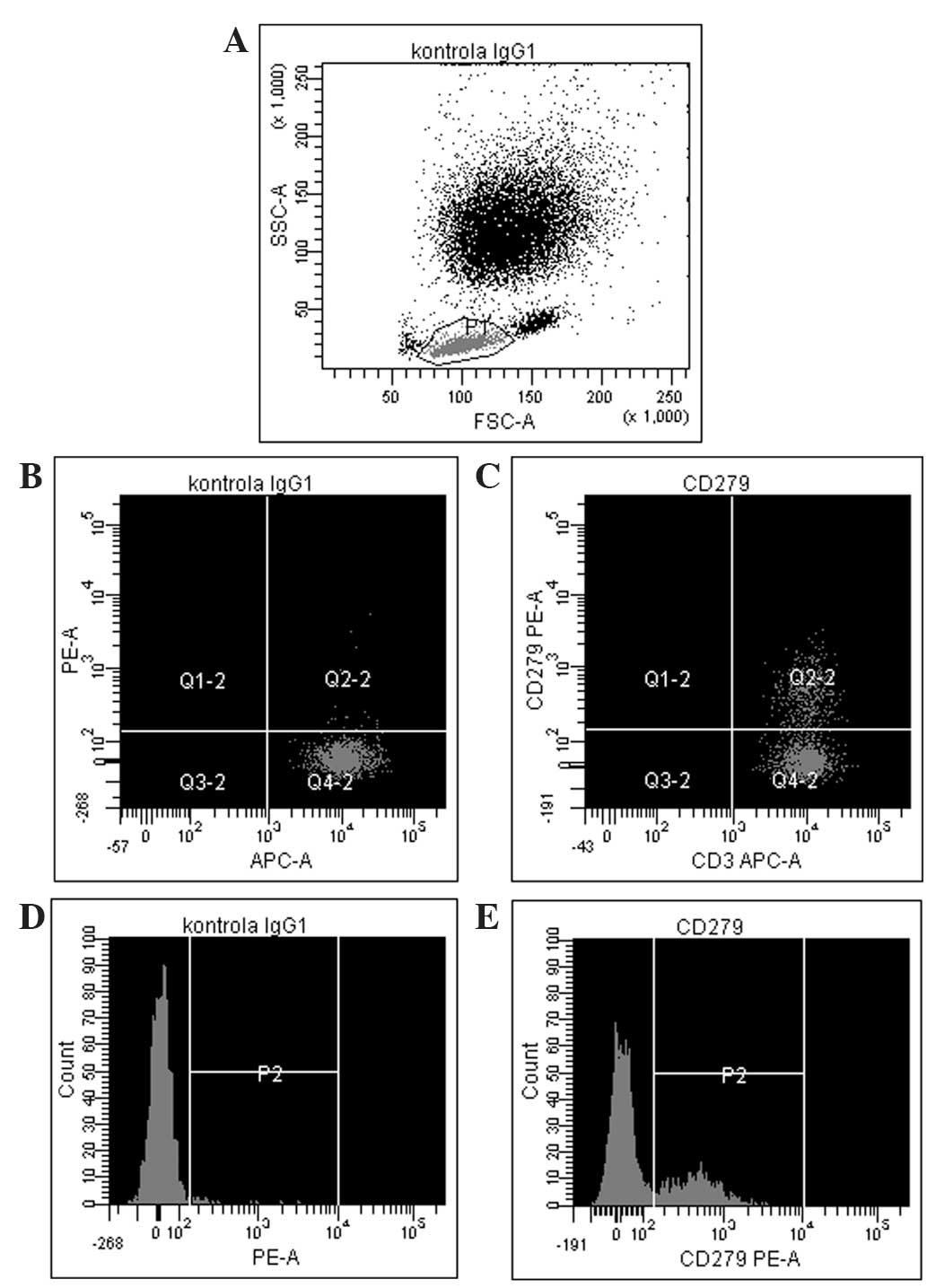
The results of the flow cytometric analysis of PD-1
(CD279) expression for a representative sample are shown in
Fig. 1. A statistically significant
difference in PD-1 expression was identified between preoperative
breast cancer patients and healthy controls (mean ± standard
deviation, 26.31±11.87% vs. 12.72±8.15%, respectively; P<0.0001;
Fig. 2, Table II). In breast cancer patients, the
expression levels of PD-1 differed significantly between the
various time-points (P=0.0458), with the difference between the
second and third point being the most significant (P=0.0007). The
median PD-1 levels were 24.80% (Q1-Q3,
17.20–34.60%) prior to surgery and 24.65%
(Q1-Q3, 19.50–36.85%) during surgery,
decreasing to 21.25% (Q1-Q3, 17.75–32.60%) 24
h after surgery. No statistically significant differences were
noted in CTLA-4 expression, which remained constant over the time
points assessed (P=0.3788). In addition, no correlation was
identified in CTLA-4 expression between the breast cancer patients
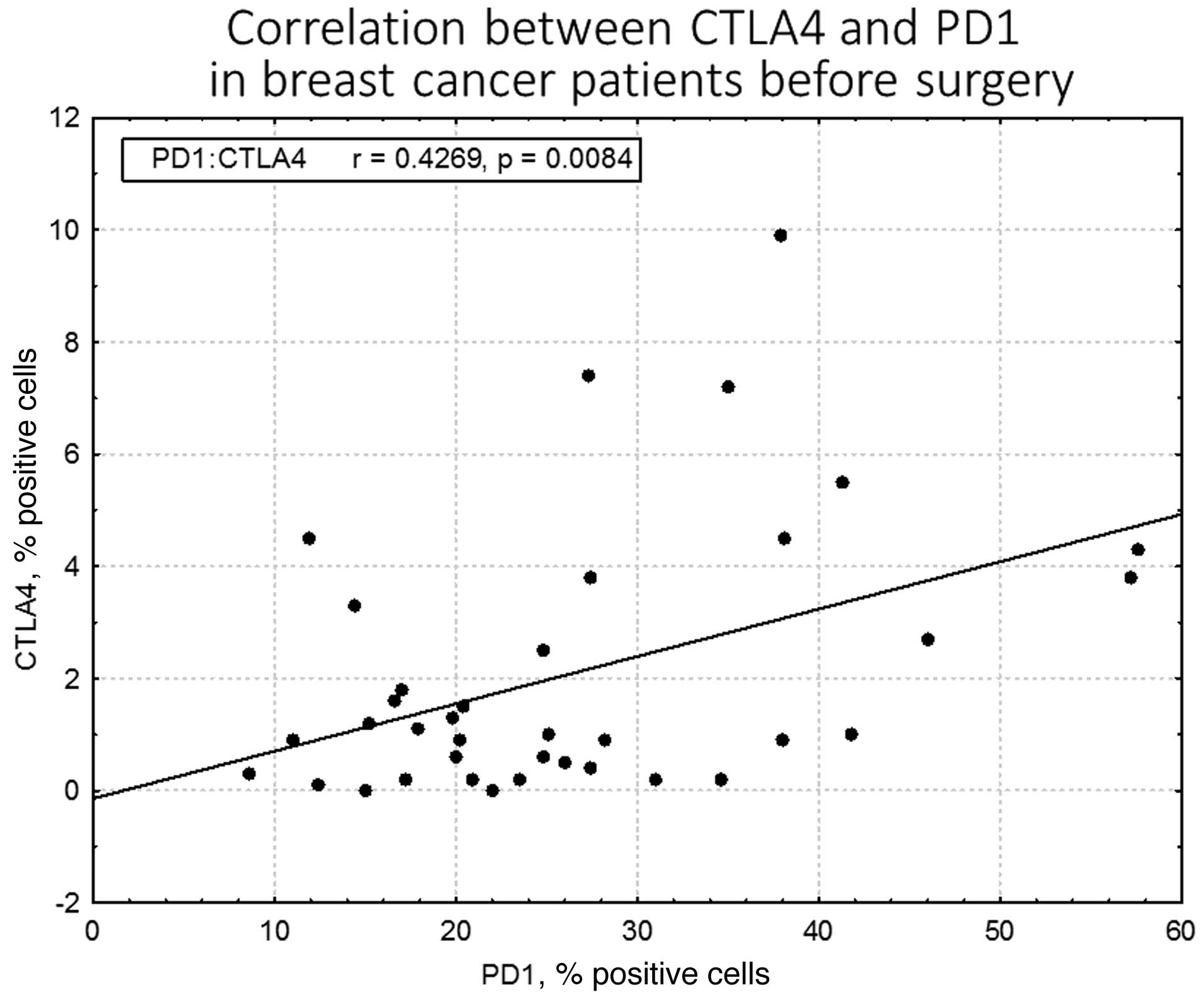
and healthy individuals. By contrast, a statistically significant
association was observed between CTLA-4 and PD-1 levels prior to
surgery in breast cancer patients (r=0.43; P=0.0084); however, this
disappeared in the two subsequent measurements, during (r=0.08;
P=0.62) and after surgery (r=0.14; P=0.43; Fig. 3). CTLA-4 expression was associated
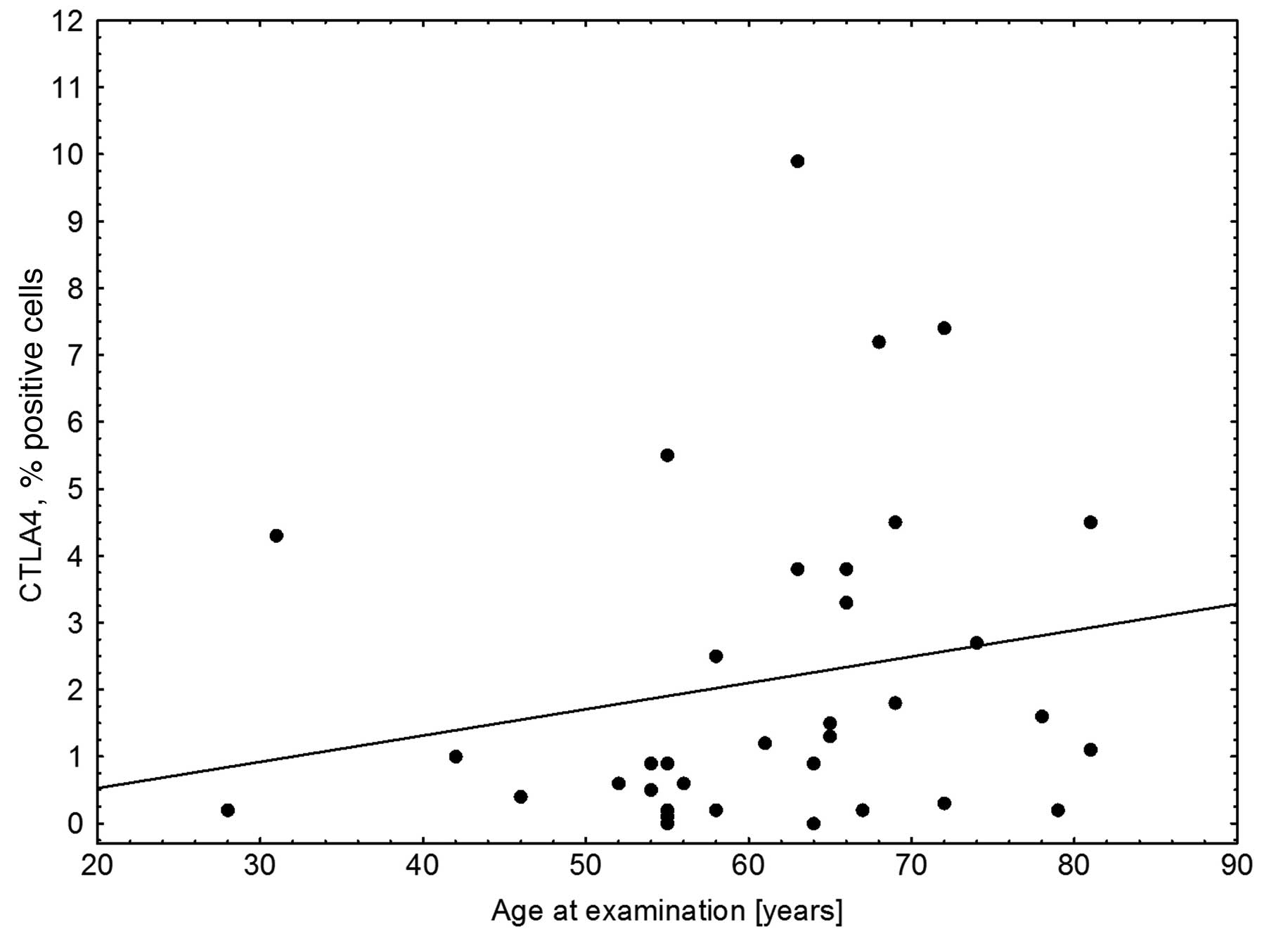
with age (r=0.33; P=0.0453), with elevated levels of CTLA-4
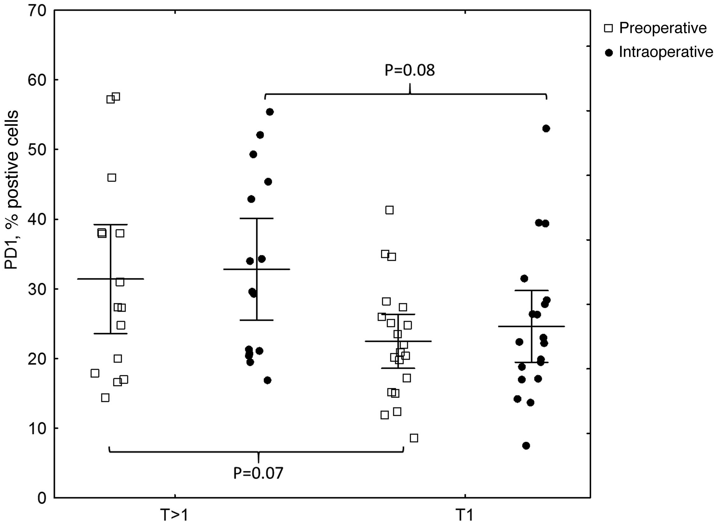
expression observed in older breast cancer patients (Fig. 4). The association of PD-1 levels and
tumor size was also analyzed, with higher levels observed in T2
tumors compared with T1 tumors prior to surgery (T2 vs. T1,
31.41±14.14% vs. 22.47±8.28%; P=0.07) and intraoperatively (T2 vs.
T1, 32.81±13.21% vs. 24.61±10.68%; P=0.08; however, the differences
were not statistically significant (Fig.
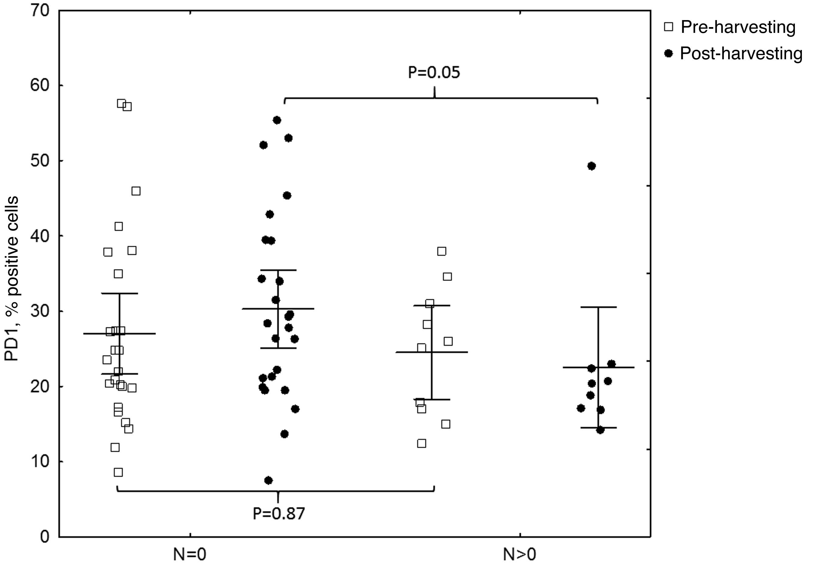
5). Furthermore, a decrease in PD-1 levels was observed
subsequent to harvesting SNs with metastasis, but not in
SN-negative patients (P=0.05; Fig.
6). A negative correlation between PD-1 expression and PR
status was detected following BCS and SNB (r=-0.39; P=0.024).
| Table II.PD-1 levels preoperatively in breast
cancer patients and healthy controls. Higher levels were found in
breast cancer patients (P<0.0001). |
Table II.
PD-1 levels preoperatively in breast
cancer patients and healthy controls. Higher levels were found in
breast cancer patients (P<0.0001).
|
|
| PD-1 expression, %
positive cells |
|
|---|
|
|
|
|
|
|---|
| Group | n | Mean | SD | Median | Q1 | Q3 | P-value |
|---|
| Cancer | 35 | 26.31 | 11.87 | 24.80 | 17.20 | 34.60 | <0.0001 |
| Control | 25 | 12.72 | 8.15 | 11.00 | 6.50 | 14.90 |
|
Discussion
In the current study, a striking difference
(P<0.0001) was observed in immune checkpoint PD-1 expression
between breast cancer patients and healthy controls, with
significantly lower levels in the latter group. This may be
expected as the immune system is often impaired in cancer patients,
and inhibitory immune pathways are predominant. This is consistent
with the observations of Legat et al (22), that expression of inhibitory
co-receptors, including PD-1, CTLA-4, LAG3 and CD160, is generally
considered a hallmark of T cell exhaustion, particularly in the
context of chronic antigen exposure, such as persistent viral
infection or cancer. Poschke et al (23) noted tumor-induced changes in the
phenotype of blood-derived and tumor-associated T cells in 43
patients with stage I and II breast cancer, compared with 10
patients with benign disease. These findings were most pronounced
in CD8+ T cells, which are key components of tumor
immune surveillance. Blood from early-stage breast cancer patients
contained fewer naïve and more antigen-experienced memory T cells
compared with healthy controls. The aforementioned observations are
consistent with those of the current study, which were also
performed in stage I and II breast cancer patients, with isolation
of CD8+ T cells and PD-1 expression associated primarily
with experienced T cells. In more advanced disease (stages III and
IV), higher tumor burden and metastatic spread induce further
inhibition and dysfunction of immune surveillance (23). Azim et al (24) demonstrated that aggressive
pregnancy-associated breast cancer patients exhibited higher
expression levels of inhibitory PD-1 (P=0.015) and its ligand PD-1L
(P=0.014) compared with non-pregnant breast cancer patients. In the
present study, patients with pregnancy-associated breast cancer
were not included.
The association between immune checkpoint expression
and age of breast cancer patients was also examined. CTLA-4
expression was found to be associated with age (r=0.33; P=0.0453),
with elevated levels present in older breast cancer patients, since
age suppresses the functions of the immune system. The theory that
age suppresses the functions of the immune system is defined as
immune senescence (functional impairments of immunity) and is an
important T cell defect associated with ageing, leading to higher
susceptibility to infections or cancer in the elderly (25,26).
In the present study, an association was observed
between PD-1 levels and tumor size prior to surgery and
intraoperatively (P=0.07 and P=0.08, respectively), with higher
levels of PD-1 present in T2 tumors compared with T1 tumors, which
is consistent with the association of immune suppression with more
advanced cancer; however, the differences were not statistically
significant. These observations are concordant with those of Stagg
et al (27), who concluded
that blockade of PD-1 reduced the tumor size in a number of
experimental cancer models. Similarly, Brahmer et al
(28) reported good tumor responses
to anti-PD-1 therapy in 37.5% of patients with melanoma, renal cell
carcinoma, prostate cancer, non-small cell lung cancer and
colorectal cancer, in a phase II clinical trial. Wolchok et
al (29) demonstrated a strong
response to a human antibody blocking PD-1 (nivolumab),
administered concurrently with ipilimumab (anti-CTLA-4) in a
substantial proportion of treated patients, with ≥80% tumor size
regression. Topalian et al (30) identified that nivolumab therapy in
melanoma patients produced durable responses.
An intriguing observation of the present study, in
light of the paradigm shift in the extent of axillary dissection in
breast cancer, was a decrease in PD-1 levels following the harvest
of SN(s) containing metastases, in contrast to SN-negative patients
(P=0.05). This implies that the removal of metastatic lymph nodes
may affect immune surveillance, altering immune checkpoints.
Furthermore, removal of healthy, non-malignant sentinel lymph nodes
may potentially be harmful, as they appear to be significant in
immune surveillance and essential for the elimination of cancer
cells, particularly in early-stage breast cancer. Due to the small
sample size of the present study, the results must be considered
preliminary and should be interpreted with caution. However, they
provide a basis for further investigation of the role of immune
checkpoints in breast cancer, as well as potential justification on
immunological grounds for purely surgical procedures, particularly
with respect to axillary lymph node surgery. In the present study,
all the patients with early-stage breast cancer were subjected to
SNB, in accordance with the current standards of care (31). A report is expected in 2022 from the
Sentinel node vs Observation after axillary UltraSouND (SOUND)
trial (6,7), confirming whether an abandonment of
nodal procedures is a safe approach in selected patients.
Therefore, no data comparing immune checkpoints in cohorts with
completely intact axillary lymph nodes is available, at present;
this is currently only feasible in patients who have positive or
negative SNs. Gentilini et al (6), as part of the SOUND trial, investigated
the outcome of breast cancer patients that did not undergo axillary
surgery and preoperative ultrasound of the axilla, in order to
identify patients with suspected lymph node involvement. The
present study attempted to provide an immunological rationale for
SN surgery on the basis of its histopathological status. In the
SOUND trial, Gentilini et al are currently exploring the
role of stem cells in the SN in breast cancer progression and
recurrence. Vallacchi et al (32) demonstrated an increase in exhausted
immune PD-1-positive cells in SNs of melanoma patients with more
advanced disease. Overall, there is little data available to
justify axillary lymph node surgery on immunological grounds and
further investigation in larger studies is urgently required.
In the present study, a negative correlation between
PD-1 expression and PR status following BCS and SNB (r=-0.39;
P=0.024) was identified. Cases with higher PR levels, which are
associated with the luminal A subtype and good prognosis, exhibited
lower PD-1 expression and, thus, decreased inhibition of the immune
system.
The current study confirmed that breast cancer
appears to be an immunogenic entity. In addition, renewed and
increasing interest exists on the role of PR. The St. Gallen
International Expert Consensus of 2013 (33) highlighted that PR added value in
distinguishing between luminal A and luminal B subtypes, derived
from the work of Prat et al (34) in which a cut-off point of ≥20%
PR-positive tumor cells corresponded to luminal A tumors. During
the San Antonio Breast Cancer Symposium (December 2013), Carroll
et al (35) reported that PR
is an essential prerequisite for the binding of ER with genes of
good prognosis, whilst loss of PR appears to be associated with ER
dysfunction. The findings of the present study that the highest
levels of inhibitory regulators PD-1 occurred in PR-negative
patients are consistent with those of Loi, Denkert, von Minckwitz
et al (13), indicating that a
targeted anti-PD-1 approach may be useful in triple negative and
non-luminal HER2-positive breast cancer patients, such as patients
without PR expression.
In conclusion, the present study demonstrated that
breast cancer patients exhibited an altered profile of immune
checkpoint markers, with higher concentrations of PD-1 in larger
and PR-negative tumors. Surgical removal of lymph nodes containing
tumor cells alters the immunologic profile by diminishing PD-1
levels. Breast cancer has not traditionally been considered
immunogenic. However, increasing evidence indicates that the
immunogenicity is important in certain molecular subtypes of breast
cancer (36). The results of the
present study provide a basis for further investigation of the role
of immune checkpoints in breast cancer and immunological
justification for purely surgical procedures, particularly axillary
lymph node surgery. Finely-tuned modulation of the immune system
may play a role in the treatment of breast cancer patients,
enhancing the effects of the already well-established multimodality
treatments (chemotherapy, endocrine therapy and anti-HER2 therapy),
and may have an impact on axillary lymph node surgery. Therefore,
further research in a large cohort of patients is required
(37–40).
Acknowledgements
The authors would like to thank Professor Andrew
Shorthouse, the Emeritus Professor of Surgery, University of
Sheffield, for proofreading the manuscript.
References
|
1
|
Urban JA and Baker HW: Radical mastectomy
in continuity with en bloc resection of the internal mammary
lymph-node chain; a new procedure for primary operable cancer of
the breast. Cancer. 5:992–1008. 1952. View Article : Google Scholar : PubMed/NCBI
|
|
2
|
Halsted WS: I. The Results of Operations
for the Cure of Cancer of the Breast Performed at the Johns Hopkins
Hospital from June, 1889, to January, 1894. Ann Surg. 20:497–555.
1894. View Article : Google Scholar : PubMed/NCBI
|
|
3
|
Giuliano AE, Kirgan DM, Guenther JM and
Morton DL: Lymphatic mapping and sentinel lymphadenectomy for
breast cancer. Ann Surg. 220:391–398. 1994. View Article : Google Scholar : PubMed/NCBI
|
|
4
|
Giuliano AE, Hunt KK, Ballman KV, Beitsch
PD, Whitworth PW, Blumencranz PW, Leitch AM, Saha S, McCall LM and
Morrow M: Axillary dissection vs no axillary dissection in women
with invasive breast cancer and sentinel node metastasis: a
randomized clinical trial. JAMA. 305:569–575. 2011. View Article : Google Scholar : PubMed/NCBI
|
|
5
|
Galimberti V, Cole BF, Zurrida S, et al
International Breast Cancer Study Group Trial 23-01 investigators:
Axillary dissection versus no axillary dissection in patients with
sentinel-node micrometastases (IBCSG 23-01): a phase 3 randomised
controlled trial. Lancet Oncol. 14:297–305. 2013. View Article : Google Scholar : PubMed/NCBI
|
|
6
|
Gentilini O and Veronesi U: Abandoning
sentinel lymph node biopsy in early breast cancer? A new trial in
progress at the European Institute of Oncology of Milan (SOUND:
Sentinel node vs Observation after axillary UltraSouND). Breast.
21:678–681. 2012. View Article : Google Scholar : PubMed/NCBI
|
|
7
|
Reimer T, Hartmann S, Stachs A and Gerber
B: Local treatment of the axilla in early breast cancer: Concepts
from the national surgical adjuvant breast and bowel project B-04
to the planned intergroup sentinel mamma trial. Breast Care Basel.
9:87–95. 2014.PubMed/NCBI
|
|
8
|
Pardoll DM: The blockade of immune
checkpoints in cancer immunotherapy. Nat Rev Cancer. 12:252–264.
2012. View
Article : Google Scholar : PubMed/NCBI
|
|
9
|
Topalian SL, Hodi FS, Brahmer JR, et al:
Safety, activity, and immune correlates of anti-PD-1 antibody in
cancer. N Engl J Med. 366:2443–2454. 2012. View Article : Google Scholar : PubMed/NCBI
|
|
10
|
Ott PA, Hodi FS and Robert C: CTLA-4 and
PD-1/PD-L1 blockade: New immunotherapeutic modalities with durable
clinical benefit in melanoma patients. Clin Cancer Res.
19:5300–5309. 2013. View Article : Google Scholar : PubMed/NCBI
|
|
11
|
Eggermont AM, Spatz A and Robert C:
Cutaneous melanoma. Lancet. 383:816–827. 2014. View Article : Google Scholar : PubMed/NCBI
|
|
12
|
Lipson EJ, Sharfman WH, Drake CG, et al:
Durable cancer regression off-treatment and effective reinduction
therapy with an anti-PD-1 antibody. Clin Cancer Res. 19:462–468.
2013. View Article : Google Scholar : PubMed/NCBI
|
|
13
|
Loi S: Tumor infiltrating lymphocytes
(TILs) indicate trastuzumab benefit in early-stage HER2-positive
breast cancer (HER2+ BC). Presented at the. San Antonio
Breast Cancer Symposium. 2013.https://www.conferencenotes.co/conferences/5467ecb588d6868d254609f6/presentations/54739f542e0507a22d1b9341?referrer=history
|
|
14
|
Beavis PA, Milenkovski N, Henderson MA, et
al: Adenosine receptor 2A blockade increases the efficacy of
anti-PD1 through enhanced antitumor T-cell responses. Cancer
Immunol Res. Feb 11–2015.(Epub ahead of print). View Article : Google Scholar : PubMed/NCBI
|
|
15
|
Denkert C, von Minckwitz G, Brase JC, et
al: Tumor-infiltrating lymphocytes and response to neoadjuvant
chemotherapy with or without carboplatin in human epidermal growth
factor receptor 2-positive and triple-negative primary breast
cancers. J Clin Oncol. 33:983–991. 2015. View Article : Google Scholar : PubMed/NCBI
|
|
16
|
Verbrugge I, Hagekyriakou J, Sharp LL, et
al: Radiotherapy increases the permissiveness of established
mammary tumors to rejection by immunomodulatory antibodies. Cancer
Res. 72:3163–3174. 2012. View Article : Google Scholar : PubMed/NCBI
|
|
17
|
Bos PD, Plitas G, Rudra D, Lee SY and
Rudensky AY: Transient regulatory T cell ablation deters
oncogene-driven breast cancer and enhances radiotherapy. J Exp Med.
210:2435–2466. 2013. View Article : Google Scholar : PubMed/NCBI
|
|
18
|
Vonderheide RH, LoRusso PM, Khalil M, et
al: Tremelimumab in combination with exemestane in patients with
advanced breast cancer and treatment-associated modulation of
inducible costimulator expression on patient T cells. Clin Cancer
Res. 16:3485–3494. 2010. View Article : Google Scholar : PubMed/NCBI
|
|
19
|
Elston CW and Ellis IO: Pathological
prognostic factors in breast cancer. I. The value of histological
grade in breast cancer: Experience from a large study with
long-term follow-up. Histopathology. 19:403–410. 1991. View Article : Google Scholar : PubMed/NCBI
|
|
20
|
Allred DC, Harvey JM, Berardo M and Clark
GM: Prognostic and predictive factors in breast cancer by
immunohistochemical analysis. Mod Pathol. 11:155–168.
1998.PubMed/NCBI
|
|
21
|
Wolff AC, Hammond ME, Hicks DG, Dowsett M,
McShane LM, Allison KH, Allred DC, Bartlett JM, Bilous M,
Fitzgibbons P, et al American Society of Clinical Oncology; College
of American Pathologists: Recommendations for human epidermal
growth factor receptor 2 testing in breast cancer: American Society
of Clinical Oncology/College of American Pathologists clinical
practice guideline update. J Clin Oncol. 31:3997–4013. 2013.
View Article : Google Scholar : PubMed/NCBI
|
|
22
|
Legat A, Speiser DE, Pircher H, Zehn D and
Fuertes Marraco SA: Inhibitory receptor expression depends more
dominantly on differentiation and activation than “exhaustion” of
human CD8 T cells. Front Immunol. 4:4552013. View Article : Google Scholar : PubMed/NCBI
|
|
23
|
Poschke I, De Boniface J, Mao Y and
Kiessling R: Tumor-induced changes in the phenotype of
blood-derived and tumor-associated T cells of early stage breast
cancer patients. Int J Cancer. 131:1611–1620. 2012. View Article : Google Scholar : PubMed/NCBI
|
|
24
|
Azim HA, Brohée S, Peccatori FA, et al:
Biology of breast cancer during pregnancy using genomic profiling.
Endocr Relat Cancer. 21:545–554. 2014. View Article : Google Scholar : PubMed/NCBI
|
|
25
|
Poland GA, Ovsyannikova IG, Kennedy RB,
Lambert ND and Kirkland JL: A systems biology approach to the
effect of aging, immunosenescence and vaccine response. Curr Opin
Immunol. 29:62–68. 2014. View Article : Google Scholar : PubMed/NCBI
|
|
26
|
Martinet KZ, Bloquet S and Bourgeois C:
Ageing combines CD4 T cell lymphopenia in secondary lymphoid organs
and T cell accumulation in gut associated lymphoid tissue. Immun
Ageing. 11:82014. View Article : Google Scholar : PubMed/NCBI
|
|
27
|
Stagg J, Andre F and Loi S:
Immunomodulation via chemotherapy and targeted therapy: A new
paradigm in breast cancer therapy? Breast Care Basel. 7:267–272.
2012. View Article : Google Scholar : PubMed/NCBI
|
|
28
|
Brahmer JR, Drake CG, Wollner I, et al:
Phase I study of single-agent anti-programmed death-1 (MDX-1106) in
refractory solid tumors: Safety, clinical activity,
pharmacodynamics, and immunologic correlates. J Clin Oncol.
28:3167–3175. 2010. View Article : Google Scholar : PubMed/NCBI
|
|
29
|
Wolchok JD, Kluger H, Callahan MK, et al:
Nivolumab plus ipilimumab in advanced melanoma. N Engl J Med.
369:122–133. 2013. View Article : Google Scholar : PubMed/NCBI
|
|
30
|
Topalian SL, Sznol M, McDermott DF, et al:
Survival, durable tumor remission, and long-term safety in patients
with advanced melanoma receiving nivolumab. J Clin Oncol.
32:1020–1030. 2014. View Article : Google Scholar : PubMed/NCBI
|
|
31
|
National Comprehensive Cancer Network, .
Clinical Practice Guidelines in Oncology. Version 2. 2015
http://www.nccn.org/professionals/physician_gls/f_guidelines.asp#breastAccessed.
March 11–2015
|
|
32
|
Vallacchi V, Vergani E, Camisaschi C, et
al: Transcriptional profiling of melanoma sentinel nodes identify
patients with poor outcome and reveal an association of CD30(+) T
lymphocytes with progression. Cancer Res. 74:130–140. 2014.
View Article : Google Scholar : PubMed/NCBI
|
|
33
|
Goldhirsch A, Winer EP, Coates AS, et al:
Role: Panel membersPersonalizing the treatment of women with early
breast cancer: Highlights of the St Gallen International Expert
Consensus on the Primary Therapy of Early Breast Cancer 2013. Ann
Oncol. 24:2206–2223. 2013. View Article : Google Scholar : PubMed/NCBI
|
|
34
|
Prat A, Cheang MC, Martín M, et al:
Prognostic significance of progesterone receptor-positive tumor
cells within immunohistochemically defined luminal A breast cancer.
J Clin Oncol. 31:203–209. 2013. View Article : Google Scholar : PubMed/NCBI
|
|
35
|
Carroll JS: Steroids, nuclear receptors
and breast cancer. Preface. Mol Cell Endocrinol. 382:6232014.
View Article : Google Scholar : PubMed/NCBI
|
|
36
|
Denkert C: The immunogenicity of breast
cancer - molecular subtypes matter. Ann Oncol. 25:1453–1455. 2014.
View Article : Google Scholar : PubMed/NCBI
|
|
37
|
Loi S, Michiels S, Salgado R, et al: Tumor
infiltrating lymphocytes are prognostic in triple negative breast
cancer and predictive for trastuzumab benefit in early breast
cancer: Results from the FinHER trial. Ann Oncol. 25:1544–1550.
2014. View Article : Google Scholar : PubMed/NCBI
|
|
38
|
von Minckwitz G, Schneeweiss A, Loibl S,
et al: Neoadjuvant carboplatin in patients with triple-negative and
HER2-positive early breast cancer (GeparSixto; GBG 66): A
randomised phase 2 trial. Lancet Oncol. 15:747–756. 2014.
View Article : Google Scholar : PubMed/NCBI
|
|
39
|
Denkert C: Diagnostic and therapeutic
implications of tumor-infiltrating lymphocytes in breast cancer. J
Clin Oncol. 31:836–837. 2013. View Article : Google Scholar : PubMed/NCBI
|
|
40
|
Loi S, Sirtaine N, Piette F, et al:
Prognostic and predictive value of tumor-infiltrating lymphocytes
in a phase III randomized adjuvant breast cancer trial in
node-positive breast cancer comparing the addition of docetaxel to
doxorubicin with doxorubicin-based chemotherapy: BIG 02–98. J Clin
Oncol. 31:860–867. 2013. View Article : Google Scholar : PubMed/NCBI
|