Introduction
Pancreatic cancer has the worst prognosis of all
major malignancies, with a 5-year survival rate of 6% (1). At present, surgical resection is the
only effective treatment in these patients; however, the 5-year
survival rate following surgical resection is only 5.5–21%
(2,3).
Gemcitabine (GEM)-based chemotherapy is the core of multimodal
therapy for pancreatic cancer and has improved patient prognosis
(3). Multimodal therapies that
include both chemotherapy and radiation therapy have been
previously investigated and are able to reportedly improve the
clinical outcome in pancreatic cancer patients (4–6). This
provides several therapeutic pathways to help reduce the high
refractoriness of pancreatic cancer. Therefore, identifying
specific predictive markers in order to determine which patients
present a poor prognosis is essential.
Cyclin G2 (CCNG2; encoded by CCNG2 gene)
belongs to a family of cyclins homologous to CCNG1 (7). Cyclins positively regulate cell
proliferation to a large extent. However, unexpectedly, CCNG2 has
been reported to regulate cell proliferation as a tumor suppressor
gene and its decreased expression is associated with malignant
phenotypes in several types of cancer (8–17).
Previous studies have reported that CCNG2 is involved in a variety
of functions associated with cancer progression, including the
regulation of cell proliferation (8–10),
chemoresistance (8), DNA repair
(11) and cell differentiation
(12). Furthermore, our previous
study demonstrated that CCNG2 was associated with chemoresistance
and cancer stemness via cell apoptosis in pancreatic cancer
(13). Recently, CCNG2 has been
reported as a novel prognostic marker in several types of cancer
(14–17). However, to date, no study has
clarified the association between CCNG2 and the prognosis of
pancreatic cancer. Therefore, in the present study, the association
between CCNG2 expression and the clinical outcomes of 36 patients
with pancreatic cancer was evaluated.
Materials and methods
Primary tumor samples
Between March 2007 and October 2012, 92 patients
underwent surgery for pancreatic cancer at the Department of
Gastroenterological Surgery in Osaka University (Osaka, Japan).
Among these patients, 36 consecutive patients who underwent
curative resection (R0) with histologically clear margins and no
preoperative therapy were enrolled in the present study. All the
patients were staged prior to and following surgery, according to
the criteria of the Union for International Cancer Control
(18). The median follow-up period
was 26.4 months (range, 3.8–79.7 months), the 5-year survival rate
was 29.0%, and recurrence of the disease was observed in 19
patients. GEM was administered in 21 patients as adjuvant
chemotherapy (1,000 mg/m2, 3 times/month for 6 months).
No radiation therapy was administered during the follow-up period.
Table I summarizes the
characteristics of the 36 patients. The use of resected samples was
approved by the Human Ethics Review Committee of the Graduate
School of Medicine, Osaka University (approval number, 08226).
Written informed consent was obtained from all the patients
included in the study.
 | Table I.Clinicopathological characteristics of
the 36 pancreatic cancer patients. |
Table I.
Clinicopathological characteristics of
the 36 pancreatic cancer patients.
| Parameter | Value |
|---|
| Mean age, years | 68.5±9.4 |
| Gender (male/female),
n | 21/15 |
| Location (Ph/Pb/Pt),
n | 10/21/5 |
| Lymphatic invasion
(+/-), n | 26/10 |
| Venous invasion
(+/-), n | 15/21 |
| Intrapancreatic
perineural invasion (+/-), n | 29/7 |
| Maximal diameter,
mm | 25.9±14.6 |
| Histology
(well/mod/poor), n | 2/30/4 |
| pT (T1/T2/T3/T4),
n | 4/4/28/0 |
| pN (+/-), n | 16/20 |
| pStage
(IA/IB/IIA/IIB/III/IV), n | 4/4/12/16/0/0 |
| CCNG2 expression
(+/-), n | 17/19 |
| Adjuvant therapy
(+/-), n | 21/15 |
| Recurrence (+/-),
n | 19/17 |
Immunohistochemical staining
Immunohistochemical staining was performed using the
method described previously (19), in
order to detect CCNG2 expression in the 36 pancreatic cancer
samples. Noncancerous pancreatic tissues obtained from the 36
patients were used as positive controls. Briefly, formalin-fixed,
paraffin-embedded 4-µm sections were deparaffinized in xylene.
Next, antigen-retrieval was performed with heat-induced epitope
retrieval, at 95°C for 40 min and then the samples were incubated
in methanol containing 0.3% hydrogen peroxide to block endogenous
peroxidase. Following incubation with normal protein block serum
(Vectastain Elite ABC kit; Vector Laboratories, Inc., Burlingame,
CA, USA), the sections were incubated overnight at 4°C with an
anti-CCNG2 antibody as the primary antibody (polyclonal rabbit
anti-human CCNG2 antibody; 1.0 µg/ml; MBL International
Corporation, Nagoya, Japan) (11).
Thereafter, the staining was revealed with avidin-biotin complex
reagents (Vector Laboratories Inc., Burlingame, CA, USA) using an
Olympus BX50 microscope (Olympus Corporation, Tokyo, Japan) and
3,3′-diaminobenzidine. All sections were counterstained with
hematoxylin (Sigma-Aldrich, St. Louis, MO, USA). Positive staining
for CCNG2 was defined as detectable nuclear staining in >50% of
the cancer cells.
Statistical analysis
Data are expressed as the mean ± standard deviation.
The clinicopathological parameters were compared using Fisher's
exact test, and the continuous variables were compared using the
Mann-Whitney U test. The survival curves were plotted using the
Kaplan-Meier method, while the differences between survival curves
were compared using the log-rank test. P<0.05 denotes the
presence of a statistically significant difference. Statistical
analysis was performed using JMP software, version 10.0.2 (SAS
Institute Inc., Cary, NC, USA).
Results
CCNG2 expression in pancreatic cancer
tissue samples
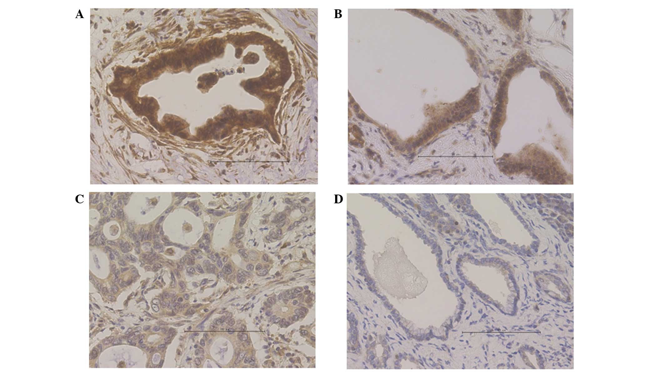
Immunohistochemical staining was performed to detect
CCNG2 expression in the 36 samples included in the present study.
The nuclei of normal pancreatic ductal cells were partially
stained; by contrast, acinar cells, which were used as positive
controls, were stained strongly in the cytoplasm and nuclei
(Fig. 1). In the cancerous sections,
the functional CCNG2 protein expression appeared to localize in the
nucleus, although CCNG2 expression has been previously demonstrated
to appear in the cytoplasm as well (12). Cases were defined as CCNG2-positive
when the cells presented diffused or spotted nuclear patterns
(>50% of cancer cells; Fig. 2A and
B), and as CCNG2-negative when the cells exhibited a
cytoplasmic pattern (no staining in the nucleus; Fig. 2C) or a negative pattern (no staining
in the nucleus or cytoplasm; Fig. 2D)
in the pancreatic cancer lesions. Among the 36 samples examined, 17
samples (47.2%) were positive for CCNG2, whereas 19 samples (52.8%)
were negative.
CCNG2 expression and
clinicopathological characteristics
The clinical and histopathological factors between
the CCNG2-positive and CCNG2-negative patients were compared to
examine the correlation between CCNG2 expression and cancer
progression (Table II). The
histopathological analysis revealed that venous invasion and the
tumor invasion depth (pT) factor (18) were significantly higher in the
CCNG2-negative group compared with the CCNG2-positive group. The
pathological stage also tended to be higher in the CCNG2-negative
group. Therefore, CCNG2 expression correlated inversely with cancer
progression in pancreatic cancer.
| Table II.Comparison of clinical and
histopathological factors between the CCNG2-positive and -negative
groups. |
Table II.
Comparison of clinical and
histopathological factors between the CCNG2-positive and -negative
groups.
| Characteristics | CCNG2-positive
(n=17) | CCNG2-negative
(n=19) | P-value |
|---|
| Mean age, years | 68.6±8.6 | 68.4±10.4 | NS |
| Gender (male/female),
n | 9/8 | 12/7 | NS |
| Tumor location
(Ph/Pb/Pt), n | 8/5/4 | 13/5/1 | NS |
| Maximal diameter,
mm | 26.6±19.5 | 25.4±8.6 | NS |
| Histology
(well/mod/poor), n | 2/12/3 | 0/18/1 | NS |
| Lymphatic invasion
(+/-), n | 10/7 | 16/3 | NS |
| Venous invasion
(+/-), n | 4/13 | 11/8 | 0.0489 |
| Intrapancreatic
perineural invasion (+/-), n | 12/5 | 17/2 | NS |
| pT (T1/T2/T3/T4),
n | 4/3/10/0 | 0/1/18/0 | 0.0242 |
| pN (+/-), n | 6/11 | 10/9 | NS |
| pStage
(IA/IB/IIA/IIB/III/IV), n | 4/3/4/6/0/0 | 0/1/8/10/0/0 | 0.0728 |
| Adjuvant therapy
(+/-), n | 11/6 | 10/9 | NS |
| Recurrence (+/-),
n | 7/10 | 12/7 | NS |
Association between CCNG2 expression
and prognosis
Predictive markers for overall survival were
assessed based on the clinicopathological details of the patients.
Upon univariate analysis, pT, lymph node metastasis (pN) (18) and CCNG2 expression were found to be
significantly associated with overall survival, as opposed to other
prognostic markers (Table III).
Furthermore, multivariate analysis identified pN and CCNG2
expression as significant and independent prognostic factors.
| Table III.Predictive markers for overall
survival in the clinicopathological information. |
Table III.
Predictive markers for overall
survival in the clinicopathological information.
| Parameter | Univariate analysis
HR (95% CI) | P-value | Multivariate
analysis HR (95% CI) | P-value |
|---|
| Mean age
(≥69/<69 years) | 2.04
(0.79–5.88) | 0.143 | – | – |
| Gender
(male/female) | 1.18
(0.47–3.19) | 0.722 | – | – |
| Maximal diameter
(≥26/<26 mm) | 0.84
(0.27–2.19) | 0.731 | – | – |
| Histology
(well/mod/poor) | 1.95
(0.40–35.23) | 0.472 | – | – |
| pT (T1, T2/T3,
T4) | 0.15
(0.023–0.55) | 0.003 | 0.22
(0.026–1.30) | 0.095 |
| pN (+/-) | 4.15
(1.62–11.08) | 0.003 | 3.18
(1.08–10.45) | 0.035 |
| Lymphatic invasion
(+/-) | 1.86
(0.67–6.54) | 0.245 |
| Venous invasion
(+/-) | 2.35
(0.94–6.11) | 0.066 | 2.43
(0.69–8.18) | 0.159 |
| Intrapancreatic
perineural invasion (+/-) | 4.41
(0.91–79.44) | 0.070 | 3.33
(0.54–64.60) | 0.217 |
| CCNG2 expression
(+/-) | 0.32
(0.11–0.84) | 0.020 | 0.32
(0.096–0.98) | 0.046 |
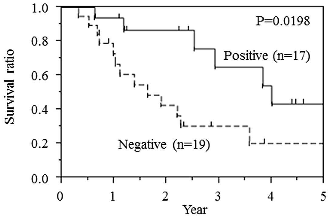
The Kaplan-Meier curve for overall survival is shown
in Fig. 3 and reveals that the
negative expression of CCNG2 was a consistent indicator of poor
prognosis in pancreatic cancer patients. The median survival time
of the CCNG2-negative group was 19.6 months, whereas that of the
CCNG2-positive group was 47.9 months.
Discussion
CCNG2 gene was initially identified in 1996
and encodes for a protein that belongs to a family of cyclins
homologous to CCNG1 (7). Previous
studies have reported that CCNG2 participates in
carcinogenesis and is a known tumor suppressor gene (15–17,20–26).
CCNG2 gene expression is downregulated in thyroid (20), oral (21), ovarian (22), breast (23,24),
gastric (16), esophageal (17), prostate (25), kidney (26) and colorectal (15) cancer cells.
Several aspects of CCNG2 behavior are associated
with antitumor effects. Antitumor agents induce CCNG2 expression,
which results in the inhibition of cancer cell proliferation
(8–10). In breast cancer, CCNG2
knockdown induces multidrug resistance (8). In colorectal cancer, CCNG2 expression
correlates with the tumor stage, lymph node metastasis, clinical
stage, histological grade and overall survival (15). In gastric cancer, CCNG2 expression
correlates with the extent of differentiation: CCNG2 expression is
high in well-differentiated adenocarcinomas and low in
poorly-differentiated adenocarcinomas (12). In our previous study, CCNG2
induced apoptosis and was partially associated with cancer stemness
in a pancreatic cancer cell line (13). In summary, CCNG2 is heavily involved
in cancer progression, including proliferation, invasion,
chemoresistance and differentiation in various cancer types.
In the present study, several histopathological
factors associated with clinical outcome were evaluated. CCNG2 was
identified as an independent novel prognostic factor in pancreatic
cancer patients. In the CCNG2-negative group, the rate of venous
invasion and pT factor were significantly higher, while the
pathological stage was also higher compared with that in the
positive group. Altogether, these findings suggest that low
expression of CCNG2 reflects, at least partially, cancer
progression and CCNG2 is an independent prognostic factor.
In conclusion, the present study demonstrated that
CCNG2 expression correlates inversely with cancer progression and
may be used as a novel, independent prognostic factor in pancreatic
cancer.
Acknowledgements
The authors would like to thank the members of our
laboratories for their contribution. This study was supported in
part by: A Grant-in-Aid for Scientific Research and a grant from
the Platform for Drug Discovery, Informatics, and Structural Life
Science, from the Ministry of Education, Culture, Sports, Science
and Technology (grant nos. 23390199, 25112708, 25134711, 30253420,
26670604; P-Direct); a Grant-in-Aid from the Third Comprehensive
10-year Strategy for Cancer Control, Ministry of Health, Labor, and
Welfare (grant no. H23-003); a grant from the Kobayashi Cancer
Research Foundation; a grant from the Princess Takamatsu Cancer
Research Fund, Japan; and a grant from the National Institute of
Biomedical Innovation, Japan.
References
|
1
|
Siegel R, Naishadham D and Jemal A: Cancer
statistics, 2012. CA Cancer J Clin. 62:10–29. 2012. View Article : Google Scholar : PubMed/NCBI
|
|
2
|
Neoptolemos JP, Stocken DD, Friess H,
Bassi C, Dunn JA, Hickey H, Beger H, Fernandez-Cruz L, Dervenis C,
Lacaine F, et al: A randomized trial of chemoradiotherapy and
chemotherapy after resection of pancreatic cancer. N Engl J Med.
350:1200–1210. 2004. View Article : Google Scholar : PubMed/NCBI
|
|
3
|
Oettle H, Neuhaus P, Hochhaus A, Hartmann
JT, Gellert K, Ridwelski K, Niedergethmann M, Zülke C, Fahlke J,
Arning MB, et al: Adjuvant chemotherapy with gemcitabine and
long-term outcomes among patients with resected pancreatic cancer:
The CONKO-001 randomized trial. JAMA. 310:1473–1481. 2013.
View Article : Google Scholar : PubMed/NCBI
|
|
4
|
Eguchi H, Nagano H, Tanemura M, Takeda Y,
Marubashi S, Kobayashi S, Kawamoto K, Wada H, Hama N, Akita H, et
al: Preoperative chemoradiotherapy, surgery and adjuvant therapy
for resectable pancreatic cancer. Hepatogastroenterology.
60:904–911. 2013.PubMed/NCBI
|
|
5
|
Evans DB, Varadhachary GR, Crane CH, Sun
CC, Lee JE, Pisters PW, Vauthey JN, Wang H, Cleary KR, Staerkel GA,
et al: Preoperative gemcitabine-based chemoradiation for patients
with resectable adenocarcinoma of the pancreatic head. J Clin
Oncol. 26:3496–3502. 2008. View Article : Google Scholar : PubMed/NCBI
|
|
6
|
Ohigashi H, Ishikawa O, Eguchi H,
Takahashi H, Gotoh K, Yamada T, Yano M, Nakaizumi A, Uehara H,
Tomita Y, et al: Feasibility and efficacy of combination therapy
with preoperative full-dose gemcitabine, concurrent
three-dimensional conformal radiation, surgery, and postoperative
liver perfusion chemotherapy for T3-pancreatic cancer. Ann Surg.
250:88–95. 2009. View Article : Google Scholar : PubMed/NCBI
|
|
7
|
Bates S, Rowan S and Vousden KH:
Characterisation of human cyclin G1 and G2: DNA damage inducible
genes. Oncogene. 13:1103–1109. 1996.PubMed/NCBI
|
|
8
|
Kasukabe T, Okabe-Kado J, Honma Y and
Cotylenin A: a new differentiation inducer and rapamycin
cooperatively inhibit growth of cancer cells through induction of
cyclin G2. Cancer Sci. 99:1693–1698. 2008. View Article : Google Scholar : PubMed/NCBI
|
|
9
|
Padua MB and Hansen PJ: Changes in
expression of cell-cycle-related genes in PC-3 prostate cancer
cells caused by ovine uterine serpin. J Cell Biochem.
107:1182–1188. 2009. View Article : Google Scholar : PubMed/NCBI
|
|
10
|
Zhao Z, Liu Y, He H, Chen X, Chen J and Lu
YC: Candidate genes influencing sensitivity and resistance of human
glioblastoma to Semustine. Brain Res Bull. 86:189–194. 2011.
View Article : Google Scholar : PubMed/NCBI
|
|
11
|
Naito Y, Yabuta N, Sato J, Ohno S, Sakata
M, Kasama T, Ikawa M and Nojima H: Recruitment of cyclin G2 to
promyelocytic leukemia nuclear bodies promotes dephosphorylation of
γH2AX following treatment with ionizing radiation. Cell Cycle.
12:1773–1784. 2013. View
Article : Google Scholar : PubMed/NCBI
|
|
12
|
Choi MG, Noh JH, An JY, Hong SK, Park SB,
Baik YH, Kim KM, Sohn TS and Kim S: Expression levels of cyclin G2,
but not cyclin E, correlate with gastric cancer progression. J Surg
Res. 157:168–174. 2009. View Article : Google Scholar : PubMed/NCBI
|
|
13
|
Hasegawa S, Eguchi H, Nagano H, Konno M,
Tomimaru Y, Wada H, Hama N, Kawamoto K, Kobayashi S, Nishida N, et
al: MicroRNA-1246 expression associated with CCNG2-mediated
chemoresistance and stemness in pancreatic cancer. Br J Cancer.
111:1572–1580. 2014. View Article : Google Scholar : PubMed/NCBI
|
|
14
|
Li WJ, Liu GL, Yu F, Xiang XX, Lu YF, Xiao
HZ and Shi YP: CCNG2 suppressor biological effects on thyroid
cancer cell through promotion of CDK2 degradation. Asian Pac J
Cancer Prev. 14:6165–6171. 2013. View Article : Google Scholar : PubMed/NCBI
|
|
15
|
Sun GG, Zhang J and Hu WN: CCNG2
expression is downregulated in colorectal carcinoma and its
clinical significance. Tumour Biol. 35:3339–3346. 2014. View Article : Google Scholar : PubMed/NCBI
|
|
16
|
Sun GG, Hu WN, Cui DW and Zhang J:
Decreased expression of CCNG2 is significantly linked to the
malignant transformation of gastric carcinoma. Tumour Biol.
35:2631–2639. 2014. View Article : Google Scholar : PubMed/NCBI
|
|
17
|
Chen JQ, Liu CJ, Wen HX, Shi CL, Zhang HS,
Li M and Sun GG: Changes in the expression of cyclin G2 in
esophageal cancer cell and its significance. Tumour Biol. 2014.
|
|
18
|
Sobin LH, Gospodarowicz MK and Wittekind
C: TNM Classification of Malignant Tumors (7th). Wiley-Blackwell.
Oxford: 2009.
|
|
19
|
Kondo M, Yamamoto H, Nagano H, Okami J,
Ito Y, Shimizu J, Eguchi H, Miyamoto A, Dono K, Umeshita K, et al:
Increased expression of COX-2 in nontumor liver tissue is
associated with shorter disease-free survival in patients with
hepatocellular carcinoma. Clin Cancer Res. 5:4005–4012.
1999.PubMed/NCBI
|
|
20
|
Ito Y, Yoshida H, Uruno T, Nakano K,
Takamura Y, Miya A, Kobayashi K, Yokozawa T, Matsuzuka F, Kuma K,
et al: Decreased expression of cyclin G2 is significantly linked to
the malignant transformation of papillary carcinoma of the thyroid.
Anticancer Res. 23:2335–2338. 2003.PubMed/NCBI
|
|
21
|
Kim Y, Shintani S, Kohno Y, Zhang R and
Wong DT: Cyclin G2 dysregulation in human oral cancer. Cancer Res.
64:8980–8986. 2004. View Article : Google Scholar : PubMed/NCBI
|
|
22
|
Fu G and Peng C: Nodal enhances the
activity of FoxO3a and its synergistic interaction with Smads to
regulate cyclin G2 transcription in ovarian cancer cells. Oncogene.
30:3953–3966. 2011. View Article : Google Scholar : PubMed/NCBI
|
|
23
|
Montagner M, Enzo E, Forcato M, Zanconato
F, Parenti A, Rampazzo E, Basso G, Leo G, Rosato A, Bicciato S, et
al: SHARP1 suppresses breast cancer metastasis by promoting
degradation of hypoxia-inducible factors. Nature. 487:380–384.
2012. View Article : Google Scholar : PubMed/NCBI
|
|
24
|
Ahmed S, Al-Saigh S and Matthews J: FOXA1
is essential for aryl hydrocarbon receptor-dependent regulation of
cyclin G2. Mol Cancer Res. 10:636–648. 2012. View Article : Google Scholar : PubMed/NCBI
|
|
25
|
Cui DW, Cheng YJ, Jing SW and Sun GG:
Effect of cyclin G2 on proliferative ability of prostate cancer
PC-3 cell. Tumour Biol. 35:3017–3024. 2014. View Article : Google Scholar : PubMed/NCBI
|
|
26
|
Cui DW, Sun GG and Cheng YJ: Change in
expression of cyclin G2 in kidney cancer cell and its significance.
Tumour Biol. 35:3177–3183. 2014. View Article : Google Scholar : PubMed/NCBI
|