Open Access

Small cell carcinoma of the rectum: A report of imaging results from four cases

  • Authors:
    • Jian Zhou
    • Jingxian Shen
    • Huali Ma
    • Yun Zhang
    • Meili Sun
    • Lie Zheng
    • Yanchun Lv
  • View Affiliations

  • Published online on: January 20, 2016     https://doi.org/10.3892/ol.2016.4136
  • Pages: 1671-1676
  • Copyright: © Zhou et al. This is an open access article distributed under the terms of Creative Commons Attribution License.

Metrics: Total Views: 0 (Spandidos Publications: | PMC Statistics: )
Total PDF Downloads: 0 (Spandidos Publications: | PMC Statistics: )


Abstract

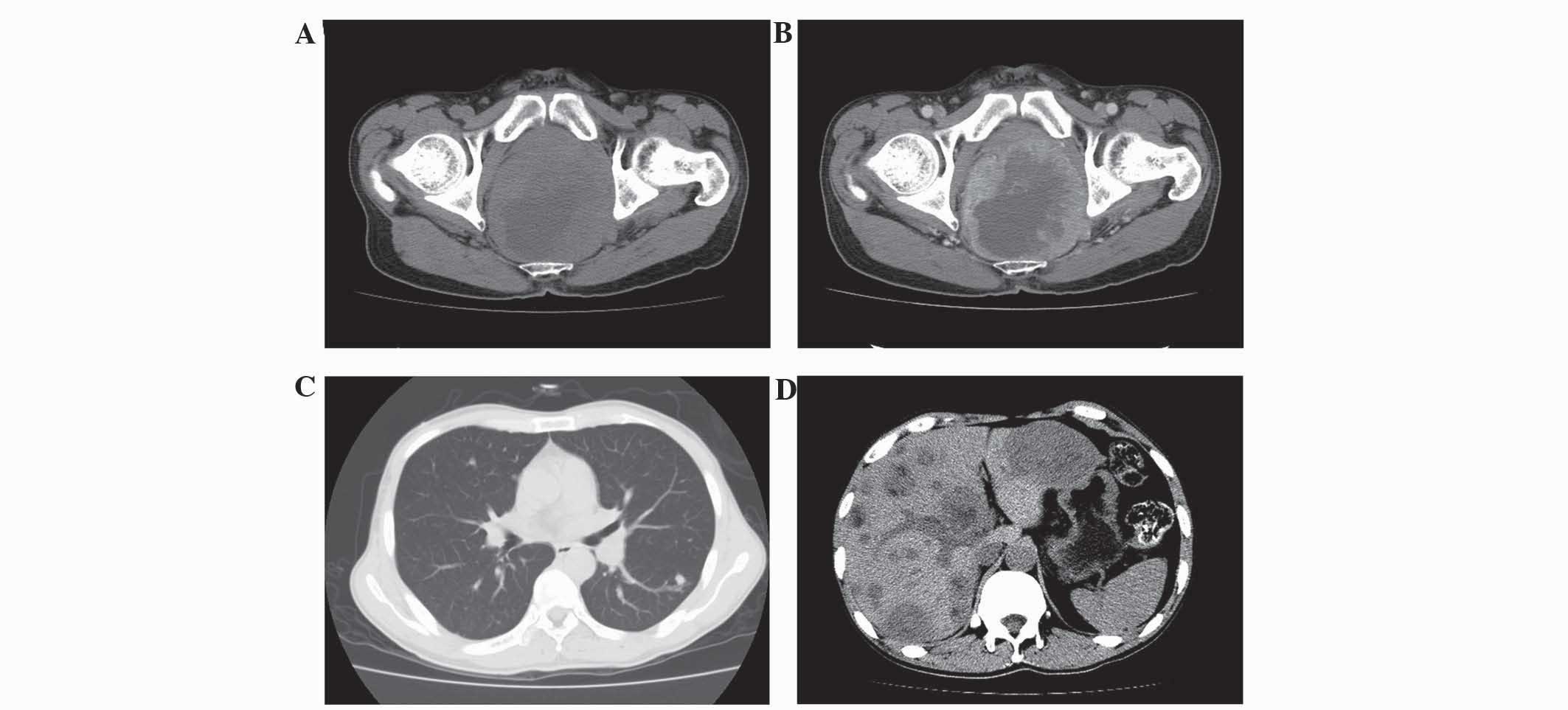
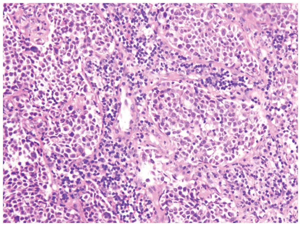
Small cell carcinoma of the rectum is extremely rare. The present study aimed to improve the recognition of computed tomography (CT) and magnetic resonance imaging (MRI) features observed in patients with small cell carcinoma of the rectum. A total of 4 cases of patients with histologically confirmed small cell carcinoma of the rectum were retrospectively reviewed. The imaging features assessed consisted of the thickening of the rectal wall, local tumor invasion, CT density, MRI signal intensity, lesion texture, contrast enhancement characteristics, lymphadenopathy and distant metastasis. All 4 patients displayed ring‑like rectal wall thickening and hypodensity in comparison to the gluteus muscles on non‑enhanced CT images. MRI demonstrated isointensity on T1‑weighted images and hyperintensity on T2‑weighted images. Thickening of the left wall of the rectum with patchy low attenuation and the presence of an exophytic mass was observed in 1 patient. Local tumor invasion was observed in the perirectal fat spaces of 4 patients, in the prostate gland and seminal vesicle of 1 patient, and in the left seminal vesicle of 1 patient. Heterogeneous attenuation was exhibited on non‑enhanced CT images in 3 lesions, and 1 lesion exhibited heterogeneous intensity on non‑enhanced MRI images. All the lesions observed demonstrated strong enhancement following contrast imaging. Lymphadenopathy was observed in 4 patients, liver metastasis in 3 patients, pulmonary metastasis in 2 patients and multiple bone metastases in 1 patient. Overall, small cell carcinoma of the rectum appeared to be more likely to metastasize to the liver, pulmonary, lymph nodes and bone. Distinguishing features of small cell carcinoma of the rectum were noted as a thickening rectal wall, the presence of soft‑tissue tumors, local tumor invasion, lymphadenopathy and distant metastases. Pre-operative CT and MRI are required as an aid in selecting the correct treatment plan and for the prognosis assessment of patients.
View Figures
View References

Related Articles

Journal Cover

March-2016
Volume 11 Issue 3

Print ISSN: 1792-1074
Online ISSN:1792-1082

Sign up for eToc alerts

Recommend to Library

Copy and paste a formatted citation
x
Spandidos Publications style
Zhou J, Shen J, Ma H, Zhang Y, Sun M, Zheng L and Lv Y: Small cell carcinoma of the rectum: A report of imaging results from four cases. Oncol Lett 11: 1671-1676, 2016
APA
Zhou, J., Shen, J., Ma, H., Zhang, Y., Sun, M., Zheng, L., & Lv, Y. (2016). Small cell carcinoma of the rectum: A report of imaging results from four cases. Oncology Letters, 11, 1671-1676. https://doi.org/10.3892/ol.2016.4136
MLA
Zhou, J., Shen, J., Ma, H., Zhang, Y., Sun, M., Zheng, L., Lv, Y."Small cell carcinoma of the rectum: A report of imaging results from four cases". Oncology Letters 11.3 (2016): 1671-1676.
Chicago
Zhou, J., Shen, J., Ma, H., Zhang, Y., Sun, M., Zheng, L., Lv, Y."Small cell carcinoma of the rectum: A report of imaging results from four cases". Oncology Letters 11, no. 3 (2016): 1671-1676. https://doi.org/10.3892/ol.2016.4136