Introduction
Anaplastic lymphoma kinase (ALK) gene
rearrangement is a driving mutation underlying the development of
non-small cell lung cancer (NSCLC) (1,2). The most
commonly observed type of ALK rearrangement is a fusion with
echinoderm microtubule associated protein like 4 (EML4) as a
result of the inversion of chromosome 2 (1,2). A total
of 5–7% of NSCLC tumors harbor EML4-ALK fusion (3). In addition to EML4, ALK
has been identified to fuse with other partner genes, including
nucleophosmin (NPM1), tropomyosin 3 and vinculin (4). Targeted therapeutic agents, including
crizotinib, have shown clinical efficacy in treating NSCLC patients
harboring EML4-ALK gene fusion (5). Furthermore, a previous study
demonstrated that crizotinib is additionally effective at treating
tumors harboring ALK fused with other partner genes,
including NPM1 (6).
A prerequisite for the effective use of targeted
therapy is the accurate detection of molecular targets in tumors.
For ALK rearrangement detection, the U.S. Food and Drug
Administration (FDA)-approved method for companion diagnostics is
fluorescence in situ hybridization (FISH), which measures
the physical integrity of the ALK gene in chromosome 2
(7). However, previous studies have
identified that FISH is prone to technical difficulties, which may
lead to non-interpretable results (8,9). In
addition, FISH is unable to detect genomic gain or overexpression
of the ALK gene (10). FISH is
also an expensive and time-consuming method, and requires a high
degree of technological expertise for its operation (11). Immunohistochemistry (IHC) is another
method commonly used clinically for ALK detection in tumor tissues
(8–10); it detects the levels of ALK protein,
rather than gene rearrangement, in tumor cells. Studies have
revealed that results obtained using IHC are highly concordant with
those obtained using FISH (8,9), and IHC is able to capture ALK-positive
tumors that are not detected using FISH (9,10).
However, IHC is a time-consuming method, its success is subject to
antibody quality and the experience of technologists, and the
interpretation of IHC results is subjective (12). Reverse transcription polymerase chain
reaction (RT-PCR) has been used for the detection of ALK
fusion (13–15). The shortcoming of RT-PCR is that
multiple reactions are typically required to cover various forms of
ALK fusions, thus requiring a relatively large quantity of
clinical samples (13). In order to
maximize the number of NSCLC patients able to benefit from
ALK-targeted therapy, there is a requirement for the development of
a simple yet comprehensive method for the detection of ALK
rearrangements.
In its rearranged forms, ALK typically breaks
between exons 19 and 20 or 20 and 21, leaving its kinase domain
intact, which is then able to fuse with a partner gene (4). Taking advantage of this feature, the
present study developed a simple technique, named the differential
expression method, to assess ALK fusion via measurement of
the relative expression levels of the kinase and non-kinase
domains. In addition, the present study developed a complementary
method, named the exon integrity method, for detection of the
integrity of exons 19–20 and 20–21 as an indicator of ALK
breaks at these loci.
Materials and methods
NSCLC cells and clinical samples
The NCI-H3122 human NSCLC cell line (ALK
fusion-positive) was provided by AstraZeneca Innovation Center
(Shanghai, China). The NCI-H2228 human NSCLC cell line (ALK
fusion-positive) and A549 cells (ALK fusion-negative) were
purchased from the American Type Culture Collection (Manassas, VA,
USA). The cells were cultured in Dulbecco's modified Eagle's medium
(Hyclone; GE Healthcare Life Sciences, Logan, UT, USA) supplemented
with 10% fetal bovine serum(Ausgenex Pty Ltd., Molindar,
Australia), 100 U/ml penicillin and 100 U/ml streptomycin (GE
Healthcare Life Sciences) at 37°C in an atmosphere of 5%
CO2.
The use of clinical samples for the present study
was approved by the Ethics Committees of the First Affiliated
Hospital of Guangzhou Medical University (Guangzhou, China) and
Ningbo University School of Medicine (Ningbo, China) and informed
consent was received from all patients. Formalin-fixed,
paraffin-embedded (FFPE) tissues from 100 NSCLC patients were
originally used for screening of ALK expression using an anti-ALK
(D5F3) IHC assay (Ventana Medical Systems, Inc., Tucson, AZ, USA)
based on the manufacturer's protocols. Sections of the IHC-positive
samples were additionally analyzed using a Vysis LSI ALK Break
Apart Rearrangement FISH Probes system based on the manufacturer's
protocols (Abbott Molecular, Des Plaines, IL, USA). In addition,
two urine samples from NSCLC patients with ALK fusion
positivity detected using FISH were obtained and used for testing
the detection methods developed in the present study.
Differential expression method
design
The pathological consequence of ALK fusion in tumor
cells is the overexpression of the ALK kinase domain compared with
the non-kinase domain. Based on this fact, a differential
expression method was designed by measuring the expression levels
of the ALK kinase and non-kinase domains simultaneously, and using
the difference in expression levels as an indicator of ALK gene
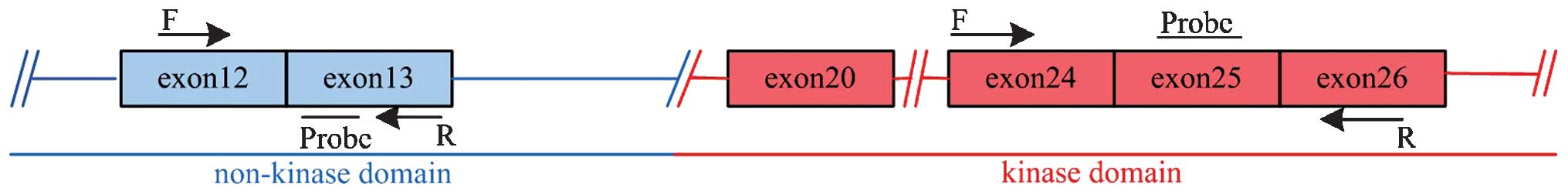
fusion. The ALK gene has 29 exons, with the kinase domain spanning
from exons 20–29 and the non-kinase domain spanning from exons 1–19
(1). Among a series of candidate
sequences, a sequence located between exons 24–26 was selected for
the kinase domain, and a sequence between exons 12–13 was selected
for the non-kinase domain. For the measurement of the two domains,
the forward and reverse primers were located in different exons,
and the probe was located between the primers (Fig. 1). The sequences of primers and probes
used are listed in Table I. The
transcript levels of the ALK kinase and non-kinase domains were
measured using TaqMan probe-based quantitative (q)PCR. The
quantification cycle (Cq) values of the non-kinase domain (NK) and
the kinase domain (K) were used for calculating the differential
expression levels of these two domains as D = 2ΔCq,
where ΔCq = Cq(NK) - Cq(K). When ΔCq was ≥1, this indicated that
the levels of ALK kinase domain were twice (when ΔCq=1, D=2) or
more than twice (when ΔCq>1, D>2) that of the non-kinase
domain, and the sample was deemed to be ALK fusion-positive.
 | Table I.Sequences of primers and probes used
in the differential expression and exon integrity method
experiments. |
Table I.
Sequences of primers and probes used
in the differential expression and exon integrity method
experiments.
| Method | Sequence (5′-3′) |
|---|
| Differential
expression |
|
| Kinase
domain | F:
GAAAACCACTTCATCCACCG |
|
| R:
TGGCACAGCCTCCCTTTCTAT |
|
| Probe:
FAM-CGTGCCAGAAACTGCCTCTTGAC-BHQ1 |
|
Non-kinase domain | F:
TGCCAGCCACCGACACCTA |
|
| R:
TTGAAGATGCCCAGCACAGA |
|
| Probe:
HEX-ATGGTGTTCTTCCCGCCTTTCCCG-BHQ1 |
| Exon integrity |
|
|
ALK exons 19–20 | F:
ACTCTCGCTGATCCTCTCTG |
|
| R:
TTGCTCAGCTTGTACTCAGG |
|
ALK exons 20–21 | F:
CCGACTACAACCCCAACTAC |
|
| R:
CCTTCATACACCTCCCCAAAG |
Exon integrity method design
The exon integrity method is based on the fact that
ALK fusion results from the break of the gene, thus destroying the
integrity of the ALK locus. ALK breakpoints are typically located
at exons 19–20 or 20–21 (1,2). By measuring the amplificability of exons
19–20 and 20–21 using qPCR, the integrity of the ALK gene may be
assessed and used as an indicator of ALK fusion. An identical
strategy is employed by FISH technology for assessing ALK fusion
(5). The sequences of primers
utilized are summarized in Table
I.
qPCR
Total RNA was isolated from cultured cells, FFPE
tissues or urine using UniGene total RNA isolation kits (UniGene
Bioscience Co., Ltd, Ningbo, China). For urine RNA isolation, 15 ml
urine was centrifuged for 15 min at 400 × g at 4°C to remove cells,
and subsequently mixed with an equal volume of 8 M guanidine
thiocyanate (Sigma-Aldrich, St. Louis, MO, USA) and loaded onto a
silica-based spin column (UniGene Bioscience Co., Ltd). Total RNA
was transcribed into complementary (c)DNA using a
reverse-transcription kit (UniGene Bioscience Co., Ltd). The levels
of complementary (c)DNA at exons 24–26 and 12–13 of ALK were
measured using qPCR systems (Roche Diagnostics, Shanghai, China)
with a Roche 480 II RT-PCR instrument (Roche Diagnostics). The
levels of cDNA at exons 19–20 and 20–21 of ALK were measured using
SYBR Green Master Mix (Roche Diagnostics). The qPCR reactions were
conducted as follows: 95°C for 10 min, amplification for 40 cycles
of 95°C for 15 sec (denaturation) and 61°C for 1 min (annealing and
extension). For each reaction, the levels of
glyceraldehyde-3-phosphate dehydrogenase transcripts were measured
as a control for the loading amount of cDNA.
Statistical analysis
Data were analyzed for statistical significance
using Student's t-test or Fisher's exact test as appropriate.
P<0.05 was considered to indicate a statistically significant
difference. All statistical analyses were performed using SPSS
software, version 18.0 (SPSS, Inc., Chicago, IL, USA).
Results
Differential expression method
demonstrates the ability to detect ALK fusion
Based on the pathological consequences of ALK
fusion, including aberrantly high expression levels, and the
activities of the ALK kinase domain driven by the high
transcription activity of the partner gene, a strategy for
assessing ALK fusion by measuring the differential expression
levels of the ALK kinase and non-kinase domains was designed. To
test this design, ALK fusion-positive H3122 and H2228 cells, and
ALK fusion-negative A549 cells were initially used to measure the
differential expression levels of the ALK kinase and non-kinase
domains. ALK gene break-apart occurs at exons 19–20 in H3122 and
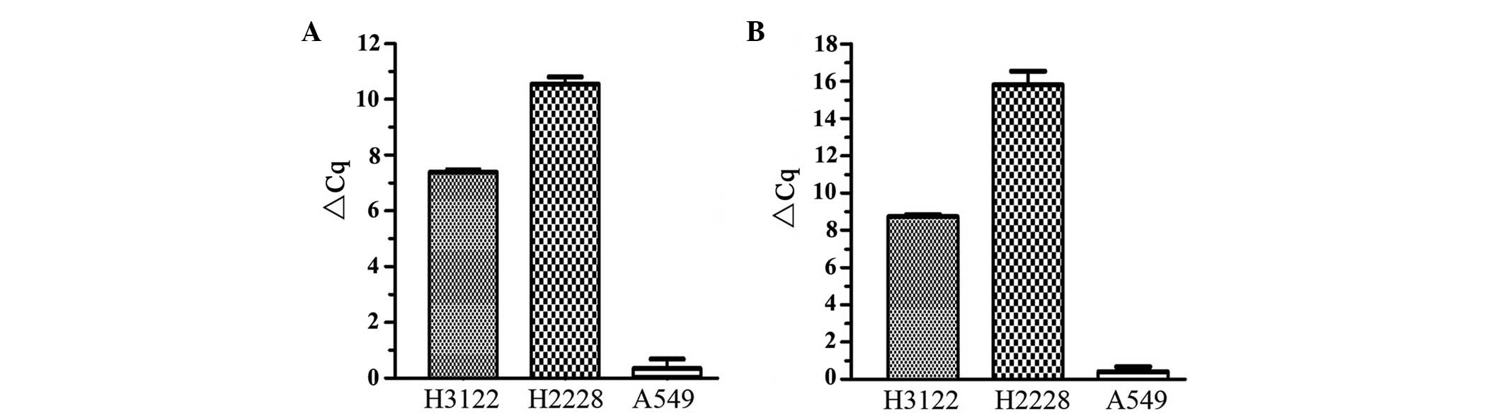
H2228 cells (12). The results of the
present study demonstrated that the average ΔCq for the H3122,
H2228 and A549 cells was 7.3, 10.3 and 0.5, respectively (Fig. 2A). Therefore, in the H3122 and H2228
cells, the levels of kinase domain transcripts were 158 times
(27.3) and 1,261 times (210.3) greater
compared with that of the non-kinase domain transcripts,
respectively. In A549 cells, there was no differential expression
observed between the kinase domain and domains of ALK, as ΔCq<1.
The effect of the input amount of cDNA was also investigated, and
it was observed that the ΔCq values were constant and independent
of input cDNA quantities (data not shown). The results of the
present study clearly demonstrated the ability of the differential
expression method to detect ALK fusion.
Exon integrity method reveals
amplification at exons 19–20 and 20–21
In addition to the differential expression method,
another method was developed to assess the integrity of exons 19–20
and 20–21, two loci that are frequently broken in ALK fusions, via
amplification of cDNA across these exons. The results showed that
for H3122 and H2228 cells, cDNA across exons 19–20 and 20–21 was
amplified. However, the levels of cDNA across exons 19–20 were 375
times (28.55) and 15,936 times (213.96) less
than that across exons 20–21 in the H3122 cells and H2228 cells,
respectively (Fig. 2B). For the A549
cells, transcripts across exons 19–20 and 20–21 were expressed at
low levels, and there were no differences between them, indicating
that there were no breakpoints at these two loci.
Differential expression and exon
integrity methods validated using clinical samples
Following the establishment of the methods using
H3122 and H2228 cells, the performance of these methods was tested
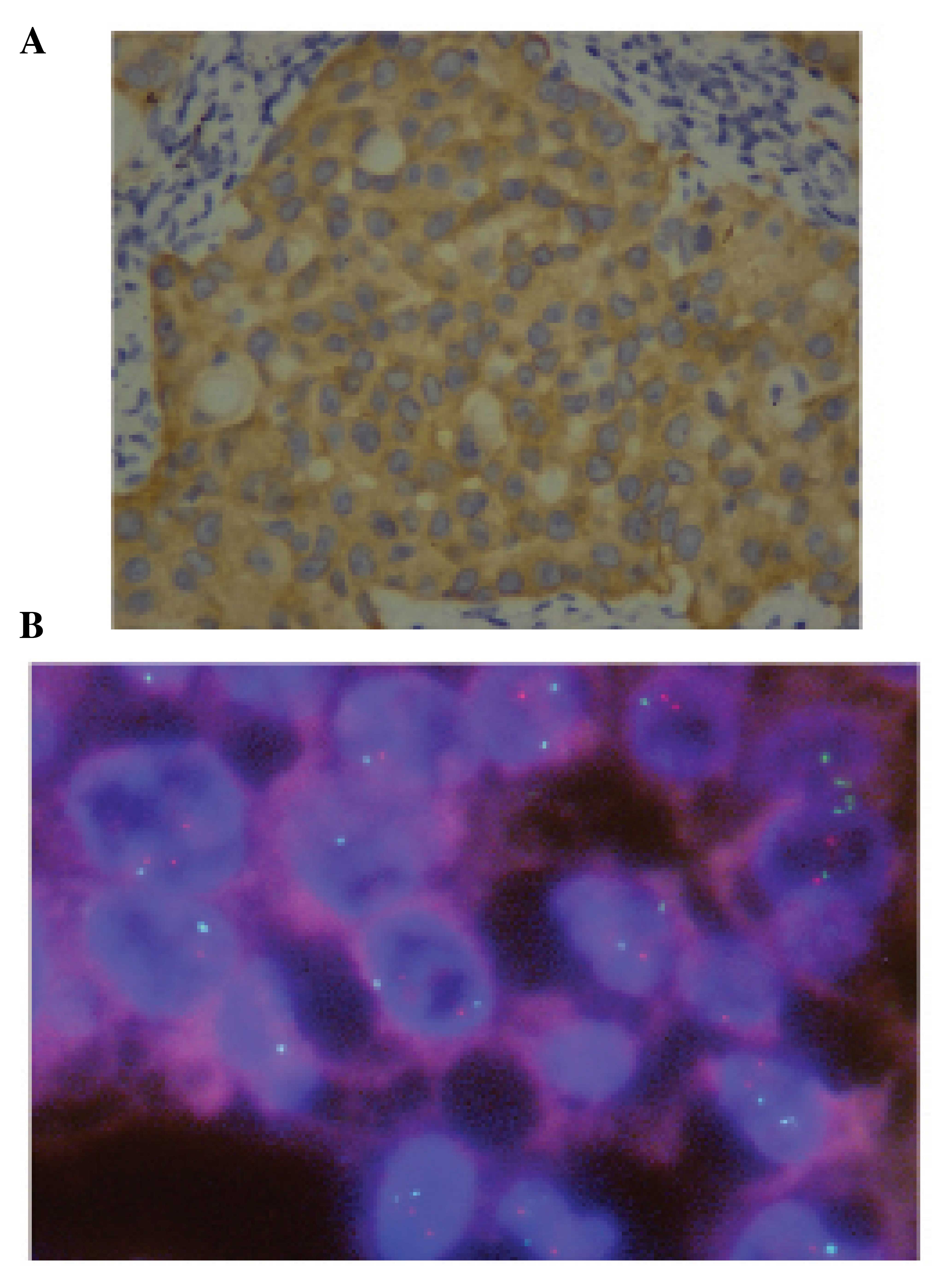
using 100 NSCLC tumor FFPE samples. Of these NSCLC samples, 10
(10%) were observed to be ALK-positive using the VENTANA ALK IHC
method (Fig. 3A). Of these 10 ALK
IHC-positive samples, 3 were further analyzed using FISH, and all
were identified to be FISH-positive (Fig.
3B).
The differential expression method was initially
used to detect ALK fusions in these NSCLC samples in a
double-blind manner. It was identified that the kinase domain was
successfully amplified in 10 samples (10%), while the non-kinase
domain was amplified in only 1 of these 10 samples. For the sample
with non-kinase domain amplification, the expression levels of
kinase domain were 9.19 times (23.2) greater compared
with that of the non-kinase domain. The results therefore indicated
that these 10 samples were ALK-fusion-positive. By checking
the identities of these samples, it was identified that the results
of the differential expression method were in 100% concordance with
the IHC results (Table II).
Subsequently, the exon integrity method was used to test the 10
positive samples, and it was identified that exon 20–21 sequences
were amplified in 8/10 samples; however, the exon 19–20 sequences
were not amplified (Table II).
| Table II.Performance of the differential
expression and exon integrity methods compared with IHC. |
Table II.
Performance of the differential
expression and exon integrity methods compared with IHC.
|
| IHC |
|---|
|
|
|
|---|
| Method | + | − |
|---|
| Differential
expression |
|
|
| + | 10 | 0 |
| − | 0 | 90 |
| Exon integrity |
|
|
| + | 8 | 0 |
| − | 2 | 90 |
The applicability of the methods for detection of
ALK fusion was additionally investigated in liquid samples.
Using cell-free urine samples collected from two NSCLC patients
whose tumors were ALK fusion-positive as detected by FISH,
the differential expression method successfully detected the
amplification of the kinase domain, but not the non-kinase domain
of ALK (Table III). The cDNA
from H3122 cells served as a positive control, and cDNA from
cell-free urine collected from two healthy women served as a
negative control.
| Table III.Detection of anaplastic lymphoma
kinase fusion in urine from non-small cell lung cancer patients
using the differential expression method. |
Table III.
Detection of anaplastic lymphoma
kinase fusion in urine from non-small cell lung cancer patients
using the differential expression method.
|
| Differential
expression method |
|---|
|
|
|
|---|
| Sample | Kinase domain | Non-kinase
domain |
|---|
| FISH+
sample 1 | 33.35 | – |
| FISH+
sample 2 | 30.13 | – |
| Healthy control
1 | – | – |
| Healthy control
2 | – | – |
| NCI-H3122 | 24.53 | 31.52 |
| H2O | – | – |
Discussion
Accurate and convenient methods for detecting drug
targets in tumor cells are critical for the implementation of
targeted therapies for the treatment of cancer. ALK fusion
with EML4 or alternative partner genes has been recognized
as an effective target for ALK inhibitors, including crizotinib,
for the treatment of NSCLC (16). As
a companion diagnostics approach approved by the FDA, FISH-based
methods detect ALK fusion in tumor cells by determining the
physical integrity of 5′ and 3′ portions of the ALK gene.
FISH-based methods have an advantage in that the general morphology
of tumor cells and the physical integrity of chromosome 2 may be
observed simultaneously. However, FISH methods are expensive,
require a high degree of technological expertise and frequently
produce non-interpretable results (7,8). By
contrast, IHC is able to detect the levels of ALK protein, and
several commercial systems with specific monoclonal antibodies
against ALK protein are available (7,8). However,
the protocol for IHC is complex and time-consuming, and final
results of IHC are subject to antibody quality and pathologist
experience (12).
Considering the limitations of FISH and IHC, two
transcript-based methods were developed for assessment of
ALK fusion in the present study. The differential expression
method was based on the pathological consequences of ALK
gene fusion, which are aberrantly high levels of the ALK kinase
domain. The exon integrity method was based on the break of the
gene locus when ALK fusion occurs, a strategy similar to the
FISH method. The results of the present study clearly demonstrated
the applicability of these two methods for the assessment of
ALK fusion. Compared with FISH and IHC, the methods used in
the present study were easier to perform and cheaper in cost.
Furthermore, the differential expression method directly measured
the levels of the ALK kinase domain in comparison with its
non-kinase domain, which gave it an advantage over FISH, which only
detects the physical integrity of chromosome 2. Similar to the
differential expression method, IHC detects the total levels of ALK
protein in tumor tissues, rather than the fusion of ALK (17). Factors leading to increases in the
protein levels of ALK include: Gene fusion, gene amplification,
genomic gain or regulations at transcriptional or
post-transcriptional levels (18).
Theoretically, ALK inhibitors would be effective in treating tumors
exhibiting high levels of ALK regardless of the causes leading to
the overexpression of this kinase. However, there is little
information regarding the comparison of performances between IHC
and FISH in terms of their power for predicting patient
responsiveness to ALK inhibitors.
Although tumor tissues are the optimum sample for
gene mutation detection, it is frequently difficult to collect a
sufficient amount of tissue samples. In certain cases, it may be
impossible to obtain tumor tissue samples. Therefore, liquid biopsy
represents a positive source for the assessment of tumor-associated
gene mutations. Liquid samples, including plasma, have been used
for the detection of mutations in epidermal growth factor receptor,
BRAF and Kirsten rat sarcoma viral oncogene homolog in NSCLC
patients (19). In the present study,
using the differential expression method, ALK fusion was
successfully detected in cell-free urine samples. To the best of
our knowledge, this is the first report indicating that urine can
be used for the assessment of ALK fusion in NSCLC patients.
Therefore, the method devised in the present study would provide a
novel non-invasive approach for the assessment of driving
mutations, including ALK fusion, and for the monitoring of
disease progression in NSCLC patients.
It should be noted that although the methods
utilized in the present study were validated using 100 NSCLC tumor
samples, these NSCLC patients did not receive ALK inhibitor in
their treatment regimens. To confirm the usefulness of the methods
developed in the present study for the prediction of patient
responsiveness to ALK inhibitors, validation of these methods is
required using samples from NSCLC patients receiving crizotinib
treatment.
In conclusion, the present study developed two
methods for the assessment of ALK fusion in NSCLC tumor and
urine samples. These methods would provide simple yet comprehensive
approaches to clinical practice for the selection of patients that
may benefit from treatment with ALK inhibitors, and the monitoring
of treatment efficacy and disease progression.
Acknowledgements
The present study was supported by the Science and
Technology Plan of Zhejiang Province of China (grant no.
2013C33165) and the National Natural Science Fund for Distinguished
Young Scholars (grant no. 81302000).
References
|
1
|
Soda M, Choi YL, Enomoto M, Takada S,
Yamashita Y, Ishikawa S, Fujiwara S, Watanabe H, Kurashina K,
Hatanaka H, et al: Identification of the transforming EML4-ALK
fusion gene in non-small-cell lung cancer. Nature. 448:561–566.
2007. View Article : Google Scholar : PubMed/NCBI
|
|
2
|
Takeuchi K, Choi YL, Soda M, Inamura K,
Togashi Y, Hatano S, Enomoto M, Takada S, Yamashita Y, Satoh Y, et
al: Multiplex reverse transcription-PCR screening for EML4-ALK
fusion transcripts. Clin Cancer Res. 14:6618–6624. 2008. View Article : Google Scholar : PubMed/NCBI
|
|
3
|
Zhou JX, Yang H, Deng Q, Gu X, He P, Lin
Y, Zhao M, Jiang J, Chen H, Lin Y, et al: Oncogenic driver
mutations in patients with non-small-cell lung cancer at various
clinical stages. Ann Oncol. 24:1319–1325. 2013. View Article : Google Scholar : PubMed/NCBI
|
|
4
|
Mano H: ALKoma: A cancer subtype with a
shared target. Cancer Discov. 2:495–502. 2012. View Article : Google Scholar : PubMed/NCBI
|
|
5
|
Kwak EL, Bang YJ, Camidge DR, Shaw AT,
Solomon B, Maki RG, Ou SH, Dezube BJ, Jänne PA, Costa DB, et al:
Anaplastic lymphoma kinase inhibition in non-small-cell lung
cancer. N Engl J Med. 363:1693–1703. 2010. View Article : Google Scholar : PubMed/NCBI
|
|
6
|
Mossé YP, Lim MS, Voss SD, Wilner K,
Ruffner K, Laliberte J, Rolland D, Balis FM, Maris JM, Weigel BJ,
et al: Safety and activity of crizotinib for paediatric patients
with refractory solid tumours or anaplastic large-cell lymphoma: A
Children's Oncology Group phase 1 consortium study. Lancet Oncol.
14:472–480. 2013. View Article : Google Scholar : PubMed/NCBI
|
|
7
|
Di Maio M, De Marinis F, Hirsch FR and
Gridelli C: Diagnostic and therapeutic issues for patients with
advanced non-small cell lung cancer harboring anaplastic lymphoma
kinase rearrangement: European vs. US perspective (Review). Int J
Oncol. 45:509–515. 2014.PubMed/NCBI
|
|
8
|
von Laffert M, Warth A, Penzel R,
Schirmacher P, Kerr KM, Elmberger G, Schildhaus HU, Büttner R,
Lopez-Rios F, Reu S, et al: Multicenter immunohistochemical
ALK-testing of non-small-cell lung cancer shows high concordance
after harmonization of techniques and interpretation criteria. J
Thorac Oncol. 9:1685–1692. 2014. View Article : Google Scholar : PubMed/NCBI
|
|
9
|
McLeer-Florin A, Moro-Sibilot D, Melis A,
Salameire D, Lefebvre C, Ceccaldi F, de Fraipont F, Brambilla E and
Lantuejoul S: Dual IHC and FISH testing for ALK gene rearrangement
in lung adenocarcinomas in a routine practice: A French study. J
Thorac Oncol. 7:348–354. 2012. View Article : Google Scholar : PubMed/NCBI
|
|
10
|
Ilie MI, Bence C, Hofman V, Long-Mira E,
Butori C, Bouhlel L, Lalvée S, Mouroux J, Poudenx M, Otto J, et al:
Discrepancies between FISH and immunohistochemistry for assessment
of the ALK status are associated with ALK ‘borderline’-positive
rearrangements or a high copy number: A potential major issue for
anti-ALK therapeutic strategies. Ann Oncol. 26:238–244. 2015.
View Article : Google Scholar : PubMed/NCBI
|
|
11
|
Ali G, Proietti A, Pelliccioni S, Niccoli
C, Lupi C, Sensi E, Giannini R, Borrelli N, Menghi M, Chella A, et
al: ALK rearrangement in a large series of consecutive non-small
cell lung cancers: Comparison between a new immunohistochemical
approach and fluorescence in situ hybridization for the screening
of patients eligible for crizotinib treatment. Arch Pathol Lab Med.
138:1449–1458. 2014. View Article : Google Scholar : PubMed/NCBI
|
|
12
|
Wong DW, Leung EL, So KK, Tam IY, Sihoe
AD, Cheng LC, Ho KK, Au JS, Chung LP and Pik WM: The EML4-ALK
fusion gene is involved in various histologic types of lung cancers
from nonsmokerswith wild-type EGFR and KRAS. Cancer. 115:1723–1733.
2009. View Article : Google Scholar : PubMed/NCBI
|
|
13
|
Soda M, Isobe K, Inoue A, Maemondo M,
Oizumi S, Fujita Y, Gemma A, Yamashita Y, Ueno T, Takeuchi K, et
al: North-East Japan Study Group; ALK Lung Cancer Study Group: A
prospective PCR-based screening for the EML4-ALK oncogene in
non-small cell lung cancer. Clin Cancer Res. 18:5682–5689. 2012.
View Article : Google Scholar : PubMed/NCBI
|
|
14
|
Pan Y, Zhang Y, Li Y, Hu H, Wang L, Li H,
Wang R, Ye T, Luo X, Zhang Y, et al: ALK, ROS1 and RET fusions in
1139 lung adenocarcinomas: A comprehensive study of common and
fusion pattern-specific clinicopathologic, histologic and cytologic
features. Lung Cancer. 84:121–126. 2014. View Article : Google Scholar : PubMed/NCBI
|
|
15
|
Koivunen JP, Mermel C, Zejnullahu K,
Murphy C, Lifshits E, Holmes AJ, Choi HG, Kim J, Chiang D, Thomas
R, et al: EML4-ALK fusion gene and efficacy of an ALK kinase
inhibitor in lung cancer. Clin Cancer Res. 14:4275–4283. 2008.
View Article : Google Scholar : PubMed/NCBI
|
|
16
|
Rolfo C, Passiglia F, Castiglia M, Raez
LE, Germonpre P, Gil-Bazo I, Zwaenepoel K, De Wilde A, Bronte G,
Russo A, et al: ALK and crizotinib: After the honeymoon…what else?
Resistance mechanisms and new therapies to overcome it. Transl Lung
Cancer Res. 3:250–261. 2014.PubMed/NCBI
|
|
17
|
Teixidó C, Karachaliou N, Peg V,
Gimenez-Capitan A and Rosell R: Concordance of IHC, FISH and RT-PCR
for EML4-ALK rearrangements. Transl Lung Cancer Res. 3:70–74.
2014.PubMed/NCBI
|
|
18
|
Weickhardt AJ, Aisner DL, Franklin WA,
Varella-Garcia M, Doebele RC and Camidge DR: Diagnostic assays for
identification of anaplastic lymphoma kinase-positive non-small
cell lung cancer. Cancer. 119:1467–1477. 2013. View Article : Google Scholar : PubMed/NCBI
|
|
19
|
Oxnard GR, Paweletz CP, Kuang Y, Mach SL,
O'Connell A, Messineo MM, Luke JJ, Butaney M, Kirschmeier P,
Jackman DM and Jänne PA: Noninvasive detection of response and
resistance in EGFR-mutant lung cancer using quantitative
next-generation genotyping of cell-free plasma DNA. Clin Cancer
Res. 20:1698–1705. 2014. View Article : Google Scholar : PubMed/NCBI
|