Introduction
Castleman's disease (CD) is a relatively rare
lymphoproliferative disease, characterized by enlarged hyperplastic
lymph node(s) (1). The disease was
initially described by Castleman and Towne in 1954 (2). The disease most commonly develops in the
mediastinum, and the cervical region is the second most common
location. Additional common nonmediastinal sites are the pelvic
cavity, axilla and retroperitoneum (3,4).
Clinically, CD may manifest as localized disease (unicentric) or
widespread disease (multicentric) (3). The unicentric type of CD typically
presents as an asymptomatic singular enlarged lymph node. By
contrast, the multicentric type of CD is associated with systemic
symptoms (3). CD is also classified
according to histopathological type into hyaline vascular, plasma
cell or mixed cell type (5). The
hyaline vascular type accounts for 90% of all CD cases, and is most
common in 30–40-year-old females. The plasma cell type accounts for
~10% of cases, and typically occurs in patients of 50–60 years of
age (6). At present there is no
standard protocol for predicting the prognosis and effectively
managing CD. The present study reports a rare case of unicentric
CD, which presented as a retroperitoneal mass and was located in a
peripancreatic location, in an asymptomatic 36-year-old male
patient.
Case report
An asymptomatic 36-year-old man with an unremarkable
past medical history received a routine physical examination in
Yiqiao Town Central Hospital (Hangzhou, China) in March 2014, and
was indicated to have an abdominal mass upon ultrasonography and
computed tomography (CT) scans. The patient was then referred to
the First Affiliated Hospital, School of Medicine, Zhejiang
University (Hangzhou, China).
A physical examination at the First Affiliated
Hospital did not reveal any abnormal results, and the patient's
full blood count and biochemical profile were normal. The result of
the human immunodeficiency virus (HIV) screening test was
negative.
An abdominal ultrasound (MyLab™Twice; Esaote Europe
BV, Cambridge, UK) revealed a 67×55-mm hypoechoic mass in the
region between the first liver segment and the pancreas, which
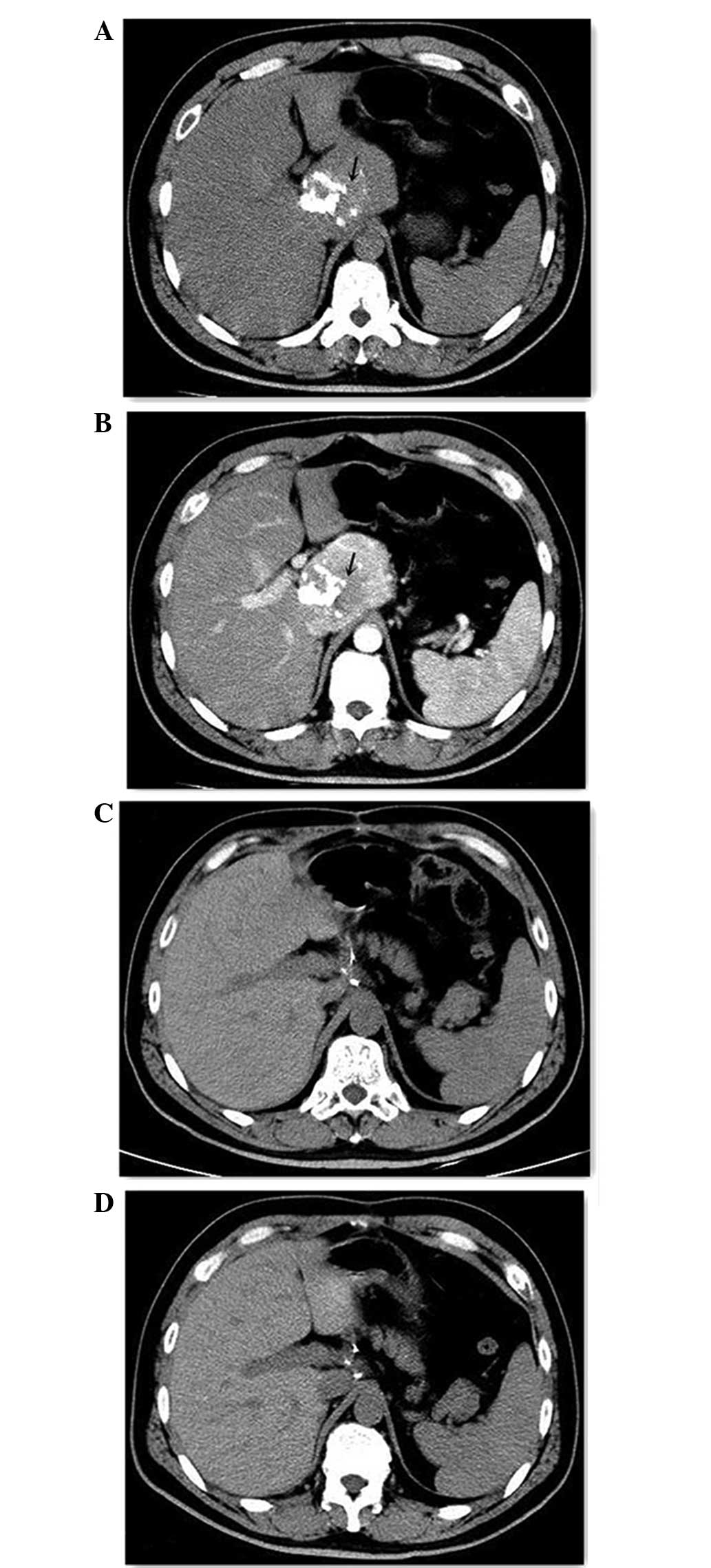
possessed calcification on the inside. The abdominal
contrast-enhanced CT scan (Brilliance iCT; Philips Healthcare,
Andover, MA, USA) reveled a 70×72-mm mass on the border of the
caudate lobe. The non-enhanced phase revealed patchy calcification
inside the mass (Fig. 1A), and
evident contrast enhancement was observed in the mass during the
arterial phase (Fig. 1B). The CT scan
also revealed possible infiltration into the head and neck of the
pancreas, indicating a solid pseudopapillary tumor of the
pancreas.
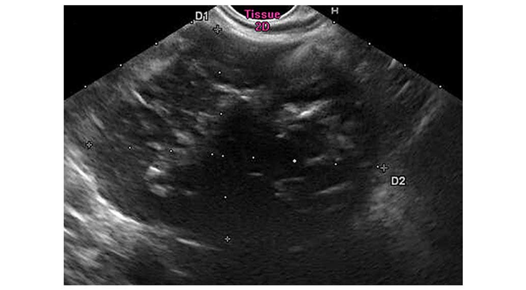
The endosonography revealed a 71×51-mm
retroperitoneal mass infiltrating into the pancreas hook,
indicating neuroendocrine carcinoma originating from the pancreas.
An endosonography-guided fine-needle aspiration biopsy revealed
chronic inflammation and lymphadenosis (Fig. 2).
On the basis of these test results, the patient
underwent an exploratory laparotomy. This procedure revealed a
well-defined mass densely adherent to the celiac trunk, inferior
vena cava and superior border of the pancreas. The mass was excised
from the adjacent organs successfully, without any
complications.
The excised mass was sectioned as follows: The
paraffin-embedded tissue was sectioned continuously into slices of
4 µm thickness using a Leica CM1850 freezing microtome (Leica
Biosystems, Wetzlar, Germany) and subsequently transferred onto the
surface of prepared glass slides (Thermo Fisher Scientific, Inc.,
Waltham, MA, USA).
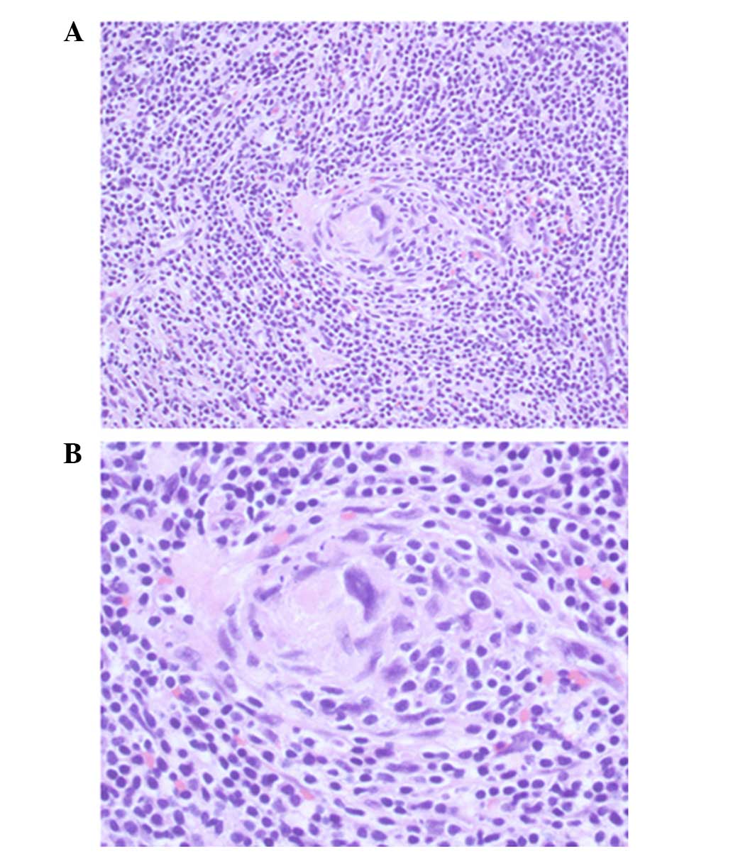
The histological examination revealed a mass
consisting of lymphoid tissue with a large amount of vascular
invasion, and laminated mantle zones with concentric rings of small
lymphocytes surrounding small atrophic germinal centers. A
hyalinized interstitium was observed between follicles (Fig. 3).
Immunohistochemistry for the biomarkers was
performed manually. Briefly, antigen retrieval was performed using
citrate buffer (pH 6.0; Sangon Biotech Co., Ltd., Shanghai, China).
Sections were subsequently incubated with the following antibodies:
Mouse monoclonal anti-human cluster of differentiation 20 (clone
7D1; dilution, 1:200; catalog no., NCL-CD20-7D1; Leica Biosystems),
mouse monoclonal anti-human cluster of differentiation 3 (clone
LN10; dilution, 1:100; catalog no., NCL-L-CD3-565; Leica
Biosystems), mouse monoclonal anti-human cluster of differentiation
21 (clone 2G9; dilution, 1:60; catalog no., NCL-CD21-2G9; Leica
Biosystems) and rabbit anti-human immunoglobulin G cluster of
differentiation 23 (clone RM104; dilution, 1:100; catalog no.,
RM-9123-S0; Thermo Fisher Scientific, Inc.), in a humidified
chamber for 1 h at 37°C. Sections were then incubated with
horseradish peroxidase-conjugated goat anti-mouse secondary
antibody (part of the EnVision+ System-HRP kit; ready to use;
catalog no., K4007; Dako, Glostrup, Denmark). Following incubation,
3,3′-diaminobenzidine (part of the EnVision+ System-HRP kit) was
applied for ~2 min. Slides were subsequently stained with
hematoxylin (Sangon Biotech Co., Ltd., Shanghai, China). A negative
control was designed by using phosphate-buffered saline (Sangon
Biotech Co., Ltd., Shanghai, China) rather than primary antibody.
The immunohistochemical analysis revealed the expression of cluster
of differentiation 3, 20, 21 and 23 subsequent to staining. Due to
these findings, the patient was diagnosed with CD of the hyaline
vascular type. The patient was followed up and is currently free of
disease 20 months subsequent to resection (Fig. 1C and D).
Discussion
CD exhibits a low incidence rate worldwide.
Histologically, there are 3 types of CD: The hyaline vascular type,
the plasma cell type and the mixed type (5,7).
Approximately 90% of cases are the hyaline vascular type (8). Clinically, according to the range of
lymph nodes involved, CD may also be divided into the unicentric
and multicentric types (9). The
hyaline vascular and plasma cell types have often been recognized
as being associated with the unicentric and multicentric
presentations, respectively (10,11).
The clinical presentations of CD vary greatly
between the unicentric and multicentric types. Patients with
unicentric disease are usually asymptomatic or may present with
lymph node swelling. Cases of the unicentric type usually have a
good prognosis and are treated with surgical excision (3). By contrast, constitutional symptoms,
including fatigue, fever, sweat, weight loss, arthralgia,
hepatomegaly, splenomegaly and systemic lymph node swelling, are
often observed in patients with multicentric disease (5,12). The
presentations of the multicentric type are occasionally accompanied
by symptoms of POEMS, including polyneuritis, organomegaly,
endocrinopathy, M protein and skin changes (13,14).
Patients with the multicentric type tend to possess an unfavorable
prognosis, and a number of cases eventually develop lymphomas
(15–17).
The etiology of CD remains unclear and has been
described in association with a reactive lymphoid hyperplasia
initiated by viral infections or a developmental growth disturbance
of the lymphoid tissue (1,18). Certain studies have indicated that CD
is associated with human herpes virus 8 (HHV-8) and HIV infection
(19,20). These viruses may lead to the
oversecretion of inflammatory mediators, particularly interleukin-6
(21,22), which results in the induction of a
hyperplasic reaction of the lymphoid system.
The initial challenge in retroperitoneal CD remains
in the establishment of a diagnosis. However, CD is often
overlooked as a possible diagnosis due to its low incidence.
Imaging tests have been shown to aid diagnosis. The possibility of
CD should be considered following the identification of a
homogeneous vascular mass with contrast kinetics that follow the
surrounding large arteries along a lymph node chain (23). CD is ultimately diagnosed by
histology, which requires either a resection or biopsy of the
lesion in order to be definitive. A number of previous cases had
diagnosed abdominal unicentric CD following post-surgical
histological examination. The preoperative diagnoses in these cases
were pheochromocytoma (6), malignant
tumor (24) and unknown mass
(25,26). The majority of CD cases receive
immediate resection, however, Bo et al (27) performed an initial exploratory
laparotomy prior to excision. Attention to the patient's history
and imaging results may assist with the diagnosis of CD (23). In the present study, a fine-needle
aspiration biopsy was not a useful diagnostic tool for CD, which is
similar to the findings of other studies (28–30). This
issue may be attributed to the fact that the histological diagnosis
of CD is based on cell architecture (30). Therefore, similar to previous cases
(3,8,23), a
surgical resection was used for the diagnosis and treatment of the
present patient. To the best of our knowledge, this strategy was
rational for the present case.
In conclusion, the rare case of a patient with a
retroperitoneal unicentric CD of hyaline vascular type was
encountered during a routine physical examination. A well-defined
mass densely adherent to the celiac trunk, inferior vena cava and
superior border of the pancreas was identified. Overall, CD has a
good prognosis following the surgical removal of the lesion. The
possible diagnosis of unicentric CD should be considered in the
presence of a solid hypervascular retroperitoneal mass. In cases
where a unicentric type of CD is suspected, a complete surgical
resection may be used to successfully treat the patient without an
unnecessarily extensive resection.
Glossary
Abbreviations
Abbreviations:
|
CD
|
Castleman's disease
|
|
HHV-8
|
human herpes virus 8 infection
|
|
HIV
|
human immunodeficiency virus
|
|
CT
|
computed tomography
|
References
|
1
|
El-Osta HE and Kurzrock R: Castleman's
disease: From basic mechanisms to molecular therapeutics.
Oncologist. 16:497–511. 2011. View Article : Google Scholar : PubMed/NCBI
|
|
2
|
Castleman B and Towne VW: Case records of
the Massachusetts General Hospital; weekly clinicopathological
exercises; founded by Richard C. Cabot. N Engl J Med. 251:396–400.
1954.PubMed/NCBI
|
|
3
|
Bucher P, Chassot G, Zufferey G, Ris F,
Huber O and Morel P: Surgical management of abdominal and
retroperitoneal Castleman's disease. World J Surg Oncol. 3:332005.
View Article : Google Scholar : PubMed/NCBI
|
|
4
|
Fu L, Wang XL, Babu SR, Zhang Y, Su AP,
Wang ZL, Hu T and Tian BL: Pancreatic Castleman's disease: Studies
of three cases and a cumulative review of the literature. Indian J
Surg. 75:34–38. 2013. View Article : Google Scholar : PubMed/NCBI
|
|
5
|
Keller AR, Hochholzer L and Castleman B:
Hyaline-vascular and plasma-cell types of giant lymph node
hyperplasia of the mediastinum and other locations. Cancer.
29:670–683. 1972. View Article : Google Scholar : PubMed/NCBI
|
|
6
|
Zhang J, Song N, Liu B, Hua L, Wang Z, Yin
C and Zhang W: A case report of unusual retroperitoneal Castleman's
disease in an old woman. Urol Int. 89:369–372. 2012. View Article : Google Scholar : PubMed/NCBI
|
|
7
|
Flendrig JA and Schillings PHM: Benign
giant lymphoma: The clinical signs and symptoms. Folia Med Neerl.
12:119–120. 1969.
|
|
8
|
Seco JL, Velasco F, Manuel JS, Serrano SR,
Tomas L and Velasco A: Retroperitoneal Castleman's disease.
Surgery. 112:850–855. 1992.PubMed/NCBI
|
|
9
|
Gaba AR, Stein RS, Sweet DL and Variakojis
D: Multicentric giant lymph node hyperplasia. Am J Clin Pathol.
69:86–90. 1978.PubMed/NCBI
|
|
10
|
Frizzera G, Banks PM, Massarelli G and
Rosai J: A systemic lymphoproliferative disorder with morphologic
features of Castleman's disease. Pathological findings in 15
patients. Am J Surg Pathol. 7:211–231. 1983. View Article : Google Scholar : PubMed/NCBI
|
|
11
|
Talat N and Schulte KM: Castleman's
disease: Systematic analysis of 416 patients from the literature.
Oncologist. 16:1316–1324. 2011. View Article : Google Scholar : PubMed/NCBI
|
|
12
|
Shahidi H, Myers JL and Kvale PA:
Castleman's disease. Mayo Clin Proc. 70:969–977. 1995. View Article : Google Scholar : PubMed/NCBI
|
|
13
|
Gracia-Ramos AE, del Cruz-Domínguez MP and
Vera-Lastra OL: Multicentric hyaline vascular Castleman's disease.
A POEMS type variant. Rev Med Inst Mex Seguro Soc. 51:464–467.
2013.(In Spanish). PubMed/NCBI
|
|
14
|
Andhavarapu S and Jiang L: POEMS syndrome
and Castleman disease. Blood. 122:1592013. View Article : Google Scholar : PubMed/NCBI
|
|
15
|
Franco V: Report of a case of localized
Castleman's disease with progression to malignant lymphoma. Am J
Clin Pathol. 100:707–708. 1993.PubMed/NCBI
|
|
16
|
Park J, Lee JE, Kim M, Lim J, Kim Y, Han
K, Park G, Jung YH, Roh SY and Hong YS: Discordant
lymphocyte-depleted classical Hodgkin's and peripheral T-cell
lymphoma arising in a patient 11 years after diagnosis of
multicentric Castleman's disease. Int J Hematol. 98:114–121. 2013.
View Article : Google Scholar : PubMed/NCBI
|
|
17
|
Rao S, Ramesh A, Rajkumar A, Arcot R and
Kuruvilla S: Persistent or recurrent Castleman's disease - Look out
for a lurking lymphoma! Indian J Med Paediatr Oncol. 32:162–164.
2011. View Article : Google Scholar : PubMed/NCBI
|
|
18
|
Ozkan H, Tolunay S, Gözü O and Ozer ZG:
Giant lymphoid hamartoma of mediastinum (Castleman's disease).
Thorac Cardiovasc Surg. 38:321–323. 1990. View Article : Google Scholar : PubMed/NCBI
|
|
19
|
Dossier A, Meignin V, Fieschi C, Boutboul
D, Oksenhendler E and Galicier L: Human herpes virus 8-related
Castleman disease in the absence of HIV infection. Clin Infect Dis.
56:833–842. 2013. View Article : Google Scholar : PubMed/NCBI
|
|
20
|
Oksenhendler E: HIV-associated
multicentric Castleman disease. Curr Opin HIV AIDS. 4:16–21. 2009.
View Article : Google Scholar : PubMed/NCBI
|
|
21
|
Yoshizaki K, Matsuda T, Nishimoto N,
Kuritani T, Taeho L, Aozasa K, Nakahata T, Kawai H, Tagoh H, Komori
T, et al: Pathogenic significance of interleukin-6 (IL-6/BSF-2) in
Castleman's disease. Blood. 74:1360–1367. 1989.PubMed/NCBI
|
|
22
|
Suthaus J, Stuhlmann-Laeisz C, Tompkins
VS, Rosean TR, Klapper W, Tosato G, Janz S, Scheller J and
Rose-John S: HHV-8-encoded viral IL-6 collaborates with mouse IL-6
in the development of multicentric Castleman disease in mice.
Blood. 119:5173–5181. 2012. View Article : Google Scholar : PubMed/NCBI
|
|
23
|
Poole PS, Chang EY and Santillan CS: Case:
172 Retroperitoneal Castleman disease (hyaline vascular type).
Radiology. 260:601–605. 2011. View Article : Google Scholar : PubMed/NCBI
|
|
24
|
Miyoshi H, Mimura S, Nomura T, Tani J,
Morishita A, Kobara H, Mori H, Yoneyama H, Deguchi A, Himoto T, et
al: A rare case of hyaline-type Castleman disease in the liver.
World J Hepatol. 5:404–408. 2013. View Article : Google Scholar : PubMed/NCBI
|
|
25
|
Otto M, Wieprzowski L, Dzwonkowski J and
Ziarkiewicz-Wróblewska B: Castleman's disease - an unusual
indication for laparoscopic adrenalectomy. Wideochir Inne Tech
Maloinwazyjne. 7:50–54. 2012.PubMed/NCBI
|
|
26
|
Kim MS, Ju JK and Kim Y: Surgical
management of unicentric castleman's disease in the abdomen. Ann
Coloproctol. 30:97–100. 2014. View Article : Google Scholar : PubMed/NCBI
|
|
27
|
Bo P, Junhua Z, Qiruo G and Hong L: A case
report of retroperitoneal Castleman disease. Can Urol Assoc J.
3:E14–E16. 2009.PubMed/NCBI
|
|
28
|
Park JB, Hwang JH, Kim H, Choe HS, Kim YK,
Kim HB and Bang SM: Castleman disease presenting with jaundice: A
case with the multicentric hyaline vascular variant. Korean J
Intern Med. 22:113–117. 2007. View Article : Google Scholar : PubMed/NCBI
|
|
29
|
Rhee KH, Lee SS and Huh JR: Endoscopic
ultrasonography-guided trucut biopsy for the preoperative diagnosis
of peripancreatic Castleman's disease: A case report. World J
Gastroenterol. 14:2115–2117. 2008. View Article : Google Scholar : PubMed/NCBI
|
|
30
|
Cecka F, Ferko A, Jon B, Subrt Z,
Kasparova P and Repak R: Pancreatic Castleman disease treated with
laparoscopic distal pancreatectomy. Hepatobiliary Pancreat Dis Int.
12:332–334. 2013. View Article : Google Scholar : PubMed/NCBI
|