Introduction
Hepatocellular carcinoma (HCC) is the sixth most
frequent cancer and the third most common cause of
cancer-associated mortality in the world (1). There are numerous risk factors
associated with the causes of liver disease, including hepatitis B
virus (HBV), hepatitis C virus (HCV), alcohol abuse (≥80 g/day),
iron overload and fatty liver, which results in HCC being
heterogeneous and complex (2–4). The prognosis of patients with HCC is
poor. Resection, liver transplantation and percutaneous treatment
may be curative for an early stage of tumor, which accounts for
≤30% of patients. For intermediate to advanced-stage HCC,
transarterial chemoembolization (TACE) is considered to be the
standard treatment by certain international guidelines (5). A meta-analysis reveals that TACE
improves survival in patients with unresectable HCC, who have been
selected as they have intermediate stage, multimodular and
Performance Status Test (PST) 0 or advanced stage, portal invasion,
lymph node 1, metastasis 1 and PST 1–2, and evidence obtained from
randomized controlled trials has confirmed the beneficial effect of
TACE in improving survival (6,7). However,
studies have also shown that not all patients with unresectable HCC
benefit from TACE. Therefore, it is crucial to differentiate
between patients that are most likely to benefit from TACE and
those that are not in a heterogeneous HCC population.
The pathogenesis of HCC is based on inflammation.
Particularly in China, the majority of HCC cases develop due to
underlying chronic HBV infection. Tumor inflammation and immunology
have previously been identified to enable cancer characteristics,
and increasing evidence supports the involvement of inflammation
and immunology in cancer progression and metastases (8,9). In
addition, the combination of hematological components of the
systemic inflammatory response have been shown to have prognostic
value in patients with a variety of cancers, including the Glasgow
prognostic score (GPS) (10–13), modified GPS (mGPS) (10–13),
neutrophil to lymphocyte ratio (NLR) (14–17),
prognostic nutritional index (PNI) (18), platelet-lymphocyte ratio (PLR)
(19) and prognostic index (PI)
(20). Of all these scores, the NLR
is the most inexpensive and easily obtained. Studies have
previously shown that an elevated NLR indicated a poor prognosis
for patients with HCC (14–17). However, in clinical trials, only the
white blood cell and neutrophil counts of the patients are commonly
entered into clinical trial databases. Therefore, Proctor et
al (21) recently implemented a
derived NLR (dNLR), which is composed of the neutrophil count and
the white blood cell count minus neutrophil count. Proctor et
al evaluated the prognostic value of dNLR on cancer outcome in
different cancer types and demonstrated that the dNLR had a similar
prognostic value to the well-established NLR, and dNLR was
suggested to be a cheaper and more easily determinable parameter
than NLR. However, the application of dNLR in HCC patients was not
fully validated. The present study was conducted to investigate the
prognostic value of the pre-treatment dNLR on overall survival (OS)
in patients with unresectable HCC undergoing TACE.
Patients and methods
Patients
Patients treated with TACE for unresectable HCC
between September 2009 and November 2011 at the Department of
Hepatobiliary Surgery of Sun Yat-sen University Cancer Canter
(Guangzhou, China) were identified using the prospective database
of the hospital. The present study was approved by the
Institutional Review Board of Sun Yat-sen University Cancer Center,
and written informed consent was obtained from all patients.
The diagnosis of HCC was based on the diagnostic
criteria for HCC used by the American Association for the Study of
the Liver guidelines (22). HCC was
diagnosed by at least two radiological images showing the
characteristic features of HCC, or one radiological image showing
characteristic features of HCC associated with elevated serum AFP
level (≥400 ng/ml) or histopathological evidence. Patients who met
all of the following criteria were included in analysis: i) No
previous treatment prior to TACE; ii) HBV-positive; iii) no HCV and
HIV expression; iv) liver function Child-Pugh grade A or B; and v)
a follow-up period ≥3 months.
All parameters were recorded and evaluated as
possible predictors of survival, such as the gender, age,
C-reactive protein (CRP) level, white blood cell count (WBC),
neutrophil count, lymphocyte count, platelet count (PLT),
α-fetoprotein (AFP) level, alkaline phosphatase (ALP) level, total
bilirubin level (TBIL), albumin (ALB) level, tumor size and number
and vascular invasion status of patients.
TACE procedure
TACE was performed using a previously reported
protocol (23). A selective 5-Fr
catheter (Terumo Corporation, Tokyo, Japan) was introduced into the
hepatic artery and visceral angiography was performed to assess the
arterial blood supply to the liver. Depending on the size, location
and arterial supply of the tumor, the tip of the catheter was
advanced into the right or left hepatic artery; if all the tumors
were fed by one enlarged independent hepatic artery branch, the tip
of catheter was introduced into this tumor-feeding artery. If the
conventional catheter could not enter the hepatic artery due to
technical reasons, a 2.9-Fr micro catheter (Terumo Corporation,
Tokyo, Japan) was used. Hepatic artery infusion chemotherapy was
performed using 300 mg carboplatin (Bristol-Myers Squibb, New York,
NY, USA). Subsequently, chemolipiodolization was performed using 50
mg epirubicin (Pfizer, Wuxi, Jiangsu, China), and 6 mg mitomycin C
(Zhejiang Hisun Pharmaceutical Co. Ltd., Taizhou, Zhejiang, China)
mixed with 5 ml lipiodol (Lipiodol Ultra-Fluide; Guerbet,
Villepinte, France). If the chemolipiodolized arterial territory
did not show stagnant flow, pure lipiodol was then injected. In
certain patients, stasis in a tumor-feeding artery was not
achieved, even subsequent to the injection of the maximum amount of
iodized oil (25 ml), due to the large size of the tumor.
Embolization was then performed in these patients with the
injection of absorbable gelatin sponge particles (Gelfoam; Hangzhou
ALC Ltd., Hangzhou, China), 1–2 mm in diameter, through the
angiographic catheter. This treatment regimen was used consistently
in the present study, regardless of tumor type and size.
Follow-up
Patients were followed carefully subsequent to
treatment. Patients underwent liver computed tomography (CT) scans
1 month subsequent to TACE, and liver CT scans were performed at
3-month intervals during the first 2 years, then every 6 months
thereafter, with physical examination, blood tests for the AFP
level and liver function. When metastasis was suspected, CT chest,
bone scintigraphy, positron emission tomography (PET) and biopsy,
if indicated, were also performed to confirm the presence of
metastasis. The end of follow-up was December 2013, which was the
time of the last follow-up, or the date of mortality.
Another session of TACE was performed every 4–10
weeks after the original administration of TACE until CT scans and
AFP levels indicated stabilization of the tumor, or until TACE was
not technically feasible, either due to hepatic artery occlusion or
impaired liver function. The OS time was defined as the interval
between the date of treatment and the date of mortality or last
follow-up of surviving patients. Causes of mortality were
determined from death certificates, medical interviews and
radiological findings.
Statistical analysis
The receiver operating characteristic (ROC) analysis
was used to determine the cut-off values of NLR and dNLR. The OS
was calculated using the Kaplan-Meier method and compared by the
log-rank test. The prognostic varieties in predicting the OS were
assessed by multivariate Cox proportional hazards regression
analysis. All covariates that affected survival at the P<0.10
level of significance in univariate analysis were included in the
multivariate Cox proportional hazards model. A ROC curve was also
generated and the area under the curve (AUC) was calculated to
evaluate the discriminatory ability. The association between the
NLR and dNLR was assessed by Spearman's rank correlation analysis.
Data are expressed as the mean ± standard deviation. All
statistical tests were two-sided, and P<0.05 was considered to
indicate a statistically significant difference. All statistical
analyses were performed using the SPSS 13.0 statistical software
(SPSS, Inc., Chicago, IL, USA).
Results
Baseline characteristics
A total of 279 consecutive patients that met the
inclusion criteria were included in present study. The baseline
characteristics of the patients are summarized in Table I. Overall, 251 patients were male
(90%) and 28 patients were female (10%), with a median age of 50
years (range, 23–80 years). The majority of the present patients
exhibited a good liver functional reserve, classified as Child-Pugh
grade A (92.5%).
 | Table I.Baseline characteristics and
univariate analysis for overall survival in 279 patients undergoing
transarterial chemoembolization for hepatocellular carcinoma. |
Table I.
Baseline characteristics and
univariate analysis for overall survival in 279 patients undergoing
transarterial chemoembolization for hepatocellular carcinoma.
| Variables | n | Value | Univariate analysis
P-value |
|---|
| Median age, years
(range) |
| 50 (23–80) |
0.049 |
|
<50 | 158 |
|
|
|
≥50 | 121 |
|
|
| Gender, n |
| NA |
0.111 |
|
Male | 251 |
|
|
|
Female | 28 |
|
|
| Mean WBC, n
x109/1 (range) |
| 6.6 (2.1–24.6) |
0.080 |
|
<9 | 36 |
|
|
| ≥9 | 243 |
|
|
| Mean neutrophil
count, n ×109/1 (range) |
| 4.2 (0.7–21.5) |
0.019 |
|
<7 | 28 |
|
|
| ≥7 | 251 |
|
|
| Mean lymphocyte
count, n ×109/1 (range) |
| 1.5 (0.3–4.8) |
0.166 |
|
<0.8 | 268 |
|
|
|
≥0.8 | 11 |
|
|
| Mean PLT count, n
×109/1 (range) |
| 182 (23–548) |
0.412 |
|
<100 | 47 |
|
|
|
≥100 | 232 |
|
|
| Mean ALT, µ/1
(range) |
| 56.6 (8–304) |
0.003 |
|
<40 | 169 |
|
|
|
≥40 | 110 |
|
|
| Mean AST, µ/1
(range) |
| 75.8
(19.3–472.6) | <0.001 |
|
<45 | 191 |
|
|
|
≥45 | 88 |
|
|
| Mean albumin, g/1
(range) |
| 39 (25–79) |
0.052 |
|
<35 | 238 |
|
|
|
≥35 | 41 |
|
|
| Mean total serum
bilirubin, µmol/1 (range) |
| 17.8
(4.8–222.9) | <0.001 |
|
<20 | 68 |
|
|
|
≥20 | 191 |
|
|
| Mean ALP, IU/1
(range) |
| 150 (13–761.5) | <0.001 |
|
<110 | 171 |
|
|
|
≥110 | 126 |
|
|
| Mean AFP, ng/ml
(range) |
| 751.2
(1.26–1,210,000) | <0.001 |
|
|
|
<400 | 154 |
|
|
|
≥400 | 125 |
|
|
| Mean AFU, U/1
(range) |
| 35.2 (13–992) |
0.022 |
|
<40 | 105 |
|
|
|
≥40 | 174 |
|
|
| Mean PT, sec
(range) |
| 12.5
(9.8–36.8) |
0.001 |
|
≤13.5 | 242 |
|
|
|
>13.5 | 37 |
|
|
| Mean diameter of
largest lesion, cm (range) |
| 10 (1.4–20.0) | <0.001 |
|
<10 | 109 |
|
|
|
≥10 | 170 |
|
|
| Tumor number,
n |
| NA |
0.041 |
|
Solitary | 83 |
|
|
|
Multiple | 196 |
|
|
| Vascular invasion,
n |
| NA | <0.001 |
|
Absent | 185 |
|
|
|
Present | 94 |
|
|
| Child-Pugh grade,
n |
| NA |
0.005 |
| A | 258 |
|
|
| B | 21 |
|
|
| NLR, n |
| NA |
0.001 |
|
<2.6 | 139 |
|
|
|
≥2.6 | 140 |
|
|
| dNLR, n |
| NA |
0.002 |
|
<1.8 | 153 |
|
|
|
≥1.8 | 126 |
|
|
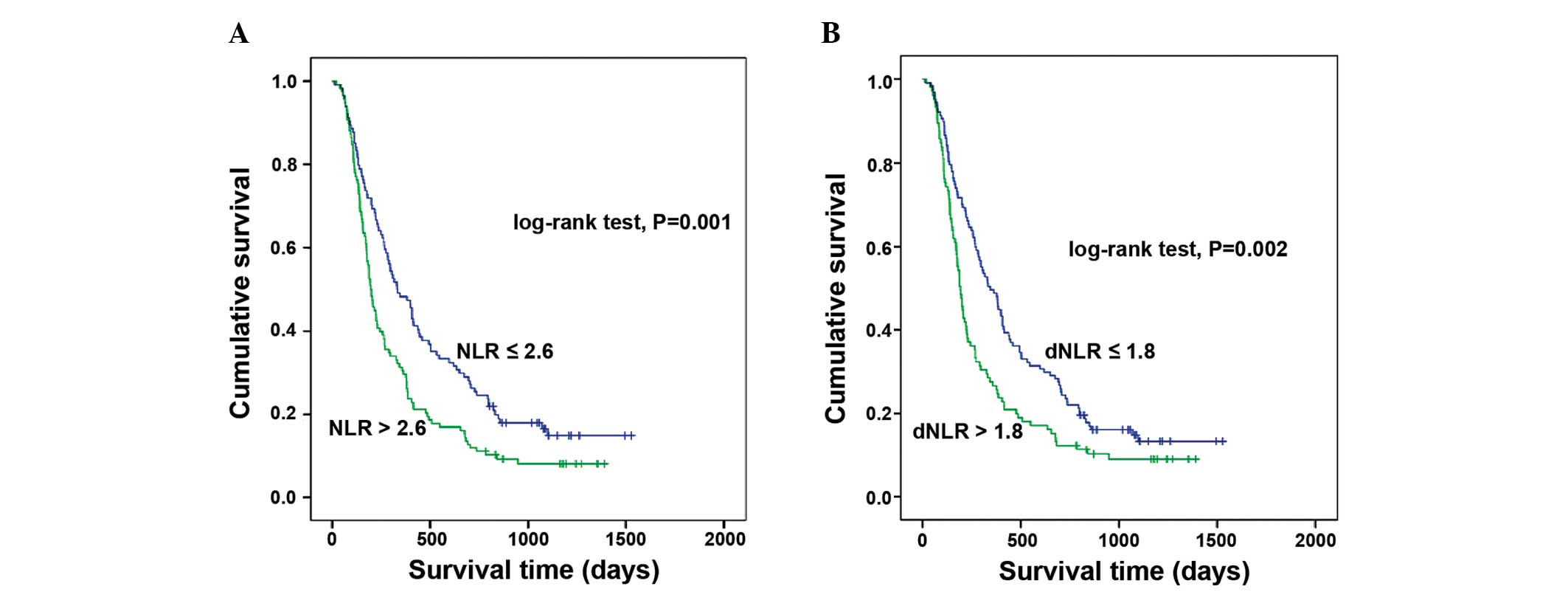
By applying the aforementioned criteria, a cut-off
value of 2.6 for NLR and 1.8 for dNLR was determined by ROC
analysis to be best to discriminate between patients' survival in
the whole cohort. In total, 140 patients had NLR ≥2.6 (50%) and 126
patients had dNLR ≥1.8 (45.2%). The association between
inflammatory scores and clinicopathological features was analyzed
(Table II). The NLR and dNLR were
each associated with patient age, tumor size, presence of vascular
invasion, and aspartate aminotransferase (AST) and ALP levels.
| Table II.Association between NLR or dNLR and
clinical variables. |
Table II.
Association between NLR or dNLR and
clinical variables.
|
| NLR, n |
| dNLR, n |
|
|---|
|
|
|
|
|
|
|---|
| Variables | ≤2.6 | >2.6 | P-value | ≤1.8 | >1.8 | P-value |
|---|
| Age |
|
| 0.03 |
|
|
0.016 |
| >50
years | 87 | 71 |
| 96 | 62 |
|
| ≤50
years | 52 | 69 |
| 57 | 64 |
|
| Gender |
|
| 0.437 |
|
|
0.179 |
|
Male | 127 | 124 |
| 141 | 110 |
|
|
Female | 12 | 16 |
| 12 | 16 |
|
| Diameter of largest
lesion |
|
| <0.001 |
|
| <0.001 |
| >10
cm | 29 | 80 |
| 35 | 74 |
|
| ≤10
cm | 106 | 56 |
| 114 | 48 |
|
| Number of
lesions |
|
| 0.096 |
|
|
0.235 |
| 1 | 35 | 48 |
| 41 | 42 |
|
|
>1 | 104 | 92 |
| 112 | 84 |
|
| Vascular
invasion |
|
| <0.001 |
|
| <0.001 |
|
Absent | 107 | 78 |
| 115 | 70 |
|
|
Present | 32 | 62 |
| 38 | 56 |
|
| ALT |
|
| 0.063 |
|
|
0.024 |
| >40
µ/1 | 76 | 93 |
| 84 | 85 |
|
| ≤40
µ/1 | 63 | 47 |
| 69 | 41 |
|
| AST |
|
| <0.001 |
|
| <0.001 |
| >45
µ/1 | 81 | 110 |
| 94 | 97 |
|
| ≤45
µ/1 | 58 | 30 |
| 59 | 29 |
|
| Total serum
bilirubin |
|
| 0.049 |
|
|
0.104 |
| >20
µmol/1 | 25 | 42 |
| 32 | 35 |
|
| ≤20
µmol/1 | 114 | 98 |
| 121 | 91 |
|
| ALP |
|
| <0.001 |
|
|
0.001 |
| >110
IU/1 | 72 | 98 |
| 82 | 88 |
|
| ≤110
IU/1 | 67 | 42 |
| 71 | 38 |
|
| AFP |
|
| 0.115 |
|
|
0.204 |
| >400
ng/ml | 70 | 83 |
| 79 | 74 |
|
| ≤400
ng/ml | 69 | 57 |
| 74 | 52 |
|
| PT |
|
| 0.012 |
|
|
0.128 |
|
Normal | 126 | 116 |
| 137 | 105 |
|
|
Abnormal | 13 | 24 |
| 16 | 21 |
|
| Child-Pugh
grade |
|
| 0.507 |
|
|
0.489 |
| A | 130 | 128 |
| 143 | 115 |
|
| B | 9 | 12 |
| 10 | 11 |
|
Survival and prognostic factors
The median follow-up period was 446 days. The 1, 2
and 3-year OS rates were 38.8, 18.5 and 11.1% respectively, and the
median OS time was 264 days. The univariate and multivariate
analyses of prognostic factors for OS were analyzed. In univariate
analysis (Table I), age (P=0.049),
CRP (P<0.001), alanine aminotransferase (ALT; P=0.003), AST
(P<0.001), ALP (P<0.001), LDH (P<0.001), α-L-fucosidase
(AFU; P=0.024), TBIL (P<0.001), AFP (P<0.001), prothrombin
time (PT; P=0.001), tumor size (P<0.001), tumor number
(P=0.041), vascular invasion (P<0.001), metastasis (P<0.001),
Child-Pugh scores (P=0.005), NLR (P=0.001) and dNLR (P=0.002) were
prognostic factors for OS.
Multivariate analysis (Table III) showed that NLR [hazard ratio
(HR), 1.382; 95% confidence interval (CI), 1.037–1.842; P=0.027],
ALT (HR, 1.472; 95% CI, 1.099–1.971; P=0.01), and AFP (HR, 1.677;
95% CI, 1.259–2.233; P<0.001) were independent prognostic
factors for OS. When NLR was replaced by dNLR, the multivariate
analysis also showed that the dNLR (HR, 1.445; 95% CI, 1.086–1.923;
P=0.012) was an independent prognostic factor for OS, along with
the ALT and AFP levels. An elevated NLR or dNLR is associated with
a poor prognosis (P=0.001 and P=0.002 respectively; Fig. 1).
| Table III.Multivariate analyses of prognostic
factors for overall survival in 279 patients undergoing TACE for
hepatocellular carcinoma. |
Table III.
Multivariate analyses of prognostic
factors for overall survival in 279 patients undergoing TACE for
hepatocellular carcinoma.
|
|
| Multivariate
analysis |
|---|
|
|
|
|
|---|
| Variables | Value | Hazard ratio (95%
CI) | P-value |
|---|
| NLRa |
|
|
|
| Mean
ALT, µ/1 (range) | 56.6 (8–304) | 1.472
(1.099–1.971) |
0.010 |
| Mean
AFP, ng/ml (range) | 751.2
(1.26–1,210,000) | 1.677
(1.259–2.233) | <0.001 |
| NLR,
n |
|
|
|
|
≤2.6 | 139 | 1.382
(1.037–1.842) |
0.027 |
|
>2.6 | 140 |
|
|
| dNLRa |
|
|
|
| Mean
ALT, µ/1 (range) | 56.6 (8–304) | 1.469
(1.098–1.966) |
0.010 |
| Mean
AFP, ng/ml (range) | 751.2
(1.26–1,210,000) | 1.720
(1.294–2.287) | <0.001 |
| dNLR,
n |
|
|
|
|
≤1.8 | 153 | 1.445
(1.086–1.923) |
0.012 |
|
>1.8 | 126 |
|
|
Association and comparison between the
NLR and dNLR
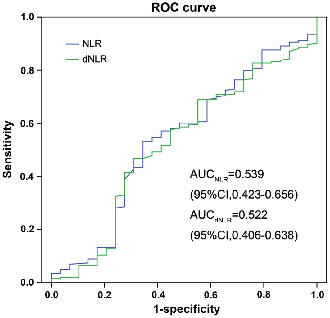
The association between the NLR and dNLR was
assessed by Spearman's rank correlation analysis. There was a
significant correlation between NLR and dNLR (R=0.875; P<0.001).
The prognostic power of NLR and dNLR was compared using AUC
analysis. As shown in Fig. 2, the AUC
of NLR was 0.539 (95% CI, 0.423–0.656) and the AUC of dNLR was
0.522 (95% CI, 0.406–638), which was similar.
Discussion
In the present study, the prognostic power of the
NLR and dNLR was evaluated in patients with HBV-associated HCC
undergoing TACE. The present results demonstrated that there was a
significant correlation between NLR and dNLR, and NLR and dNLR each
predicted the prognosis of patients with a similar prognostic
power. Thus, the dNLR may be used as an alternative to NLR.
Previous studies (10–13) have
shown that inflammation scores, such as the GPS, mGPS, NLR, PLR, PI
and PNI, are associated with the prognosis of patients with HCC
undergoing surgical resection, transplantation, TACE and RFA. Among
these inflammation-based scores, NLR is inferior to other measures
of the systemic inflammatory response, including mGPS, but it is
less expensive and more readily available in day-to-day oncological
practice (24). It is therefore
notable that the NLR has been shown to have prognostic value in
patients with a variety of cancers, and dynamic changes in the NLR
may predict the prognosis of patients (25). Proctor et al (21) evaluated the prognostic value of the
dNLR in a large cohort of 12,118 patients with different cancer
types, including hepatopancreaticobiliary cancer (n=721). This
study clearly demonstrated that the dNLR has a similar predictive
ability for prognosis as the NLR, with patients with an elevated
dNLR demonstrating a poor clinical outcome, which can be equally
used to predict survival (21). The
advantage of the dNLR compared with the NLR is that the dNLR
remains available in the absence of the lymphocyte count, and may
therefore be widely used on the basis of clinical trial
databases.
It is generally accepted that inflammatory processes
in the tumor microenvironment play a crucial role in promoting the
proliferation, invasion and metastasis of malignant cells (26,27). The
infiltrating leucocytes are important factors in this process
(26). There are two elements to the
dNLR, consisting of the neutrophil count, and the white blood cell
count minus the neutrophil count. The latter count is dominated by
lymphocytes and monocytes. Neutrophils in the peripheral blood or
in the tumor microenvironment have been shown to produce
pro-angiogenic factors, including vascular endothelial growth
factor, to stimulate tumor development and progression (27). The cytokines involved in
cancer-associated inflammation, including interleukin-6 (IL-6) and
tumor necrosis factor-α (TNFα), may induce neutrophilia (28,29). The
para-neoplastic production of myeloid growth factors by cancer
cells may act as an additional cause of neutrophilia (30). Therefore, a high peripheral neutrophil
level may indicate a cancer-associated inflammation or tumor
progression, and predict poor clinical outcome. In addition, the
neutrophils and leucocytes are mostly composed of lymphocytes.
Immune cells that infiltrate into or around the tumor engage in
dynamic and extensive crosstalk with cancer cells (31). Over the past decade, there has been
growing evidence that lymphocytes act as crucial components of the
adaptive immune system and are the cellular basis of cancer
immuno-surveillance and immuno-editing (32,33).
Furthermore, infiltrating lymphocytes have been reported to
indicate the generation of an effective anti-tumor cellular immune
response (34,35). Therefore, a low lymphocyte count may
be responsible for an inadequate immunological reaction to the
tumor, and consequently, a weakened defense against cancer,
resulting in a poor prognosis (35).
The peripheral monocyte count is known to be increased in cancer
patients (36–38). Schmidt et al (36) created a prognostic model in metastatic
melanoma based on independent prognostic factors in 321 patients
receiving IL-2-based immunotherapy. This study showed that an
elevated monocyte count may replace an elevated neutrophil count as
an independent prognostic factor for poor survival (36). Leitch et al (39) compared the prognostic value of an
inflammation-based prognostic score in 149 patients with colorectal
cancer and concluded that the monocyte count was independently
associated with cancer-specific survival. One possible hypothesis
is that macrophages express chemokine (C-X-C motif) (CXC) receptors
1 and 2, corresponding with CXC ligand (CXCL)1, also termed Gro-α,
and CXCL8, also termed IL-8, respectively. These chemokines may be
involved in tumor invasion and angiogenesis. However, monocytes
only account for <8% of leucocytes, with limited effect on the
dNLR or NLR.
In the present study the association between the NLR
and the dNLR was analyzed by Spearman's rank correlation analysis
and it is not notable that a significant correlation was identified
between the NLR and the dNLR. The prognostic power of the NLR and
the dNLR was also compared, and AUC analysis showed that the
prognostic power was similar between the two. In the Cox
proportional hazards regression analysis for OS, either NLR or dNLR
were considered to be an independent prognostic factor, with a
similar hazard ratio. All these results indicated that the dNLR may
be used as an alternative to NLR in these patients.
The elevated ALT and AFP levels were also revealed
as independent prognostic factors for poor outcome, as has been
reported in previous studies (40–42).
Notably, in patients with chronic hepatitis and cirrhosis, an
increase in the AST/ALT ratio is associated with progressive liver
functional impairment (43,44). As a major mammalian embryo-specific
and tumor-associated protein, AFP has been used for the diagnosis
and screening of HCC worldwide. An increased AFP level is connected
with larger tumors and lower hypohepatia, reflecting an aggressive
biology (45).
There are potential limitations of the present
study, as follows: i) It is a retrospective, small sample,
single-institution study; ii) only patients treated with TACE were
recruited; and iii) the patient population is biased due to the
prevalence of HBV infection, which is unusual in Western countries.
Therefore, a large-scale prospective validation study is required
to confirm the present results.
The current results revealed that an elevated dNLR
predicted poor prognosis with a similar prognostic power to the NLR
in patients with HBV-associated HCC undergoing TACE. Due to the
dNLR being an easily available and inexpensive marker in clinical
studies, the dNLR should be considered as a novel prognostic marker
for patients with HBV-associated HCC in routine practice.
References
|
1
|
Kamangar F, Dores GM and Anderson WF:
Patterns of cancer incidence, mortality and prevalence across five
continents: Defining priorities to reduce cancer disparities in
different geographic regions of the world. J Clin Oncol.
24:2137–2150. 2006. View Article : Google Scholar : PubMed/NCBI
|
|
2
|
Farazi PA and DePinho RA: Hepatocellular
carcinoma pathogenesis: From genes to environment. Nat Rev Cancer.
6:674–687. 2006. View
Article : Google Scholar : PubMed/NCBI
|
|
3
|
Chalasani N, Horlander JC Sr, Said A, Hoen
H, Kopecky KK, Stockberger SM Jr, Manam R, Kwo PY and Lumeng L:
Screening for hepatocellular carcinoma in patients with advanced
cirrhosis. Am J Gastroenterol. 94:2988–2993. 1999. View Article : Google Scholar : PubMed/NCBI
|
|
4
|
Tsukuma H, Hiyama T, Tanaka S, Nakao M,
Yabuuchi T, Kitamura T, Nakanishi K, Fujimoto I, Inoue A, Yamazaki
H, et al: Risk factors for hepatocellular carcinoma among patients
with chronic liver disease. N Engl J Med. 328:1797–1801. 1993.
View Article : Google Scholar : PubMed/NCBI
|
|
5
|
Saraswat VA, Pandey G and Shetty S:
Treatment algorithms for managing hepatocellular carcinoma. J Clin
Exp Hepatol. 4:S80–S89. 2014. View Article : Google Scholar : PubMed/NCBI
|
|
6
|
Llovet JM, Burroughs A and Bruix J:
Hepatocellular carcinoma. Lancet. 362:1907–1917. 2003. View Article : Google Scholar : PubMed/NCBI
|
|
7
|
Llovet JM and Bruix J: Systematic review
of randomized trials for unresectable hepatocellular carcinoma:
Chemoembolization improves survival. Hepatology. 37:429–442. 2003.
View Article : Google Scholar : PubMed/NCBI
|
|
8
|
Colotta F, Allavena P, Sica A, Garlanda C
and Mantovani A: Cancer-related inflammation, the seventh hallmark
of cancer: Links to genetic instability. Carcinogenesis.
30:1073–1081. 2009. View Article : Google Scholar : PubMed/NCBI
|
|
9
|
Hanahan D and Weinberg RA: Hallmarks of
cancer: The next generation. Cell. 144:646–674. 2011. View Article : Google Scholar : PubMed/NCBI
|
|
10
|
Forrest LM, McMillan DC, McArdle CS,
Angerson WJ, Dagg K and Scott HR: A prospective longitudinal study
of performance status, an inflammation-based score (GPS) and
survival in patients with inoperable non-small-cell lung cancer. Br
J Cancer. 92:1834–1836. 2005. View Article : Google Scholar : PubMed/NCBI
|
|
11
|
Hashimoto K, Ikeda Y, Korenaga D, Tanoue
K, Hamatake M, Kawasaki K, Yamaoka T, Iwatani Y, Akazawa K and
Takenaka K: The impact of preoperative serum C-reactive protein on
the prognosis of patients with hepatocellular carcinoma. Cancer.
103:1856–1864. 2005. View Article : Google Scholar : PubMed/NCBI
|
|
12
|
Ishizuka M, Kubota K, Kita J, Shimoda M,
Kato M and Sawada T: Usefulness of a modified inflammation-based
prognostic system for predicting postoperative mortality of
patients undergoing surgery for primary hepatocellular carcinoma. J
Surg Oncol. 103:801–806. 2011. View Article : Google Scholar : PubMed/NCBI
|
|
13
|
Kinoshita A, Onoda H, Imai N, Iwaku A,
Oishi M, Fushiya N, Koike K, Nishino H and Tajiri H: Comparison of
the prognostic value of inflammation-based prognostic scores in
patients with hepatocellular carcinoma. Br J Cancer. 107:988–993.
2012. View Article : Google Scholar : PubMed/NCBI
|
|
14
|
Gomez D, Farid S, Malik HZ, Young AL,
Toogood GJ, Lodge JP and Prasad KR: Preoperative
neutrophil-to-lymphocyte ratio as a prognostic predictor after
curative resection for hepatocellular carcinoma. World J Surg.
32:1757–1762. 2008. View Article : Google Scholar : PubMed/NCBI
|
|
15
|
Halazun KJ, Hardy MA, Rana AA, Woodland DC
IV, Luyten EJ, Mahadev S, Witkowski P, Siegel AB, Brown RS Jr and
Emond JC: Negative impact of neutrophil-lymphocyte ratio on outcome
after liver transplantation for hepatocellular carcinoma. Ann Surg.
250:141–151. 2009. View Article : Google Scholar : PubMed/NCBI
|
|
16
|
Huang ZL, Luo J, Chen MS, Li JQ and Shi M:
Blood neutrophil-to-lymphocyte ratio predicts survival in patients
with unresectable hepatocellular carcinoma undergoing transarterial
chemoembolization. J Vasc Interv Radiol. 22:702–709. 2011.
View Article : Google Scholar : PubMed/NCBI
|
|
17
|
Chen TM, Lin CC, Huang PT and Wen CF:
Neutrophil-to-lymphocyte ratio associated with mortality in early
hepatocellular carcinoma patients after radiofrequency ablation. J
Gastroenterol Hepatol. 27:553–561. 2012. View Article : Google Scholar : PubMed/NCBI
|
|
18
|
Pinato DJ, North BV and Sharma R: A novel,
externally validated inflammation-based prognostic algorithm in
hepatocellular carcinoma: The prognostic nutritional index (PNI).
Br J Cancer. 106:1439–1445. 2012. View Article : Google Scholar : PubMed/NCBI
|
|
19
|
Kasymjanova G, MacDonald N, Agulnik JS,
Cohen V, Pepe C, Kreisman H, Sharma R and Small D: The predictive
value of pre-treatment inflammatory markers in advanced
non-small-cell lung cancer. Curr Oncol. 17:52–58. 2010. View Article : Google Scholar : PubMed/NCBI
|
|
20
|
Smith RA, Bosonnet L, Raraty M, Sutton R,
Neoptolemos JP, Campbell F and Ghaneh P: Preoperative
platelet-lymphocyte ratio is an independent significant prognostic
marker in resected pancreatic ductal adenocarcinoma. Am J Surg.
197:466–472. 2009. View Article : Google Scholar : PubMed/NCBI
|
|
21
|
Proctor MJ, McMillan DC, Morrison DS,
Fletcher CD, Horgan PG and Clarke SJ: A derived neutrophil to
lymphocyte ratio predicts survival in patients with cancer. Br J
Cancer. 107:695–699. 2012. View Article : Google Scholar : PubMed/NCBI
|
|
22
|
Kusumanto YH, Dam WA, Hospers GA, Meijer C
and Mulder NH: Platelets and granulocytes, in particular the
neutrophils, form important compartments for circulating vascular
endothelial growth factor. Angiogenesis. 6:283–287. 2003.
View Article : Google Scholar : PubMed/NCBI
|
|
23
|
Chen MS, Li JQ, Zhang YQ, Lu LX, Zhang WZ,
Yuan YF, Guo YP, Lin XJ and Li GH: High-dose iodized oil
transcatheter arterial chemoembolization for patients with large
hepatocellular carcinoma. World J Gastroenterol. 8:74–78. 2002.
View Article : Google Scholar : PubMed/NCBI
|
|
24
|
Proctor MJ, Morrison DS, Talwar D, Balmer
SM, Fletcher CD, O'Reilly DS, Foulis AK, Horgan PG and McMillan DC:
A comparison of inflammation-based prognostic scores in patients
with cancer. A Glasgow Inflammation Outcome Study. Eur J Cancer.
47:2633–2641. 2011. View Article : Google Scholar : PubMed/NCBI
|
|
25
|
Dan J, Zhang Y, Peng Z, Huang J, Gao H, Xu
L and Chen M: Postoperative neutrophil-to-lymphocyte ratio change
predicts survival of patients with small hepatocellular carcinoma
undergoing radiofrequency ablation. PloS One. 8:e581842013.
View Article : Google Scholar : PubMed/NCBI
|
|
26
|
Mantovani A, Allavena P, Sica A and
Balkwill F: Cancer-related inflammation. Nature. 454:436–444. 2008.
View Article : Google Scholar : PubMed/NCBI
|
|
27
|
Coussens LM and Werb Z: Inflammation and
cancer. Nature. 420:860–867. 2002. View Article : Google Scholar : PubMed/NCBI
|
|
28
|
Ulich TR, del Castillo J, Keys M, Granger
GA and Ni RX: Kinetics and mechanisms of recombinant human
interleukin 1 and tumor necrosis factor-alpha-induced changes in
circulating numbers of neutrophils and lymphocytes. J Immunol.
139:3406–3415. 1987.PubMed/NCBI
|
|
29
|
Ulich TR, del Castillo J and Guo KZ: In
vivo hematologic effects of recombinant interleukin-6 on
hematopoiesis and circulating numbers of RBCs and WBCs. Blood.
73:108–110. 1989.PubMed/NCBI
|
|
30
|
Teramukai S, Kitano T, Kishida Y, Kawahara
M, Kubota K, Komuta K, Minato K, Mio T, Fujita Y, Yonei T, et al:
Pretreatment neutrophil count as an independent prognostic factor
in advanced non-small-cell lung cancer: An analysis of Japan
multinational trial organisation LC00-03. Eur J Cancer.
45:1950–1958. 2009. View Article : Google Scholar : PubMed/NCBI
|
|
31
|
Grivennikov SI, Greten FR and Karin M:
Immunity, inflammation and cancer. Cell. 140:883–899. 2010.
View Article : Google Scholar : PubMed/NCBI
|
|
32
|
Dunn GP, Old LJ and Schreiber RD: The
immunobiology of cancer immunosurveillance and immunoediting.
Immunity. 21:137–148. 2004. View Article : Google Scholar : PubMed/NCBI
|
|
33
|
Dunn GP, Ikeda H, Bruce AT, Koebel C,
Uppaluri R, Bui J, Chan R, Diamond M, White JM, Sheehan KC and
Schreiber RD: Interferon-gamma and cancer immunoediting. Immunol
Res. 32:231–245. 2005. View Article : Google Scholar : PubMed/NCBI
|
|
34
|
Rabinowich H, Cohen R, Bruderman I,
Steiner Z and Klajman A: Functional analysis of mononuclear cells
infiltrating into tumors: Lysis of autologous human tumor cells by
cultured infiltrating lymphocytes. Cancer Res. 47:173–177.
1987.PubMed/NCBI
|
|
35
|
Hoffmann TK, Dworacki G, Tsukihiro T,
Meidenbauer N, Gooding W, Johnson JT and Whiteside TL: Spontaneous
apoptosis of circulating T lymphocytes in patients with head and
neck cancer and its clinical importance. Clin Cancer Res.
8:2553–2562. 2002.PubMed/NCBI
|
|
36
|
Schmidt H, Bastholt L, Geertsen P,
Christensen IJ, Larsen S, Gehl J and von der Maase H: Elevated
neutrophil and monocyte counts in peripheral blood are associated
with poor survival in patients with metastatic melanoma: A
prognostic model. Br J Cancer. 93:273–278. 2005. View Article : Google Scholar : PubMed/NCBI
|
|
37
|
Cho H and Kim JH: Multiplication of
neutrophil and monocyte counts (MNM) as an easily obtainable tumour
marker for cervical cancer. Biomarkers. 14:161–170. 2009.
View Article : Google Scholar : PubMed/NCBI
|
|
38
|
Millrud CR, Kvarnhammar Månsson A, Uddman
R, Björnsson S, Riesbeck K and Cardell LO: The activation pattern
of blood leukocytes in head and neck squamous cell carcinoma is
correlated to survival. PloS One. 7:e511202012. View Article : Google Scholar : PubMed/NCBI
|
|
39
|
Leitch EF, Chakrabarti M, Crozier JE,
McKee RF, Anderson JH, Horgan PG and McMillan DC: Comparison of the
prognostic value of selected markers of the systemic inflammatory
response in patients with colorectal cancer. Br J Cancer.
97:1266–1270. 2007. View Article : Google Scholar : PubMed/NCBI
|
|
40
|
Hernaez R, Yeh HC, Lazo M, Chung HM,
Hamilton JP, Koteish A, Potter JJ, Brancati FL and Clark JM:
Elevated ALT and GGT predict all-cause mortality and hepatocellular
carcinoma in Taiwanese male: A case-cohort study. Hepatol Int.
7:1040–1049. 2013. View Article : Google Scholar : PubMed/NCBI
|
|
41
|
Tarao K, Rino Y, Takemiya S, Tamai S,
Ohkawa S, Sugimasa Y, Miyakawa K, Morinaga S, Yoshida M, Shibuya A,
et al: Close association between high serum ALT and more rapid
recurrence of hepatocellular carcinoma in hepatectomized patients
with HCV-associated liver cirrhosis and hepatocellular carcinoma.
Intervirology. 43:20–26. 2000. View Article : Google Scholar : PubMed/NCBI
|
|
42
|
El-Serag HB, Kramer JR, Chen GJ, Duan Z,
Richardson PA and Davila JA: Effectiveness of AFP and ultrasound
tests on hepatocellular carcinoma mortality in HCV-infected
patients in the USA. Gut. 60:992–997. 2011. View Article : Google Scholar : PubMed/NCBI
|
|
43
|
Giannini E, Botta F, Fasoli A, Ceppa P,
Risso D, Lantieri PB, Celle G and Testa R: Progressive liver
functional impairment is associated with an increase in AST/ALT
ratio. Dig Dis Sci. 44:1249–1253. 1999. View Article : Google Scholar : PubMed/NCBI
|
|
44
|
Giannini E, Risso D, Botta F, Chiarbonello
B, Fasoli A, Malfatti F, Romagnoli P, Testa E, Ceppa P and Testa R:
Validity and clinical utility of the aspartate
aminotransferase-alanine aminotransferase ratio in assessing
disease severity and prognosis in patients with hepatitis C
virus-related chronic liver disease. Arch Intern Med. 163:218–224.
2003. View Article : Google Scholar : PubMed/NCBI
|
|
45
|
Carr BI, Guerra V, Giannini EG, Farinati
F, Ciccarese F, Rapaccini GL, Di Marco M, Benvegnù L, Zoli M,
Borzio F, et al: Significance of platelet and AFP levels and liver
function parameters for HCC size and survival. Int J Biol Markers.
29:e215–e223. 2014.PubMed/NCBI
|