Introduction
Nasopharyngeal carcinoma (NPC) is one of the most
common malignant head and neck tumors. Due to the fast metastasis
to the lymph nodes, swelling is frequently indicated in the neck,
retropharynx and parapharynx (1). The
survival rate of terminal stage patients ranges between 50–60%, and
patients with NPC possess a relatively poor prognosis (2). The prevertebral space is located at the
retropharynx or behind the esophagus, with the prevertebral fascia
in front. The prevertebral fascia is a thick and dense tissue that
extends between the deep surface of the sternocleidomastoid muscle,
attached to the skull base, and the thoracic mediastinum, of which
one side attaches to the cervical transverse position in front of
the vertebrae and the prevertebral muscle and the rear side
attaches to the cervical acantha. In addition, retropharyngeal
lymph nodes are located in front of the prevertebral fascia
(3). The present study reports a rare
clinical case, in which effusion of the prevertebral space has been
caused by the breaking of swollen lymphonodi retropharynici.
Case report
A 20-year-old male patient was admitted to the
Department of Radiotherapy, The Linyi Cancer Hospital (Linyi,
China) on 16 January 2015 due to nasal obstruction and associated
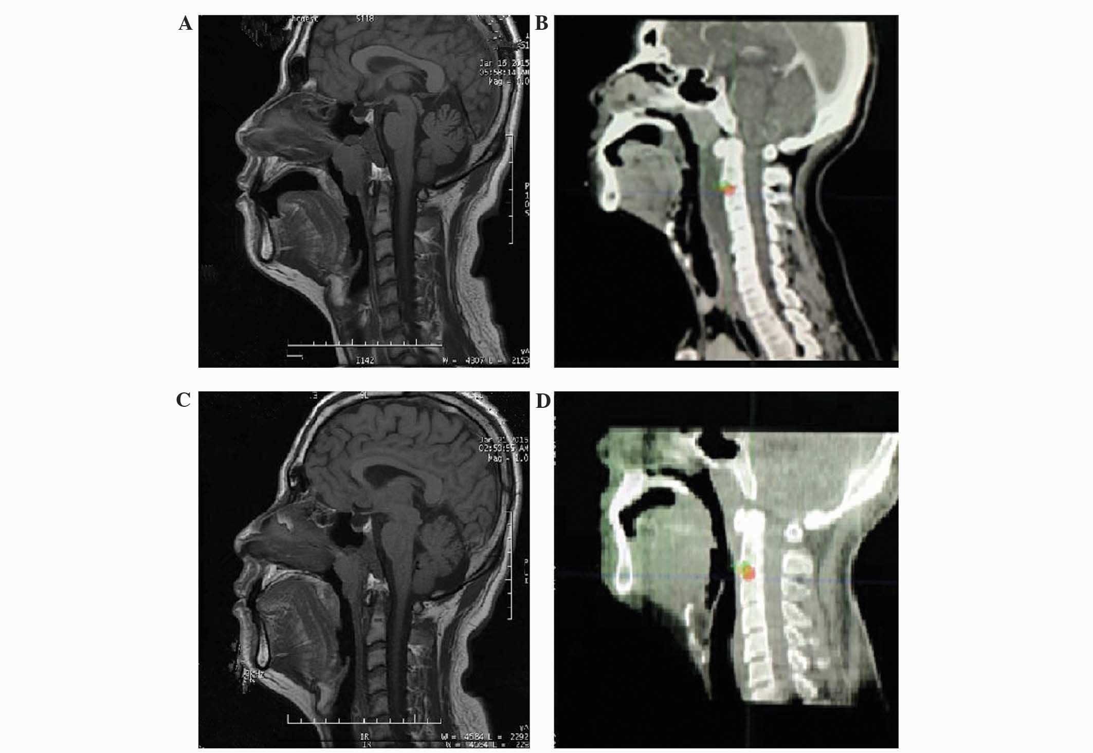
epistaxis for a period of 6 months. A magnetic resonance imaging
(MRI) scan of the nasopharynx was completed the same day, and the
results revealed abnormal masses on the posterior wall of the
nasopharynx, the disappearance of the fossa and lateral pharyngeal
wall on each side, multiple swollen lymph nodes on each side of the
parapharynx, the retropharynx and the neck, and a normal
prevertebral space (Fig. 1A). A
pathological biopsy was performed on the nasopharyngeal mass, and
consequently the patient was diagnosed with NPC.
The patient coughed violently and constantly due to
passive smoking, and on 18 January 2015, the coughing was
accompanied by a sudden pain behind the right ear and significant
feeling of swelling on the retropharynx wall. The patient was
administered anti-inflammatory treatments under the consideration
of acute mumps. On the following day, a localized computed
tomography (CT) scan revealed that the lymph nodes on the right
side had decreased in size. This finding was accompanied by an
evident hypodense shadow at the prevertebral space, which had an
anteroposterior diameter of 2 cm (Fig.
1B). The violent cough was considered to have increased the
pressure at the retropharynx wall, which then led to the breaking
of the swollen lymph nodes in the right pharynx. The broken lymph
nodes allowed necrotic liquid to seep into the prevertebral space
and cause effusion; therefore, the patient was administered 40 mg
methylprednisolone via an intravenous drip. Feedback from the
patient subsequent to treatment indicated that the pain on the
right parotid gland and the swelling at the retropharynx wall were
notably relieved. MRI, which was performed in the re-examination on
21 January 2015, revealed that the prevertebral space was unusually
filled. However in comparison with the localized CT scan that was
performed 2 days previously, the size of the lesion had evidently
decreased to ~1 cm in diameter (Fig.
1C). Subsequent to continuous treatment with methylprednisolone
(ivgtt qd, d1-5), on 26 January 2015, the results of the cone bean
computed tomography identification, which was performed prior to
the administration of radiotherapy, indicated that the swelling on
the retropharynx wall had improved and the prevertebral space had
returned to normal (Fig. 1D). In
addition, the patient confirmed that the feeling of swelling on the
right parotid gland and the retropharynx wall had gone. At present,
the patient is undergoing radiotherapy (total dose, 70 Gy; 35
fractions of 2 Gy over 6 weeks).
Discussion
Effusion in the prevertebral space is generally
considered to be associated with the stimulation of local chronic
inflammation. Common causes of effusion in the prevertebral space
include odontogenic and pharyngeal infections, sialadenitis,
nasosinusitis, wounds and foreign matter on the upper respiratory
and upper gastrointestinal tracts, lymphadenitis colli, and
fistulae and cysts of the neck (4).
Of all the head and neck tumors, NPC is the type that may invade
the prevertebral space and cause effusion. In accordance with the
study conducted by Liao et al (5), the MRI and CT scans estimated that the
occurring rates of the prevertebral muscle in the prevertebral
space being attacked are 36.0 and 18.4%, respectively (P<0.001).
In the study conducted by Zhou et al (6), patients with NPC were treated using
intensity modulated radiation therapy (IMR), and it was identified
that patients with an invaded prevertebral space had dramatically
decreased overall survival, distant metastasis-free survival and
local recurrence-free survival rates compared with patients with no
invaded prevertebral space (6).
Therefore, T4 staging is recommended for invasion into the
prevertebral space in NPC. For the patient in the present study,
the prevertebral space appeared normal in the nasopharyngeal MRI,
so the invasion of the prevertebral space has been ruled out as the
cause of effusion. Following a comprehensive analysis of the
effusion, necrosis may have occurred due to interior ischemia of
the swollen retropharyngeal lymph nodes. The tissue structures
became loose due to the stress on the pharynx wall, produced by the
violent cough, which caused the lymph nodes to break. Subsequently,
the necrotic fluid seeped into the prevertebral space through the
prevertebral fascia and caused effusion. The treatments used were
anti-inflammatory and promoted the absorption of
methylprednisolone, and were accompanied by appropriate IMR of the
target region clinical target volume 1 (CTV1), including the
prevertebral fascia and the prevertebral space.
The prevertebral space is located next to the
retropharyngeal space, linking to the skull base at the top, the
orpharynx at the bottom and the parapharyngeal space at the sides.
Therefore, the sclerotin of the skull base and the oral pharynx and
parapharyngeal space may be easily affected by effusion. However,
as the rear of the prevertebral space is close to the brainstem and
the medulla spinalis, the fluid may often cause stress and necrosis
in these vital organs. In addition, implantation metastasis and
blood metastasis are highly possible results of the fluid released
from the broken swollen lymph nodes, which is provided with a T4
staging. The prevertebral fascia and prevertebral space are
inclusive in the CTV1 of the radiotherapy target region, but this
requires validation from additional clinical studies with a large
number of patients. Therefore, for patients with malignant head and
neck tumors with swollen lymph nodes on the retropharynx and
parapharynx, the clinical symptoms include sudden swelling and pain
the behind ears and on the retropharynx. Treatments may be actively
administered in order to prevent the occurrence of prevertebral
effusion caused by lymph node breakage.
References
|
1
|
Wu TT, Chen C, Chen SM, Xu Y, Wang Y, Chen
Z, Wang F, Xiao BK and Tao ZZ: Nuclear translocation of telomerase
reverse transcriptase is a critical process in lymphatic metastasis
of nasopharyngeal carcinoma. Oncol Lett. 9:265–269. 2015.PubMed/NCBI
|
|
2
|
Wong EY, Wong SC, Chan CM, Lam EK, Ho LY,
Lau CP, Au TC, Chan AK, Tsang CM, Tsao SW, et al: TP53-induced
glycolysis and apoptosis regulator promotes proliferation and
invasiveness of nasopharyngeal carcinoma cells. Oncol Lett.
9:569–574. 2015.PubMed/NCBI
|
|
3
|
Mills MK and Shah LM: Imaging of the
perivertebral space. Radiol Clin North Am. 53:163–180. 2015.
View Article : Google Scholar : PubMed/NCBI
|
|
4
|
Regueiro Villarín S, Vázquez Barro JC and
Herranz González-Botas J: Deep neck infections: Etiology,
bacteriology and treatment. Acta Otorrinolaringol Esp. 57:342–348.
2006.(In Spanish).
|
|
5
|
Liao XB, Mao YP, Liu LZ, Tang LL, Sun Y,
Wang Y, Lin AH, Cui CY, Li L and Ma J: How does magnetic resonance
imaging influence staging according to AJCC staging system for
nasopharyngeal carcinoma compared with computed tomography? Int J
Radiat Oncol Biol Phys. 72:1368–1377. 2008. View Article : Google Scholar : PubMed/NCBI
|
|
6
|
Zhou GQ, Mao YP, Chen L, Li WF, Liu LZ,
Sun Y, Chen Y, Tian L, Lin AH, Li L and Ma J: Prognostic value of
prevertebral space involvement in nasopharyngeal carcinoma based on
intensity-modulated radiotherapy. Int J Radiat Oncol Biol Phys.
82:1090–1097. 2012. View Article : Google Scholar : PubMed/NCBI
|