Introduction
Multidrug resistance (MDR) is a significant obstacle
to successful cancer chemotherapy in numerous types of human tumour
(1,2).
MDR occurs predominantly as a result of overexpression of the
transmembrane permeability glycoprotein (Pgp) encoded by the MDR
protein 1 gene (3). Pgp is a member
of the ATP-binding cassette transporter family that, in resistant
cancer cells, has a role as an efflux pump for various structurally
and functionally unrelated chemotherapeutic drugs, including the
anthracyclines, vinca alkaloids and taxans, thereby reducing their
concentration in the cell cytoplasm and limiting the effectiveness
of cancer chemotherapy (4–7).
Venous thromboembolic events represent an important
complication in patients with cancer due to haemostatic system
activation caused by aggressive chemotherapy (8–11).
Furthermore, D-dimer levels, which are used as coagulation markers
in cancer patients, have been observed to be higher in advanced
disease and decreased in patients in post-treatment remission,
which suggests that pro-coagulant activity in cancer patients could
predict response or disease progression (12). The expression of tissue factor (TF),
which is a transmembrane glycoprotein and the major cellular
initiator of blood coagulation in vivo, on the surface of
neoplastic cells varies across different cell types (13–15). TF
binds plasma factor II/VIIa (FII/FVIIa) to form a bimolecular
complex that actives factor X and factor IX, leading to the
generation of fibrin (16,17). Hypercoagulability has been associated
with chemoresistance, and TF may have a role in this association
(16,17). In addition, previous studies reported
that Pgp and TF tend to be coexpressed in numerous types of
neoplastic cell, potentially in response to common stimuli such as
chemotherapy (18). Therefore,
evaluation of TF-dependent coagulation activity associated with the
presence of TF on the surface of tumour cells may act as a
predictive biomarker for the indirect identification of
Pgp-mediated MDR in cancer patients.
In vitro and in vivo studies have
demonstrated that malignant cells release a large number of
microscopic cell membrane-derived vesicles, which are 0.1–1.0 µm in
diameter and called microparticles (MPs), in response to
chemotherapy or stimulation/induction of apoptosis (19). MPs carry various surface proteins that
are characteristic of their parental cells (20). In addition, clinical studies have
reported that TF is exposed on the surface of circulating MPs from
patients with cancer, and that high levels of MP-associated TF
activity in the plasma of cancer patients predicted an increased
risk for thrombosis and poor prognosis (21–25). The
evaluation of circulating MP-associated TF activity in cancer
patients during chemotherapy could be used to predict thrombosis
and the development of MDR. Therefore, this analysis, in
association with tumour markers or biopsies, could have a
prognostic value for cancer patients.
The present study aimed to investigate whether the
MPs released by the plasma membrane of cancer cells during
chemotherapy showed high levels of Pgp and TF coexpression on their
surface, and whether a rise in circulating MPs coexpressing Pgp and
TF may be indirectly predictive for the development of MDR and
thromboembolic complications. MPs were isolated from the blood of
50 patients with a variety of malignant tumours who were receiving
cancer chemotherapy, and were analysed for TF activity and Pgp
overexpression. The results of this analysis were compared with
those obtained for 10 healthy volunteers matched for age and
gender, who were considered as negative controls.
Materials and methods
Reagents and antibodies
The murine anti-human cluster of differentiation 243
(CD243) monoclonal antibody (clone, UIC2; IgG2a; dilution, 1:20;
catalog no., MCA2671A488) that recognizes an extracellular
conformational epitope of Pgp was purchased from Bio-Rad
Laboratories, Inc. (Hemel Hempstead, UK). The rabbit anti-human
FVII polyclonal antibody (clone CLBVII-I; IgG1; dilution, 1:100;
catalog no., MW1899) was obtained from Sanquin (Amsterdam, The
Netherlands).
Collection of blood samples
The patients used in the present study were enrolled
at the Department of Oncology, S.S. Annunziata Hospital (affiliated
to ‘G. d'Annunzio’ University of Chieti-Pescara; Chieti, Italy).
Between February 2012 and November 2014, ~4 ml peripheral blood was
collected from 50 cancer patients with solid tumours (including
pancreatic, breast, gastrointesyinal and lung cancer) through
venepuncture with a BD Vacutainer® blood collecting
system (BD Biosciences, Franklin Lakes, NJ, USA), and placed into
4.5 ml polypropylene tubes containing 3.8% sodium citrate. Whole
blood samples were processed immediately at room temperature to
inhibit cell activation. Blood samples were centrifuged at 3,000 ×
g for 20 min at room temperature in a swing-out rotor without break
to isolate erythrocytes and platelets. Platelet-poor plasma (PPP)
was then gently removed and coagulation tests were performed
immediately. The remaining plasma was distributed in 250 µl
aliquots, snap frozen in liquid nitrogen and finally stored at
−80°C until use. Plasma samples from 10 healthy volunteers (5
female and 5 male; mean age, 43 years; range, 23–46 years) were
similarly processed and assessed as negative controls. Informed
consent was obtained from all patients, who received detailed
information regarding the study, adhering to the Declaration of
Helsinki.
Isolation of MPs from plasma
samples
To isolate MPs, PPP obtained from whole blood was
thawed at 37°C, transferred to microtubes (1.5 ml) and further
centrifuged at 5,000 × g at room temperature for 10 min to remove
residual platelets. Subsequently, PPP was centrifuged at 19,800 × g
for 15 min at 20°C with a fixed-angle, 45° rotor (Centrifuge 5417R;
Eppendorf, Hamburg, Germany) to obtain a fraction enriched in
cell-derived MPs. Subsequently, MP-free plasma was removed from
each tube and the resultant MP pellets (25–30 µl) were washed twice
in Ca2+/Mg2+-free phosphate-buffered saline
(PBS), suspended in 0.3 ml Ca2+/Mg2+-free PBS
and used immediately. Ca2+-free PBS solution was used to
prevent fibrin generation in the solutions. Equal volumes (250 µl)
of plasma taken from healthy volunteers were run in parallel as
controls.
Pro-coagulant activity of
MP-associated TF
The pro-coagulant activity of TF associated with MPs
was evaluated in the presence and absence of the anti-FVIIa
antibody. The analysis was performed using a spectrophotometer
microplate reader (Spectra Max 190; Molecular Devices, LLC,
Sunnyvale, CA, USA) equipped to analyse kinetic data in a 96-well
format. The instrument consists of an incubator cabinet with a
photometer. Data acquisition and analysis were performed with
SoftMax® Pro 4.0 software (Molecular Devices, LLC).
Fresh PPP (90 µl) was pipetted into four wells of a 96-well plate.
Two wells were incubated with 3 µl anti-human FVIIa antibody (1
mg/ml) to inhibit TF-mediated coagulation, while the remaining two
wells were incubated with 3 µl physiological saline solution. The
96-well plate was transferred into the automated microplate reader
and incubated at 37°C for 5 min. Clotting was initiated by
simultaneous addition to each well of 15 µl of 0.1 M
CaCl2 reagent pre-warmed at 37°C. Following 5 sec of
agitation, fibrin formation was monitored by measuring the optical
density (OD) of the plasma at 1 min intervals for 30 min at 405 nm.
Equal volumes of plasma taken from 10 healthy volunteers were run
in parallel as controls. A plot of OD versus time was generated,
and the half-maximal value (1/2 Vmax) of the OD change during
clotting was determined and expressed as the anti-FVII
antibody-induced lengthening of the clotting time, as compared with
the saline samples. The following formula was used: Lengthening
time (%) = (anti-FVII clotting time - saline clotting time/saline
clotting time) × 100. The clotting time obtained for the same
re-calcified plasma (20 mmol/l CaCl2) in the presence
and absence of excess (1 mg/ml) rabbit anti-human FVII IgG
polyclonal antibody was compared to establish the
TF-FVII-dependence of the fibrin generation. The lag time of fibrin
generation was defined as the time interval required for the
solution turbidity to reach the 1/2 Vmax.
Immunohistochemical analysis of Pgp
expression on MPs
For the evaluation of Pgp expression, MP pellets
were prepared from 50 cancer patients. Briefly, plasma containing
MPs was centrifuged at 4,200 × g at room temperature for 10 min and
the MP pellet was cytocentrifuged at 400 × g at room temperature
for 10 min (Shandon CytoSpin 2 Cytocentrifuge; Thermo Fisher
Scientific, Inc., Waltham, MA, USA). To assess MP samples for CD243
expression, the Histostain®-Plus 112 kit was used
(Invitrogen; Thermo Fisher Scientific, Inc.), according to the
manufacturer's protocol. Briefly, slides were fixed for 10 min at
room temperature with 3% paraformaldehyde in Dulbecco's 1X PBS (pH
7.6). To prevent non-specific binding, slides were incubated for 10
min with serum blocking solution, followed by incubation for 45 min
at room temperature with murine anti-CD243 monoclonal antibody
(1:20). Following washing three times with 1X PBS, slides were
incubated for 10 min with the enzyme conjugate provided in the kit.
Following further washing with PBS, samples were incubated for 10
min with 3,3′-diaminobenzidine, washed with distilled water and
coverslipped with mounting medium. Subsequently, the slides were
observed under a Zeiss Axioscope light microscope (Carl Zeiss AG,
Oberkochen, Germany) equipped with a CoolSNAP camera and the
MetaMorph Image Analysis system (Universal Imaging Corp,
Downingtown, PA, USA).
Statistical analysis
Data are expressed as the mean ± standard deviation
and were analysed for statistical significance using the Student's
t-test, with Excel 2011 (Microsoft Corporation, Redmond, WA, USA).
P<0.05 was considered to indicate a statistically significant
difference.
Results
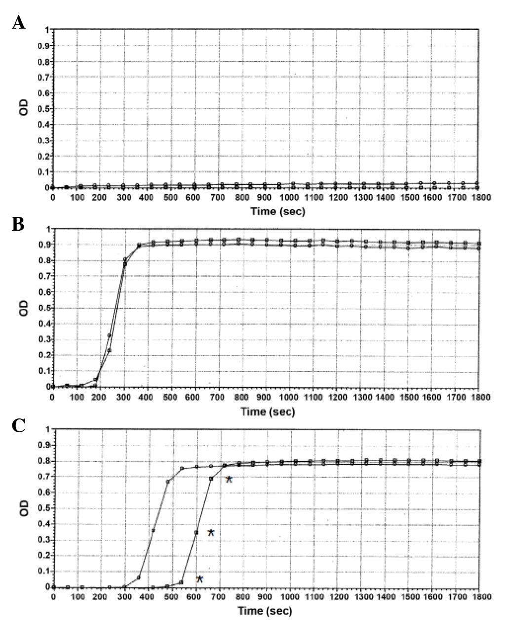
Clotting time assay or fibrin
generation assay
The present study analysed 50 PPP samples from
cancer patients, of which 20 samples showed high levels of
MP-associated TF activity, as demonstrated by the clotting time
measurements. As is shown in a representative curve obtained using
plasma from one healthy subject (Fig.
1A), no clotting was observed in the presence or absence of an
anti-FVIIa antibody when plasma was re-calcified to induce
coagulation for at least 1,800 sec. In 30 of the 50 plasma samples,
the addition of anti-FVIIa antibody did not significantly prolong
the clotting time; the percentage of lengthening between the two
curves was <10% (Fig. 1B).
However, in 20 of the 50 plasma samples, the time interval for the
solution turbidity to reach the 1/2 Vmax was significantly
different between samples treated with saline and anti-FVIIa
antibody (P=0.01). Fig. 1C shows a
representative microplate assay curve obtained using plasma
incubated with or without anti-FVII antibody, and demonstrates a
time delay of thrombin generation (427.1 vs. 609.9 sec,
respectively). Therefore, the addition of anti-FVIIa antibody
induced a significant lengthening of clotting time (~43%; P=0.02),
indicating the presence of a high amount of MP-associated TF within
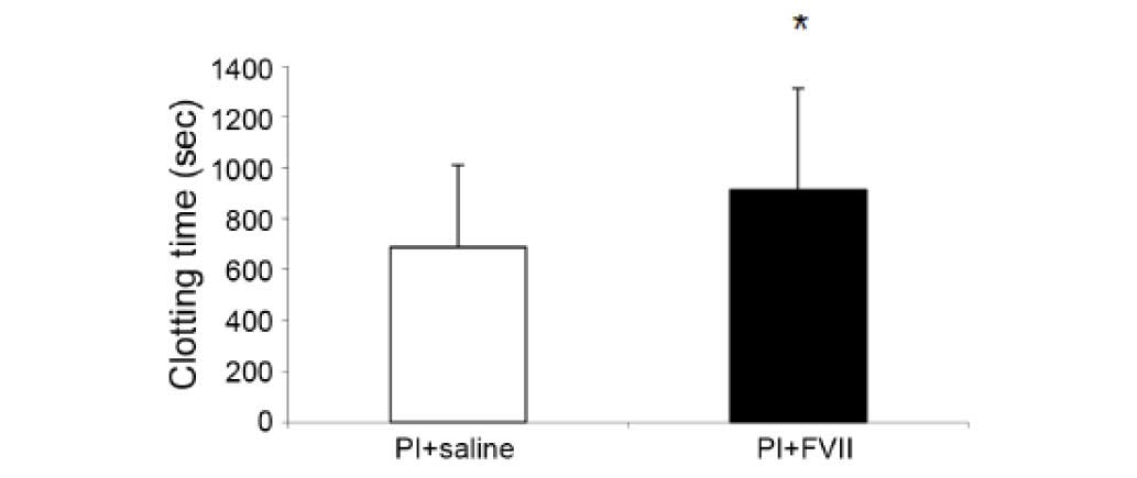
the plasma sample. Generally, the treatment of 20 TF-positive
samples with an anti-FVIIa antibody induced a significant
lengthening of clotting time compared with the controls treated
with saline alone (700±280 and 950±350 sec, respectively; P=0.01;
Fig. 2).
Pgp expression on the surface of
MPs
In order to determine whether TF and Pgp were
coexpressed on the surface of MPs, Pgp expression on MPs isolated
from plasma samples with a significant lengthening of clotting time
following inhibition of TF activity with an anti-FVIIa antibody was
evaluated. MPs obtained from cancer patients that showed
TF/FVII-dependent coagulation were compared with MPs from plasma
samples with <10% mean lengthening and MPs from plasma samples
of healthy controls. As is shown in Fig.
3A, MPs were rarely detected in the plasma samples of healthy
controls, and those that were detected were negative for Pgp
expression. Similarly, very low or absent expression of Pgp was
detected for the MPs isolated from plasma samples with no
significant TF activity (lengthening, <10%) (Fig. 3B). Conversely, a high number of MPs
were strongly labelled for Pgp expression in plasma samples from
cancer patients showing TF positivity (Fig. 3C).
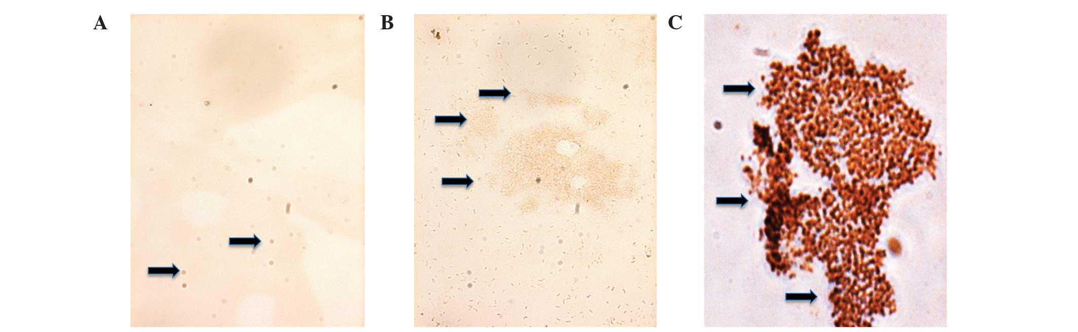 | Figure 3.Immunohistochemical detection of
permeability glycoprotein expression in cytocentrifuged
microparticles from human plasma (magnification, ×40). (A) There
were few MPs in the plasma from healthy patients, and those MPs
were negative for Pgp expression. (B) Numerous, clustered MPs were
detected in the plasma samples from 30 cancer patients without TF
activity, although these MPs showed very low expression of Pgp. (C)
Numerous, clustered MPs were detected in the plasma samples from 20
cancer patients with elevated TF activity, and these MPs showed a
high expression of Pgp, as indicated by the intense brown
colouration. Black arrows indicate MPs and clusters of MPs. Pgp,
permeability glycoprotein; MPs, micrcoparticles; TF, tissue
factor. |
Discussion
The present study demonstrated that Pgp and TF
glycoproteins were associated with circulating tumour cell-derived
MPs in blood collected from cancer patients undergoing
chemotherapy. TF expression was identified on the surface of MPs by
evaluating the TF-mediated pro-coagulant activity of plasma samples
from 50 cancer patients by means of an anti-FVIIa antibody to
distinguish fibrin generation via TF-FVII-dependent pathways. It
was demonstrated that 20 of the 50 cancer patients (>30%) had
high levels of circulating MP-associated TF activity. Notably,
treatment of these 20 plasma samples with an anti-FVIIa antibody
significantly prolonged (P<0.05) the clotting time of these
patients by up to 36.5±16%, as compared with the clotting time of
the same samples treated with saline alone. These results suggested
that tumour cell-derived MPs contribute to the activation of
coagulation in a TF-dependent manner, and indicated the specificity
of this assay to inhibit the TF-dependent pro-coagulant activity
via the FVII pathway. Conversely, in the remaining plasma samples
that were negative for MP-associated TF (n=30), the clot reaction
curves of plasma samples treated with anti-FVIIa antibody were
similar to those obtained following treatment of plasma samples
with saline alone. These results suggested that the clotting times
were unaffected by anti-FVIIa treatment, and that fibrin generation
was independent of this factor. Therefore, in these samples,
anti-FVII treatment did not prolong the fibrin generation.
Furthermore, the total absence of coagulation in the human plasma
obtained from healthy individuals incubated with anti-FVIIa
antibody suggested the absence of MPs or the presence of very low
levels of MPs in the blood of healthy subjects, as compared with
the plasma samples of cancer patients.
These results were consistent with the analysis of
the expression of Pgp on the surface of cell-derived MPs. Notably,
high levels of Pgp-positive MPs were observed in the 20 samples
that showed high TF pro-coagulant activity. Conversely, in the
TF-negative samples, the MPs were lower in number and showed a
reduced fibrin-generating capacity, as compared with the TF- and
Pgp-positive MPs. No coagulant activity and Pgp expression was
observed in plasma samples from healthy individuals. These results
indicated that the TF activity was significantly increased on the
surface of circulating MPs derived from the blood of 20 cancer
patients undergoing chemotherapy, and that this was closely
associated with Pgp overexpression. This is consistent with
previous studies that demonstrated that high levels of
MP-associated TF activity were associated with various types of
tumour, including breast cancer and pancreatic adenocarcinoma, and
with a poor response to chemotherapy (26–31).
Therefore, the analysis of MP-associated TF activity could be used
as a potential positive predictor for Pgp-mediated MDR, thereby
generating novel avenues for assessing MDR in cancer patients
during chemotherapy.
The present study demonstrated that, in cancer
patients undergoing chemotherapy, the measurement of MP-associated
TF pro-coagulant activity using a rapid and simple turbidimetric
assay may represent an important strategy for the identification of
patients with a potential Pgp-mediated MDR phenotype. Such a
strategy may permit the collation of useful information on MDR
progression, and allow early intervention with novel combinations
of anticancer drugs, including those that are structurally modified
analogues of MDR-associated agents so as to avoid unnecessary
therapy, or Pgp inhibitors to inhibit or modulate MDR development
(32,33). Therefore, TF-positive MPs may be
considered useful biomarkers for the prediction of therapeutic
response and for the development of personalized therapeutic
strategies for cancer patients.
In addition, the analysis of MP-associated TF
activity could be used alongside other markers of coagulation,
including prothrombin time and activated partial thromboplastin
time, to provide additional information on the pro-coagulant
potential of MPs released from cancer cells and to eventually
develop an effective and safe anti-thrombotic therapy.
In conclusion, the present study demonstrated that
there was an association between TF-activity and Pgp expression on
the surface of circulating MPs in cancer patients, which suggested
that the levels of TF-positive MPs may be a useful predictive
biomarker for the identification of patients with an increased risk
for MDR, and to monitor the response to targeted therapeutics.
However, these observations require verification in a larger study
population, in which cut-off values for TF-positive MP activity may
be elucidated. The results of the present study provided a
rationale for assessing alternative therapeutic strategies for
cancer patients with high plasma levels of TF-positive MPs, in
order to overcome Pgp-mediated MDR.
References
|
1
|
Ling V: Multidrug resistance: Molecular
mechanism and clinical relevance. Cancer Chemother Pharmacol.
40:(Suppl). S3–S8. 1997. View Article : Google Scholar : PubMed/NCBI
|
|
2
|
Gottesman MM: Mechanisms of cancer drug
resistance. Annu Rev Med. 53:615–627. 2002. View Article : Google Scholar : PubMed/NCBI
|
|
3
|
Sauna ZE, Smith MM, Müller M, Kerr KM and
Ambudkar SV: The mechanism of action of multigrug-resistance-linked
P-glycoprotein. J Bioenerg Biomembr. 33:481–491. 2001. View Article : Google Scholar : PubMed/NCBI
|
|
4
|
Gottesmann MM, Fojo T and Bates SE:
Multidrug resistance in cancer: Role of ATP-dependent transporters.
Nat Rev Cancer. 2:48–58. 2002. View
Article : Google Scholar : PubMed/NCBI
|
|
5
|
Gillet JP and Gottesman MM: Mechanisms of
multidrug resistance in cancer. Methods Mol Biol. 596:47–76. 2010.
View Article : Google Scholar : PubMed/NCBI
|
|
6
|
Higgins CF: Multiple molecular mechanism
for multidrug resistance transporters. Nature. 446:749–757. 2007.
View Article : Google Scholar : PubMed/NCBI
|
|
7
|
Ward AB, Szewczyk P, Grimard V, Lee CW,
Martinez L, Doshi R, Caya A, Villaluz M, Pardon E, Cregger C, et
al: Structures of P-glycoprotein reveal its conformational
flexibility and an epitope on the nucleotide-binding domain. Proc
Natl Acad Sci USA. 110:13386–13391. 2013. View Article : Google Scholar : PubMed/NCBI
|
|
8
|
Lyman GH, Khorana AA, Kuderer NM, Lee AY,
Arcelus JI, Balaban EP, Clarke JM, Flowers CR, Francis CW, Gates
LE, et al: American Society of Clinical Oncology Practice: Venous
thromboembolism prophilaxis and treatment in patients with cancer:
American Society of Clinical Oncology clinical practice guideline
update. J Clin Oncol. 31:2189–2204. 2013. View Article : Google Scholar : PubMed/NCBI
|
|
9
|
Sørensen HT, Mellemkjaer L, Olsen JH and
Baron JA: Prognosis of cancers associated with venous
thromboembolism. N Engl J Med. 343:1846–1850. 2000. View Article : Google Scholar : PubMed/NCBI
|
|
10
|
Heit JA, Silverstein MD, Mohr DN,
Petterson TM, O'Fallon WM and Melton LJ III: Risk factors for deep
vein thrombosis and pulmonary embolism: A population based
case-control study. Arch Intern Med. 160:809–815. 2000. View Article : Google Scholar : PubMed/NCBI
|
|
11
|
Khorana AA, Francis CW, Culakova E,
Kuderer NM and Lyman GH: Frequency, risk factors, and trends for
venous thromboembolism among hospitalized cancer patients. Cancer.
110:2339–2346. 2007. View Article : Google Scholar : PubMed/NCBI
|
|
12
|
Beer JH, Haeberli A, Vogt A, Woodtli K,
Henkel E, Furrer T and Fey MF: Coagulation markers predict survival
in cancer patients. Thromb Haemost. 88:745–749. 2002.PubMed/NCBI
|
|
13
|
Callader NS, Varki N and Rao LV:
Immunohistochemical identification of tissue factor in solid
tumors. Cancer. 70:1194–1201. 1992. View Article : Google Scholar : PubMed/NCBI
|
|
14
|
Kataoka H, Uchino H, Asada Y, Hatakeyama
K, Nabeshima K, Sumivoshi A and Koono M: Analysis of tissue factor
and tissue factor pathway inhibitor expression in humanoid
colorectal carcinoma cell lines and metastatic sublines to the
liver. Int J Cancer. 72:878–884. 1997. View Article : Google Scholar : PubMed/NCBI
|
|
15
|
Uno K, Homma S, Satoh T, Nakanishi K, Abe
D, Matsumoto K, Oki A, Tsunoda H, Yamaguchi I, Nagasawa T, et al:
Tissue factor expression as a possible determinant of
thromboembolism in ovarian cancer. Br J Cancer. 96:290–295. 2007.
View Article : Google Scholar : PubMed/NCBI
|
|
16
|
Antoniou D, Pavlakou G, Stathopoulos GP,
Karvdis I, Chondrou E, Papageorgiou C, Dariotaki F, Chaimala D and
Veslemes M: Predictive value of D-dimer plasma levels in response
and progressive disease in patients with lung cancer. Lung Cancer.
53:205–210. 2006. View Article : Google Scholar : PubMed/NCBI
|
|
17
|
Tomimaru Y, Yano M, Takachi K, Kishi K,
Miyashiro I, Ohue M, Ohigashi H, Sasaki Y, Ishikawa O and Imaoka S:
Correlation between pretreatment d-dimer levels and response to
neoadjuvant chemotherapy in patients with advanced esophageal
cancer. Dis Esophagus. 21:281–287. 2008. View Article : Google Scholar : PubMed/NCBI
|
|
18
|
Lwaleed BA and Cooper AJ: Tissue factor
expression and multidrug resistance in cancer: Two aspects of a
common cellular response to a hostile milieu. Med Hypotheses.
55:470–473. 2000. View Article : Google Scholar : PubMed/NCBI
|
|
19
|
Piccin A, Murphy WG and Smith OP:
Circulating microparticles: Pathophysiology and clinical
implications. Blood Rev. 21:157–171. 2007. View Article : Google Scholar : PubMed/NCBI
|
|
20
|
Hugel B, Martínez MC, Kunzelmann C and
Freyssinet JM: Membrane microparticles: Two sides of the coin.
Physiology (Bethesda). 20:22–27. 2005. View Article : Google Scholar : PubMed/NCBI
|
|
21
|
Haubold K, Rink M, Spath B, Friedrich M,
Chun FK, Marx G, Amirkhosravi A, Francis JL, Bokemeyer C, Eifrig B
and Langer F: Tissue factor procoagulant activity of plasma
microparticles is increased in patients with early-stage prostate
cancer. Thromb Haemost. 101:1147–1155. 2009.PubMed/NCBI
|
|
22
|
Hron G, Kollars M, Weber H, Sagaster V,
Quehenberger P, Eichinger S, Kyrle PA and Welterman A: Tissue
factor-positive microparticles: Cellular origin and association
with coagulation activation in patients with colorectal cancer.
Thromb Haemost. 97:119–123. 2007.PubMed/NCBI
|
|
23
|
Zwicker JI, Kos CA, Johnston KA, Liebman
HA, Furie BC and Furie B: OC-06 tissue factor-bearing
microparticles are associated with an increased risk of venous
thromboembolic events in cancer patients. Thromb Res. 120:(Suppl).
S1432007. View Article : Google Scholar
|
|
24
|
Zwicker JI, Liebman HA, Neuberg D, Lacroix
R, Bauer KA, Furie BC and Furie B: Tumor-derived tissue
factor-bearing microparticles are associated with venous
thromboembolic events in malignancy. Clin Cancer Res. 15:6830–6840.
2009. View Article : Google Scholar : PubMed/NCBI
|
|
25
|
Manly DA, Wang J, Glover SL, Kasthuri R,
Liebman HA, Key NS and Mackman N: Increased microparticle tissue
factor activity in cancer patients with venous thromboembolism.
Thromb Res. 125:511–512. 2010. View Article : Google Scholar : PubMed/NCBI
|
|
26
|
Lechner D and Weltermann A: Circulating
tissue factor-exposing microparticules. Thromb Res. 122(Suppl 1):
S42–S54. 2008.PubMed/NCBI
|
|
27
|
Thaler J, Ay C, Mackman N, Bertina RM,
Kaider A, Marosi C, Key NS, Barcel DA, Scheithauer W, Kornek G, et
al: Microparticle-associated tissue factor activity, venous
thromboembolism and mortality in pancreatic, gastric, colorectal
and brain cancer patients. J Thromb Haemost. 10:1363–1370. 2012.
View Article : Google Scholar : PubMed/NCBI
|
|
28
|
Thaler J, Ay C, Mackman N, Metz-Schimmerl
S, Stift J, Kaider A, Müllauer L, Gnant M, Scheithauer W and
Pabinger I: Microparticle-associated tissue factor activity in
patients with pancreatic cancer: Correlation with
clinicopathological features. Eur J Clin Invest. 43:277–285. 2013.
View Article : Google Scholar : PubMed/NCBI
|
|
29
|
Bharthuar A, Khorana AA, Hutson A, Wang
JG, Key NS, Mackman N and Iyer R: Circulating microparticle tissue
factor, thromboembolism and survival in pancreaticobiliary cancers.
Thromb Res. 132:180–184. 2013. View Article : Google Scholar : PubMed/NCBI
|
|
30
|
Tesselaar ME, Romijn FP, Van Der Linden
IK, Prins FA, Bertina RM and Osanto S: Microparticle-associated
tissue factor activity: A link between cancer and thrombosis? J
Thromb Haemost. 5:520–527. 2007. View Article : Google Scholar : PubMed/NCBI
|
|
31
|
Nitori N, Ino Y, Nakanishi Y, Yamada T,
Honda K, Yanagihara K, Kosuge T, Kanai Y, Kitajima M and Hirohashi
S: Prognostic significance of tissue factor in pancreatic ductal
adenocarcinoma. Clin Cancer Res. 11:2531–2539. 2005. View Article : Google Scholar : PubMed/NCBI
|
|
32
|
Angelini A, Di Pietro R, Centurione L,
Castellani ML, Conti P, Porreca E and Cuccurullo F: Inhibition of
P-glycoprotein-mediated transport by S-adenosylmethionine and
cynarin in multidrug-resistant human uterine sarcoma MES-SA/Dx5
cells. J Biol Regul Homeost Agents. 26:495–504. 2012.PubMed/NCBI
|
|
33
|
Angelini A, Centurione L, Sancilio S,
Castellani ML, Conti P, Di Ilio C, Porreca E, Cuccurullo F and Di
Pietro R: The effect of the plasticizer diethylhexyl phthalate on
transport activity and expression of P-glycoprotein in parental and
doxo-resistant human sarcoma cell lines. J Biol Regul Homeost
Agents. 25:203–211. 2011.PubMed/NCBI
|