Introduction
Prostate cancer (Pca) is the most common malignancy
affecting American men, with estimated 180,890 new cases and 26,120
mortalities in 2015 (1). A number of
studies have reported that remodeling of the tumor cell
microenvironment-extracellular matrix (ECM) is closely associated
with invasion and metastasis (2).
However, the association between ECM remodeling and malignant
behavior in Pca remains unclear.
The ECM is an omnipresent, three-dimensional
structure located between tumor cells and their surrounding tissues
(3). The function of the ECM is to
provide more than just physical support for tissue elasticity and
integrity, since the ECM is a flexible structure that is
continuously remodeled to regulate tissue homeostasis (4). Collagen I is the most abundant ECM
scaffolding protein, whose deposition, degradation and
cross-linking affects cell growth and survival, and also promotes
migration (5). In previous decades, a
number of studies demonstrated that lysyl oxidase (LOX) and matrix
metalloproteinases (MMPs) are crucial components of ECM remodeling
(6–8).
However, MMP inhibitors have failed clinically, indicating that
other ECM remodeling parameters, including LOX, may instead
regulate malignancy (9,10).
LOX is a secreted, copper-dependent amine oxidase,
which covalently cross-links elastin and collagen in the ECM
(11–13). LOX is implicated in a number of
pathological processes that affect the connective tissue (14,15),
including cancer progression and metastasis (16). LOX protein has been reported to
possess intracellular and extracellular functions (17). Notably, previous studies have reported
contradictory results regarding the expression of LOX in various
forms of cancer (18). However, the
associations between LOX expression, clinicopathological
characteristics and Pca prognosis remain to be elucidated.
Therefore, the present study analyzed the expression
of LOX in benign prostate hyperplasia (BPH) and Pca tissues, and
compared the expression data with the pathological and clinical
tumor features, and with patient prognosis. Furthermore, the
current study aimed to explain the association between LOX and the
malignant behavior of Pca.
Patients and methods
Patients and specimens
A total of 113 patients participated in the present
study, with 36 patients having previously undergone transurethral
resection of the prostate (TURP) or suprapubic transvesical
prostatectomy for BPH, and 77 patients having undergone TURP or
radical prostatectomy for Pca. The mean patient age was 72.3±7.8
years, and the mean follow-up period was 33.1 months. None of the
patients received chemotherapy or radiotherapy prior to surgery.
The surgeries were performed at the Department of Urology, First
Affiliated Hospital of Dalian Medical University (Dalian, China)
between January 2007 and December 2009. The use of the samples was
approved by the Ethics Committee of Dalian Medical University, and
all patients provided written informed consent prior to surgery.
Gleason scores were examined by two senior pathologists who were
experienced in Pca diagnosis. The 36 patients with BPH formed the
control group, while the Pca group was divided into two subgroups
based on Gleason score (32 patients with Gleason score 2–7 and 45
patients with Gleason 8–10) and presence or absence of metastasis
(50 patients, non-metastasis; and 27 patients, metastasis). The
2002 Tumor-Node-Metastasis classification system was used for
pathological staging (19).
Immunohistochemical staining in human
prostate tissues
Reagents, including anti-LOX (1:200; ab31238) and
anti-collagen I (1:100; ab34710) polyclonal antibodies, were
purchased from Abcam (Cambridge, UK). The BPH and Pca tissues (3–4
µm thick) were immediately fixed in 10% formalin, followed by
paraffin embedding, deparaffinization and hydration. For antigen
retrieval, all slides were placed in 10 mM buffer (pH 9.0 LOX; pH
6.0 collagen I) and boiled for 20 min. Subsequently, 3% hydrogen
peroxide in methanol was used to destroy endogenous peroxidase
activity. The tissue specimens were incubated with primary
antibodies at room temperature for 1 h, followed by incubation with
the secondary horseradish peroxidase-conjugated anti-rabbit/mouse
immunoglobulin G antibody (Santa Cruz Biotechnology, Inc., Dallas,
TX, USA). For immunohistochemical staining, an EliVision™ Plus kit
(Fuzhou Maixin Biotech Co., Ltd., Fuzhou, China) was used. The
tissues were stained with 3,3′-diaminobenzidine and subsequently
counterstained with hematoxylin.
Evaluation of immunostaining
The extent of expression of the target protein was
calculated by two independent pathologists who were blinded to the
clinicopathological features of the patients. For cytoplasm LOX
assessment, staining intensity was scored as follows: 0, negative;
+1, weak; +2, medium; and +3, strong. For nuclear LOX and for ECM
LOX and collagen I assessment, the staining intensity was
calculated by averaging five randomized microscopic fields, and was
scored according to the percentage of positively stained cells out
of the total number of tumor cells or matrix cells. The scoring
system was as follows: 0, 0%; +1, 1–25%; +2, 26–50%; and +3,
>50%.
Statistical analysis
SPSS software version 19.0 (IBM SPSS, Armonk, NY,
USA) was used to perform the statistical analyses. Differences in
LOX and collagen I expression in Pca and BPH tissues were assessed
using the non-parametric Mann-Whitney U test. The associations
between LOX and collagen I expression in the ECM and the
clinicopathological features of Pca were also assessed by the
non-parametric Mann-Whitney U test. Survival functions and
differences were calculated using the Kaplan-Meier method and
assessed using the log-rank test. Associations between LOX-ECM
expression and collagen I were assessed using Spearman's rank
correlation coefficient and linear regression analysis, and
multivariate survival analyses were performed using the Cox
proportional hazards regression model. P<0.05 was considered to
indicate a statistically significant difference.
Results
Clinical and pathological variable
analysis
Clinicopathological variables for the patients are
summarized in Table I. A total of 77
patients with Pca who were between 53–86 years of age (mean,
72.3±7.8 years) were included in the present study. According to
the pathological classification for tumor staging, 33 patients with
Pca were considered low grade and 44 patients were high grade.
There were 32 low-to-medium grade cases (Gleason score, 2–7) and 45
high-grade cases (Gleason score, 8–10). Follow-up time ranged from
6 to 60 months (mean, 33.01 months).
 | Table I.Expression of LOX-cytoplasm,
LOX-nuclear and LOX-ECM in BPH and Pca tissues. |
Table I.
Expression of LOX-cytoplasm,
LOX-nuclear and LOX-ECM in BPH and Pca tissues.
|
|
| LOX-cytoplasm |
| LOX-nuclear |
| LOX-ECM |
|
|---|
|
|
|
|
|
|
|
|
|
|---|
| Characteristic | N | 0 | +1 | +2 | +3 | P-value | 0 | +1 | +2 | +3 | P-value | 0 | +1 | +2 | +3 | P-value |
|---|
| BPH | 36 | 3 | 5 | 18 | 10 | 0.674 | 8 | 2 | 5 | 21 | 0.433 | 2 | 5 | 12 | 17 | 0.001 |
| Pca | 77 | 5 | 11 | 32 | 24 |
| 21 | 6 | 11 | 39 |
| 15 | 31 | 19 | 12 |
|
LOX and collagen I expression in BPH
and Pca
Immunohistochemistry was performed to detect the
presence and location of LOX and collagen I expression in BPH and
Pca tissues. The results demonstrated that LOX was expressed in the
cytoplasm, nucleus and ECM (Table I),
while collagen I was expressed in the ECM (Table II). The cytoplasmic and nuclear
expression of LOX was not significantly different between BPH and
Pca tissues (P=0.674 and P=0.433, respectively). A significant
decrease was observed in LOX and collagen I expression in the ECM
in the Pca tissues compared with the BPH tissues (P=0.001; Table II).
| Table II.Expression of collagen I in the ECM
in BPH and Pca tissues. |
Table II.
Expression of collagen I in the ECM
in BPH and Pca tissues.
|
|
| Collagen I |
|
|---|
|
|
|
|
|
|---|
| Characteristic | No. | 0 | +1 | +2 | +3 | P-value |
|---|
| BPH | 36 | 1 | 2 | 6 | 27 | 0.001 |
| Pca | 77 | 6 | 45 | 23 | 3 |
|
LOX staining features in the ECM and
Pca
The associations between clinical and pathological
variables and LOX expression in the ECM are presented in Table III. No significant associations were
observed between LOX expression and age (P=0.826), tumor metastasis
(P=0.272) or prostate-specific antigen (PSA; P=0.54). However, LOX
staining in the ECM was identified to be significantly associated
with tumor stage (P=0.004) and Gleason score (P=0.004).
| Table III.Association between
clinicopathological parameters and LOX-ECM expression. |
Table III.
Association between
clinicopathological parameters and LOX-ECM expression.
|
|
| LOX-ECM |
|
|---|
|
|
|
|
|
|---|
| Variable | No. | 0 | +1 | +2 | +3 | P-value |
|---|
| Age, years |
|
|
|
|
| 0.826 |
|
<72.3 | 37 | 7 | 14 | 11 | 5 |
|
|
>72.3 | 40 | 8 | 17 | 8 | 7 |
|
| Stage |
|
|
|
|
| 0.004 |
|
T1-T2 | 33 | 3 | 12 | 10 | 8 |
|
|
T3-T4 | 44 | 12 | 19 | 9 | 4 |
|
| Gleason score |
|
|
|
|
| 0.004 |
|
2–7 | 32 | 4 | 10 | 9 | 9 |
|
|
8–10 | 45 | 11 | 21 | 10 | 3 |
|
| Metastasis |
|
|
|
|
| 0.272 |
| No | 50 | 9 | 19 | 12 | 10 |
|
|
Yes | 27 | 6 | 12 | 7 | 2 |
|
| PSA, ng/ml |
|
|
|
|
| 0.540 |
|
≤20 | 29 | 5 | 11 | 8 | 5 |
|
|
>20 | 48 | 10 | 20 | 11 | 7 |
|
Association between LOX expression and
cancer prognosis
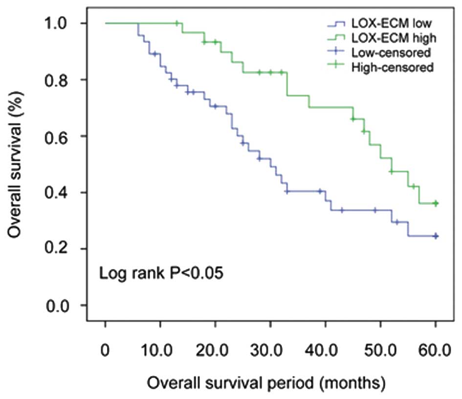
Associations between overall survival time and LOX
expression parameters were assessed by Kaplan-Meier survival
analysis with a log-rank test. As presented in Fig. 1, the mean overall survival time
significantly decreased in the low ECM LOX expression group
compared with the high ECM LOX expression group (low LOX in ECM:
Mean overall survival, 27.89 months; high LOX in ECM: Mean overall
survival, 40.61 months; P=0.023).
A multivariate analysis was performed based on the
Cox proportional hazards regression model that assessed the
independent predictive value of LOX expression in the ECM, in
addition to relevant clinical and pathological parameters,
including tumor stage, age, Gleason score, PSA and distant
metastasis. As presented in Table
IV, decreased LOX expression in the ECM was identified as an
independent prognostic marker for overall survival (P=0.032), in
addition to tumor stage (P=0.005) and Gleason score (P=0.008).
| Table IV.Cox regression analysis of prognostic
parameters for overall survival in patients with prostate
cancer. |
Table IV.
Cox regression analysis of prognostic
parameters for overall survival in patients with prostate
cancer.
| Overall survival
parameters | B | SE | Wald | df | P-value | HR | 95% CI |
|---|
| LOX-ECM | −0.760 | 0.355 | 4.594 | 1 | 0.032a | 0.468 | 0.233–0.937 |
| Gleason score | 1.175 | 0.440 | 7.117 | 1 | 0.008a | 3.238 | 1.366–7.675 |
| Stage | 1.389 | 0.491 | 8.002 | 1 | 0.005a | 4.013 | 1.532–10.508 |
Taken together, these results suggest that the
expression of LOX in the ECM has a prognostic effect independent of
Gleason score and tumor stage, and a decreased expression of LOX in
the ECM predicts an increased risk of tumor-associated
mortality.
Association between ECM LOX and
collagen I expression
To investigate whether LOX expression in the ECM was
associated with collagen I, the most important component of the ECM
(20), the current study analyzed the
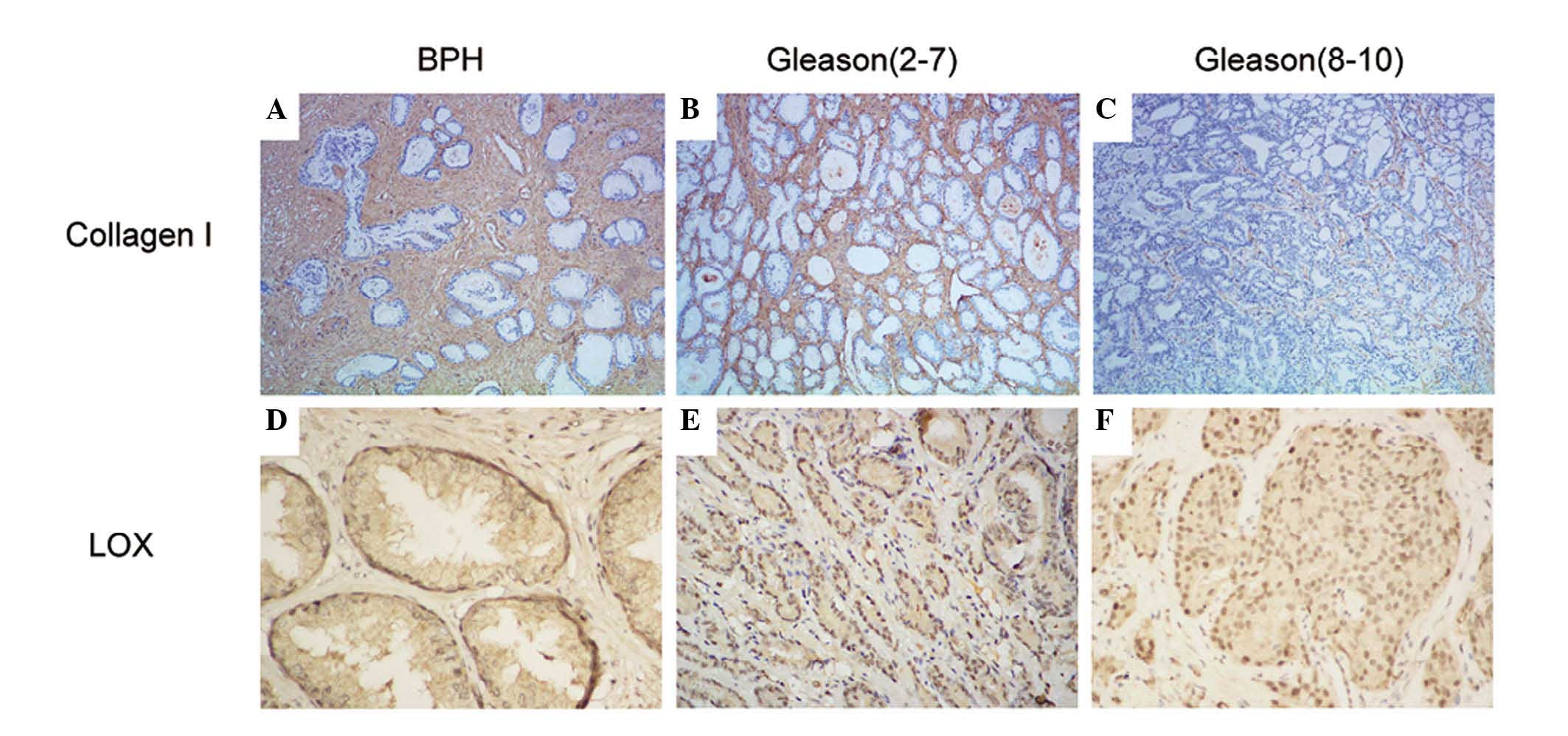
expression of collagen I. As presented in Fig. 2, collagen I was primarily expressed in
the ECM, and its expression levels were significantly reduced in
the Pca tissues compared with the BPH tissues (P=0.001). The
expression of collagen I in the ECM is in agreement with that of
LOX, which suggests that they are closely associated. Spearman's
rank correlation coefficient analysis identified a significant
correlation between ECM LOX percentage and collagen I percentage
(r=0.722; P=0.001).
Discussion
Over the last few years, a number of researchers
have focused on the association between solid tumor ECM remodeling
and malignant behavior (21,22). Previously published results have
indicated that the remodeling of the ECM is important in the
progression of Pca (23–25). LOX is well known for its role in ECM
remodeling, whereby the protein initiates cross-linking of elastin
and collagen I (26). However, LOX
contradictory functions have been observed intra- and
extracellularly in different solid malignancies; for instance, LOX
enhanced breast cancer metastasis but had a inhibitory effect on
hepatic and lung cancer (27). Thus,
the current study analyzed the expression levels of LOX and
collagen I in Pca and BPH tissues by immunohistochemistry, and
aimed to determine the association between LOX and the malignant
behavior of Pca.
Collagen I is the most abundant scaffold protein
observed in the ECM, and through its organization, orientation and
chemical modification, the malignant behavior of tumor cells may be
altered (28). The mechanism of
collagen remodeling has been studied in a number of solid tumors.
For example, following the deposition and cross-linking of collagen
induced by LOX, it was observed that phosphoinositide 3-kinase
signaling was activated, which promoted invasion and tumor
progression in breast cancer (29).
However, in accordance with a previous study (30), the current study demonstrated that the
expression of collagen I was significantly decreased in Pca
tissues, particularly in advanced Pca, compared with BPH tissues
(Fig. 2A-C). These results
demonstrate that the mechanism of ECM remodeling may vary in
different malignancies.
It is widely understood that collagen cross-linking
is primarily mediated by LOX and LOX-like enzymes, which are
commonly upregulated in various forms of cancer and at metastatic
sites, and that patients with high LOX expression have poor
survival in comparison with those with low LOX expression (31). In the current study, it was observed
that LOX exhibited a differential expression pattern between BPH
and Pca tissues. In the BPH tissues, LOX was located in the
cytoplasm, nucleus and ECM, whereas in the Pca tissues, a lower
level of LOX was secreted into the ECM. In addition, the expression
of LOX was higher in the low-grade Pca tissues compared with the
high-grade Pca tissues (Fig. 2D-F).
The expression patterns of LOX and collagen I corresponded to tumor
stage and Gleason score (Table IV).
These results demonstrate that the remodeling effect induced by LOX
was present in the ECM in Pca tissues, which is in accordance with
previous studies (32,33).
Multiple studies have reported that the LOX family
functions intra- and extracellularly in cancer tissues (34). To maintain homeostasis and tissue
stability, LOX is expressed intra- and extracellularly in normal
tissues (35). When tissue suffers
from injury or damage, the oxidative effect induced by LOX results
in the cross-linking of collagen and elastin in what is known as a
host response (36). This response is
similar to the formation of a ‘shell’, repairing the damage and
inhibiting tumor cell migration (37). Notably, a high expression of LOX was
observed in basal cells, which functions as another form of ‘shell’
to maintain glandular structure integrity. In the present study, it
was observed that, with the progression of Pca, LOX expression in
the ECM significantly decreased. Subsequently, it was speculated
that during the initial stages of Pca development, LOX may drive
collagen to form a host response to repair the damage arising from
tumorigenesis. However, with the progression of Pca, certain
hydrolytic enzymes such as MMPs may inhibit this process through
the degradation of collagen, resulting in a loss of the stimulatory
element. This may be the reason that the expression of LOX
decreases in the ECM with Pca progression.
To the best of our knowledge, LOX, MMPs and
fibronectin are key molecules in the ECM remodeling signaling
pathway (38,39). Following the initiation of Pca, the
tumor cells transform to acquire a malignant phenotype. Certain
important proteases, including LOX and MMPs, are secreted from the
intracellular space to the ECM, which may result in collagen
deposition, cross-linking and degradation. These changes in
components result in the remodeling of the cellular biomechanical
microenvironment. The signal transduction of fibronectin inside
tumor cells functions via integrins, which are molecular switches
located on the cell membrane. In addition, the secretion of focal
adhesion kinase and LOX strengthens the malignant phenotype.
Simultaneously, due to limited space, hyperproliferative cancer
cells compress against each other and generate tension that may
result in cytoskeletal deformation. The change in the biomechanical
properties of tumor cells may also affect the expression of
integrins. Finally, with the simulation of ECM remodeling and
biomechanical changes, the invasive and metastatic ability of Pca
cells are likely to be enhanced (34,40–42).
In conclusion, the present study identified that LOX
expression in the ECM is correlated with Pca stage, Gleason score
and tumor-associated mortality. Expression of LOX protein also
appeared to function as a marker for Pca prognosis. Thus, LOX may
operate as a cancer suppressor in Pca. However, in vivo
experiments are required to confirm the function of LOX in Pca
detection. Furthermore, additional studies are warranted to further
understand the mechanism of LOX in the regulation of Pca cell
apoptosis, proliferation, invasion and metastasis, in addition to
elucidate the association between LOX and the ECM.
Acknowledgements
The present study was supported by grants from the
Youth Fund of the National Natural Science Foundation of China
(Beijing, China; grant no. 81202030) and the National Natural
Science Foundation of China (grant nos. 81271603 and 81372761).
Glossary
Abbreviations
Abbreviations:
|
BPH
|
benign prostatic hyperplasia
|
|
Pca
|
prostate cancer
|
|
MMP
|
matrix metalloproteinase
|
|
ECM
|
extracellular matrix
|
References
|
1
|
Winchester DA, Gurel B, Till C, Goodman
PJ, Tangen CM, Santella RM, Johnson-Pais TL, Leach RJ, Thompson IM,
Xu J, et al: Key genes involved in the immune response are
generally not associated with intraprostatic inflammation in men
without a prostate cancer diagnosis: Results from the prostate
cancer prevention trial. Prostate. 76:565–574. 2016. View Article : Google Scholar : PubMed/NCBI
|
|
2
|
Bonnans C, Chou J and Werb Z: Remodelling
the extracellular matrix in development and disease. Nat Rev Mol
Cell Biol. 15:786–801. 2014. View
Article : Google Scholar : PubMed/NCBI
|
|
3
|
Chester D and Brown AC: The role of
biophysical properties of provisional matrix proteins in wound
repair. Matrix Biol S0945-053X. 30185–30188. 2016.
|
|
4
|
Hynes RO: The extracellular matrix: Not
just pretty fibrils. Science. 326:1216–1219. 2009. View Article : Google Scholar : PubMed/NCBI
|
|
5
|
Lo CM, Wang HB, Dembo M and Wang YL: Cell
movement is guided by the rigidity of the substrate. Biophys J.
79:144–152. 2000. View Article : Google Scholar : PubMed/NCBI
|
|
6
|
Yamada S, Wang KY, Tanimoto A and Sasaguri
Y: Novel function of histamine signaling in hyperlipidemia-induced
atherosclerosis: Histamine H1 receptors protect and H2 receptors
accelerate atherosclerosis. Pathol Int. 65:67–80. 2015. View Article : Google Scholar : PubMed/NCBI
|
|
7
|
Boufraqech M, Zhang L, Nilubol N, Sadowski
SM, Kotian S, Quezado M and Kebebew E: Lysyl oxidase (LOX)
transcriptionally regulates SNAI2 expression and TIMP4 secretion in
human cancers. Clin Cancer Res. 30:2016.(Epub ahead of print).
|
|
8
|
Palladini G, Ferrigno A, Richelmi P,
Perlini S and Vairetti M: Role of matrix metalloproteinases in
cholestasis and hepatic ischemia/reperfusion injury: A review.
World J Gastroenterol. 21:12114–12124. 2015. View Article : Google Scholar : PubMed/NCBI
|
|
9
|
Coussens LM, Fingleton B and Matrisian LM:
Matrix metalloproteinase inhibitors and cancer: Trials and
tribulations. Science. 295:2387–2392. 2002. View Article : Google Scholar : PubMed/NCBI
|
|
10
|
Levental KR, Yu H, Kass L, Lakins JN,
Egeblad M, Erler JT, Fong SF, Csiszar K, Giaccia A, Weninger W, et
al: Matrix crosslinking forces tumor progression by enhancing
integrin signaling. Cell. 139:891–906. 2009. View Article : Google Scholar : PubMed/NCBI
|
|
11
|
Kagan HM and Li W: Lysyl oxidase:
Properties, specificity, and biological roles inside and outside of
the cell. J Cell Biochem. 88:660–672. 2003. View Article : Google Scholar : PubMed/NCBI
|
|
12
|
Vadasz Z, Kessler O, Akiri G,
Gengrinovitch S, Kagan HM, Baruch Y, Izhak OB and Neufeld G:
Abnormal deposition of collagen around hepatocytes in Wilson's
disease is associated with hepatocyte specific expression of lysyl
oxidase and lysyl oxidase like protein-2. J Hepatol. 43:499–507.
2005. View Article : Google Scholar : PubMed/NCBI
|
|
13
|
Hase H, Jingushi K, Ueda Y, Kitae K, Egawa
H, Ohshio I, Kawakami R, Kashiwagi Y, Tsukada Y, Kobayashi T, et
al: LOXL2 status correlates with tumor stage and regulates integrin
levels to promote tumor progression in ccRCC. Mol Cancer Res.
12:1807–1817. 2014. View Article : Google Scholar : PubMed/NCBI
|
|
14
|
Cenizo V, André V, Reymermier C, Sommer P,
Damour O and Perrier E: LOXL as a target to increase the elastin
content in adult skin: A dill extract induces the LOXL gene
expression. Exp Dermatol. 15:574–581. 2006. View Article : Google Scholar : PubMed/NCBI
|
|
15
|
Liu X, Zhao Y, Gao J, Pawlyk B, Starcher
B, Spencer JA, Yanagisawa H, Zuo J and Li T: Elastic fiber
homeostasis requires lysyl oxidase-like 1 protein. Nat Genet.
36:178–182. 2004. View
Article : Google Scholar : PubMed/NCBI
|
|
16
|
Xiao Q and Ge G: Lysyl oxidase,
extracellular matrix remodeling and cancer metastasis. Cancer
Microenviron. 5:261–273. 2012. View Article : Google Scholar : PubMed/NCBI
|
|
17
|
Barker HE, Cox TR and Erler JT: The
rationale for targeting the LOX family in cancer. Nat Rev Cancer.
12:540–552. 2012. View
Article : Google Scholar : PubMed/NCBI
|
|
18
|
Erler JT, Bennewith KL, Nicolau M,
Dornhöfer N, Kong C, Le QT, Chi JT, Jeffrey SS and Giaccia AJ:
Lysyl oxidase is essential for hypoxia-induced metastasis. Nature.
440:1222–1226. 2006. View Article : Google Scholar : PubMed/NCBI
|
|
19
|
Chang SS and Amin MB: Utilizing the
tumor-node-metastasis staging for prostate cancer: The sixth
edition, 2002. CA Cancer J Clin. 58:54–59. 2008. View Article : Google Scholar : PubMed/NCBI
|
|
20
|
Liu W, Burdick JA and van Osch GJ:
Plant-derived recombinant human collagen: A strategic approach for
generating safe human ECM-based scaffold. Tissue Eng Part A.
19:1489–1490. 2013. View Article : Google Scholar : PubMed/NCBI
|
|
21
|
Alarab M, Kufaishi H, Lye S, Drutz H and
Shynlova O: Expression of extracellular matrix-remodeling proteins
is altered in vaginal tissue of premenopausal women with severe
pelvic organ prolapse. Reprod Sci. 21:704–715. 2014. View Article : Google Scholar : PubMed/NCBI
|
|
22
|
Wood SL, Pernemalm M, Crosbie PA and
Whetton AD: The role of the tumor-microenvironment in lung
cancer-metastasis and its relationship to potential therapeutic
targets. Cancer Treat Rev. 40:558–566. 2014. View Article : Google Scholar : PubMed/NCBI
|
|
23
|
Taddei ML, Giannoni E, Comito G and
Chiarugi P: Microenvironment and tumor cell plasticity: An easy way
out. Cancer Lett. 341:80–96. 2013. View Article : Google Scholar : PubMed/NCBI
|
|
24
|
Menen R, Pinney E, Hassanein MK, Kolostova
K, Bobek V, Suetsugu A, Zhang N, Bouvet M, Naughton GK and Hoffman
RM: Inhibition of metastasis of circulating human prostate cancer
cells in the chick embryo by an extracellular matrix produced by
foreskin fibroblasts in culture. Anticancer Res. 32:1573–1577.
2012.PubMed/NCBI
|
|
25
|
Palumbo A Jr, Ferreira LB, de Souza PA
Reis, Oliveira FL, Pontes B, Viana NB, Machado DE, Palmero CY,
Alves LM, Gimba ER and Nasciutti LE: Extracellular matrix secreted
by reactive stroma is a main inducer of pro-tumorigenic features on
LNCaP prostate cancer cells. Cancer Lett. 321:55–64. 2012.
View Article : Google Scholar : PubMed/NCBI
|
|
26
|
Przybyt E, van Luyn MJ and Harmsen MC:
Extracellular matrix components of adipose derived stromal cells
promote alignment, organization, and maturation of cardiomyocytes
in vitro. J Biomed Mater Res A. 103:1840–1848. 2014. View Article : Google Scholar : PubMed/NCBI
|
|
27
|
Grimsby JL, Lucero HA, Trackman PC, Ravid
K and Kagan HM: Role of lysyl oxidase propeptide in secretion and
enzyme activity. J Cell Biochem. 111:1231–1243. 2010. View Article : Google Scholar : PubMed/NCBI
|
|
28
|
Barcus CE, Holt EC, Keely PJ, Eliceiri KW
and Schuler LA: Dense collagen-I matrices enhance pro-tumorigenic
estrogen-prolactin crosstalk in MCF-7 and T47D breast cancer cells.
PLoS One. 10:e01168912015. View Article : Google Scholar : PubMed/NCBI
|
|
29
|
Li Y, Zhao H, Wang Y, Zheng H, Yu W, Chai
H, Zhang J, Falck JR, Guo AM, Yue J, et al: Isoliquiritigenin
induces growth inhibition and apoptosis through downregulating
arachidonic acid metabolic network and the deactivation of PI3K/Akt
in human breast cancer. Toxicol Appl Pharmacol. 272:37–48. 2013.
View Article : Google Scholar : PubMed/NCBI
|
|
30
|
Wang X, Wang J, Liu Y, Zong H, Che X,
Zheng W, Chen F, Zhu Z, Yang D and Song X: Alterations in
mechanical properties are associated with prostate cancer
progression. Med Oncol. 31:8762014. View Article : Google Scholar : PubMed/NCBI
|
|
31
|
Erler JT, Bennewith KL, Cox TR, Lang G,
Bird D, Koong A, Le QT and Giaccia AJ: Hypoxia-induced lysyl
oxidase is a critical mediator of bone marrow cell recruitment to
form the premetastatic niche. Cancer Cell. 15:35–44. 2009.
View Article : Google Scholar : PubMed/NCBI
|
|
32
|
Liu J, Ping W, Zu Y and Sun W:
Correlations of lysyl oxidase with MMP2/MMP9 expression and its
prognostic value in non-small cell lung cancer. Int J Clin Exp
Pathol. 7:6040–6047. 2014.PubMed/NCBI
|
|
33
|
Takahashi T, Friedmacher F, Takahashi H,
Hofmann A Daniel and Puri P: Lysyl oxidase expression is decreased
in the developing diaphragm and lungs of nitrofen-induced
congenital diaphragmatic hernia. Eur J Pediatr Surg. 25:15–19.
2015. View Article : Google Scholar : PubMed/NCBI
|
|
34
|
Cox TR, Bird D, Baker AM, Barker HE, Ho
MW, Lang G and Erler JT: LOX-mediated collagen crosslinking is
responsible for fibrosis-enhanced metastasis. Cancer Res.
73:1721–1732. 2013. View Article : Google Scholar : PubMed/NCBI
|
|
35
|
Wei L, Song XR, Sun JJ, Wang XW, Xie L and
Lv LY: Lysyl oxidase may play a critical role in hypoxia-induced
NSCLC cells invasion and migration. Cancer Biother Radiopharm.
27:672–677. 2012. View Article : Google Scholar : PubMed/NCBI
|
|
36
|
Tuinstra HM, Margul DJ, Goodman AG,
Boehler RM, Holland SJ, Zelivyanskaya ML, Cummings BJ, Anderson AJ
and Shea LD: Long-term characterization of axon regeneration and
matrix changes using multiple channel bridges for spinal cord
regeneration. Tissue Eng Part A. 20:1027–1037. 2014. View Article : Google Scholar : PubMed/NCBI
|
|
37
|
Burns-Cox N, Avery NC, Gingell JC and
Bailey AJ: Changes in collagen metabolism in prostate cancer: A
host response that may alter progression. J Urol. 166:1698–1701.
2001. View Article : Google Scholar : PubMed/NCBI
|
|
38
|
Kothapalli D, Liu SL, Bae YH, Monslow J,
Xu T, Hawthorne EA, Byfield FJ, Castagnino P, Rao S, Rader DJ, et
al: Cardiovascular protection by ApoE and ApoE-HDL linked to
suppression of ECM gene expression and arterial stiffening. Cell
Reports. 2:1259–1271. 2012. View Article : Google Scholar : PubMed/NCBI
|
|
39
|
Helleman J, Jansen MP, Ruigrok-Ritstier K,
van Staveren IL, Look MP, Meijer-van Gelder ME, Sieuwerts AM, Klijn
JG, Sleijfer S, Foekens JA, et al: Association of an extracellular
matrix gene cluster with breast cancer prognosis and endocrine
therapy response. Clin Cancer Res. 14:5555–5564. 2008. View Article : Google Scholar : PubMed/NCBI
|
|
40
|
Cho BA, Cho NH, Seong SY, Choi MS and Kim
IS: Intracellular invasion by Orientia tsutsugamushi is mediated by
integrin signaling and actin cytoskeleton rearrangements. Infect
Immun. 78:1915–1923. 2010. View Article : Google Scholar : PubMed/NCBI
|
|
41
|
Baker AM, Bird D, Lang G, Cox TR and Erler
JT: Lysyl oxidase enzymatic function increases stiffness to drive
colorectal cancer progression through FAK. Oncogene. 32:1863–1868.
2013. View Article : Google Scholar : PubMed/NCBI
|
|
42
|
Taubenberger AV, Bray LJ, Haller B,
Shaposhnykov A, Binner M, Freudenberg U, Guck J and Werner C: 3D
extracellular matrix interactions modulate tumour cell growth,
invasion and angiogenesis in engineered tumour microenvironments.
Acta Biomater. 36:73–85. 2016. View Article : Google Scholar : PubMed/NCBI
|