Introduction
Oral cancer is one the leading causes of cancer
mortality worldwide, and early diagnosis of high-risk, potentially
malignant lesions is a high priority for reducing both morbidity
and mortality (1–3). Oral leukoplakia (OL) is a clinically
diagnosed preneoplastic lesion of the oral cavity with a frequency
of malignant transformation of 17–24% (4–6). However,
the risk of malignant transformation of OL is still difficult to
assess. Although lesions with dysplastic features are considered to
be at a higher risk for malignant transformation, the majority of
oral cancers develop from lesions that lack dysplastic changes
(4,5).
Therefore, objective biomarkers are required to evaluate the risk
of malignant transformation in OL as well as for prophylactic
intervention and proper management of the high-risk patient group.
The cancer stem cell theory indicates that the initiation,
progression, recurrence and metastasis of head and neck squamous
cell carcinoma (HNSCC) are related to the behavior of a small
subpopulation of cancer stem cells (7–9). The
identification of these cells is important, not only for a better
understanding of early detection and progression of cancer, but
also for the development of more effective therapies. Diverse cell
surface markers have been used for the identification of cancer
stem cells in human tumors (10).
ALDH1 and podoplanin are two well-studied cancer stem cell markers
that have been implicated in several solid tumor types, including
oral cancer (11,12).
ALDH1 is an isoform of aldehyde dehydrogenase, which
is expressed in humans as a cytosolic detoxifying isoenzyme that
oxidizes intracellular aldehydes and contributes to the oxidation
of retinol to retinoic acid in early stem cell differentiation
(13). Previous studies have shown
that ALDH1 is a specific marker for the identification of head and
neck cancer stem cells, and serves a crucial role in maintaining
the self-renewal properties and tumorigenicity of HNSCC-derived
cancer stem cells (11,14–16).
Furthermore, ALDH1 overexpression was observed in oral dysplasia
(17), and it was suggested to be a
promising biomarker for distinguishing malignant from premalignant
OL (17) and other oral dysplasia
(18). In addition, ALDH1
immunoexpression is present in tissue samples of human epithelial
cancer, including HNSCC, and is a prognostic marker for HNSCC
survival (19–21).
Podoplanin is a mucin-type transmembrane
glycoprotein that is specifically expressed in lymphatic
endothelial cells but not in blood endothelial cells (22). High podoplanin expression was reported
in HNSCC with lymph node metastasis and poor survival (23). Wicki and Christofori (12) suggested that podoplanin may act as a
mediator of tumor cell invasion and metastasis. In addition,
overexpression of podoplanin in OL and lichen planus suggested the
use of podoplanin as a biomarker for oral cancer risk in patients
with oral premalignancy (24,25). Podoplanin is also expressed in a
number of hyperplastic and dysplastic areas adjacent to the primary
oral cancer, indicating that its abnormal expression occurs early
in oral tumorigenesis, and may serve a role in malignant
transformation (26). In the present
study, it was hypothesized that the expression patterns of ALDH1
and podoplanin in OL would have predictive value for early
detection of oral cancer. Therefore, the immunoexpression of ALDH1
and podoplanin was examined in samples from 79 patients with OL,
and their usefulness as biomarkers for cancer risk assessment was
evaluated.
Materials and methods
Patients and tissue specimens
All the medical records of 79 patients diagnosed
with OL from Janauray 2002 to July 2012 at Hokkaido University
Hospital (Sapporo, Japan) were retrieved and reviewed at the
Department of Oral Pathology and Biology, Hokkaido University
Graduate School of Dental Medicine (Sapporo, Japan). The clinical
data were obtained from the medical records, while the biopsy
specimens were obtained from formalin-fixed paraffin-embedded
tissues. The expression pattern of ALDH1 and podoplanin was
determined immunohistochemically in 79 OL patients during the
follow-up period. The latency of the progressor was defined as the
interval from the first biopsy of OL to subsequent OSCC
development. A minimum of 6 months latency was maintained in all
cases. Associations between the protein expression patterns and
clinicopathological parameters, including oral cancer development
during the follow-up period, were analyzed statistically. In the
present retrospective follow-up study, malignant transformation vs.
non-transformation was considered as a surrogate for clinical
outcomes of patients with OL. The present study was approved by the
institutional review board.
Histologic examination
Hematoxylin and eosin-stained slides were cut for
routine diagnostics. The World Health Organization criteria
(27) for OL and epithelial dysplasia
were used when examining the histopathology of the sections. The
presence of dysplasia was graded as mild, moderate or severe.
Dysplasia was mild when dysplastic changes were present in less
than a third of the epithelia, moderate when two thirds of the
epithelia were affected and severe when the whole thickness of the
epithelia was involved. During statistical analysis, mild dysplasia
was considered as low-grade dysplasia (LGD), while
moderate-to-severe dysplasia was considered as high-grade dysplasia
(HGD).
Tissue processing and
immunohistochemistry
Immunohistochemical staining was performed using the
streptavidin-peroxidase methods described previously (28). In brief, immunostaining was conducted
using 5-µm paraffin-embedded tissue serial sections. The slides
were deparaffinized in xylene, rehydrated in graded alcohol and
subjected to antigen retrieval by heat treatment in Tris-EDTA
buffer. To inhibit endogenous peroxidase activity, the slides were
immersed in 3% hydrogen peroxide for 5 min, followed by blocking
solution [1% bovine serum albumin (A8327; Sigma-Aldrich; Merck
Millipore, Darmstadt, Germany) in PBS) for 30 min. The slides were
then incubated with anti-ALDH1 (clone 44/ALDH1; 1:100 dilution; BD
Transduction Laboratories™; BD Biosciences, Franklin Lakes, NJ,
USA) and anti-podoplanin (clone D2-40; 1:100 dilution; Vector
Laboratories, Inc., Burlingame, CA, USA) monoclonal antibodies at
4°C overnight. The sections were then treated with
N-Histofine® Simple Stain Rat MAX PO (M) (Nichirei
Bioscience, Inc., Tokyo, Japan) at 37°C for 30 min. Visualization
was conducted using ChemMate™ DAKO EnVision™/HRP (Dako North
America, Inc., Carpinteria, CA, USA). Cytoplasmic and/or cell
membrane immunoreactivity in the epithelium was considered to
indicate positive ALDH1 and podoplanin expression, respectively. To
analyze the prognostic value for cancer development, the ALDH1 and
podoplanin expression levels were categorized as follows: Lesions
with no expression or expression restricted to the basal layer of
the oral epithelium were considered to be negative, while
immunoexpression extending to the suprabasal layer of the oral
epithelium was considered to be ALDH1 and podoplanin positive.
Statistical analysis
The associations between protein (ALDH1 and
podoplanin) expression and clinicopathological variables were
assessed using the Wilcoxon rank-sum test for continuously
distributed variables and the χ2 test for categorical
variables. Kaplan-Meier survival analysis was used to estimate the
events of interest for oral cancer-free survival (OCFS) and the
time interval from histopathological diagnosis to the development
of oral squamous cell carcinoma (OSCC). Patients who did not
develop invasive OSCC were censored at their last date of
follow-up. The log-rank test was used to compare survival times
among patients with different characteristics. The Cox regression
model was applied to evaluate the hazard ratio (HR) for malignant
transformation of OL. HRs with a 95% confidence interval (CI) and
P-values were reported. The likelihood ratio test was applied to
evaluate the point-prevalence value (PPV). All tests were two
sided, and P<0.05 was considered to indicate a statistically
significant difference. JMP® Pro version 10.0.2 (SAS
Institute Inc., Cary, NC, USA) was used for statistical
analysis.
Results
Patient characteristics, and ALDH1 and
podoplanin expression
According to the follow-up record, patients
primarily diagnosed as OL were grouped into untransformed (UT;
n=42) and malignant transformed OL (MT; n=37) lesions. Patient with
untransformed OL was defined as who did not develop into OSCC
during the follow-up period, while patient with malignant
transformed OL was defined as who subsequently developed into OSCC
during the follow-up period. Patients' baseline characteristics and
associations between expression of the proteins (ALDH1 and
podoplanin) and clinicopathological parameters are summarized in
Tables I and II, respectively. A significant difference
between protein expression patterns and degree of dysplasia was
noted, whereas differences in age, gender or lesion site were not
significant.
 | Table I.Patients' baseline
characteristics. |
Table I.
Patients' baseline
characteristics.
| Characteristics | N (%) |
|---|
| All patients | 79 |
| Age, years |
|
| Mean ±
SD | 70±12 |
|
Median | 72 |
| Gender |
|
|
Female | 54 (68) |
| Male | 25 (32) |
| Follow-up,
months |
|
| Mean ±
SD | 42.1±34.1 |
|
Median | 25 |
| Site |
|
|
Tongue | 28 (35) |
|
Gingiva | 18 (23) |
| BM | 21 (27) |
| FOM | 5 (6) |
|
Other | 7 (9) |
| Dysplasia |
|
| LGD | 27 (34) |
|
HGD | 52 (66) |
| ALDH1
expression |
|
|
Positive | 48 (61) |
|
Negative | 31 (39) |
| Podoplanin
expression |
|
|
Positive | 53 (67) |
|
Negative | 26 (33) |
| Co-expression of
ALDH1 and podoplanin |
|
|
Positive | 35 (44) |
|
Negative | 44 (56) |
| Expression of
either ALDH1 or podoplanin |
|
|
Positive | 66 (84) |
|
Negative | 13 (16) |
| Malignant
transformation |
|
|
Yes | 37 (47) |
| No | 42 (53) |
| Table II.Association between protein
expression (ALDH1 and podoplanin) and clinicopathological
parameters. |
Table II.
Association between protein
expression (ALDH1 and podoplanin) and clinicopathological
parameters.
|
|
| ALDH1
expression |
| Podoplanin
expression |
|
|---|
|
|
|
|
|
|
|
|---|
|
|
| Negative | Positive |
| Negative | Positive |
|
|---|
|
|
|
|
|
|
|
|
|
|---|
|
Characteristics | Patients, n | N | % | N | % | P-value | N | % | N | % | P-value |
|---|
| All patients | 79 | 31 | 39 | 48 | 61 |
| 26 | 33 | 53 | 67 |
|
| Age (years) |
|
|
|
|
| 0.557 |
|
|
|
| 0.475 |
| Mean ±
SD |
| 71±11 |
| 69±13 |
|
| 66±17 |
| 72±08 |
|
|
|
Median |
| 72 |
| 72 |
|
| 71 |
| 72 |
|
|
|
Range |
| 43–90 |
| 23–95 |
|
| 23–90 |
| 54–95 |
|
|
| Gender |
|
|
|
|
| 0.925 |
|
|
|
| 0.906 |
|
Female | 54 | 21 | 68 | 33 | 69 |
| 18 | 69 | 36 | 68 |
|
|
Male | 25 | 10 | 32 | 15 | 31 |
| 8 | 31 | 17 | 32 |
|
| Site |
|
|
|
|
| 0.045 |
|
|
|
| 0.015 |
|
Tongue | 28 | 11 | 35 | 17 | 35 |
| 12 | 46 | 16 | 30 |
|
|
Gingiva | 18 | 2 | 6 | 16 | 33 |
| 2 | 8 | 16 | 30 |
|
| BM | 21 | 12 | 39 | 9 | 20 |
| 7 | 27 | 14 | 26 |
|
|
FOM | 5 | 2 | 7 | 3 | 6 |
| 0 | 0 | 5 | 9 |
|
|
Other | 7 | 4 | 13 | 3 | 6 |
| 5 | 19 | 2 | 5 |
|
| Dysplasia |
|
|
|
|
| 0.243 |
|
|
|
| <0.001 |
|
LGD | 27 | 13 | 42 | 14 | 29 |
| 18 | 69 | 9 | 17 |
|
|
HGD | 52 | 18 | 58 | 34 | 71 |
| 8 | 31 | 44 | 83 |
|
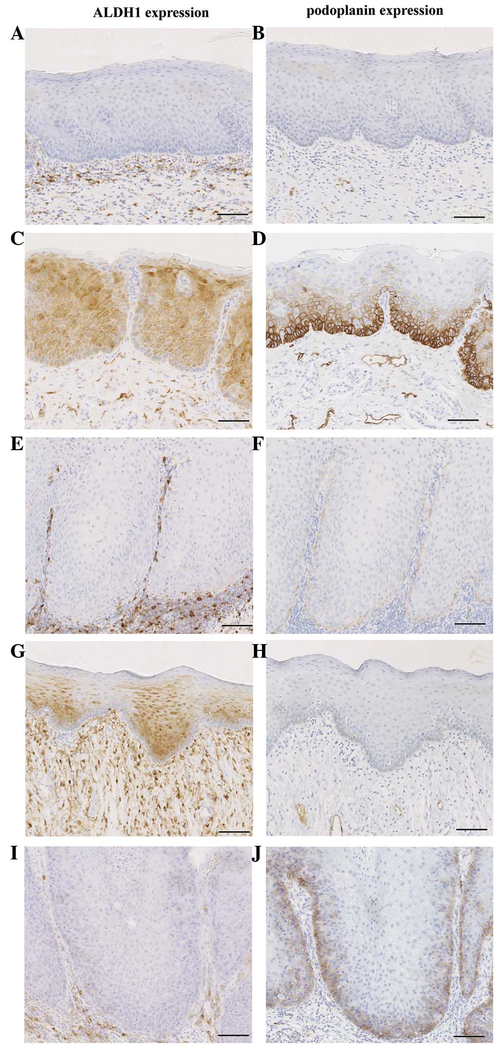
The expression patterns of ALDH1 and podoplanin in
normal epithelium and serial tissue samples of OL are illustrated
in Fig. 1. Among the 79 cases, 48
(61%) and 53 (67%) displayed positive expression of ALDH1 and
podoplanin, respectively. ALDH1 expression was observed in 21 of
the 42 (50%) patients with UT lesions and in 27 of the 37 (73%)
patients with MT lesions (P=0.037). Concurrently, podoplanin
expression was detected in 22 of the 42 (52%) patients with UT
lesions and in 31 of 37 (84%) patients with MT lesions (P=0.003).
Of interest, significant differences in co-expression of ALDH1 and
podoplanin (P=0.003), and expression of either ALDH1 or podoplanin
(P=0.013), were noted between cases of UT OL and cases of MT OL,
respectively (Fig. 2A).
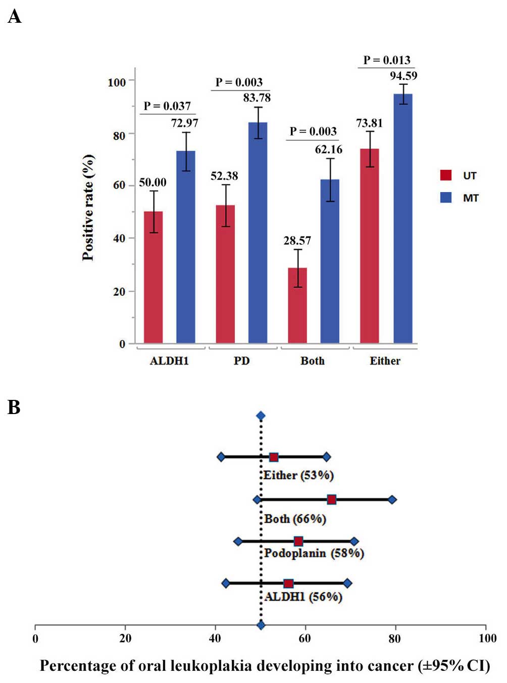 | Figure 2.Association between the percentages of
positive protein expression and malignant transformation. (A)
Percentages of ALDH1- and podoplanin-positive expression in
patients with UT lesions and patients with MT lesions of OL. (B)
Point-prevalence analysis of the incidence of oral cancer
development from OL with ALDH1 and podoplanin expression as PPVs
and the 95% CI. The PPVs of ALDH1 expression, podoplanin expression
and protein co-expression were 56, 58 and 66%, respectively. The
vertical dashed line indicates the 50% of this cohort that
developed malignancy. ALDH1, aldehyde dehydrogenase 1; PD,
podoplanin; MT, malignant transformed; UT, untransformed; PPV,
point-prevalence value; CI, confidence interval; OL, oral
leukoplakia. |
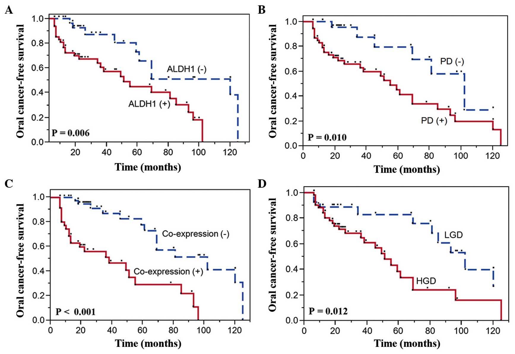
ALDH1 and podoplanin expression, and
risk of oral cancer
To estimate the time for malignant transformation of
OL, OCFS was analyzed by the Kaplan-Meier method using
clinicopathological factors, and ALDH1 and podoplanin expression.
In this analysis, the expression levels of ALDH1 and podoplanin,
and the grade of dysplasia were observed to be significant
indicators using the log-rank test. All findings are summarized in
Table III and illustrated in
Fig. 3. Patients with positive ALDH1
(Fig. 3A) and podoplanin expression
(Fig. 3B) experienced a significantly
higher oral cancer incidence than those with negative expression of
these proteins. Among the 79 cases, 10 of the 31 (32%)
ALDH1-negative cases and 27 of the 48 (56%) ALDH1-positive cases of
OL transformed into oral cancer (P=0.006), similarly to 6 of the 26
(23%) podoplanin-negative cases and 31 of the 53 (58%)
podoplanin-positive cases (P=0.010). Of interest, co-expression of
ALDH1 and podoplanin (P<0.001) in patients with OL was observed
to be strongly prognostic of malignant transformation (Fig. 3C). In addition, in 27 of the 52 (52%)
patients with HGD OL, malignant transformation occurred in a
significantly shorter period of time than in those with LGD lesions
(P=0.012) (Table III and Fig. 3D).
| Table III.Univariate survival analysis by
significant prognostic factors using the Kaplan-Meier model. |
Table III.
Univariate survival analysis by
significant prognostic factors using the Kaplan-Meier model.
|
|
| Patients not
transformed into malignancy | Patients
transformed into malignancy |
|
|
|---|
|
|
|
|
|
|
|
|---|
| Prognostic
factors | Patients, n | N | % | N | % | Median survival
time, months (95% CI) | P-value |
|---|
| ALDH1
expression |
|
|
|
|
|
| 0.006 |
|
Negative | 31 | 21 | 68 | 10 | 32 | 120 (59–125) |
|
|
Positive | 48 | 21 | 44 | 27 | 56 | 51 (34–85) |
|
| Podoplanin
expression |
|
|
|
|
|
| 0.010 |
|
Negative | 26 | 20 | 77 | 6 | 23 | 102
(45-~)a |
|
|
Positive | 53 | 22 | 42 | 31 | 58 | 55 (36–85) |
|
| Dysplasia |
|
|
|
|
|
| 0.012 |
|
LGD | 27 | 17 | 63 | 10 | 37 | 102
(69-~)a |
|
|
HGD | 52 | 25 | 48 | 27 | 52 | 51 (36–69) |
|
| Co-expression of
proteins (ALDH1 and podoplanin) |
|
|
|
|
|
| <0.001 |
|
Absent | 44 | 30 | 68 | 14 | 32 | 102 (69–125) |
|
|
Present | 35 | 12 | 34 | 23 | 66 | 38 (13–55) |
|
To evaluate the oral cancer risk in patients with
OL, clinicopathological parameters, and ALDH1 and podoplanin
expression were analyzed using the Cox proportional hazards model
(Table IV). In univariate analysis,
the expression levels of ALDH1 (HR=2.91; 95% CI=1.37–6.92; P=0.005)
and podoplanin (HR=2.95; 95% CI=1.32–7.89; P=0.007), and the grade
of dysplasia (HR=2.57; 95% CI=1.23–5.77; P=0.011) were
significantly associated with an increased risk of malignant
transformation. Multivariate analysis was performed to assess the
factors that had a significant impact on OCFS in univariate
analysis (histology, ALDH1 expression and podoplanin expression).
In multivariate analysis, the adjusted HR for malignant
transformation was 3.02 for ALDH1 expression (95% CI=1.39–7.38;
P=0.004). Notably, when histology and co-expression of ALDH1 and
podoplanin were considered as cofactors, the risk of malignant
transformation was considerably higher for OL with positive
co-expression of the above proteins compared with OL with negative
expression of both proteins (HR=3.64; 95% CI=1.71–8.25;
P<0.001).
| Table IV.Cox proportional hazards regression
models to estimate cancer development. |
Table IV.
Cox proportional hazards regression
models to estimate cancer development.
|
Characteristics | HR | 95% CI | P-value |
|---|
| Univariate
analysis |
|
|
|
| Age
(>70 vs. ≤70 years) | 1.44 | 0.74–2.89 | 0.285 |
| Gender
(male vs. female) | 1.58 | 0.72–3.29 | 0.239 |
|
Histology (HGD vs. LGD) | 2.57 | 1.23–5.77 | 0.011 |
| ALDH1
expression (positive vs. negative) | 2.91 | 1.37–6.92 | 0.005 |
|
Podoplanin expression
(positive vs. negative) | 2.95 | 1.32–7.89 | 0.007 |
| Multivariate
analysis (histology, ALDH1 and podoplanin expression) |
|
|
|
|
Histology (HGD vs. LGD) | 1.55 | 0.68–3.81 | 0.306 |
| ALDH1
expression (positive vs. negative) | 3.02 | 1.39–7.38 | 0.004 |
|
Podoplanin expression
(positive vs. negative) | 2.62 | 1.04–7.59 | 0.039 |
| Multivariate
analysis (histology, and co-expression of ALDH1 and
podoplanin) |
|
|
|
|
Histology (HGD vs. LGD) | 1.61 | 0.72–3.84 | 0.253 |
| Co
expression of ALDH1 and podoplanin | 3.64 | 1.71–8.25 | <0.001 |
| Present
vs. absent |
|
|
|
To determine the value of ALDH1 and podoplanin for
predicting OL malignant transformation, the expression of ALDH1 and
podoplanin was evaluated using their PPV. ALDH1 positivity (56%)
and podoplanin positivity (58%) were associated with higher
incidence of oral cancer development compared with the 50.0% cohort
average. Importantly, the expression of both ALDH1 and podoplanin
was a strong indicator for oral cancer development (66% of PPV with
marginal significance). In addition, 53% of the lesions with either
ALDH1 or podoplanin expression developed oral cancer (Fig. 2B).
Discussion
The current study attempted to determine the
usefulness of ALDH1 and podoplanin expression for predicting the
transformation of OL with varying grades of dysplasia into OSCC,
and to assess the clinical implications of ALDH1 and podoplanin
expression in patients with OL on the basis of the cancer stem cell
theory. Braakhuis et al (29)
proposed a patch-field carcinoma progression model of oral cancer,
and hypothesized that oral cancer development started with a ‘patch
stem cell’ that developed into an expanding subpopulation of stem
cells escaping growth control, eventually resulting in malignant
transformation.
ALDH1 has been demonstrated to be a cancer stem cell
marker in several solid tumor types, including OSCC (11,14). ALDH1
was reported to serve a crucial role in maintaining the
self-renewal properties and tumorigenicity of HNSCC-derived cancer
stem cells (30), and its
immunoexpression is associated with a poor prognosis of patients
with HNSCC (19,20). Visus et al (18) observed ALDH1 overexpression in samples
of oral dysplasia and HNSCC, and suggested that it was a marker for
distinguishing malignant from premalignant cells in HNSCC, in
addition to being an essential epitope for developing ALDH1-based
vaccines for HNSCC therapy. The present study observed that the
overexpression of ALDH1 in OL samples was a significant predictor
of malignant transformation.
Although the role of podoplanin in carcinogenesis is
still a matter of debate, it has been reported that podoplanin is
expressed in approximately 90% of OSCCs (21). Podoplanin is involved in the
remodeling of the cell cytoskeleton mediated by actin, and may
promote cell invasion by increasing cell motility (22). Podoplanin-positive cells in the
epithelial layers may represent upward clonal expansion of stem
cells during carcinogenesis and oral disorders, and such clonal
expansion may represent a higher risk of malignant transformation.
Kawaguchi et al (26) reported
that podoplanin was a marker of malignant transformation in OL and
other oral precancerous lesions. In agreement with that study, the
present study also demonstrated that podoplanin expression in OL
was associated with an increased risk of malignant transformation.
Other studies also support a relevant role for podoplanin in early
oral tumorigenesis, even though podoplanin expression alone may not
be sufficient to promote carcinogenesis (25,26,31).
In the present study, immunohistochemical staining
of both ALDH1 and podoplanin was performed to evaluate the oral
cancer risk in patients with OL. Kaplan-Meier analysis indicated a
significant impact for the 5-year OCFS rate, and demonstrated that
55% of patients with ALDH1 positivity developed OSCC compared with
27% of those with ALDH1 negativity. In addition, 55% of patients
with podoplanin positivity developed OSCC compared with 20% of
those with podoplanin negativity. Multivariate analysis revealed
that the expression of ALDH1 and podoplanin was associated with a
3.02- and 2.62-fold increased risk of transformation, respectively.
Taken together, these data not only support the potential
importance of ALDH1 and podoplanin in oral carcinogenesis, but also
suggest that both proteins may be used as biomarkers for evaluating
the malignant transformation risk in oral premalignancy. However,
contrary to previous findings, 10 (32%) ALDH1- and 6 (23%)
podoplanin-negative patients also developed cancers (although later
than expression-positive patients) in the present study. Their
delayed but cancerous transformation can be attributed to the
following possible reasons: The lesions were biopsied prior to when
the abnormality occurred, or those cancers originated from lesions
not clinically visible at the time of biopsy, which therefore
remained unexamined. Another possibility is that the biopsies were
obtained from different clonal sites than those from which the
cancers eventually developed (26).
Although there is general agreement that the rate of
malignant transformation increases with the severity of the
dysplasia, certain authors did not observe a significant
association between epithelial dysplasia and malignant
transformation (32). The present
study demonstrated that the grade of dysplasia was a significant
risk factor for malignant transformation; however, the prognostic
values of ALDH1 and podoplanin proteins were superior to that of
histological grading, which was associated with a 1.55-fold
increased risk. Furthermore, significant differences in the
co-expression of ALDH1 and podoplanin proteins, and histological
examination were also noted in malignant transformation of OL. This
suggested that the co-expression of both biomarkers may be more
informative than the histological examination alone. Therefore,
immunohistochemical staining of ALDH1 and podoplanin could help to
augment the predictability and reliability of cancer risk
assessment for OL in association with histopathologic assessment of
epithelial dysplasia.
Cancer of the oral cavity results in severe
morbidity, limited quality of life and short overall survival, and
there is a strong requirement to understand oral carcinogenesis and
to establish accurate and reliable predictors of oral cancer risk
(1–3,33). The
interpretation of the protein expression of significant biomarkers
is relatively simple in routine/diagnostic laboratories. Therefore,
immunohistochemical staining of biomarkers is promising for the
evaluation of oral cancer risk (17).
To the best of our knowledge, the present report is
the first study investigating the combined expression of ALDH1 and
podoplanin for oral cancer risk assessment in patients with OL. Our
data revealed that positive expression of ALDH1 and podoplanin in
OL was significantly associated with the risk of malignant
transformation and, consequently, such leukoplakia should be
followed carefully. However, further studies are required to fully
define the functional roles of these biomarkers in oral cancer
initiation and disease progression.
In summary, ALDH1 and podoplanin can be used as
biomarkers for risk assessment of oral malignant transformation in
patients with OL.
Acknowledgements
The authors thank Mr. Yohei Murayama and Ms. Tomomi
Takahashi (Support Section for Education and Research, Hokkaido
University Graduate School of Dental Medicine, Sapporo, Japan) for
their technical assistance.
References
|
1
|
Zini A, Czerninski R and Sgan-Cohen HD:
Oral cancer over four decades: Epidemiology, trends, histology, and
survival by anatomical sites. J Oral Pathol Med. 39:299–305. 2010.
View Article : Google Scholar : PubMed/NCBI
|
|
2
|
Ganly I, Patel S and Shah J: Early stage
squamous cell cancer of the oral tongue-clinicopathologic features
affecting outcome. Cancer. 118:101–111. 2012. View Article : Google Scholar : PubMed/NCBI
|
|
3
|
Warnakulasuriya S: Living with oral
cancer: Epidemiology with particular reference to prevalence and
life-style changes that influence survival. Oral Oncol. 46:407–410.
2010. View Article : Google Scholar : PubMed/NCBI
|
|
4
|
Papadimitrakopoulou VA, Hong WK, Lee JS,
Martin JW, Lee JJ, Batsakis JG and Lippan SM: Low-dose isotretinoin
versus beta-carotene to prevent oral carcinogenesis: Long-term
follow-up. J Natl Cancer Inst. 89:257–258. 1997. View Article : Google Scholar : PubMed/NCBI
|
|
5
|
Silverman S Jr, Gorsky M and Lozada F:
Oral leukoplakia and malignant transformation. A follow-up study of
257 patients. Cancer. 53:563–568. 1984. View Article : Google Scholar : PubMed/NCBI
|
|
6
|
Lee JJ, Hong WK, Hittelman WN, Mao L,
Lotan R, Shin DM, Benner SE, Xu XC, Lee JS, Papadimitrakopoulou VM,
et al: Predicting cancer development in oral leukoplakia: Ten years
of translational research. Clin Cancer Res. 6:1702–1710.
2000.PubMed/NCBI
|
|
7
|
Sayed SI, Dwivedi RC, Katna R, Garg A,
Pathak KA, Nutting CM, Rhys-Evans P, Harrington KJ and Kazi R:
Implications of understanding cancer stem cell (CSC) biology in
head and neck squamous cell cancer. Oral Oncol. 47:237–243. 2011.
View Article : Google Scholar : PubMed/NCBI
|
|
8
|
Zhang Z, Filho MS and Nör JE: The biology
of head and neck cancer stem cells. Oral Oncol. 48:1–9. 2012.
View Article : Google Scholar : PubMed/NCBI
|
|
9
|
Krishnamurthy S and Nör JE: Head and neck
cancer stem cells. J Dent Res. 91:334–340. 2012. View Article : Google Scholar : PubMed/NCBI
|
|
10
|
Gires O: Lessons from common markers of
tumor-initiating cells in solid cancers. Cell Mol Life Sci.
68:4009–4022. 2011. View Article : Google Scholar : PubMed/NCBI
|
|
11
|
Chen YC, Chen YW, Hsu HS, Tseng LM, Huang
PI, Lu KH, Chen DT, Tai LK, Yung MC, Chang SC, et al: Aldehyde
dehydrogenase 1 is a putative marker for cancer stem cells in head
and neck squamous cancer. Biochem Biophys Res Commun. 385:307–313.
2009. View Article : Google Scholar : PubMed/NCBI
|
|
12
|
Wicki A and Christofori G: The potential
role of podoplanin in tumor invasion. Br J Cancer. 96:1–5. 2007.
View Article : Google Scholar : PubMed/NCBI
|
|
13
|
Marcato P, Dean CA, Giacomantonio CA and
Lee PW: Aldehyde dehydrogenase: Its role as a cancer stem cell
marker comes down to the specific isoform. Cell Cycle.
10:1378–1384. 2011. View Article : Google Scholar : PubMed/NCBI
|
|
14
|
Clay MR, Tabor M, Owen JH, Carev TE,
Bradford CR, Golf GT, Wicha MS and Prince ME: Single-marker
identification of head and neck squamous cell carcinoma cancer stem
cells with aldehyde dehydrogenase. Head Neck. 32:1195–1201. 2010.
View Article : Google Scholar : PubMed/NCBI
|
|
15
|
Chen YC, Chang CJ, Hsu HS, Chen YW, Tai
LK, Tseng LM, Chiou GY, Chang SC, Kao SY, Chiou SH and Lo WL:
Inhibition of tumorigenicity and enhancement of
radiochemosensitivity in head and neck squamous cell cancer-derived
ALDH1-positive cells by knockdown of Bmi-1. Oral Oncol. 46:158–165.
2010. View Article : Google Scholar : PubMed/NCBI
|
|
16
|
Chen C, Wei Y, Hummel M, Hoffmann TK,
Gross M, Kaufmann AM and Albers AE: Evidence for
epithelial-mesenchymal transition in cancer stem cells of head and
neck squamous cell carcinoma. PLoS One. 6:e164662011. View Article : Google Scholar : PubMed/NCBI
|
|
17
|
Liu W, Wu L, Shen XM, Shi LJ, Zhang CP, Xu
LQ and Zhou ZT: Expression patterns of cancer stem cell markers
ALDH1 and CD133 correlate with a high risk of malignant
transformation of oral leukoplakia. Int J Cancer. 132:868–874.
2013. View Article : Google Scholar : PubMed/NCBI
|
|
18
|
Visus C, Ito D, Amoscato A,
Macieiewska-Franczak M, Abdelsalem A, Dhir R, Shin DM, Donnenbera
VS, Whiteside TL and DeLeo AB: Identification of human aldehyde
dehydrogenase 1 family member A1 as a novel CD8+ T-cell-defined
tumor antigen in squamous cell carcinoma of the head and neck.
Cancer Res. 67:10538–10545. 2007. View Article : Google Scholar : PubMed/NCBI
|
|
19
|
Chen YW, Chen KH, Huang PI, Chen YC, Chiou
GY, Lo WL, Tseng LM, Hsu HS, Chang KW and Chiou SH: Cucurbitacin I
suppressed stem-like property and enhanced radiation-induced
apoptosis in head and neck squamous carcinoma-derived CD44(+)
ALDH1(+) cells. Mol Cancer Ther. 9:2879–2892. 2010. View Article : Google Scholar : PubMed/NCBI
|
|
20
|
Luo WR, Gao F, Li SY and Yao KT: Tumor
budding and the expression of cancer stem cell marker aldehyde
dehydrogenase 1 in nasopharyngeal carcinoma. Histopathology.
61:1072–1081. 2012. View Article : Google Scholar : PubMed/NCBI
|
|
21
|
Deng S, Yang X, Lassus H, Liang S, Kaur S,
Ye Q, Li C, Wang LP, Roby KF, Orsulic S, et al: Distinct expression
levels and patterns of stem cell marker, aldehyde dehydrogenase
isoform 1 (ALDH1), in human epithelial cancers. PLoS One.
5:e102772010. View Article : Google Scholar : PubMed/NCBI
|
|
22
|
Kahn HJ and Marks A: A new monoclonal
antibody, D2-40, for detection of lymphatic invasion in primary
tumors. Lab Invest. 82:1255–1257. 2002. View Article : Google Scholar : PubMed/NCBI
|
|
23
|
Yuan P, Temam S, El-Naggar A, Zhou X, Liu
DD, Lee JJ and Mao L: Over expression of podoplanin in oral cancer
and its association with poor clinical outcome. Cancer.
107:563–569. 2006. View Article : Google Scholar : PubMed/NCBI
|
|
24
|
Shi P, Liu W, Zhou ZT, He QB and Jiang WW:
Podoplanin and ABCG2: Malignant transformation risk markers for
oral lichen planus. Cancer Epidemiol Biomarkers Prev. 19:844–849.
2010. View Article : Google Scholar : PubMed/NCBI
|
|
25
|
Saintigny P, El-Naggar AK,
Papadimitrakopoulou V, Ren H, Fan YH, Feng L, Lee JJ, Kim ES, Hong
WK, Lippman SM and Mao L: DeltaNp63 over expression, alone and in
combination with other biomarkers, predicts the development of oral
cancer in patients with leukoplakia. Clin Cancer Res. 15:6284–6291.
2009. View Article : Google Scholar : PubMed/NCBI
|
|
26
|
Kawaguchi H, El-Naggar AK,
Papadimitrakopoulou V, Ren H, Fan YH, Feng L, Lee JJ, Kim E, Hong
WK, Lippman SM and Mao L: Podoplanin: A novel marker for oral
cancer risk in patients with oral premalignancy. J Clin Oncol.
26:354–360. 2008. View Article : Google Scholar : PubMed/NCBI
|
|
27
|
Pindborg JJ, Reichart PA, Smith CJ and Van
der Waal I: Histological Typing of Cancer and Precancer of the Oral
Mucosa. World Health Organization. Springer; Berlin: pp. 25–26.
1997
|
|
28
|
Habiba U, Kitamura T, Yanagawa-Matsuda A,
Hida K, Higashno F, Ohiro Y and Shindoh M: Cytoplasmic expression
of HuR may be a valuable diagnostic tool for determining the
potential for malignant transformation of oral verrucous borderline
lesions. Oncol Rep. 31:1547–1554. 2014.PubMed/NCBI
|
|
29
|
Braakhuis BJ, Leemans CR and Brakenhoff
RH: A genetic progression model of oral cancer: Current evidence
and clinical implications. J Oral Patho Med. 33:317–322. 2004.
View Article : Google Scholar
|
|
30
|
Wu A, Luo W, Zhang Q, Yang Z, Zhang G, Li
S and Yao K: Aldehyde dehydrogenase 1, a functional marker for
identifying cancer stem cells in human nasopharyngeal carcinoma.
Cancer Lett. 330:181–189. 2013. View Article : Google Scholar : PubMed/NCBI
|
|
31
|
Inoue H, Miyazaki Y, Kikuchi K, Yoshida N,
Ide F, Ohmori Y, Tomomura A, Sakashita H and Kusama K: Podoplanin
expression during dysplasia-carcinoma sequence in the oral cavity.
Tumor Biol. 33:181–194. 2012. View Article : Google Scholar
|
|
32
|
Holmstrup P, Vedtofte P, Reibel J and
Stoltze K: Long term treatment outcome of oral premalignant
lesions. Oral Oncol. 42:461–474. 2006. View Article : Google Scholar : PubMed/NCBI
|
|
33
|
de Vicente J Carlos, Palbo J Rodrigo,
Santamarta-Rodriguez T, Lequerica-Fernández P, Allonca E and
García-Pedrero JM: Podoplanin expression in oral leukoplakia:
Tumorigenic role. Oral Oncol. 49:598–603. 2013. View Article : Google Scholar : PubMed/NCBI
|