Spandidos Publications Logo
  • About
    • About Spandidos
    • Aims and Scopes
    • Abstracting and Indexing
    • Editorial Policies
    • Reprints and Permissions
    • Job Opportunities
    • Terms and Conditions
    • Contact
  • Journals
    • All Journals
    • Oncology Letters
      • Oncology Letters
      • Information for Authors
      • Editorial Policies
      • Editorial Board
      • Aims and Scope
      • Abstracting and Indexing
      • Bibliographic Information
      • Archive
    • International Journal of Oncology
      • International Journal of Oncology
      • Information for Authors
      • Editorial Policies
      • Editorial Board
      • Aims and Scope
      • Abstracting and Indexing
      • Bibliographic Information
      • Archive
    • Molecular and Clinical Oncology
      • Molecular and Clinical Oncology
      • Information for Authors
      • Editorial Policies
      • Editorial Board
      • Aims and Scope
      • Abstracting and Indexing
      • Bibliographic Information
      • Archive
    • Experimental and Therapeutic Medicine
      • Experimental and Therapeutic Medicine
      • Information for Authors
      • Editorial Policies
      • Editorial Board
      • Aims and Scope
      • Abstracting and Indexing
      • Bibliographic Information
      • Archive
    • International Journal of Molecular Medicine
      • International Journal of Molecular Medicine
      • Information for Authors
      • Editorial Policies
      • Editorial Board
      • Aims and Scope
      • Abstracting and Indexing
      • Bibliographic Information
      • Archive
    • Biomedical Reports
      • Biomedical Reports
      • Information for Authors
      • Editorial Policies
      • Editorial Board
      • Aims and Scope
      • Abstracting and Indexing
      • Bibliographic Information
      • Archive
    • Oncology Reports
      • Oncology Reports
      • Information for Authors
      • Editorial Policies
      • Editorial Board
      • Aims and Scope
      • Abstracting and Indexing
      • Bibliographic Information
      • Archive
    • Molecular Medicine Reports
      • Molecular Medicine Reports
      • Information for Authors
      • Editorial Policies
      • Editorial Board
      • Aims and Scope
      • Abstracting and Indexing
      • Bibliographic Information
      • Archive
    • World Academy of Sciences Journal
      • World Academy of Sciences Journal
      • Information for Authors
      • Editorial Policies
      • Editorial Board
      • Aims and Scope
      • Abstracting and Indexing
      • Bibliographic Information
      • Archive
    • International Journal of Functional Nutrition
      • International Journal of Functional Nutrition
      • Information for Authors
      • Editorial Policies
      • Editorial Board
      • Aims and Scope
      • Abstracting and Indexing
      • Bibliographic Information
      • Archive
    • International Journal of Epigenetics
      • International Journal of Epigenetics
      • Information for Authors
      • Editorial Policies
      • Editorial Board
      • Aims and Scope
      • Abstracting and Indexing
      • Bibliographic Information
      • Archive
    • Medicine International
      • Medicine International
      • Information for Authors
      • Editorial Policies
      • Editorial Board
      • Aims and Scope
      • Abstracting and Indexing
      • Bibliographic Information
      • Archive
  • Articles
  • Information
    • Information for Authors
    • Information for Reviewers
    • Information for Librarians
    • Information for Advertisers
    • Conferences
  • Language Editing
Spandidos Publications Logo
  • About
    • About Spandidos
    • Aims and Scopes
    • Abstracting and Indexing
    • Editorial Policies
    • Reprints and Permissions
    • Job Opportunities
    • Terms and Conditions
    • Contact
  • Journals
    • All Journals
    • Biomedical Reports
      • Information for Authors
      • Editorial Policies
      • Editorial Board
      • Aims and Scope
      • Abstracting and Indexing
      • Bibliographic Information
      • Archive
    • Experimental and Therapeutic Medicine
      • Information for Authors
      • Editorial Policies
      • Editorial Board
      • Aims and Scope
      • Abstracting and Indexing
      • Bibliographic Information
      • Archive
    • International Journal of Epigenetics
      • Information for Authors
      • Editorial Policies
      • Editorial Board
      • Aims and Scope
      • Abstracting and Indexing
      • Bibliographic Information
      • Archive
    • International Journal of Functional Nutrition
      • Information for Authors
      • Editorial Policies
      • Editorial Board
      • Aims and Scope
      • Abstracting and Indexing
      • Bibliographic Information
      • Archive
    • International Journal of Molecular Medicine
      • Information for Authors
      • Editorial Policies
      • Editorial Board
      • Aims and Scope
      • Abstracting and Indexing
      • Bibliographic Information
      • Archive
    • International Journal of Oncology
      • Information for Authors
      • Editorial Policies
      • Editorial Board
      • Aims and Scope
      • Abstracting and Indexing
      • Bibliographic Information
      • Archive
    • Medicine International
      • Information for Authors
      • Editorial Policies
      • Editorial Board
      • Aims and Scope
      • Abstracting and Indexing
      • Bibliographic Information
      • Archive
    • Molecular and Clinical Oncology
      • Information for Authors
      • Editorial Policies
      • Editorial Board
      • Aims and Scope
      • Abstracting and Indexing
      • Bibliographic Information
      • Archive
    • Molecular Medicine Reports
      • Information for Authors
      • Editorial Policies
      • Editorial Board
      • Aims and Scope
      • Abstracting and Indexing
      • Bibliographic Information
      • Archive
    • Oncology Letters
      • Information for Authors
      • Editorial Policies
      • Editorial Board
      • Aims and Scope
      • Abstracting and Indexing
      • Bibliographic Information
      • Archive
    • Oncology Reports
      • Information for Authors
      • Editorial Policies
      • Editorial Board
      • Aims and Scope
      • Abstracting and Indexing
      • Bibliographic Information
      • Archive
    • World Academy of Sciences Journal
      • Information for Authors
      • Editorial Policies
      • Editorial Board
      • Aims and Scope
      • Abstracting and Indexing
      • Bibliographic Information
      • Archive
  • Articles
  • Information
    • For Authors
    • For Reviewers
    • For Librarians
    • For Advertisers
    • Conferences
  • Language Editing
Login Register Submit
  • This site uses cookies
  • You can change your cookie settings at any time by following the instructions in our Cookie Policy. To find out more, you may read our Privacy Policy.

    I agree
Search articles by DOI, keyword, author or affiliation
Search
Advanced Search
presentation
Oncology Letters
Join Editorial Board Propose a Special Issue
Print ISSN: 1792-1074 Online ISSN: 1792-1082
Journal Cover
January-2017 Volume 13 Issue 1

Full Size Image

Sign up for eToc alerts
Recommend to Library

Journals

International Journal of Molecular Medicine

International Journal of Molecular Medicine

International Journal of Molecular Medicine is an international journal devoted to molecular mechanisms of human disease.

International Journal of Oncology

International Journal of Oncology

International Journal of Oncology is an international journal devoted to oncology research and cancer treatment.

Molecular Medicine Reports

Molecular Medicine Reports

Covers molecular medicine topics such as pharmacology, pathology, genetics, neuroscience, infectious diseases, molecular cardiology, and molecular surgery.

Oncology Reports

Oncology Reports

Oncology Reports is an international journal devoted to fundamental and applied research in Oncology.

Experimental and Therapeutic Medicine

Experimental and Therapeutic Medicine

Experimental and Therapeutic Medicine is an international journal devoted to laboratory and clinical medicine.

Oncology Letters

Oncology Letters

Oncology Letters is an international journal devoted to Experimental and Clinical Oncology.

Biomedical Reports

Biomedical Reports

Explores a wide range of biological and medical fields, including pharmacology, genetics, microbiology, neuroscience, and molecular cardiology.

Molecular and Clinical Oncology

Molecular and Clinical Oncology

International journal addressing all aspects of oncology research, from tumorigenesis and oncogenes to chemotherapy and metastasis.

World Academy of Sciences Journal

World Academy of Sciences Journal

Multidisciplinary open-access journal spanning biochemistry, genetics, neuroscience, environmental health, and synthetic biology.

International Journal of Functional Nutrition

International Journal of Functional Nutrition

Open-access journal combining biochemistry, pharmacology, immunology, and genetics to advance health through functional nutrition.

International Journal of Epigenetics

International Journal of Epigenetics

Publishes open-access research on using epigenetics to advance understanding and treatment of human disease.

Medicine International

Medicine International

An International Open Access Journal Devoted to General Medicine.

Journal Cover
January-2017 Volume 13 Issue 1

Full Size Image

Sign up for eToc alerts
Recommend to Library

  • Article
  • Citations
    • Cite This Article
    • Download Citation
    • Create Citation Alert
    • Remove Citation Alert
    • Cited By
  • Similar Articles
    • Related Articles (in Spandidos Publications)
    • Similar Articles (Google Scholar)
    • Similar Articles (PubMed)
  • Download PDF
  • Download XML
  • View XML
Article Open Access

The diagnostic utility of PAX8 immunostaining of malignant peritoneal mesothelioma presenting as serous ovarian carcinoma: A single‑center report of two cases

  • Authors:
    • Kohei Nakamura
    • Kentaro Nakayama
    • Risa Nagaoka
    • Kiyoka Nishisako
    • Masako Ishikawa
    • Hiroshi Katagiri
    • Tomoka Ishibashi
    • Emi Sato
    • Chika Amano
    • Satoru Kyo
  • View Affiliations / Copyright

    Affiliations: Department of Obstetrics and Gynecology, Shimane University School of Medicine, Izumo, Shimane 6938501, Japan, Department of Organ Pathology, Shimane University School of Medicine, Izumo, Shimane 6938501, Japan
    Copyright: © Nakamura et al. This is an open access article distributed under the terms of Creative Commons Attribution License.
  • Pages: 263-266
    |
    Published online on: November 30, 2016
       https://doi.org/10.3892/ol.2016.5444
  • Expand metrics +
Metrics: Total Views: 0 (Spandidos Publications: | PMC Statistics: )
Metrics: Total PDF Downloads: 0 (Spandidos Publications: | PMC Statistics: )
Cited By (CrossRef): 0 citations Loading Articles...

This article is mentioned in:


Abstract

Malignant peritoneal mesotheliomas (MPMs) are rare and progressive tumors, which may present similarly to primary peritoneal carcinoma or ovarian carcinoma (OC). The current study reports two cases of MPM that initially presented with the features of OC, for which paired box 8 (PAX8) immunostaining was found to be useful for diagnosis. The two patients were women, aged 58 and 56 years, respectively. The primary presenting symptoms and clinical findings included prolonged abdominal pain, abdominal swelling and cough. The two cases were initially diagnosed as OC and were treated with primary debulking surgery. The patient in case 1 had no history of asbestos exposure, while the patient in case 2 did. Final diagnoses were determined based on histological and immunohistochemical results, which included negative PAX8 immunostaining, and which were consistent with MPM. The present cases demonstrated that PAX8 negativity may be a useful diagnostic biomarker for differentiating MPM from OC.

Introduction

Malignant peritoneal mesothelioma (MPM) is a rare disease that typically demonstrates a poor prognosis, with an average survival time of 6–12 months (1). The incidence in the United States is 200–400 novel cases annually (2). Although asbestos exposure is the primary risk factor for the development of MPM, only 30% of reported cases possess a history of asbestos exposure (3).

The clinical and morphological distinction between MPM and serous ovarian carcinoma (OC) may be difficult due to their overlapping morphological features. This is particularly true when the latter is of low morphological grade and is associated with diffuse invasive peritoneal implants, or when high-grade serous carcinoma has metastasized to the pleura (4). Due to this, a number of studies have attempted to use a variety of immunohistochemical markers to distinguish between the two diseases (5,6). However, few immunostaining markers have proven to be sufficiently specific or sensitive for either type of cancer. Calretinin, Wilms tumor 1 (WT1), D2-40 and mesothelin are expressed in the majority of mesotheliomas (high sensitivity); however, these markers may additionally be expressed in a significant subset of serous OC cases (low specificity) (5). Alternative markers, including Ber-EP4, human epididymis protein 4, cluster of differentiation (CD)15 and B72.3, have been demonstrated to be expressed more frequently in serous OC compared with mesothelioma; however, poor sensitivity or specificity of these markers has limited their use as reliable discriminators (5).

Paired box 8 (PAX8) is a member of the paired box family of transcription factors, and is significant in organogenesis of the Müllerian system (7). In the Müllerian system, PAX8 is expressed in a variety of ovarian tumors, particularly serous carcinoma. Secretory cells of the normal fallopian tube are positive for PAX8 expression, and these cells are thought to be the origin of serous OC in a high proportion of cases (7). Previous studies have suggested that PAX8 immunostaining may be useful for differentiating MPM from serous OC with high specificity and sensitivity (5,6).

A delayed diagnosis of MPM is common due to the long interval between initial asbestos exposure and the onset of symptoms (3). Furthermore, the symptoms, including abdominal pain, ascites and abdominal distention without abdominal pain, are non-specific. Therefore, exact diagnosis of MPM is difficult, and it may appear to present as primary peritoneal carcinoma or OC (3). In the present study, two cases of MPM that were distinguished from OC by immunostaining for PAX8 are discussed.

Case report

Case 1
Patient

A 58-year-old (gravida 2, para 2) woman presented with abdominal distension. The patient had no history of exposure to asbestos, and no significant past medical or family history. The serum cancer antigen 125 (CA125) level was 90 U/ml (normal, <35 U/ml). The sialyl-Tn (STn) antigen level was within normal limits, and the general examination was also normal.

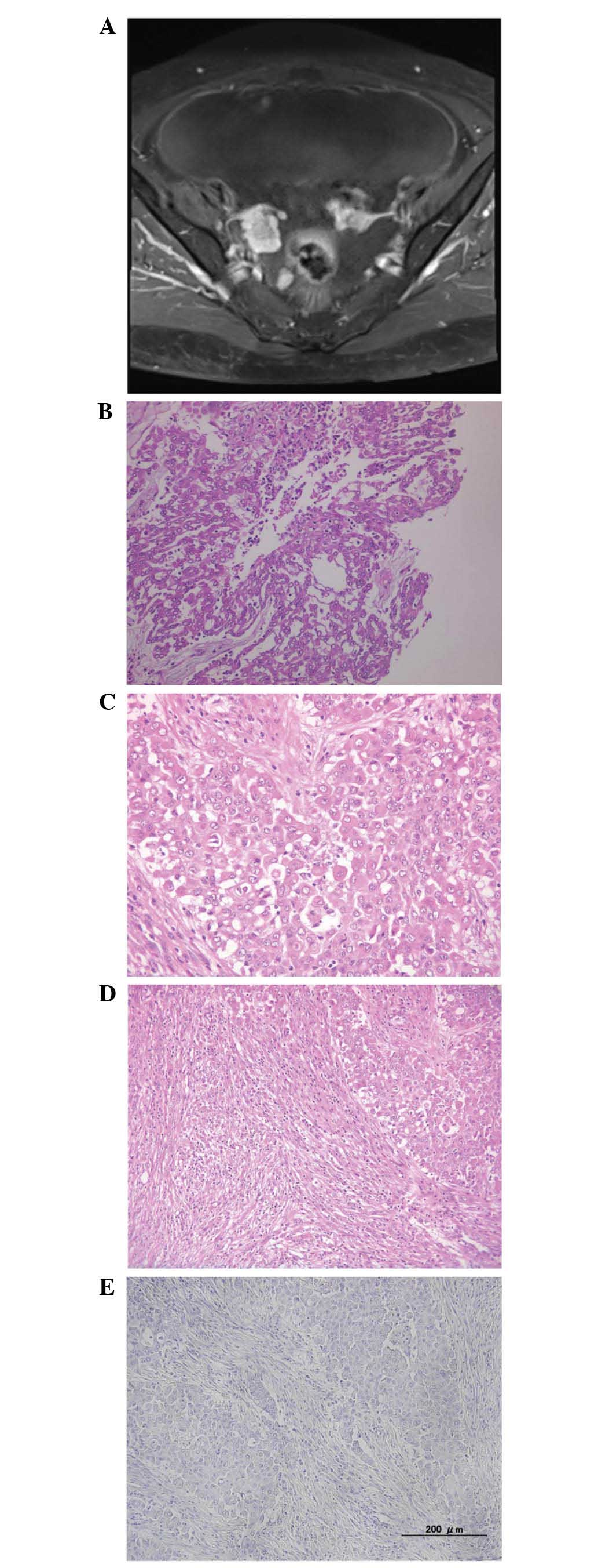
Pelvic magnetic resonance imaging (MRI) revealed small cysts in both ovaries, and lobular nodules at the surface of the ovaries (Fig. 1A). MRI additionally revealed peritonitis carcinomatosa, ascites, disseminated nodules and metastasis to the omentum. Contrast-enhanced computed tomography (CT) revealed a number of small lung nodules, and these findings were considered to represent metastasis to the lung. No lymphadenopathy was observed. Positron emission tomography (PET)-CT demonstrated abnormal fludeoxyglucose (FDG) uptake in the ovarian tumor, disseminated nodules and omentum. These findings suggested a diagnosis of OC.

Figure 1.

Case 1. (A) Magnetic resonance imaging of the pelvis showing small cysts in the ovaries and lobular nodules at the surface of the ovaries. (B) Cells with eosinophilic cytoplasm formed a tubular, papillary and solid arrangement (H&E; magnification, ×100). (C) Epithelial-like cells. (H&E; magnification, ×200). (D) Sarcomatoid-like cells (H&E; magnification, ×100). (E) Negative paired box 8 immunohistochemical staining in malignant peritoneal mesothelioma (H&E; magnification, ×100). H&E, hematoxylin and eosin staining.

A total abdominal hysterectomy (TAH), bilateral salpingo-oophorectomy (BSO) and omentectomy were performed. At the conclusion of primary debulking surgery (PDS), the residual tumor size was <1 cm in diameter. The resected specimens were reviewed by a pathologist and the mass was subsequently diagnosed as MPM.

The patient was discharged from hospital on the 14th postoperative day following an uneventful postoperative period. The patient was treated with chemotherapy (75 mg/m2 cisplatin and 500 mg/m2 pemetrexed) following PDS and remains alive without disease progression, 1 year subsequent to the completion of the first-line chemotherapy.

Pathological findings

Macroscopic examination revealed numerous nodular lesions in the pelvic cavity. Metastatic findings included metastasis to the omentum, measuring ~20 cm (omental cake). The tumor exhibited a number of clusters composed of cuboidal cells with eosinophilic cytoplasm, forming a tubular, papillary and solid arrangement (Fig. 1B). The clusters appeared to include two types of cells (epithelial-like and sarcomatoid-like cells; Fig. 1C and D). In the ovarian specimen, a number of clusters that included epithelial-like cells were observed at the surface of the ovary and infiltrated into the parenchyma of the ovary. There was no evidence of malignant cells in the oviduct or the fimbriae.

Immunohistochemical findings

Epithelial-like cells were positive for calretinin and CAM5.2. Thrombomodulin, D2-40, and CD10 were partially expressed. These cells were negative for WT1, carcinoembryonic antigen (CEA), estrogen receptor (ER), progesterone receptor, Ber-EP4 and PAX8.

Spindle cells were strongly positive for calretinin, CAM5.2 and vimentin. Thrombomodulin and D2-40 were partially expressed. These cells were negative for WT1, CEA, Ber-EP4, desmin, CD10 and PAX8 (Fig. 1E).

Case 2
Patient

A 56-year-old (gravida 1, para 1) woman presented with a cough. The patient had a history of exposure to asbestos, and had no significant past medical or family history. Serum CA125, CA19-9, CEA and STn antigen levels were within normal limits, and the general examination was additionally normal.

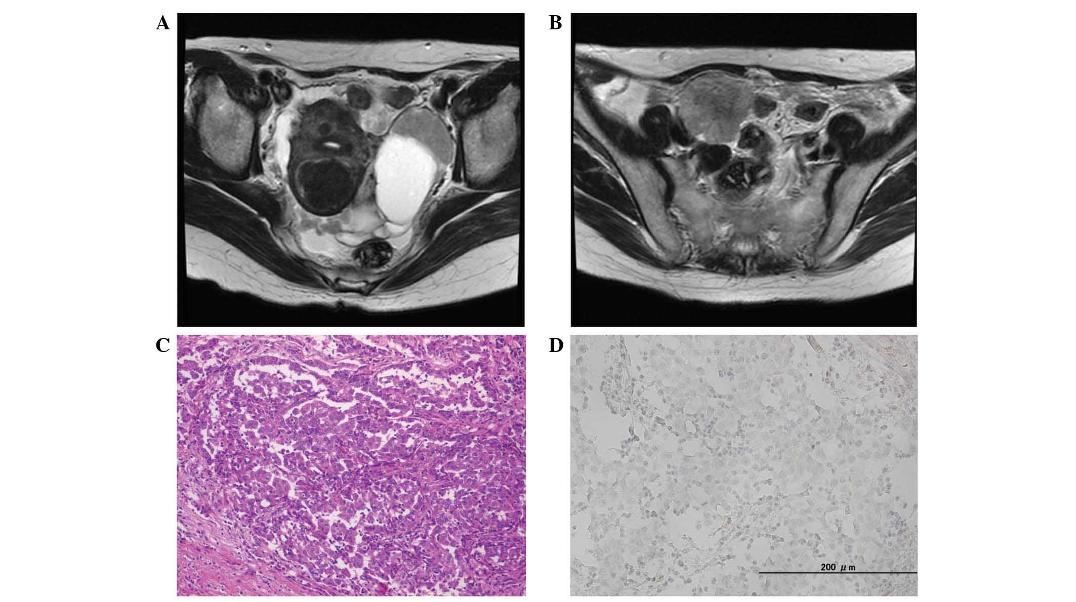
Pelvic MRI revealed a mass with two cysts, measuring 99×42 mm, in the left adnexal region (Fig. 2A), and a solid mass, measuring 46×38 mm in the right adnexal region (Fig. 2B). It additionally revealed peritonitis carcinomatosa, ascites, disseminated nodules and metastasis to the omentum. No lymphadenopathy was observed. Contrast-enhanced CT suggested the possibility of thickened pleura, but there was no indication of metastasis to the lung. PET-CT revealed abnormal FDG uptake in the adnexal tumor, but no abnormal uptake in the pleura and lung. These results suggested a diagnosis of primary OC.

Figure 2.

Case 2. (A) MRI T2-weighted imaging of the pelvis showing a mass with two cysts, measuring 99 × 42 mm, in the left adnexal region. (B) MRI T2-weighted imaging of the pelvis showing a solid mass, measuring 46×38 mm, in the right adnexal region. (C) Cells with eosinophilic cells formed a papillary and solid arrangement (hematoxylin and eosin staining; magnification, ×50). (D) Negative paired box 8 immunohistochemical staining in malignant peritoneal mesothelioma (magnification, ×200). MRI, magnetic resonance imaging.

TAH, BSO and omentectomy were performed as the PDS. At the conclusion of surgery, residual tumors were <1 cm in diameter. The resected specimens were reviewed by a pathologist and the mass was subsequently diagnosed as MPM.

The patient was discharged from hospital on day 14 subsequent to surgery, following an uneventful postoperative period. The patient was treated with chemotherapy (75 mg/m2 cisplatin and 500 mg/m2 pemetrexed) following surgery, which was well-tolerated. The patient remains alive without disease progression 3 years subsequent to completion of first-line chemotherapy.

Pathological findings

Macroscopic examination revealed numerous nodular lesions in the abdominal cavity, including the ovary, omentum and ileum. The tumor exhibited a number of clusters composed of eosinophilic cells, forming a papillary and solid arrangement (Fig. 2C). Identical findings were observed in the ovary and ileum specimens.

Immunohistochemical findings

Tumor cells were strongly positive for calretinin, cytokeratin (CK)7, CK20, D2-40 and CK5/6. CEA was partially expressed. The cells were negative for WT1, ER, Ber-EP4 and PAX8 expression (Fig. 2D).

Discussion

MPM is a rare malignancy of the peritoneum that typically remains confined to the abdominal cavity until the advanced stages of tumor progression. According to the World Health Organization classification, histological subtypes of MPM include epithelioid, sarcomatoid and biphasic (mixed epithelioid and sarcomatoid) (8). Treatment methods for MPM include cytoreductive surgery and hyperthermic intraperitoneal chemotherapy; these therapies have resulted in an improvement in the survival of affected patients, with median survival times ranging from 29.5 to 94 months (9). All patients provided written informed consent to be included in the present study.

Patients with MPM do not present with distinctive symptoms, and the non-specific symptoms commonly observed make diagnosis and early treatment difficult. MPM is frequently misdiagnosed as OC. The clinical and morphological distinction between these malignancies may be difficult due a number of overlapping morphological features, including papillary structures, that exist between the two (4).

Imaging is a significant tool for diagnosis. However, pathological examination of biopsy or resection material is essential to confirm the diagnosis. In the present cases, it was not possible to differentiate MPM from OC by imaging alone.

Immunohistochemistry has a significant role in the distinction between MPM and serous OC. A panel of immunohistochemical antibodies is used to exclude malignant mesotheliomas; this panel includes WT1, calretinin, CK5/6 and BerEP4. An additional panel is typically performed to exclude adenocarcinoma of unknown origin and includes CK7/CK20, thyroid transcription factor 1, caudal type homeobox 2, gross cystic disease fluid protein 15 and WT1 (6). Although the aforementioned immunomarkers are useful, they have a number of limitations. Therefore, it is necessary to identify a marker with high sensitivity and specificity that may be added to 
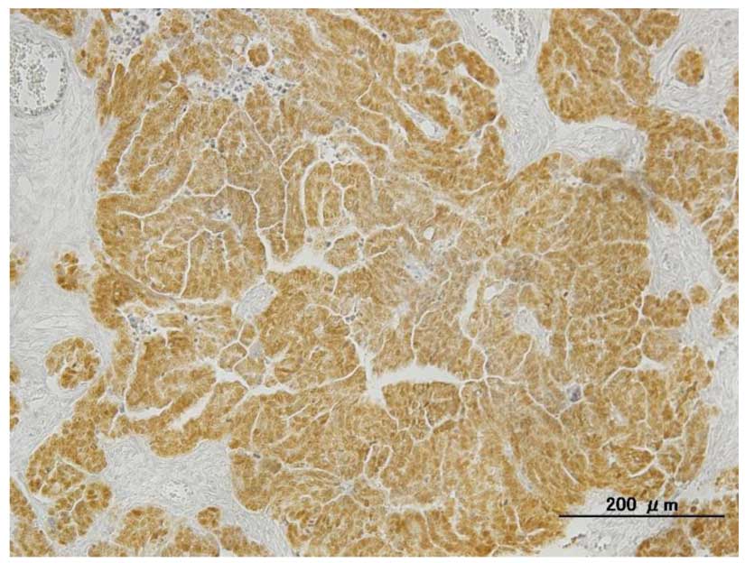
the traditional immunohistochemistry panel of antibodies. PAX8 is a member of the PAX family of transcription factors, and previous studies have demonstrated that high levels of PAX8 expression are specific to serous adenocarcinoma, while all mesotheliomas are PAX8-negative (5–7). Our group has observed PAX8 nuclear positivity (Fig. 3) in 65/67 cases of serous adenocarcinoma (97%) (unpublished data).

Figure 3.

Strong positive paired box 8 immunohistochemical staining in a high-grade serous carcinoma (magnification, ×100).

In the present two cases, a diagnosis of MPM was confirmed through immunohistochemical evaluation, which revealed that both were negative for PAX8. The present immunohistological analyses were consistent with the aforementioned results regarding PAX8-negativity in mesotheliomas.

In conclusion, the present cases indicated that PAX8 immunostaining is a useful tool for differentiating MPM from serous OC, and it is important to consider rare clinical conditions, including peritoneal mesotheliomas, in patients exhibiting common and non-specific symptoms, including abdominal pain, ascites and abdominal distension without abdominal pain. Although MPM is a rare disease, the possibility of MPM should be considered in patients presenting with the aforementioned symptoms. In addition, a history of asbestos exposure is not essential for the disease to occur, and radiological assessment and traditional immunostaining evaluation may lead to misdiagnosis as it may be difficult to differentiate MPM from serous OC. Thus, a thorough comprehensive approach that includes PAX8 immunostaining is important for achieving a precise diagnosis and for the correct treatment of patients exhibiting MPM.

References

1 

Ahmed I, Koulaouzidis I, Iqbal J and Tan WC: Malignant peritoneal mesothelioma as a rare cause of ascites: A case report. J Med Case Rep. 2:1212008. View Article : Google Scholar : PubMed/NCBI

2 

Price B and Ware A: Time trend of mesothelioma incidence in the United States and projection of future cases: An update based on SEER data for 1973 through 2005. Crit Rev Toxicol. 39:576–588. 2009. View Article : Google Scholar : PubMed/NCBI

3 

Bridda A, Padoan I, Mencarelli R and Frego M: Peritoneal mesothelioma: A review. MedGenMed. 9:322007.PubMed/NCBI

4 

Tangjitgamol S, Warnnissorn M, Attakettaworn K and Puripat N: Huge peritoneal malignant mesothelioma mimicking primary ovarian carcinoma. J Med Assoc Thai. 96:107–111. 2013.PubMed/NCBI

5 

Laury AR, Hornick JL, Perets R, Krane JF, Corson J, Drapkin R and Hirsch MS: PAX8 reliably distinguishes ovarian serous tumors from malignant mesothelioma. Am J Surg Pathol. 34:627–635. 2010.PubMed/NCBI

6 

Ordóñez NG: Value of PAX8, PAX2, claudin-4, and h-caldesmon immunostaining in distinguishing peritoneal epithelioid mesotheliomas from serous carcinomas. Mod Pathol. 26:553–562. 2013. View Article : Google Scholar : PubMed/NCBI

7 

Bowen NJ, Logani S, Dickerson EB, Kapa LB, Akhtar M, Benigno BB and McDonald JF: Emerging roles for PAX8 in ovarian cancer and endosalpingeal development. Gynecol Oncol. 104:331–337. 2007. View Article : Google Scholar : PubMed/NCBI

8 

Lu Z and Chen J: Introduction of WHO classification of tumours of female reproductive organs, fourth edition. Zhonghua Bing Li Xue Za Zhi. 42:649–650. 2014.(In Chinese).

9 

Sebbag G, Yan H, Shmookler BM, Chang D and Sugarbaker PH: Results of treatment of 33 patients with peritoneal mesothelioma. Br J Surg. 87:1587–1593. 2008. View Article : Google Scholar

Related Articles

  • Abstract
  • View
  • Download
  • Twitter
Copy and paste a formatted citation
Spandidos Publications style
Nakamura K, Nakayama K, Nagaoka R, Nishisako K, Ishikawa M, Katagiri H, Ishibashi T, Sato E, Amano C, Kyo S, Kyo S, et al: The diagnostic utility of PAX8 immunostaining of malignant peritoneal mesothelioma presenting as serous ovarian carcinoma: A single‑center report of two cases. Oncol Lett 13: 263-266, 2017.
APA
Nakamura, K., Nakayama, K., Nagaoka, R., Nishisako, K., Ishikawa, M., Katagiri, H. ... Kyo, S. (2017). The diagnostic utility of PAX8 immunostaining of malignant peritoneal mesothelioma presenting as serous ovarian carcinoma: A single‑center report of two cases. Oncology Letters, 13, 263-266. https://doi.org/10.3892/ol.2016.5444
MLA
Nakamura, K., Nakayama, K., Nagaoka, R., Nishisako, K., Ishikawa, M., Katagiri, H., Ishibashi, T., Sato, E., Amano, C., Kyo, S."The diagnostic utility of PAX8 immunostaining of malignant peritoneal mesothelioma presenting as serous ovarian carcinoma: A single‑center report of two cases". Oncology Letters 13.1 (2017): 263-266.
Chicago
Nakamura, K., Nakayama, K., Nagaoka, R., Nishisako, K., Ishikawa, M., Katagiri, H., Ishibashi, T., Sato, E., Amano, C., Kyo, S."The diagnostic utility of PAX8 immunostaining of malignant peritoneal mesothelioma presenting as serous ovarian carcinoma: A single‑center report of two cases". Oncology Letters 13, no. 1 (2017): 263-266. https://doi.org/10.3892/ol.2016.5444
Copy and paste a formatted citation
x
Spandidos Publications style
Nakamura K, Nakayama K, Nagaoka R, Nishisako K, Ishikawa M, Katagiri H, Ishibashi T, Sato E, Amano C, Kyo S, Kyo S, et al: The diagnostic utility of PAX8 immunostaining of malignant peritoneal mesothelioma presenting as serous ovarian carcinoma: A single‑center report of two cases. Oncol Lett 13: 263-266, 2017.
APA
Nakamura, K., Nakayama, K., Nagaoka, R., Nishisako, K., Ishikawa, M., Katagiri, H. ... Kyo, S. (2017). The diagnostic utility of PAX8 immunostaining of malignant peritoneal mesothelioma presenting as serous ovarian carcinoma: A single‑center report of two cases. Oncology Letters, 13, 263-266. https://doi.org/10.3892/ol.2016.5444
MLA
Nakamura, K., Nakayama, K., Nagaoka, R., Nishisako, K., Ishikawa, M., Katagiri, H., Ishibashi, T., Sato, E., Amano, C., Kyo, S."The diagnostic utility of PAX8 immunostaining of malignant peritoneal mesothelioma presenting as serous ovarian carcinoma: A single‑center report of two cases". Oncology Letters 13.1 (2017): 263-266.
Chicago
Nakamura, K., Nakayama, K., Nagaoka, R., Nishisako, K., Ishikawa, M., Katagiri, H., Ishibashi, T., Sato, E., Amano, C., Kyo, S."The diagnostic utility of PAX8 immunostaining of malignant peritoneal mesothelioma presenting as serous ovarian carcinoma: A single‑center report of two cases". Oncology Letters 13, no. 1 (2017): 263-266. https://doi.org/10.3892/ol.2016.5444
Follow us
  • Twitter
  • LinkedIn
  • Facebook
About
  • Spandidos Publications
  • Careers
  • Cookie Policy
  • Privacy Policy
How can we help?
  • Help
  • Live Chat
  • Contact
  • Email to our Support Team