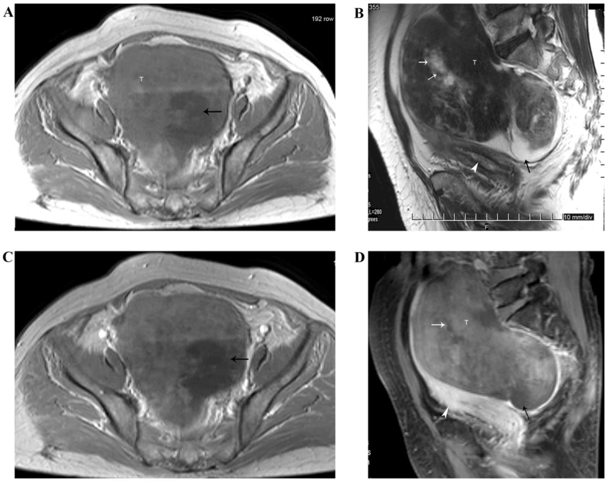
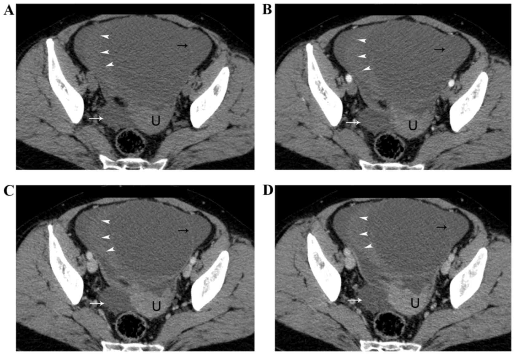
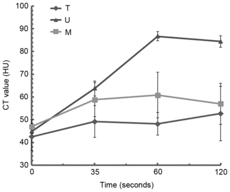
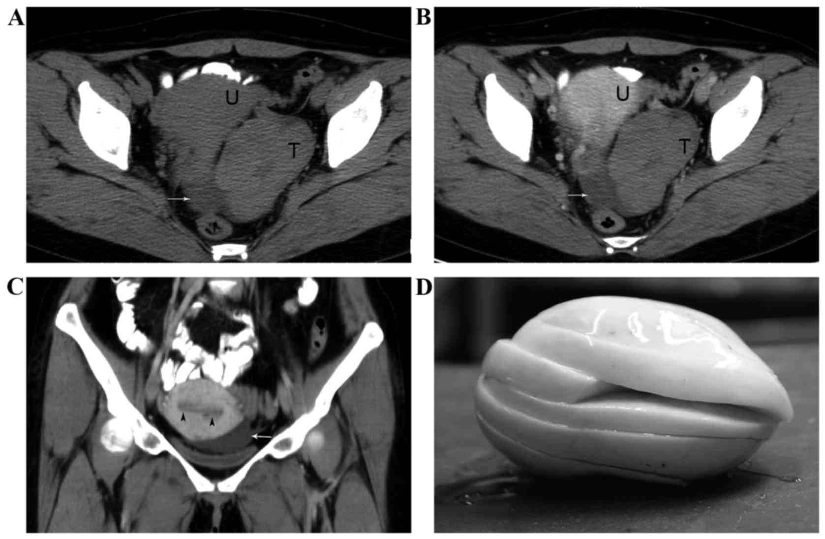
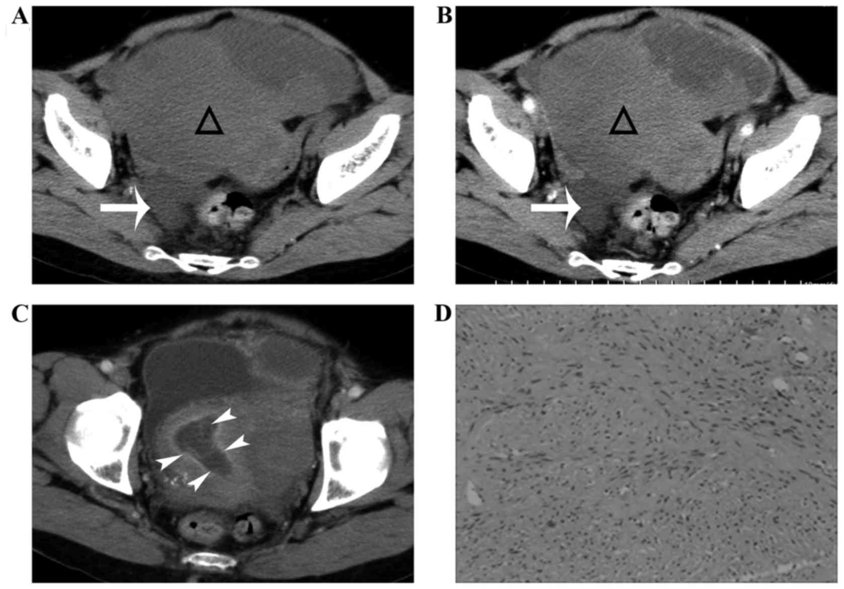
|
1
|
Zhang Z, Wu Y and Gao J: CT diagnosis in
the thecoma-fibroma group of the ovarian stromal tumors. Cell
Biochem Biophys. 71:937–943. 2015. View Article : Google Scholar : PubMed/NCBI
|
|
2
|
Zhang H, Zhang GF, Wang TP and Zhang H:
Value of 3.0 T diffusion-weighted imaging in discriminating thecoma
and fibrothecoma from other adnexal solid masses. J Ovarian Res.
6:582013. View Article : Google Scholar : PubMed/NCBI
|
|
3
|
Li X, Zhang W, Zhu G, Sun C, Liu Q and
Shen Y: Imaging features and pathologic characteristics of ovarian
thecoma. J Comput Assist Tomogr. 36:46–53. 2012. View Article : Google Scholar : PubMed/NCBI
|
|
4
|
Shinagare AB, Meylaerts LJ, Laury AR and
Mortele KJ: MRI features of ovarian fibroma and fibrothecoma with
histopathologic correlation. AJR Am J Roentgenol. 198:W296–W303.
2012. View Article : Google Scholar : PubMed/NCBI
|
|
5
|
Yen P, Khong K, Lamba R, Corwin MT and
Gerscovich EO: Ovarian fibromas and fibrothecomas: Sonographic
correlation with computed tomography and magnetic resonance
imaging: A 5-year single-institution experience. J Ultrasound Med.
32:13–18. 2013. View Article : Google Scholar : PubMed/NCBI
|
|
6
|
Björkholm E and Silfverswärd C: Theca-cell
tumors. Clinical features and prognosis. Acta Radiol Oncol.
19:241–244. 1980. View Article : Google Scholar : PubMed/NCBI
|
|
7
|
Wu B, Peng WJ, Gu YJ, Cheng YF and Mao J:
MRI diagnosis of ovarian fibrothecomas: Tumour appearances and
oestrogenic effect features. Br J Radiol. 87:201306342014.
View Article : Google Scholar : PubMed/NCBI
|
|
8
|
Nocito AL, Sarancone S, Bacchi C and
Tellez T: Ovarian thecoma: Clinicopathological analysis of 50
cases. Ann Diagn Pathol. 12:12–16. 2008. View Article : Google Scholar : PubMed/NCBI
|
|
9
|
Buy JN and Ghossain M: Sex cord-stromal
tumorsGynecological Imaging. Springer; Berlin, Heidelberg: pp.
329–375. 2013, View Article : Google Scholar
|
|
10
|
Magoffin DA: Ovarian theca cell. Int J
Biochem Cell Biol. 37:1344–1349. 2005. View Article : Google Scholar : PubMed/NCBI
|
|
11
|
Outwater EK, Wagner BJ, Mannion C,
McLarney JK and Kim B: Sex cord-stromal and steroid cell tumors of
the ovary. Radiographics. 18:1523–1546. 1998. View Article : Google Scholar : PubMed/NCBI
|
|
12
|
Tanaka YO, Tsunoda H, Kitagawa Y, Ueno T,
Yoshikawa H and Saida Y: Functioning ovarian tumors: Direct and
indirect findings at MR imaging. Radiographics. 24 Suppl
1:S147–S166. 2004. View Article : Google Scholar : PubMed/NCBI
|
|
13
|
Kato H, Kanematsu M, Ono H, Yano R, Furui
T, Morishige K and Hatano Y: Ovarian fibromas: MR imaging findings
with emphasis on intratumoral cyst formation. Eur J Radiol.
82:e417–e421. 2013. View Article : Google Scholar : PubMed/NCBI
|
|
14
|
Chung BM, Park SB, Lee JB, Park HJ, Kim YS
and Oh YJ: Magnetic resonance imaging features of ovarian fibroma,
fibrothecoma, and thecoma. Abdom Imaging. 40:1263–1272. 2015.
View Article : Google Scholar : PubMed/NCBI
|
|
15
|
Malek M, Pourashraf M, Mousavi AS, Rahmani
M, Ahmadinejad N, Alipour A, Hashemi FS and Shakiba M:
Differentiation of benign from malignant adnexal masses by
functional 3 tesla MRI techniques: Diffusion-weighted imaging and
time-intensity curves of dynamic contrast-enhanced MRI. Asian Pac J
Cancer Prev. 16:3407–3412. 2015. View Article : Google Scholar : PubMed/NCBI
|
|
16
|
Robertson DM, Pruysers E and Jobling T:
Inhibin as a diagnostic marker for ovarian cancer. Cancer Lett.
249:14–17. 2007. View Article : Google Scholar : PubMed/NCBI
|
|
17
|
Winking H, Gerdes J and Traut W:
Expression of the proliferation marker Ki-67 during early mouse
development. Cytogenet Genome Res. 105:251–256. 2004. View Article : Google Scholar : PubMed/NCBI
|
|
18
|
Horta M and Cunha TM: Sex cord-stromal
tumors of the ovary: A comprehensive review and update for
radiologists. Diagn Interv Radiol. 21:277–286. 2015. View Article : Google Scholar : PubMed/NCBI
|
|
19
|
Genç M, Solak A, Genç B, Sivrikoz ON,
Kurtulmuş S, Turan A, Sahin N and Gür EB: A diagnostic dilemma for
solid ovarian masses: The clinical and radiological aspects with
differential diagnosis of 23 cases. Eur J Gynaecol Oncol.
36:186–191. 2015.PubMed/NCBI
|
|
20
|
Cornitescu FI, Tănase F, Simionescu C and
Iliescu D: Clinical, histopathological and therapeutic
considerations in non-neoplastic abnormal uterine bleeding in
menopause transition. Rom J Morphol Embryol. 52:759–765.
2011.PubMed/NCBI
|
|
21
|
Outwater EK, Siegelman ES, Kim B,
Chiowanich P, Blasbalg R and Kilger A: Ovarian Brenner tumors: MR
imaging characteristics. Magn Reson Imaging. 16:1147–1153. 1998.
View Article : Google Scholar : PubMed/NCBI
|
|
22
|
Arnogiannaki N, Grigoriadis C, Zygouris D,
Terzakis E, Sebastiadou M and Tserkezoglou A: Proliferative Brenner
tumor of the ovary. clinicopathological study of two cases and
review of the literature. Eur J Gynaecol Oncol. 32:576–578.
2011.PubMed/NCBI
|
|
23
|
Zhang P, Cui Y, Li W, Ren G, Chu C and Wu
X: Diagnostic accuracy of diffusion-weighted imaging with
conventional MR imaging for differentiating complex solid and
cystic ovarian tumors at 1.5T. World J Surg Oncol. 10:2372012.
View Article : Google Scholar : PubMed/NCBI
|