Introduction
Esophageal carcinoma, one of the most common types
of mortality-inducing cancer globally, ranked ninth in cancer
incidence and was the sixth leading cause of cancer-associated
mortality worldwide in 2013 (1).
Esophageal carcinoma is divided into two major histological
subtypes: Esophageal squamous cell carcinoma (ESCC) and esophageal
adenocarcinoma. In Asia and the United States, ESCC represents ~90
and ~26% of all cases of esophageal cancer, respectively (2). Current treatment options for patients
with esophageal carcinoma include surgery, chemotherapy and
radiotherapy. However, the 5-year survival rate for patients with
ESCC remains poor (3–5). These data suggest that novel treatment
approaches for advanced esophageal carcinoma are required.
Tumor metastasis is associated with complex
interactions between primary tumor cells prior to invasion,
intravasation, immune escape and extravasation of the circulatory
system, followed by lymphangiogenesis/angiogenesis and migration
towards target organs (6). The
epithelial-mesenchymal transition (EMT), a key event of tumor
metastasis, enables epithelial cells to cease exhibiting certain
epithelial properties, obtain a mesenchymal phenotype and thereby
potentially migrate and invade. This process is associated with
alterations in biomarkers, including the proteins of the cell
membrane, cytoskeleton, extracellular matrix (ECM), and certain
transcription factors (7,8). ECM degradation provides biochemical and
mechanical barriers to cell movement and is a crucial process in
tumor metastasis. Matrix metallopeptidases (MMPs), a multigene
family of zinc-dependent endopeptidases, degrade the majority of
ECM components. MMP9 is secreted at the primary tumor site and, by
degrading ECM components, may provide tumor cells access to the
vasculature and thereby facilitate tumor cell migration and
invasion into the target organ (9).
Previous studies have suggested that MMP9 activity is increased in
multiple metastatic carcinomas, including those occurring in cases
of esophageal cancer (10,11).
Metformin, one of the most widely prescribed oral
hypoglycemic agents, inhibits hepatic gluconeogenesis and induces
the uptake and use of glucose in skeletal muscle and adipose
tissue. Metformin was originally established as a glucose
level-decreasing drug and became available for human consumption in
the 1950s. In 2005, Evans et al (12) demonstrated that in addition to its
antidiabetic function, metformin may significantly decrease tumor
incidence among patients with type 2 diabetes. Previously, multiple
studies have demonstrated that metformin may inhibit proliferation,
induce apoptosis and arrest the cell cycle in ESCC cells (13–15).
Furthermore, metformin may inhibit malignant cell migration, and
the invasion of ovarian, pancreatic cancer and melanoma cells in
vitro (16–19).
Materials and methods
Cell lines and culture
The human ESCC cell line EC109 was obtained from the
Type Culture Collection of the Chinese Academy of Sciences
(Shanghai, China). All cells were cultured in Dulbecco's modified
Eagle's medium (DMEM) with 10% fetal bovine serum (FBS) (both from
Gibco, Thermo Fisher Scientific, Inc., Waltham, MA, USA) without
antibiotics at 37°C in a humidified, 5% CO2 atmosphere.
Cells were inoculated onto a Petri dish (106 cells/6 cm)
in DMEM with 10% FBS and allowed to attach overnight prior to drug
treatment. EC109 cells were treated with 100 nM insulin [a protein
kinase B (AKT) activator] and 10 µM LY294002 (a PI3K inhibitor)
dissolved in dimethyl sulfoxide (DMSO) for 30 min or 24 h at 37°C
in a humidified, 5% CO2 atmosphere, separately or in
combination. The EC109 cells treated with metformin in combination
with insulin were pretreated with metformin for 23.5 h and
subsequently cultured in DMEM containing 10% FBS and 100 nM insulin
for 30 min at 37°C in a humidified, 5% CO2 atmosphere.
DMSO and ddH2O were used as the negative controls.
Metformin and LY294002, a phosphoinositide 3-kinase (PI3K)
inhibitor, were purchased from Sigma-Aldrich; Merck KGaA
(Darmstadt, Germany). Human recombinant insulin was also from
Sigma-Aldrich; Merck KGaA.
Western blot analysis
To extract total cell protein, the EC109 cells
(5×10⁵) were sonicated at 50 Hz twice for 3 sec, with 20
sec in between sonication, in 100 µl RIPA buffer supplemented with
NaF (1 mM), sodium orthovanadate (1 mM), and phenylmethane sulfonyl
fluoride (1 mM), and then homogenized for 30 min in 4°C, and then
centrifuged at 4°C and 12,000 × g for 12 min. Cell nuclear protein
extraction was performed as previously described (9), and protein concentration was determined
using a BCA Protein Assay kit (Pierce; Thermo Fisher Scientific,
Inc.). A total of 30 µg protein for each sample was separated using
SDS-PAGE and transferred onto a polyvinylidene fluoride (PVDF)
membrane (EMD Millipore, Billerica, MA, USA). Subsequently, the
membranes were then blocked with 10% non-fat milk in Tris-buffered
saline with Tween-20 (TBST) for 1 h at room temperature. The
membranes were incubated with the following primary antibodies: AKT
(catalog no. 9272s), phosphorylated (p)-AKT (Ser473; catalog no.
4060s), nuclear factor (NF)-κB (p65; catalog no. 8242s), epithelial
(E)-cadherin (catalog no. 3195s), neural (N)-cadherin (catalog no.
4061s) (dilution, 1:1,000; Cell Signaling Technology, Inc.,
Danvers, MA, USA) and MMP9 (dilution, 1:1,000; catalog no. ab38898;
Abcam, Cambridge, UK) overnight at 4°C. Membranes were washed three
times with TBST and incubated with a HRP-conjugated goat
anti-rabbit secondary antibodies (dilution, 1:3,000; catalog no.
AS006; Asbio Technology, Inc., Danvers, MA, USA) for 1 h at room
temperature, then washed and developed with the ECL Plus Western
Blot Detection System kit (Amersham, Piscataway, NJ USA).
Antibodies against TATA-box binding protein (catalog no. 8515s) and
GAPDH (catalog no. 5014s; dilution, 1:1,000; Cell Signaling
Technology, Inc.) were used as loading controls.
RNA extraction and reverse
transcription-quantitative polymerase chain reaction (RT-qPCR)
Total RNA was extracted from the EC109 cells
(2×10⁵) using TRIzol reagent (Invitrogen; Thermo Fisher
Scientific, Inc.), and 2 µg RNA was reverse transcribed using the
PrimeScript™ RT reagent kit, 20 µl volume (Takara Biotechnology
Co., Ltd., Dalian, China) according to the supplier's protocol. The
reactions were set up with 12.5 µl SYBR-Green PCR Master Mix
(Takara Biotechnology Co., Ltd.), 1.0 µl primer mixture (10 µM) and
2 µl cDNA template, and analyzed using a Bio-Rad iCycler system
(version 2.1; Bio-Rad Laboratories, Inc., Hercules, CA, USA), in
accordance with the manufacturer's cycling protocol: Initial
denaturation at 95°C for 30 sec, followed by 40 cycles at 95°C for
5 sec and at 60°C for 30 sec. Gene expression was measured using
the SYBR Premix Ex Taq kit (Takara Biotechnology Co., Ltd.). The
mRNA expression level of MMP9 for each sample was normalized to
that of the housekeeping gene GAPDH by using the 2−ΔΔCq
method (20). Primer sequences were
as follows: MMP9 forward, 5′-CTTTGACAGCGACAAGAAGTGG-3′ and reverse,
5′-GGCACTGAGGAATGATCTAAGC-3′; GAPDH forward,
5′-GAGCCAAAAGGGTCATCATCTC-3′ and reverse,
5′-AAAGGTGGAGGAGTGGGTGTC-3′.
Migration and invasion assay
In the migration assay, EC109 cell suspensions (each
with 105 cells in 200 µl serum-free DMEM) were
inoculated into the upper chamber of a 24-well cell culture
Transwell insert (pore size, 8 µm; Corning Incorporated, Corning,
NY, USA), and the lower chamber was filled with 600 µl DMEM
supplemented with 10% FBS. Following 20 h of incubation at 37°C,
non-migratory cells in the upper chamber were removed from the
upper surface of the filters using a PBS-soaked cotton swab, and
cells that had migrated through the membrane were fixed using 92%
ice methanol (10 min), stained with 0.2% hematoxylin (7 min) and
0.5% eosin (2 min) at room temperature, and then washed and dried
in air. Images of ten randomly selected fields of the
migrated/invaded cells were captured and counted under an upright
fluorescence microscope (magnification, ×200; Ni-E; Nikon
Corporation, Tokyo, Japan). Invasion assays were performed using
the same protocol, except with Matrigel-coated Transwell chambers
and incubating for 22 h at 37°C. Cells that were pretreated with
ddH2O and the same concentration of DMSO as LY294002
were used as the negative control.
Statistical analysis
Data were expressed as the mean ± standard error of
the mean of three independent experiments and analyzed using
GraphPad Prism version 6.0 software (GraphPad, Inc., La Jolla, CA,
USA). One-way ANOVA with the Student-Newman-Keuls as the post hoc
test was used to determine statistical differences between
parametric data. P<0.05 and P<0.01 were considered to
indicate a statistically significant difference.
Results
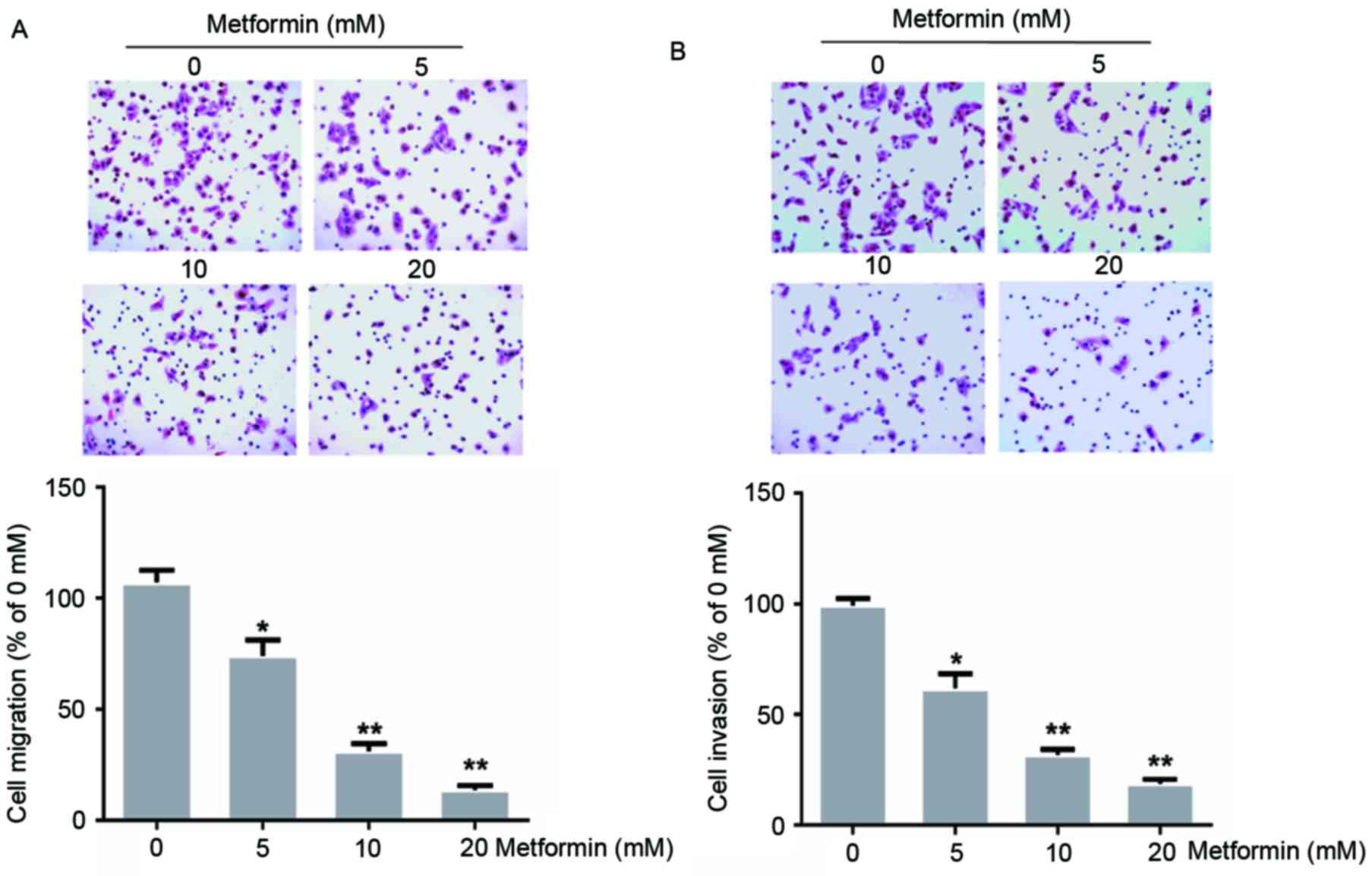
Effects of metformin on migration and
invasion in human EC109 cells
A previous study demonstrated that treatment with 20
mM metformin for 72 h suppressed EC109 cell proliferation (13). To evaluate whether metformin treatment
influenced EC109 cell metastasis, the present study evaluated the
ability of EC109 cells to migrate through a Transwell membrane
coated with Matrigel to assess invasion, or without to assess
migration, following treatment with 0, 5, 10 or 20 mM metformin for
24 h. In the present study, metformin treatment decreased the
migration and invasion of EC109 cells dose-dependently (Fig. 1). Treatment with 20 mM metformin
inhibited EC109 cell migration and invasion by 87 and 81%,
respectively (P<0.01).
Effect of metformin on the expression
of p-AKT and NF-κB in human EC109 cells
AKT, a key downstream effector of the PI3K signaling
pathway, is associated with metastasis in colorectal cancer,
intrahepatic cholangiocarcinoma, lung, breast cancer (21–24) and
esophageal carcinoma (25). In these
human epithelial malignancies, AKT is a key mediator of EMT
induction. In addition, previous studies have reported that NF-κB
is a target of AKT (26,27). Therefore, the present study used
western blot analysis to assess the expression of p-AKT and NF-κB
(p65) in EC109 cells following metformin treatment (Fig. 2). Following treatment with 0, 5, 10 or
20 mM metformin for 24 h, the expression of p-AKT and NF-κB (p65)
in EC109 cells decreased dose dependently (Fig. 2A). Treatment with 20 mM metformin
decreased the expression of p-AKT by 70% and that of NF-κB (p65), a
p-AKT target, by 68% in EC109 cells, compared with untreated EC109
cells.
Effects of metformin on the expression
of E-cadherin, N-cadherin and MMP9 in human EC109 cells
EMT results in epithelial cells acquiring
fibroblast-like properties, expressing mesenchymal markers,
including N-cadherin and vimentin, and decreasing the expression of
epithelial markers, including E-cadherin. The present study
evaluated whether metformin is associated with the regulation of
EMT, a key step in tumor invasion. The results of the present study
indicated that MMP9 transcription was dose-dependently decreased by
metformin (Fig. 3A). In addition, the
result revealed that metformin treatment upregulated and
downregulated E-cadherin and N-cadherin expression, respectively,
in a dose-dependent manner (Fig.
3B).
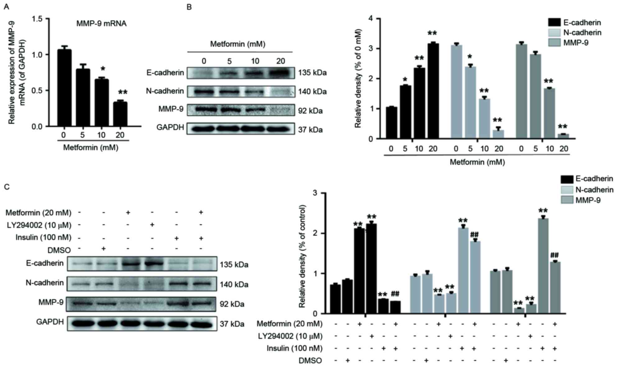 | Figure 3.Effects of metformin, LY294002 or
insulin, separately or in combination on the expression of
E-cadherin, N-cadherin and MMP9 in EC109 cells. (A) Cells were
treated with 0, 5, 10 or 20 mM metformin for 24 h and the mRNA
expression of MMP9 was subsequently measured. (B) Cells were
treated with 0, 5, 10 or 20 mM metformin for 24 h, lysed prior to
western blot analysis and probed with antibodies against
E-cadherin, N-cadherin and MMP9 proteins. (C) Cells were treated
with 20 mM metformin, 10 µM LY294002 for 24 h or 100 nM insulin for
30 min, lysed prior to western blot analysis and probed with
antibodies against E-cadherin, N-cadherin and MMP9 proteins. GAPDH
was used as the loading control. Data were presented as the mean ±
standard error of three independent experiments performed in
triplicate. *P<0.05 and **P<0.01 vs. control cells;
##P<0.01 vs. metformin. E, epithelial, N, neural,
MMP, matrix metallopeptidase. |
MMP9 may represent a key mediator in tumor cell
migration and invasion by stimulating ECM degradation, and
increased MMP9 expression is associated with tumor progression
(28,29). Therefore, the present study assessed
whether metformin affected MMP9 protein expression in EC109 cells,
and demonstrated that MMP9 expression in EC109 cells was decreased
by metformin treatment, particularly at 20 mM metformin, compared
with that in untreated EC109 cells (Fig.
3B).
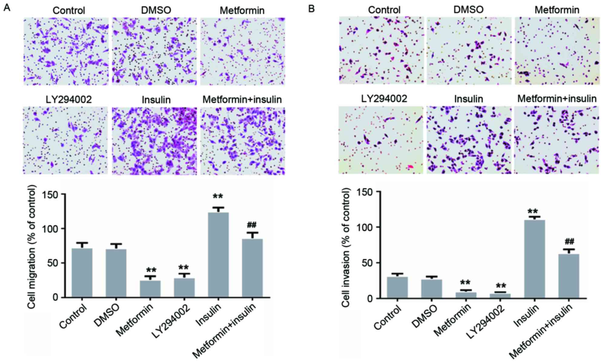
Metformin inhibits the migration and
invasion of EC109 cells by targeting the AKT signaling pathway
To further explore whether the AKT signaling pathway
is associated with EC109 cell migration and invasion, EC109 cells
were treated with 100 nM insulin (an AKT activator) and 10 µM
LY294002 (a PI3K inhibitor) for 30 min or 24 h, separately or in
combination. The EC109 cells treated with metformin in combination
with insulin were pretreated with metformin for 23.5 h and
subsequently cultured in DMEM containing 10% FBS and 100 nM insulin
for 30 min (Fig. 4). The results of
the present study demonstrated that LY294002 inhibited the
migration and invasion of the EC109 cells (Fig. 4). However, this metformin-mediated
inhibition was decreased following treatment with insulin for 30
min (Fig. 4). The results of the
present study suggested that metformin inhibited the migration and
invasion of EC109 cells via the AKT pathway.
Metformin inhibits the expression of
migration- and invasion-associated proteins in EC109 cells by
targeting the AKT signaling pathway
EC109 cells were treated with LY294002 for 24 h or
with insulin for 30 min, separately or in combination. Compared
with the control cells, the expression of p-AKT/AKT and NF-κB (p65)
was decreased (Fig. 2B), the protein
expression of MMP9 and N-cadherin was downregulated, and the
expression of E-cadherin was increased in LY294002-treated cells
(Fig. 3C). Compared with the control
cells, the expression of p-AKT/AKT and NF-κB (p65) in the
insulin-treated EC109 cells was increased and the metformin-induced
suppression of p-AKT/AKT and NF-κB (p65) was decreased (Fig. 2B). In addition, the expression of MMP9
and N-cadherin significantly increased, and the expression of
E-cadherin decreased, in EC109 cells treated with insulin for 30
min compared with the control cells (Fig.
3C). The results of the present study suggested that the AKT
signaling pathway may be associated with the inhibition of
migration- and invasion-associated protein expression in EC109
cells by metformin.
Discussion
Esophageal carcinoma is characterized by a decreased
cure rate and the potential development of invasive properties. The
increased mortality rate with which esophageal carcinoma is
associated is primarily due to metastasis and the side effects and
increased rate of recurrence associated with treatment. Therefore,
the present study assessed the antimetastatic effect of metformin
on human esophageal cancer cells.
Patients treated with metformin, a nontoxic and
antihyperglycemic drug rarely report the side effect of lactic
acidosis. Previous studies have evaluated the effects metformin may
induce through AMP-activated protein kinase-dependent mechanisms,
and have demonstrated that metformin decreases the expression of
p-AKT in bladder cancer, NRAS proto-oncogene mutant melanoma cell
line DO4 and squamous cell carcinoma (SCC) cell line A431 (30–32). In
the present study, metformin inhibited EC109 cell migration and
invasion. Previous studies have demonstrated that the mechanisms
underlying these effects were associated with the downregulation of
p-AKT expression (33,34). Consistent with these previous studies,
the results of the present study suggested that metformin inhibited
invasion in EC109 cells by inhibiting AKT activation. The present
study also demonstrated that the PI3K inhibitor LY294002 inhibited
EC109 cell migration and invasion, while the AKT activator insulin
decreased metformin-induced metastasis inhibition in EC109 cells.
These results suggested that the AKT pathway was suppressed in the
metformin-treated EC109 cells, which may indicate that metformin
inhibits migration and invasion in ESCC cells. To the best of our
knowledge, the present study is the first to demonstrate in
vitro that metformin inhibits ESCC metastasis by inhibiting AKT
activation.
In tumor cells, the nuclear transcription factor
NF-κB is formed as an inactive complex in the cytoplasm by p65, p50
and NFKB inhibitor α (NFKBIA). Once this complex is activated, IκB
kinase (IKK) phosphorylates NFKBIA, p65 is subsequently released
and NF-κB translocates to the nucleus and regulates the expression
of multiple genes (35), including
MMPs, resulting in tumor cell invasion, migration and metastasis
(36,37). AKT, which phosphorylates IKKα, is
associated with NF-κB activation (38). In the gastric cancer cell line
BGC-823, the overexpression of death associated protein kinase 3
(DAPK3) induced AKT activation and increased the phosphorylation of
IKKα, NFKBIA and NF-κB, while the AKT inhibitor LY294002
significantly decreased the phosphorylation of IKKα, NFKBIA, NF-κB,
and prevented DAPK3-induced BGC-823 cell migration and invasion
(39). In the present study, NF-κB
(p65) expression was decreased in the EC109 cells treated with
metformin or LY294002, but increased in insulin-treated EC109 cells
pretreated with or without metformin.
MMPs participate in normal connective tissue
homeostasis and remodeling, and are associated with ECM
degradation, alterations to cell-cell and cell-ECM interactions,
tumor formation, invasion and metastasis (40,41). Since
AKT promoted invasion in the human fibrosarcoma cell line HT1080 by
increasing cell motility and MMP9 expression in a previous study
(42), LY294002-induced suppression
of MMP9 expression may have resulted in the inhibition of EC109
cell invasion demonstrated in the present study. The present study
evaluated the expression of MMP9 in the EC09 cells following
metformin treatment for 24 h. The expression of MMP9 was inhibited
by metformin (Fig. 3A and B) and
LY294002 (Fig. 3C). Therefore, the
suppression of the AKT pathway by metformin may have resulted in
the decrease in MMP9 expression in the EC09 cells identified in the
present study.
E-cadherin is a Ca2+-dependent cell
adhesion molecule expressed by epithelial cells. The positive
expression of E-cadherin is involved in establishing cell-cell
connections, and maintaining epithelial polarity and structural
integrity. The absence of E-cadherin in epithelial cells results in
decreased cell-cell adhesion and induces tumor cell invasion and
metastasis (43). N-cadherin, a
transmembrane glycoprotein, is expressed by numerous types of cell,
including neuroepithelial cells, neurons and mesenchymal cells.
N-cadherin mediates Ca2+-dependent cell-cell adhesion
through the homophilic interactions of its extracellular domains
during mesenchymal condensation (11). A previous study suggested that
N-cadherin is associated with increased invasion in tumor cells and
may contribute to the mesenchymal-scattered phenotype associated
with decreased E-cadherin and cadherin 3 expression in SCC cells
(44). Consistent with this previous
study, the expression of E-cadherin and N-cadherin in the present
study was regulated in the metformin-treated EC109 cells.
The results of the present study suggested that
metformin inhibited EC109 cell migration and invasion by
downregulating the AKT pathway and that p-AKT suppression may have
induced the mesenchymal-to-epithelial reverting transition in the
EC109 cells by inhibiting the NF-κB signaling pathway and
decreasing MMP9 expression. Therefore, metformin potentially
represents a useful therapeutic tool for controlling metastasis in
patients with esophageal carcinoma.
Acknowledgements
The present study was supported by the National
Natural Science Foundation of China (grant no. 81172263).
References
|
1
|
Global Burden of Disease Cancer
Collaboration, ; Fitzmaurice C, Dicker D, Pain A, Hamavid H,
Moradi-Lakeh M, MacIntyre MF, Allen C, Hansen G, Woodbrook R, et
al: The global burden of cancer 2013. JAMA Oncol. 1:505–527. 2015.
View Article : Google Scholar : PubMed/NCBI
|
|
2
|
Torre LA, Bray F, Siegel RL, Ferlay J,
Lortet-Tieulent J and Jemal A: Global cancer statistics, 2012. CA
Cancer J Clin. 65:87–108. 2015. View Article : Google Scholar : PubMed/NCBI
|
|
3
|
Ekman S, Dreilich M, Lennartsson J,
Wallner B, Brattström D, Sundbom M and Bergqvist M: Esophageal
cancer: Current and emerging therapy modalities. Expert Rev
Anticancer Ther. 8:1433–1448. 2008. View Article : Google Scholar : PubMed/NCBI
|
|
4
|
Li LY, Jiang H, Xie YM, Liao LD, Cao HH,
Xu XE, Chen B, Zeng FM, Zhang YL, Du ZP, et al: Macrolide analog
F806 suppresses esophageal squamous cell carcinoma (ESCC) by
blocking β1 integrin activation. Oncotarget. 6:15940–15952.
2015.PubMed/NCBI
|
|
5
|
Zhao H and Gu X: Silencing of insulin-like
growth factor-1 receptor enhances the radiation sensitivity of
human esophageal squamous cell carcinoma in vitro and in vivo.
World J Surg Oncol. 12:3252014. View Article : Google Scholar : PubMed/NCBI
|
|
6
|
Miovic M and Block S: Psychiatric
disorders in advanced cancer. Cancer. 110:1665–1676. 2007.
View Article : Google Scholar : PubMed/NCBI
|
|
7
|
Tsai JH, Donaher JL, Murphy DA, Chau S and
Yang J: Spatiotemporal regulation of epithelial-mesenchymal
transition is essential for squamous cell carcinoma metastasis.
Cancer Cell. 22:725–736. 2012. View Article : Google Scholar : PubMed/NCBI
|
|
8
|
Cui Y, Wang Y, Li H, Li Q, Yu Y, Xu X, Xu
B and Liu T: Asparaginyl endopeptidase promotes the invasion and
metastasis of gastric cancer through modulating
epithelial-to-mesenchymal transition and analysis of their
phosphorylation signaling pathways. Oncotarget. 7:34356–34370.
2016. View Article : Google Scholar : PubMed/NCBI
|
|
9
|
Fong Y, Shen KH, Chiang TA and Shih YW:
Acacetin inhibits TPA-induced MMP-2 and u-PA expressions of human
lung cancer cells through inactivating JNK signaling pathway and
reducing binding activities of NF-kappaB and AP-1. J Food Sci.
75:H30–H38. 2010. View Article : Google Scholar : PubMed/NCBI
|
|
10
|
Roy R, Yang J and Moses MA: Matrix
metalloproteinases as novel biomarkers and potential therapeutic
targets in human cancer. J Clin Oncol. 27:5287–5297. 2009.
View Article : Google Scholar : PubMed/NCBI
|
|
11
|
Alizadeh AM, Shiri S and Farsinejad S:
Metastasis review: From bench to bedside. Tumour Biol.
35:8483–8523. 2014. View Article : Google Scholar : PubMed/NCBI
|
|
12
|
Evans JM, Donnelly LA, Emslie-Smith AM,
Alessi DR and Morris AD: Metformin and reduced risk of cancer in
diabetic patients. BMJ. 330:1304–1305. 2005. View Article : Google Scholar : PubMed/NCBI
|
|
13
|
Cai X, Hu X, Tan X, Cheng W, Wang Q, Chen
X, Guan Y, Chen C and Jing X: Metformin induced AMPK activation,
G0/G1 phase cell cycle arrest and the inhibition of growth of
esophageal squamous cell carcinomas in vitro and in vivo. PLoS One.
10:e01333492015. View Article : Google Scholar : PubMed/NCBI
|
|
14
|
Kobayashi M, Kato K, Iwama H, Fujihara S,
Nishiyama N, Mimura S, Toyota Y, Nomura T, Nomura K, Tani J, et al:
Antitumor effect of metformin in esophageal cancer: In vitro study.
Int J Oncol. 42:517–524. 2013. View Article : Google Scholar : PubMed/NCBI
|
|
15
|
Feng Y, Ke C, Tang Q, Dong H, Zheng X, Lin
W, Ke J, Huang J, Yeung SC and Zhang H: Metformin promotes
autophagy and apoptosis in esophageal squamous cell carcinoma by
downregulating Stat3 signaling. Cell Death Dis. 5:e10882014.
View Article : Google Scholar : PubMed/NCBI
|
|
16
|
Wu B, Li S, Sheng L, Zhu J, Gu L, Shen H,
La D, Hambly BD, Bao S and Di W: Metformin inhibits the development
and metastasis of ovarian cancer. Oncol Rep. 28:903–908. 2012.
View Article : Google Scholar : PubMed/NCBI
|
|
17
|
Rattan R, Graham RP, Maguire JL, Giri S
and Shridhar V: Metformin suppresses ovarian cancer growth and
metastasis with enhancement of cisplatin cytotoxicity in vivo.
Neoplasia. 13:483–491. 2011. View Article : Google Scholar : PubMed/NCBI
|
|
18
|
Bao B, Wang Z, Ali S, Ahmad A, Azmi AS,
Sarkar SH, Banerjee S, Kong D, Li Y, Thakur S and Sarkar FH:
Metformin inhibits cell proliferation, migration and invasion by
attenuating CSC function mediated by deregulating miRNAs in
pancreatic cancer cells. Cancer Prev Res (Phila). 5:355–364. 2012.
View Article : Google Scholar : PubMed/NCBI
|
|
19
|
Cerezo M, Tichet M, Abbe P, Ohanna M,
Lehraiki A, Rouaud F, Allegra M, Giacchero D, Bahadoran P,
Bertolotto C, et al: Metformin blocks melanoma invasion and
metastasis development in AMPK/p53-dependent manner. Mol Cancer
Ther. 12:1605–1615. 2013. View Article : Google Scholar : PubMed/NCBI
|
|
20
|
Livak KJ and Schmittgen TD: Analysis of
relative gene expression data using real-time quantitative PCR and
the 2(-Delta Delta C(T)) method. Methods. 25:402–408. 2001.
View Article : Google Scholar : PubMed/NCBI
|
|
21
|
Rui X, Yan XI and Zhang K: Baicalein
inhibits the migration and invasion of colorectal cancer cells via
suppression of the AKT signaling pathway. Oncol Lett. 11:685–688.
2016. View Article : Google Scholar : PubMed/NCBI
|
|
22
|
Xie K, Nian J, Zhu X, Geng X and Liu F:
Modulatory role of garlicin in migration and invasion of
intrahepatic cholangiocarcinoma via PI3K/AKT pathway. Int J Clin
Exp Pathol. 8:14028–14033. 2015.PubMed/NCBI
|
|
23
|
Yoneyama R, Aoshiba K, Furukawa K, Saito
M, Kataba H, Nakamura H and Ikeda N: Nicotine enhances hepatocyte
growth factor-mediated lung cancer cell migration by activating the
α7 nicotine acetylcholine receptor and phosphoinositide
kinase-3-dependent pathway. Oncol Lett. 11:673–677. 2016.
View Article : Google Scholar : PubMed/NCBI
|
|
24
|
Li J, Song Z, Wang Y, Yin Y, Liu Y, Yuan R
and Nan X: Overexpression of SphK1 enhances cell proliferation and
invasion in triple-negative breast cancer via the PI3K/AKT
signaling pathway. Tumour Biol. 37:10587–10593. 2016. View Article : Google Scholar : PubMed/NCBI
|
|
25
|
Tantai JC, Zhang Y and Zhao H:
Heterophyllin B inhibits the adhesion and invasion of ECA-109 human
esophageal carcinoma cells by targeting PI3K/AKT/β-catenin
signaling. Mol Med Rep. 13:1097–1104. 2016. View Article : Google Scholar : PubMed/NCBI
|
|
26
|
Romashkova JA and Makarov SS: NF-kappaB is
a target of AKT in anti-apoptotic PDGF signalling. Nature.
401:86–90. 1999. View
Article : Google Scholar : PubMed/NCBI
|
|
27
|
Li Z, Yang Z, Passaniti A, Lapidus RG, Liu
X, Cullen KJ and Dan HC: A positive feedback loop involving
EGFR/Akt/mTORC1 and IKK/NF-κB regulates head and neck squamous cell
carcinoma proliferation. Oncotarget. 7:31892–31906. 2016.PubMed/NCBI
|
|
28
|
Qin LX and Tang ZY: The prognostic
molecular markers in hepatocellular carcinoma. World J
Gastroenterol. 8:385–392. 2002. View Article : Google Scholar : PubMed/NCBI
|
|
29
|
Hsieh SC, Tsai JP, Yang SF, Tang MJ and
Hsieh YH: Metformin inhibits the invasion of human hepatocellular
carcinoma cells and enhances the chemosensitivity to sorafenib
through a downregulation of the ERK/JNK-mediated NF-κB-dependent
pathway that reduces uPA and MMP-9 expression. Amino Acids.
46:2809–2822. 2014. View Article : Google Scholar : PubMed/NCBI
|
|
30
|
Peng M, Su Q, Zeng Q, Li L, Liu Z, Xue L,
Cheng Y, Huang Y, Tao T, Lv H, et al: High efficacy of intravesical
treatment of metformin on bladder cancer in preclinical model.
Oncotarget. 7:9102–9117. 2016.PubMed/NCBI
|
|
31
|
Vujic I, Sanlorenzo M, Posch C,
Esteve-Puig R, Yen AJ, Kwong A, Tsumura A, Murphy R, Rappersberger
K and Ortiz-Urda S: Metformin and trametinib have synergistic
effects on cell viability and tumor growth in NRAS mutant cancer.
Oncotarget. 6:969–978. 2015. View Article : Google Scholar : PubMed/NCBI
|
|
32
|
Liu Y, Zhang Y, Jia K, Dong Y and Ma W:
Metformin inhibits the proliferation of A431 cells by modulating
the PI3K/Akt signaling pathway. Exp Ther Med. 9:1401–1406. 2015.
View Article : Google Scholar : PubMed/NCBI
|
|
33
|
Liao H, Zhou Q, Gu Y, Duan T and Feng Y:
Luteinizing hormone facilitates angiogenesis in ovarian epithelial
tumor cells and metformin inhibits the effect through the mTOR
signaling pathway. Oncol Rep. 27:1873–1878. 2012.PubMed/NCBI
|
|
34
|
Tan BK, Adya R, Chen J, Lehnert H, Sant
Cassia LJ and Randeva HS: Metformin treatment exerts antiinvasive
and antimetastatic effects in human endometrial carcinoma cells. J
Clin Endocrinol Metab. 96:808–816. 2011. View Article : Google Scholar : PubMed/NCBI
|
|
35
|
Wu D, Wu P, Zhao L, Huang L, Zhang Z, Zhao
S and Huang J: NF-κB expression and outcomes in solid tumors: A
systematic review and meta-analysis. Medicine (Baltimore).
94:e16872015. View Article : Google Scholar : PubMed/NCBI
|
|
36
|
Chen Z and Li Z, Chang Y, Ma L, Xu W, Li
M, Li J, Zhang W, Sun Q, An X and Li Z: Relationship between NF-κB,
MMP9, and MICA expression in pituitary adenomas reveals a new
mechanism of pituitary adenomas immune escape. Neurosci Lett.
597:77–83. 2015. View Article : Google Scholar : PubMed/NCBI
|
|
37
|
Maier HJ, Schmidt-Strassburger U, Huber
MA, Wiedemann EM, Beug H and Wirth T: NF-kappaB promotes
epithelial-mesenchymal transition, migration and invasion of
pancreatic carcinoma cells. Cancer Lett. 295:214–228. 2010.
View Article : Google Scholar : PubMed/NCBI
|
|
38
|
Ozes ON, Mayo LD, Gustin JA, Pfeffer SR,
Pfeffer LM and Donner DB: NF-kappaB activation by tumour necrosis
factor requires the Akt serine-threonine kinase. Nature. 401:82–85.
1999. View Article : Google Scholar : PubMed/NCBI
|
|
39
|
Li J, Deng Z, Wang Z, Wang D, Zhang L, Su
Q, Lai Y, Li B, Luo Z, Chen X, et al: Zipper-interacting protein
kinase promotes epithelial-mesenchymal transition, invasion and
metastasis through AKT and NF-κB signaling and is associated with
metastasis and poor prognosis in gastric cancer patients.
Oncotarget. 6:8323–8338. 2015.PubMed/NCBI
|
|
40
|
Martano M, Corteggio A, Restucci B, De
Biase ME, Borzacchiello G and Maiolino P: Extracellular matrix
remodeling in equine sarcoid: An immunohistochemical and molecular
study. BMC Vet Res. 12:242016. View Article : Google Scholar : PubMed/NCBI
|
|
41
|
Stallings-Mann M and Radisky D: Matrix
metalloproteinase-induced malignancy in mammary epithelial cells.
Cells Tissues Organs. 185:104–110. 2007. View Article : Google Scholar : PubMed/NCBI
|
|
42
|
Kim D, Kim S, Koh H, Yoon SO, Chung AS,
Cho KS and Chung J: Akt/PKB promotes cancer cell invasion via
increased motility and metalloproteinase production. FASEB J.
15:1953–1962. 2001. View Article : Google Scholar : PubMed/NCBI
|
|
43
|
Christofori G and Semb H: The role of the
cell-adhesion molecule E-cadherin as a tumour-suppressor gene.
Trends Biochem Sci. 24:73–76. 1999. View Article : Google Scholar : PubMed/NCBI
|
|
44
|
Hazan RB, Phillips GR, Qiao RF, Norton L
and Aaronson SA: Exogenous expression of N-cadherin in breast
cancer cells induces cell migration, invasion, and metastasis. J
Cell Biol. 148:779–790. 2000. View Article : Google Scholar : PubMed/NCBI
|