Examination of the use of needle biopsy to perform laparoscopic surgery safely on uterine smooth muscle tumors

  • Authors:
    • Makoto Murakami
    • Tomoyuki Ichimura
    • Mari Kasai
    • Makiko Matsuda
    • Naoki Kawamura
    • Takeshi Fukuda
    • Toshiyuki Sumi
  • View Affiliations

  • Published online on: April 4, 2018     https://doi.org/10.3892/ol.2018.8425
  • Pages: 8647-8651
Metrics: Total Views: 0 (Spandidos Publications: | PMC Statistics: )
Total PDF Downloads: 0 (Spandidos Publications: | PMC Statistics: )


Abstract

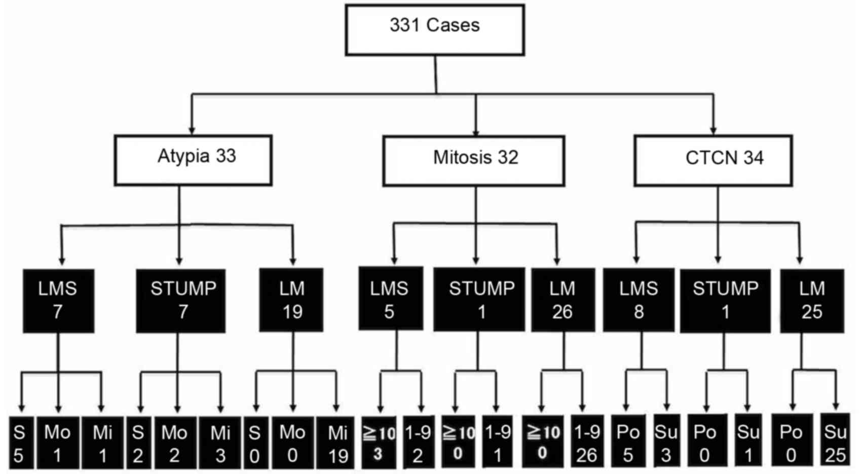
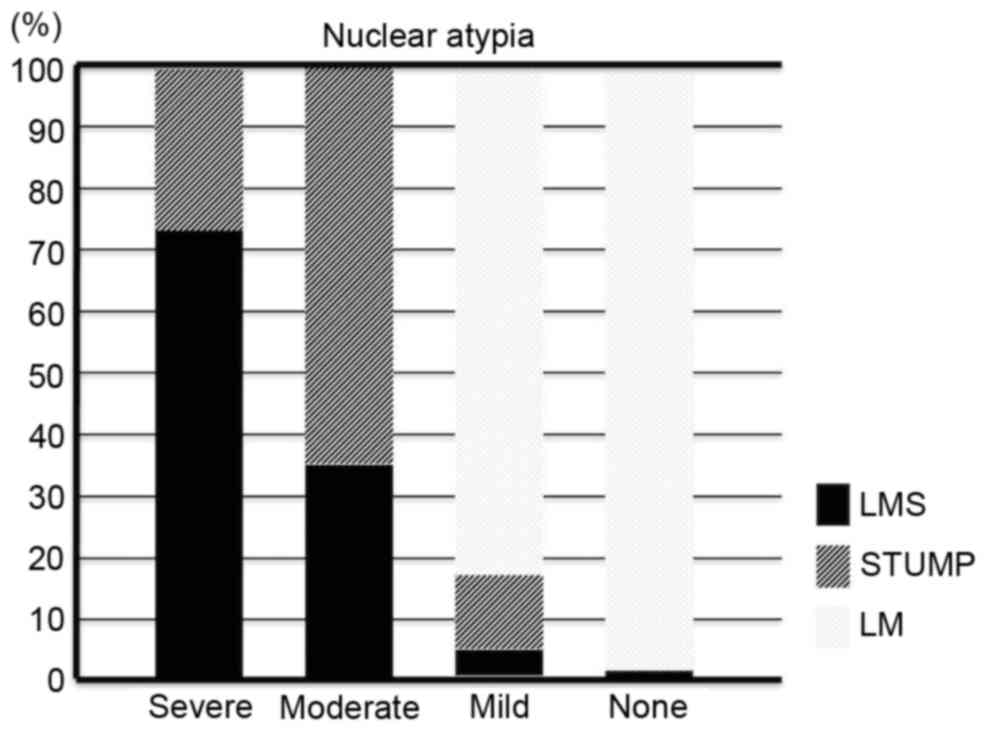
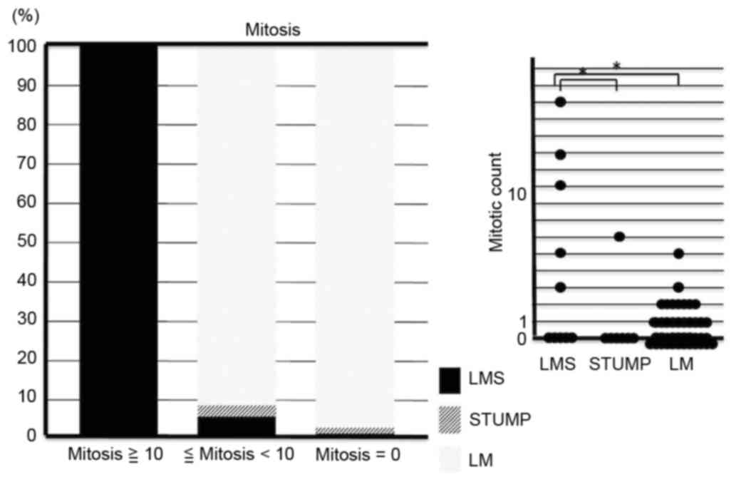
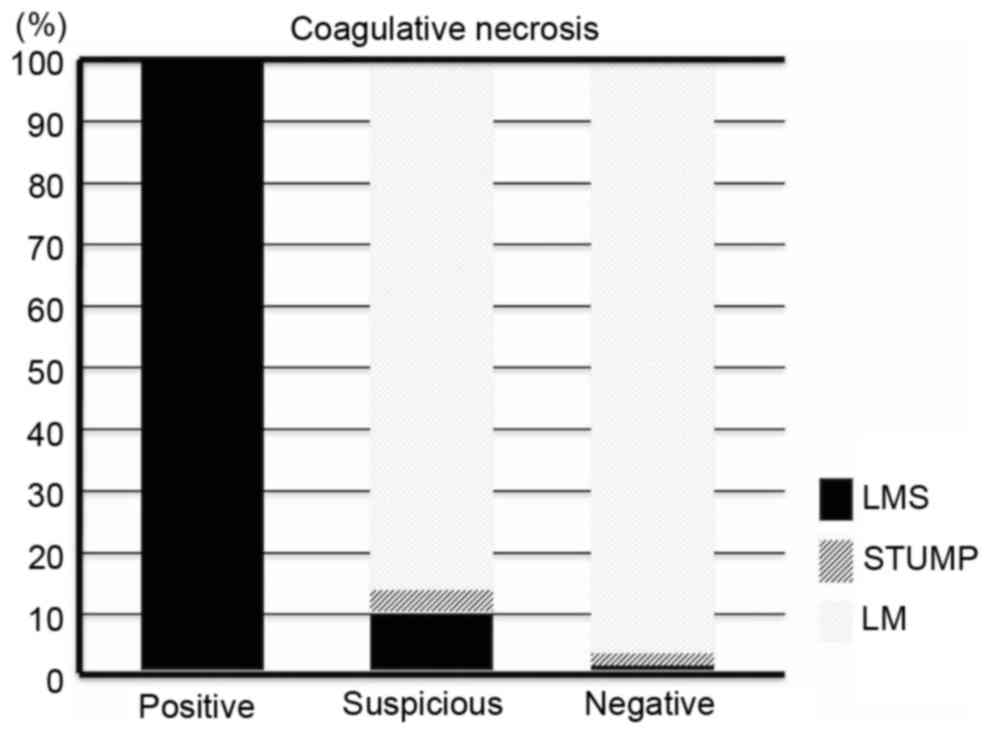
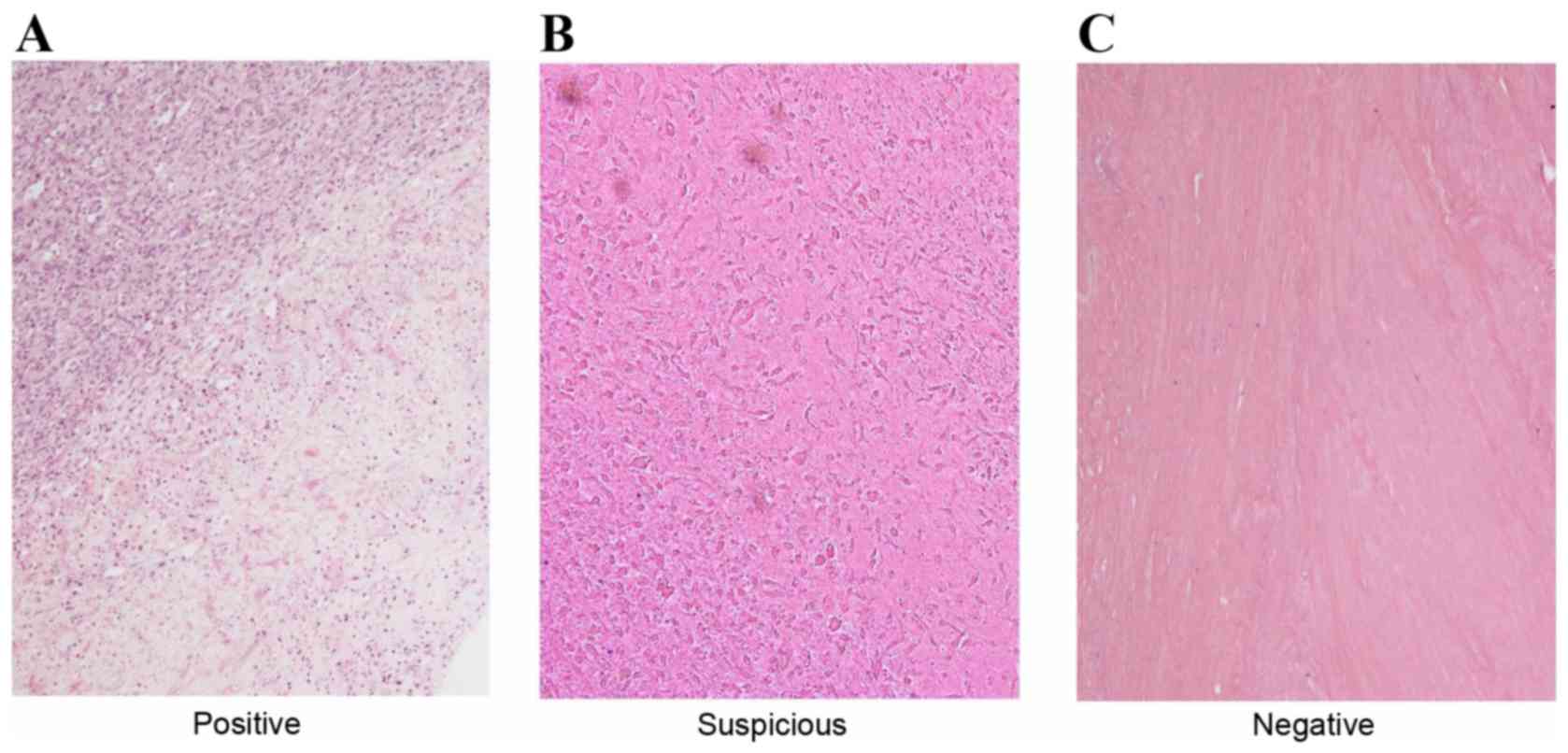
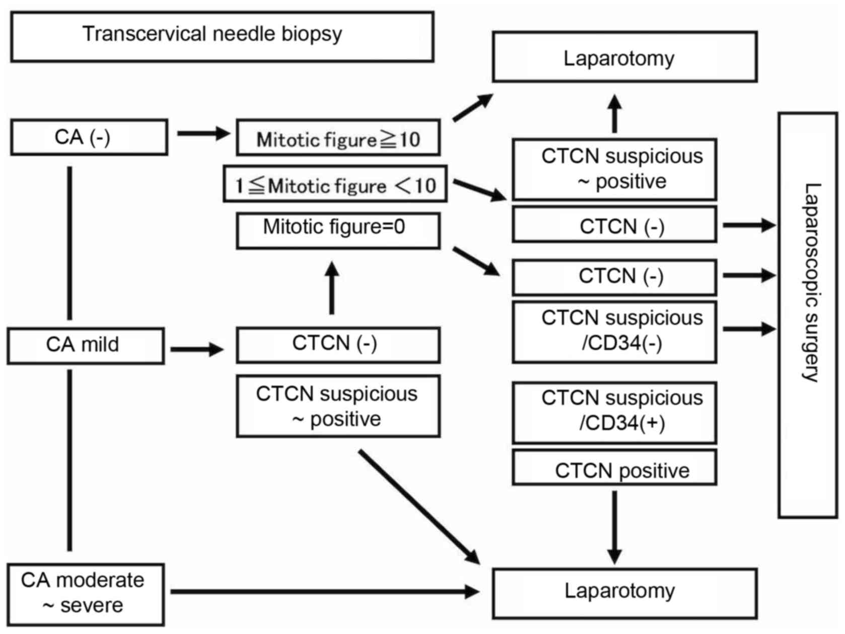
The warning statement issued by the United States Food and Drug Administration against the use of laparoscopic power morcellators prompted a discussion about the methods of preoperative diagnosis of uterine myometrial lesions. Since 1994, transcervical needle biopsies have been performed to differentiate between uterine leiomyomas and leiomyosarcomas. Needle biopsies are also useful for performing laparoscopic surgery on uterine smooth muscle tumors with histopathological safety. In the present study, data from hematoxylin and eosin (HE)‑stained specimens obtained by transcervical needle biopsies from 331 patients with smooth muscle tumors and high intensity regions on T1 weighted images (WI) and/or T2WI from magnetic resonance imaging (MRI) scans were retrospectively examined. From a total of 10 patients with moderate or severe cytological atypia, 4 exhibited smooth muscle tumors of uncertain malignant potential and 6 exhibited leiomyosarcomas. The final diagnosis in 3 patients with ≥10 mitotic figures/high‑power field was leiomyosarcoma. A total of 5 patients with coagulative tumor cell necrosis exhibited final diagnoses of leiomyosarcoma. Patients without cytological atypia, mitotic figures or coagulative tumor cell necrosis were not diagnosed with either leiomyosarcomas or smooth muscle tumors of uncertain malignant potential. The present study revealed that laparoscopic surgery is safe when HE‑stained specimens obtained by transcervical needle biopsy from areas of high intensity on an MRI scan are negative for all three criteria assessed‑cytological atypia, mitotic figures and coagulative tumor cell necrosis.
View Figures
View References

Related Articles

Journal Cover

June-2018
Volume 15 Issue 6

Print ISSN: 1792-1074
Online ISSN:1792-1082

Sign up for eToc alerts

Recommend to Library

Copy and paste a formatted citation
x
Spandidos Publications style
Murakami M, Ichimura T, Kasai M, Matsuda M, Kawamura N, Fukuda T and Sumi T: Examination of the use of needle biopsy to perform laparoscopic surgery safely on uterine smooth muscle tumors. Oncol Lett 15: 8647-8651, 2018
APA
Murakami, M., Ichimura, T., Kasai, M., Matsuda, M., Kawamura, N., Fukuda, T., & Sumi, T. (2018). Examination of the use of needle biopsy to perform laparoscopic surgery safely on uterine smooth muscle tumors. Oncology Letters, 15, 8647-8651. https://doi.org/10.3892/ol.2018.8425
MLA
Murakami, M., Ichimura, T., Kasai, M., Matsuda, M., Kawamura, N., Fukuda, T., Sumi, T."Examination of the use of needle biopsy to perform laparoscopic surgery safely on uterine smooth muscle tumors". Oncology Letters 15.6 (2018): 8647-8651.
Chicago
Murakami, M., Ichimura, T., Kasai, M., Matsuda, M., Kawamura, N., Fukuda, T., Sumi, T."Examination of the use of needle biopsy to perform laparoscopic surgery safely on uterine smooth muscle tumors". Oncology Letters 15, no. 6 (2018): 8647-8651. https://doi.org/10.3892/ol.2018.8425