Introduction
Gliomas are the most common primary malignant brain
tumor with poor clinical outcome and account for 40–50% of primary
intracranial neoplasms (1). The
etiology and pathogenesis of gliomas remain to be elucidated.
Despite the advances in therapeutic approaches, treatment offers
limited aid in prolonging survival (2). In view of the overall poor outcome with
current therapies, a better understanding of glioma etiology is
crucial for future development of more effective treatments to cure
this rapidly progressing disease.
It has been demonstrated that the growth of cancer
cells is primarily dependent on the glycolysis pathway (3). Phosphoglycerate mutase 1 (PGAM1)
catalyzes the conversion of 3-phosphoglycerate (3-PG) to
2-phosphoglycerate (2-PG) to release energy and is one of the key
enzymes in the glycolytic pathway. A previous study by Ramanathan
et al (3) suggested that
knocking out the glycolysis pathway may cause extreme damage to
tumor cells. However, the role of PGAM1 in glioma is poorly
investigated.
In the present study, PGAM1 mRNA expression in rat
C6 glioma cells and astrocytes, and the protein expression in human
infiltrating astrocytomas of different World Health Organization
(WHO) grades (4) and peritumoral
brain tissue were investigated, and the role of PGAM1 in glioma
proliferation and progression was discussed.
Materials and methods
Animals and cells
A total of 40 male adult Sprague-Dawley rats about 3
months old, weight 250±30 g, were provided by the Experimental
Animal Center of Shandong University (Jinan, China), and maintained
in a climate-controlled and sound isolated room (21°C) under a
12:12-h light:dark cycle, with 40–60% relative humidity and fed on
laboratory chow and water ad libitum. Following 1-week
acclimation, rats were sacrificed by intraperitoneal injection of
sodium pentobarbital (45 mg/kg). Brain tissue was obtained for the
experiments. The glioma C6 cell lines were purchased from the
Institute of Biochemistry and Cell Biology, Chinese Academy of
Sciences (Shanghai, China). Ethical approval was provided by the
Ethics Committee of Qilu Hospital (Shandong, China).
Patient specimens
A total of 102 cases of astrocytomas treated in the
Department of Neurosurgery, Shandong Cancer Hospital (Jinan, China)
and Qilu Hospital of Shandong University (Jinan, China) between
December 2011 and June 2013 were collected (59 males and 43
females; median age, 47 years; age range, 28–66 years). According
to WHO 2007 criteria, 38 cases were WHO grade II, 30 cases were WHO
grade III and 34 were WHO grade IV (4). A total of 11 cases of peritumoral brain
tissue were also collected. No patients had ever received
chemotherapy or radiation therapy prior to surgery.
Reagents
TRIzol was purchased from Invitrogen (Thermo Fisher
Scientific, Inc. Waltham, MA, USA). Chloroform, isopropanol and
diethyl pyrocarbonate-treated water were purchased from Shanghai
Sangon Biological Engineering Technology Services Ltd. (Shanghai,
China). Ethidium bromide (EB) was purchased from Sigma-Aldrich;
Merck KGaA (Darmstadt, Germany); the reverse
transcription-polymerase chain reaction (RT-PCR)
PrimeScript™ RT reagent kit (cat no. RR037A) was
purchased from Takara Biotechnology Co., Ltd. (Dalian, China),
deoxynucleotide triphosphates (dNTPs), Taq DNA polymerase and
CASsuper 7Kb DNA Marker were purchased from Promega Corporation
(Madison, WI, USA). Poly-L-lysine, immunohistochemical
hypersensitivity kit, goat anti-human PGAM1 antibody (sc-376638),
3,3′-diaminobenzidine (DAB) dye (sc-24982) and antibody dilutions
(sc-2023) were purchased from Santa Cruz Biotechnology, Inc.
(Dallas, TX, USA).
Culture, passage and identification of
astrocytes
Bilateral brain cortex tissue (~0.5 g) from
1-week-old SD rats was collected and washed with
Ca2+/Mg2+-free Hanks solution (sc-391061;
Santa Cruz Biotechnology, Inc.) twice. The brain tissue was
shredded and kept at room temperature for 10 min with trypsin
(sc-391055; 1:250; Santa Cruz Biotechnology, Inc.). The resulting
brain tissue was transferred into the centrifuge tube and
centrifuged at 1,000 × g for 3 min at 4°C for cell separation. Cell
suspension was collected and transferred into a new tube and
centrifuged at 1,500 × g for 5 min at 4°C. The second cell
suspension was seeded into a culture flask that was previously
coated (4°C, overnight) with poly-L-lysine (sc-286689; 0.1%; Santa
Cruz Biotechnology, Inc.). Cells were cultured with Dulbecco's
modified Eagle's medium (DMEM)/Ham's F12 with 15% fetal bovine
serum (FBS; 16250086; Thermo Fisher Scientific, Inc.) at 37°C in a
5% CO2 incubator. Finally, astrocytes were identified
with glial fibrillary acidic protein (GFAP; sc-33673; Santa Cruz
Biotechnology, Inc.) immunofluorescence (5).
The immunostaining steps were as follows: Coverslips
with their attached cells were removed from the culture medium and
washed three times in phosphate buffered saline (PBS; sc-286634;
Santa Cruz Biotechnology, Inc.) before being fixed in 4%
paraformaldehyde (sc-281692; Santa Cruz Biotechnology, Inc.) for 1
h at room temperature. The coverslips were then washed three times
in cold PBS (5 min each). Prior to staining, cells were
permeabilised in PBS containing 0.01% Triton for 10 min at room
temperature and then washed three times in cold PBS (3 min each).
The coverslips were blocked with 10% normal goat serum for 1 h at
37°C. Each coverslip was incubated with 20 µl mouse anti-GFAP
primary antibody (cat no. sc-33673; Santa Cruz Biotechnology, Inc.)
1:200 at 4°C overnight. Negative controls were incubated
identically but without primary antibody. Coverslips were then
washed three times in PBS, then incubated with a goat anti-rabbit
secondary antibody conjugated to FITC (sc-2010; 1:100; Santa Cruz
Biotechnology, Inc.) for 30 min at room temperature and washed
three times in cold PBS 5 min each. Then the coverslips were
incubated with 20 µl DAPI (sc-3598; Santa Cruz Biotechnology, Inc.)
for 5 min at room temperature in the dark. Images were captured
using a confocal microscope (PerkinElmer, Inc., Waltham, MA, USA)
at a wavelength of 520 nm.
Culture of C6 glioma cells
C6 glioma cells were cultured in DMEM/Ham's F12
containing 15% FBS at 37°C in a 5% CO2 incubator.
Following cells reaching exponential phase, they were subcultured
into two culture flasks.
Total RNA extraction and detection of PGAM1 mRNA
expression in rat astrocytes and C6 glioma cells
Extraction of total RNA from C6 and
astrocytes
Once the cultured cells were confluent in the
culture flasks, the culture medium was removed and cells were
washed for 30 sec with PBS twice. Total RNA was extracted using a
TRIzol RNA extraction kit (Thermo Fisher Scientific, Inc.)
according to the manufacturer's protocol. RNA concentration and
quality were determined using a NanoDrop ND-1000 spectrophotometer
(A260/A280 values were 1.80–2.0; Thermo
Fisher Scientific, Inc.) and gel electrophoresis.
Reverse transcription (first-strand
cDNA synthesis)
A total of 2 µg total RNA, 2.0 µl 10 µmol/l oligo
dNTPs, 2.0 µl 10X PCR buffer, 2.0 µl 5 µmol/l dNTPmix, 1.0 µl dNTP
mixture, RNasin® Ribonuclease Inhibitor (4 U/µl;
Invitrogen; Thermo Fisher Scientific, Inc.), and an appropriate
amount of diethyl pyrocarbonate-treated water was added to a total
volume of 20 µl, and thenplaced into a 0.5 ml Eppendorf tube. The
mixture was incubated at 37°C for 60 min, and the reaction was
stopped by heating at 70°C for 15 min. Then, the reaction tube was
transferred to ice, and 20 µl Tris-EDTA buffer (Invitrogen; Thermo
Fisher Scientific, Inc.) was added.
PCR and electrophoresis of PCR
products
PCR primers were synthesized by Beyotime Institute
of Biotechnology (Shanghai, China). The PGAM1 forward primer
sequence was 5′-CCTCCTGTGAGAGCCTGAAG-3′ (nt452-nt471) and the PGAM1
reverse primer sequence was 5′-CTTCTTCACCTTGCCCTGAG-3′
(nt762-nt743). β-actin was used as an internal reference. A total
of 2 µl cDNA, 2.5 µl 10X PCR buffer, 1.5 µl MgCl2, 1.0
µl dNTP mixture (2 mmol/l), 0.5 µl forward primer (10 µmol/l), 0.5
µl reverse primer (10 µmol/l), 0.5 µl Taq enzyme (2 U/µl) and 16.5
µl double-distilled water were added into a 0.2 ml Eppendorf PCR
tube on ice. Following PCR (pre-denaturation at 94°C for 7 min,
denaturation at 94°C for 30 sec, annealing at 56°C for 30 sec,
extension at 72°C for 30 sec), 5 µl PCR product was analyzed on
1.5% agarose gel electrophoresis at 100 V for 15 min. The image was
captured under UV light (WFH-204A; JK Technology, Shanghai, China).
The intensity of these bands was semi-quantified using an imaging
analyzer (ImageJ version 1.48; National Institutes of Health,
Bethesda, MD, USA).
Immunohistochemistry on
paraffin-embedded tissue sections
Sections of paraffin tissue 5-µm thick were
deparaffinized by using xylene and rehydrated through a graded
alcohol series to water. The dewaxed slides underwent antigen
retrieval in citrate buffer (Beyotime Institute of Biotechnology,
Shanghai, China) for 30 min by microwaving (Midea, Guangdong,
China) and then cooled to room temperature. Peroxidase blocking
solution (Beyotime Institute of Biotechnology, Shanghai, China) was
used to block endogenous peroxidase activity at room temperature
for 10 min. Tissue sections were incubated with anti-PGAM1 antibody
(1:100 dilution) at 4°C overnight, and then incubated with
biotinylated rabbit anti-Goat antibody (1:1,000; cat no. sc-2768;
Santa Cruz Biotechnology, Inc.) at room temperature for 10 min. A
drop of streptavidin-biotin-peroxidase solution Beyotime Institute
of Biotechnology, Shanghai, China was added onto each section and
incubated at room temperature for 10 min. Tissue sections were
incubated with freshly prepared DAB chromogenic reagent for 1–2 min
at room temperature, then counterstained with hematoxylin (1 µM;
Beyotime Institute of Biotechnology, Shanghai, China) for 3 min at
room temperature. Ten low power fields (magnification, ×100) were
selected randomly and immunostaining was detected at a high power
(magnification, ×400) using a light microscope (BX43; Olympus
Corporation, Tokyo, Japan). The immunohistochemical stain results
were divided into negative (<10% of cells with marked
cytoplasmic staining), positive (10–70% cells with marked
cytoplasmic staining) and strongly positive (>70% cells with
marked cytoplasmic staining). Positive and strongly positive
results were statistically counted as positive.
Statistical analysis
Data are expressed as mean ± standard deviation.
Statistical analysis was performed using SPSS software version 14.0
(SPSS, Inc., Chicago, IL, USA). The relative absorbance of the
astrocytes and C6 glioma cells was analyzed by unpaired Student's
t-test. For the positive expression rates of PGAM1 in astrocytoma
tissues and peritumoral brain tissue, the χ2 test was
used, and P<0.05 was considered to indicate a statistically
significant difference.
Results
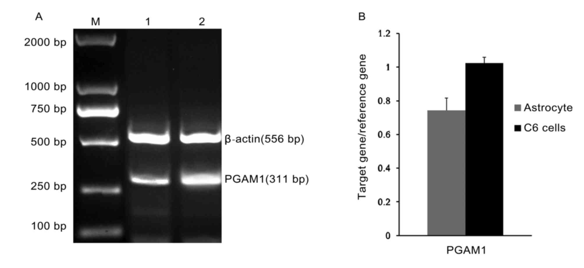
Expression of PGAM1 mRNA in C6 glioma
cells and normal astrocytes
The RT-PCR PGAM1 products from astrocytes and C6
glioma were visualized by gel electrophoresis and the intensity of
these bands was semi-quantified (Fig. 1A
and B). Using SPSS version 14.0 statistical software, 2 samples
in triplicate (C6 glioma cells vs. normal astrocytes) were
compared. It was identified that the expression of PGAM1 in C6
glioma cells (1.26±0.05) was significantly increased compared with
that of normal astrocytes (0.75±0.07). The difference was
statistically significant (P<0.05; Table I).
 | Table I.Comparison of phosphoglycerate mutase
1 expression in C6 glioma cells and astrocytes. |
Table I.
Comparison of phosphoglycerate mutase
1 expression in C6 glioma cells and astrocytes.
| Group | No. of cases | Expression quantity
(mean ± SD) |
|---|
| C6 glioma cells | 30 | 1.26±0.05 |
| Astrocytes | 10 | 0.75±0.07 |
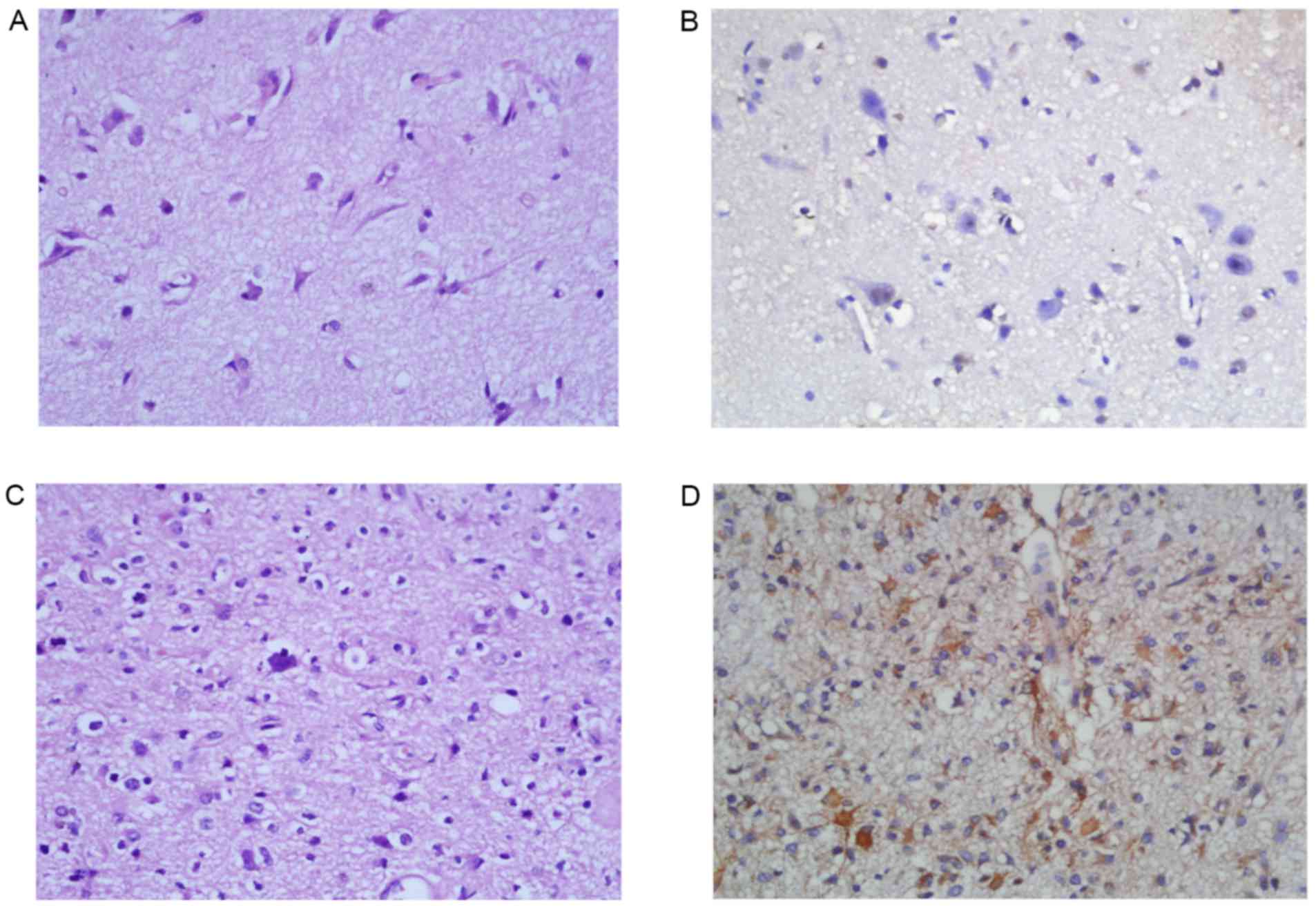
PGAM1 immunohistochemistry of
astrocytoma and peritumoral brain tissue
A total of 8/11 (73.8%) peritumoral brain tissues
were negative for PGAM1 (Fig. 2A and
B). Only 3 peritumoral brain tissues were positive for PGAM1
(3/11; 27.2%). In 102 cases of astrocytoma tissues, 74 cases
(70.6%) were positive for PGAM1 and 28 cases were negative for
PGAM1. There was a statistically significant difference in PGAM1
positive rates between the astrocytoma group and the peritumoral
brain tissue group (χ2=8.35; P<0.01; Table II). The expression of PGAM1 in the
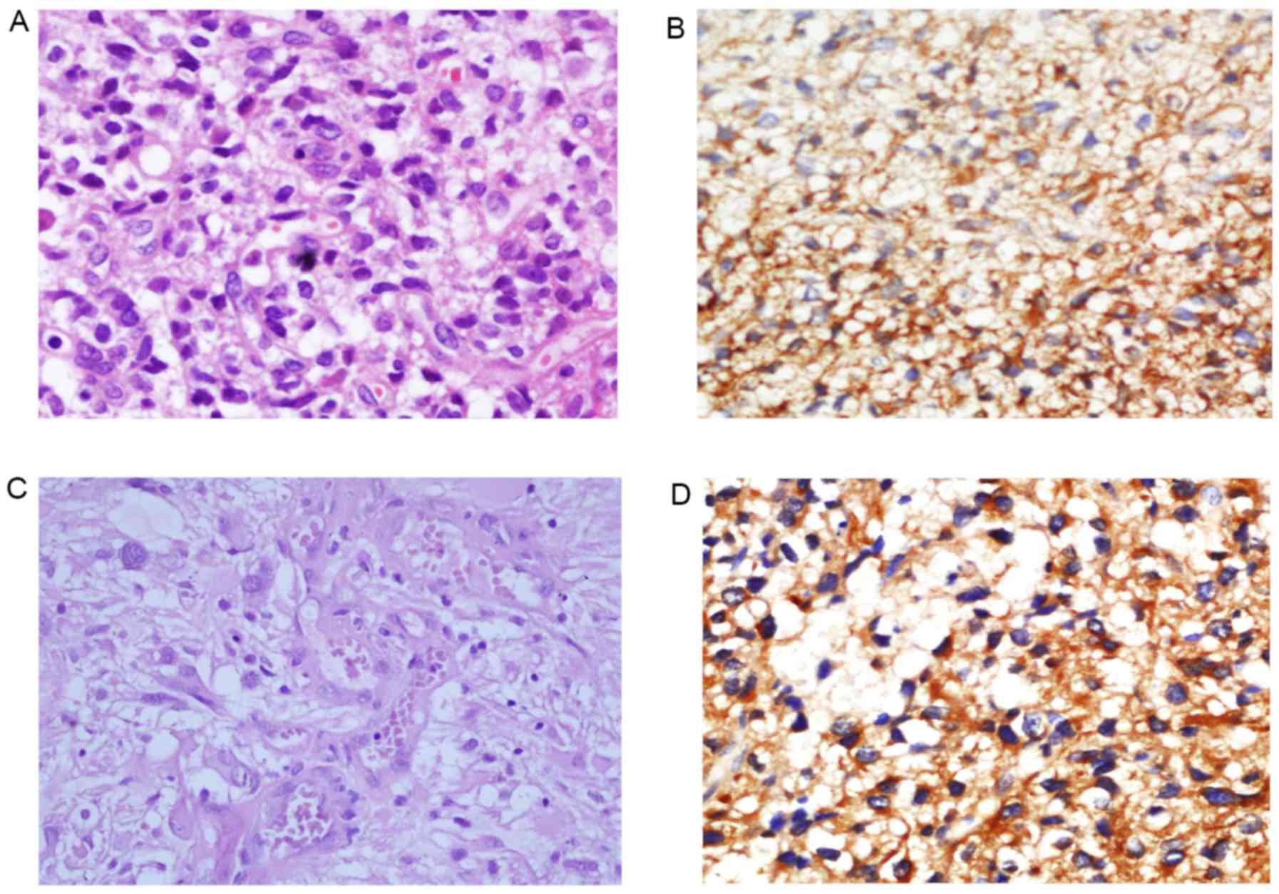
low-grade diffuse astrocytomas (WHO Grade II; Fig. 2C and D) and high-grade astrocytomas
(WHO Grade III–IV; Fig. 3) was
compared. PGAM1 was predominantly confined to the cytoplasm of
tumor cells (Figs. 2 and 3). The results demonstrated that 18/38
(47.4%) cases of WHO Grade II astrocytoma were positive for PGAM1.
A total of 54/64 cases (84.4%) of high-grade astrocytomas were
positive for PGAM1. The PGAM1 positive rate was significantly
different between low-grade astrocytomas (WHO grade II) and
high-grade astrocytomas (WHO grade III–IV) (χ2=15.73;
P<0.01; Table III).
| Table II.Comparison of phosphoglycerate mutase
1 protein in glioma tissue and peritumoral brain tissue. |
Table II.
Comparison of phosphoglycerate mutase
1 protein in glioma tissue and peritumoral brain tissue.
| Group | No. of cases | Negative | Positive | Positive rate
(%) |
|---|
| Normal brain
tissue | 11 | 8 | 3 | 27.2 |
| Glioma tissue | 102 | 30 | 72 | 70.6 |
| Table III.Association between phosphoglycerate
mutase 1 expression and the grades of gliomas. |
Table III.
Association between phosphoglycerate
mutase 1 expression and the grades of gliomas.
| Group | No. of cases | Negative | Positive | Positive rate
(%) |
|---|
| Grade II | 38 | 20 | 18 | 47.4 |
| Grade III–IV | 64 | 10 | 54 | 84.4 |
Discussion
As one of the key enzymes in the glycolytic pathway,
PGAM1 primarily catalyzes the conversion of 3-phosphoglycerate into
2-phosphoglycerate. PGAM1 may be in muscle-derived form (m-type)
and brain-derived form (b-type) in mammals (6). B-type PGAM1 has been identified
previously (7). Tumor cells are
distinct from normal cells by their unique feature of the glucose
metabolism: Tumor cells obtain energy primarily through the
glycolysis pathway, whereas normal cells primarily metabolize
glucose through aerobic oxidation (8). Bustamante et al (9) identify that the hexokinase activity is
markedly low in normal tissue, whereas the activity of this enzyme
in tumor cells is significantly increased. Previous studies have
indicated that certain enzymes which inhibit glycolysis may
effectively kill liver cancer cells and other cancer cells
(10–12).
PGAM1 is the only enzyme in the glycolytic pathway
that is regulated at the transcriptional level by the tumor
suppressor gene p53 (13), In 2005,
Evans et al (14) suggest that
PGAM1 may be a novel target for cancer treatment. PGAM1 is
significantly upregulated in 66.7% of hepatocellular carcinomas,
and markedly associated with the poorer survival of patients and
poor differentiation of cancer cells (15). PGAM1 protein is overexpressed in
breast cancer and prostate cancer (16,17). The
expression of PGAM1 is upregulated in esophageal cancer (18) and glioma cells (19,20). In
the present study, PGAM1 is expressed in astrocytes and C6 glioma
cells using RT-semi-quantitative PCR, but that the expression level
in C6 glioma cells is significantly increased compared with that of
astrocytes. Using immunohistochemical staining, it is identified
that the level of PGAM1 protein in astrocytoma tissue is
significantly increased compared with that in peritumoral brain
tissue. Furthermore, high-grade astrocytomas exhibit higher levels
of PGAM1 positivity compared with low-grade astrocytomas.
Therefore, PGAM1 may play an important role in the tumorigenesis
and progression of malignant gliomas. The results indicate that
PGAM1 inhibitors may be potential anticancer drugs, but extensive
future studies are required.
In summary, the expression of PGAM1 is upregulated
in glioma cell lines and astrocytoma tissues. This suggests that
PGAM1 may be involved in the tumorigenesis and progression of
malignant gliomas. PGAM1 may become a novel target for glioma
treatment in the future.
Acknowledgements
The authors would like to thank Mr. Kuan Zhen
(Department of Neurosurgery, Second Hospital of Shandong
University), Mr. Yan Song (Department of Neurosurgery, Qilu
Hospital of Shandong University, Brain Science Research Institute
of Shandong University) and Mr. Baibin Bi (Department of
Neurosurgery, Qilu Hospital of Shandong University, Brain Science
Research Institute of Shandong University) for administrative and
operational support.
Funding
The present study was supported by intradepartmental
funding.
Availability of data and materials
All data generated during the present study are
available from the corresponding author upon reasonable
request.
Authors' contributions
ZGL, JD and CD conducted the immunohistochemistry
and immunofluorescence experiments. ZGL drafted the manuscript. NX,
ELW and YYW participated in PCR experiments. YYW proofread the
manuscript. ZGL and JD participated in data analysis, and
statistical analysis. JYL made contributions to analysis and
interpretation of data for histopathology and immunostain results,
made the pathology figures and proofread the manuscript. JMY
participated in experimental design and coordination of the study.
All authors read and approved the final manuscript.
Ethics approval and consent to
participate
Ethical approval was provided by the Ethics
Committee of Qilu Hospital (Shandong, China).
Consent for publication
Not applicable.
Competing interests
The authors declare that they have no competing
interests.
References
|
1
|
Zhou L: Editor in chief: Modern
Neurosurgery. Shanghai: Fudan University Press. Front Page; pp.
376–377. 2004
|
|
2
|
Ma LN, Song B and Cai JY: Research of the
killing effects of cinobufacini on MCF-7cells. Chin Pharmacol Bull.
27:37–40. 2011.(In Chinese).
|
|
3
|
Ramanathan A, Wang C and Schreiber SL:
Perturbational profiling of a cell-line model of tumorigenesis by
using metabolic measurements. Proc Natl Acad Sci USA.
102:5992–5997. 2005. View Article : Google Scholar : PubMed/NCBI
|
|
4
|
Louis DN, Ohgaki H, Wiestler OD, Cavenee
WK, Burger PC, Jouvet A, Scheithauer BW and Kleihues P: The 2007
WHO classification of tumours of the central nervous system. Acta
Neuropathol. 114:97–109. 2007. View Article : Google Scholar : PubMed/NCBI
|
|
5
|
Schildge S, Bohrer C, Beck K and
Schachtrup C: Isolation and culture of mouse cortical astrocytes. J
Vis Exp. 19:pii: 50079. 2013.
|
|
6
|
Grisolia S and Cleland WW: Influence of
salt, substrate, and cofactor concentrations on the kinetic and
mechanistic behavior of phosphoglycerate mutase. Biochemistry.
7:1115–1121. 1968. View Article : Google Scholar : PubMed/NCBI
|
|
7
|
Scatena R, Bottoi P, Pontoglio A,
Mastrototaro L and Giardina B: Glycolytic enzyme inhibitors in
cancer treatment. Expert Opin Investig Drugs. 17:1533–1545. 2008.
View Article : Google Scholar : PubMed/NCBI
|
|
8
|
Jones RG and Thompson CB: Tumor
suppressors and cell metabolism: A recipe for cancer growth. Genes
Dev. 23:537–548. 2009. View Article : Google Scholar : PubMed/NCBI
|
|
9
|
Bustamante E, Morris HP and Pedersen PL:
Energy metabolism of tumor cell. Requirement for a form of
hexokinase with a propensity for mitochondrial binding. J Biol
Chem. 256:8699–8704. 1981.PubMed/NCBI
|
|
10
|
Ko YH, Pedersen PL and Geschwind JF:
Glucose catabolism in the rabbit VX2 tumor model for liver cancer:
Characterization and targeting hexokinase. Cancer Lett. 173:83–91.
2001. View Article : Google Scholar : PubMed/NCBI
|
|
11
|
Xu RH, Pelicano H, Zhou Y, Carew JS, Feng
L, Bhalla KN, Keating MJ and Huang P: Inhibition of glycolysis in
cancer cells: A novel strategy to overcome drug resistance
associated with mitochondrial respiratory defect and hypoxia.
Cancer Res. 65:613–621. 2005.PubMed/NCBI
|
|
12
|
Gwak GY, Yoon JH, Kim KM, Lee HS, Chung JW
and Gores GJ: Hypoxia stimulates proliferation of human hepatoma
cells through the induction of hexokinase II expression. J Hepatol.
42:358–364. 2005. View Article : Google Scholar : PubMed/NCBI
|
|
13
|
Cheung EC and Vousden KH: The role of p53
in glucose metabolism. Curr Opin Cell Biol. 22:186–191. 2010.
View Article : Google Scholar : PubMed/NCBI
|
|
14
|
Evans MJ, Saghatelian A, Sorensen EJ and
Cravatt BF: Target discovery in small molecule cell-based screens
by in situ proteome reactivity profiling. Nat Biotechnol.
23:1303–1307. 2005. View
Article : Google Scholar : PubMed/NCBI
|
|
15
|
Ren F, Wu H, Lei Y, Zhang H, Liu R, Zhao
Y, Chen X, Zeng D, Tong A, Chen L, et al: Quantitative proteomics
identification of phosphoglycerate mutase 1 as a novel therapeutic
target in hepatocellular carcinoma. Mol Cancer. 9:812010.
View Article : Google Scholar : PubMed/NCBI
|
|
16
|
Cortesi L, Barchetti A, De Matteis E,
Rossi E, Della Casa L, Marcheselli L, Tazzioli G, Lazzaretti MG,
Ficarra G, Federico M and Iannone A: Identification of protein
clusters predictive of response to chemotherapy in breast cancer
patients. J Proteome Res. 8:4916–4933. 2009. View Article : Google Scholar : PubMed/NCBI
|
|
17
|
Zhang XB, Tang ZY, Zu XB, Qi L and Ruan
JD: Modified serum-guided immunoblotting for differential study of
prostate cancer. Zhonghua Nan Ke Xu. 16:438–444. 2010.(In
Chinese).
|
|
18
|
Saeki H, Kitao H, Yoshinaga K, Nakanoko T,
Kubo N, Kakeji Y, Morita M and Maehara Y: Copy-neutral loss of
heterozygosity at the p53 locus in carcinogenesis of esophageal
squamous cell carcinomas associated with p53 mutations. Clin Cancer
Res. 17:1731–1740. 2011. View Article : Google Scholar : PubMed/NCBI
|
|
19
|
Gao H, Yu B, Yan Y, Shen J, Zhao S, Zhu J,
Qin W and Gao Y: Correlation of expression levels of ANXA2, PGAM1,
and CALR with glioma grade and prognosis. J Neurosurg. 118:846–853.
2013. View Article : Google Scholar : PubMed/NCBI
|
|
20
|
Jain R, Kulkarni P, Dhali S, Rapole S and
Srivastava S: Quantitative proteomic analysis of global effect of
LLL12 on U87 cell's proteome: An insight into the molecular
mechanism of LLL12. J Proteomics. 113:127–142. 2015. View Article : Google Scholar : PubMed/NCBI
|