Introduction
Nasopharyngeal carcinoma (NPC) is a squamous cell
malignancy that originates from the nasopharyngeal epithelium
(1,2).
In 2001, the incidence rates of NPC in China at 25–30 per 100,000
were the highest globally (3), as are
the mortality rates for NPC in China (4). NPC has no typical symptoms in the early
stage of the disease and is characterized by gross invasion; thus,
the majority of patients with NPC are diagnosed at an advanced
disease stage (5). Thus, there is a
requirement for further research into the molecular mechanisms
involved in the carcinogenesis of NPC, which may aid the
identification of an independent biomarker for the early detection
and treatment of NPC (1,6).
In the process of the transcription and translation
of genetic information, 98.5% of all RNAs do not possess the
ability to be translated into a protein (7,8). Long
non-coding RNAs (lncRNAs) are one such type of these RNAs, and are
>200 nucleotides in length (9).
lncRNAs were considered to be transcriptional noise for a number of
years (10); however, previous
studies have revealed that abnormally expressed lncRNAs influence
the progression of human diseases, particularly cancer, via gene
silencing, chromatin remodeling, splicing regulation and cell cycle
regulation (10,11). Furthermore, the lncRNAs HOX transcript
antisense RNA, H19 and metastasis associated lung adenocarcinoma
transcript 1 (MALAT1) were closely associated with human
tumorigenesis (12–16).
lncRNA ZNF674-1 (OTTHUMG00000021416) was
dysfunctionally expressed in primary NPC tissues, which indicates
that it may serve a role in the tumorigenesis of NPC (17). In the present study, the expression of
lncRNA ZNF674-1 in the NPC 6-10B cell line and 24 primary NPC
tissues was assessed. Next, the expression of lncRNA ZNF674-1 was
overexpressed and knocked down in vitro in order to identify
its role in the proliferation, invasion, migration and apoptosis of
NPC cells.
Materials and methods
Cell lines and tissues
A total of 24 primary NPC specimens (mean age, 43
years; age range, 25–64 years; 18 men, 6 women) and 24 normal
nasopharyngeal epithelial specimens were obtained from Peking
University Shenzhen Hospital (Shenzhen, China) between May 2013 and
September 2015. The patients were not age- and sex-matched. Fresh
clinical specimens were obtained by nasal endoscope and stored
immediately in liquid nitrogen following collection from the
Ear-Nose-Throat Department. Ethical approval was obtained from the
Research Ethics Board of Peking University Shenzhen Hospital. Each
of the patients with NPC had no history of radiotherapy,
chemotherapy or surgery prior to the experiment. Written informed
consent was obtained from all patients prior to the study.
Clinicopathological information for the patients is presented in
Table I. The clinical staging of all
patients were determined using the 2010 Cancer Staging Standard
published by the America Joint Committee (18).
 | Table I.Clinicopathological characteristics
of patients. |
Table I.
Clinicopathological characteristics
of patients.
|
Characteristics | Patients, n |
|---|
| Sex |
|
|
Male | 18 |
|
Female | 6 |
| Degree of
differentiation |
|
|
Undifferentiated | 15 |
|
Differentiated | 7 |
| Histology |
|
|
Squamous | 24 |
|
Other | 0 |
| Lymph node
metastasis |
|
|
Positive | 17 |
|
Negative | 7 |
| Distal
metastasis |
|
|
Positive | 0 |
|
Negative | 24 |
| Clinical TNM
stage |
|
|
I–II | 4 |
|
III–IV | 20 |
Cell culture
An NPC 6-10B cell line and the human normal
nasopharyngeal NP460 cell line were obtained from Peking University
Shenzhen Hospital and Southern Medical University (Guangzhou,
China). The two cell lines were maintained in RPMI-1640 (Gibco;
Thermo Fisher Scientific, Inc., Waltham, MA, USA) supplemented with
50 U/ml penicillin (Gibco; Thermo Fisher Scientific, Inc.), 50 U/ml
streptomycin (Gibco; Thermo Fisher Scientific, Inc.) and 10% fetal
bovine serum (Gibco; Thermo Fisher Scientific, Inc.) in a
humidified incubator at 37°C with 5% CO2.
RNA extraction and reverse
transcription-quantitative polymerase chain reaction (RT-qPCR)
Total RNA was extracted from clinical tissues and
cell lines using TRIzol reagent (Invitrogen; Thermo Fisher
Scientific, Inc.), according to the manufacturer's protocol. Next,
the purity of RNA was examined using the SmartSpec Plus
Spectrophotometer (Bio-Rad Laboratories, Inc., Hercules, CA, USA).
For RT-qPCR, the Reverse Transcription kit (Takara Biotechnology,
Co., Ltd., Dalian, China) was used to reverse transcribe the RNA to
cDNA according to the manufacturer's protocol. The Roche
Lightcycler 480 Real-Time PCR (Roche Applied Science, Penzberg,
Germany) system was used to detect the expression of lncRNA
ZNF674-1 in clinical tissues and cell lines using the Synergy
Brands Green kit (Takara Biotechnology, Co., Ltd.). The
thermocycling conditions for RT-qPCR were: 94°C for 3 min, 94°C for
30 sec, 55°C for 30 sec and 72°C for 2 min for 40 cycles and
finally 72°C for 10 min. All samples were tested in triplicate. The
expression of lncRNA ZNF674-1 were normalized using GAPDH, which
was set as an internal reference. The primer sequences for lncRNA
ZNF674-1 were as follows: Forward, 5′-AGCACTTGGCCCTAAAGAGA-3′ and
reverse, 5′-AACATACTGGCCCAAACAGA-3′. The primer sequences for GAPDH
were as follows: Forward, 5′-TCCAAAATCAAGTGGGGCGGA-3′ and reverse,
5′-TGATGACCCTTTTGGCTCCCC-3′. The 2−ΔΔCq method was used
to calculate the relative expression of lncRNA ZNF674-1 in clinical
tissues and cell lines (19).
Knockdown and overexpression of lncRNA
ZNF674-1
One non-targeting control (si-NC) and four small
interfering RNAs (siRNAs) against lncRNA ZNF674-1 (si-ZNF674-1)
were used. All siRNAs were synthesized by GenePharma (Shangahi
GenePharma Co., Ltd., Shanghai, China). The four si-ZNF674-1 were:
sense, 5′-CCCUAAUCUUGAUGGCCAUTT-3′ and antisense,
5′-AUGGCCAUCAAGAUUAGGGTT-3′; sense, 5′-CCUGAUAUCUGAAGUAACATT-3′ and
antisense, 5′-UGUUACUUCAGAUAUCAGGTT-3′; sense,
5′-GGCGGAUUCAUUACAGUUATT-3′ and antisense,
5′-UCAAUGAGAUGCAGGUAUGTT-3′; sense, 5′-CUACUCUAUAAGGCAUCUATT-3′ and
antisense, 5′-UAGAUGCCUUAUAGAGUAGTT-3′.
A total of 3×10⁶ 6-10B cells were
transfected with si-NC (100 nM) and si-ZNF674-1 (100 nM) using
Lipofectamine® 2000 (Invitrogen; Thermo Fisher
Scientific, Inc.) following seeding in a six-well plate overnight.
After 48 h of transfection, the interfering efficiency of the four
si-ZNF674-1 was examined. Consequently, si-ZNF674-1, which
exhibited the highest knockdown efficiency of lncRNA ZNF674-1
expression (>78.8%) of the four si-ZNF674-1, was selected for
further experimentation. The sequences of the si-ZNF674-1 selected
were as follows: Sense, 5′-CCCUAAUCUUGAUGGCCAUTT-3′; antisense,
5′-AUGGCCAUCAAGAUUAGGGTT-3′; si-NC sense,
5′-GAGGCGUGGAGUCUUGUUUTT-3′; antisense,
5′-AAACAAGACUCCACGCCUCTT-3′.
ADV4 was used as a template to synthesize the
adenovirus (ADV4-lncRNA-ZNF674-1), which was produced by Shanghai
GenePharma Co., Ltd. (Shanghai, China), and contained the target
sequence to overexpress the lncRNA ZNF674-1. A total of
3×10⁶ 6-10B cells were seeded in a 6-well plate for 24 h
and then 10 µl ADV4-lncRNA-ZNF674-1 (9×10⁹ PFU/ml) or
ADV4-NC (9×10⁹ PFU/ml) was added into the medium. Then,
48 h later, the overexpression efficiency of ADV4-lncRNA-ZNF674-1
was examined.
Cell proliferation assays
Cell proliferation was assessed using the Cell
Counting Kit-8 (CCK8; Dojindo Molecular Technologies, Inc.,
Kumamoto, Japan) according to the manufacturer's protocol. A total
of 3,000 6-10B cells/well were plated into a 96-well plate with 10
replicates. Next, half of the wells were transfected with
si-ZNF674-1 or infected with ADV4-lncRNA-ZNF674-1, and the other
half were transfected with si-NC or infected with ADV4-NC. Cellular
proliferation was assessed at 450 nm every 24 h
post-transfection.
Cell scratch assay
6-10B cells (2×10⁶ cells/well) were
seeded in a 6-well plate. Cells were transfected with si-ZNF674-1
or infected with ADV4-lncRNA-ZNF674-1 or the respective controls
when they grew to 90–100% confluence. Next, a sterile 200-µl
pipette tip was used to scratch a line through the well at 6 h
post-transfection. Finally, serum-free RPMI-1640 medium was used to
replace the serum-containing medium. Images of migration distance
were captured and assessed using an inverted microscope
(magnification, ×100; Olympus Corporation, Tokyo, Japan) at 0 and
24 h after scratch assay.
Transwell assay
6-10B cells were harvested at 24 h after
transfection with the aforementioned siRNAs or lentiviruses. For
the migration assays, a total of 1×10⁴ cells in 100-µl
serum-free RPMI-1640 medium were seeded into the upper chamber
(8-µm pore size; EMD Millipore, Billerica, MA, USA). For the
invasion assays, a total of 1×10⁴ cells in 100-µl
serum-free RPMI-1640 medium were seeded in the upper chamber, which
was coated with Matrigel (1:5; 50 µl/well; BD Biosciences, Franklin
Lakes, NJ, USA). The lower chambers were filled with 500 µl
RPMI-1640 medium which contained 10% FBS. Subsequent to culturing
for 24 h, the cells that migrated or invaded the membrane were
fixed using 4% paraformaldehyde for 25 min at room temperature and
stained using 0.1% crystal violet for 25 min at room temperature in
the dark. Finally, the stained cells were imaged using an inverted
confocal microscope (magnification, ×100; Olympus Corporation) and
the total stained cells in 5 fields of view were counted by using
an inverted confocal microscope (magnification, ×100; Olympus
Corporation).
Cell apoptosis assay
6-10B cells were plated into a 6-well plate and
transfected with aforementioned siRNAs or lentiviruses. A total of
48 h after transfection, the cells were collected and washed three
times with pre-chilled phosphate buffered saline. Next, the cells
were double stained using an Annexin V-fluorescein
isothiocyanate/propidium iodide detection kit (Invitrogen; Thermo
Fisher Scientific, Inc.) for 15 min at 4°C, according to the
manufacturer's protocol. Finally, a flow cytometer (EPICS XI-4;
Beckman Coulter, Inc., Brea, CA, USA) was used to detect the
frequency of apoptotic cells using the FCS Express 3.0 software (De
Novo Software, Glendale, CA, USA).
Statistical analysis
All experiments were performed at least three times
and the data are presented as the mean ± standard deviation. All
statistical analyses were performed using SPSS software version
17.0 (SPSS, Inc., Chicago, IL, USA). The differences in expression
between the NPC and matched normal clinical specimens and between
the two cell lines were analyzed using an unpaired Student's
t-test. The cell proliferation, cell scratch assay, Transwell assay
and cell apoptosis assay data were analyzed by one-way analysis of
variance. P<0.05 was considered to indicate a statistically
significant difference.
Results
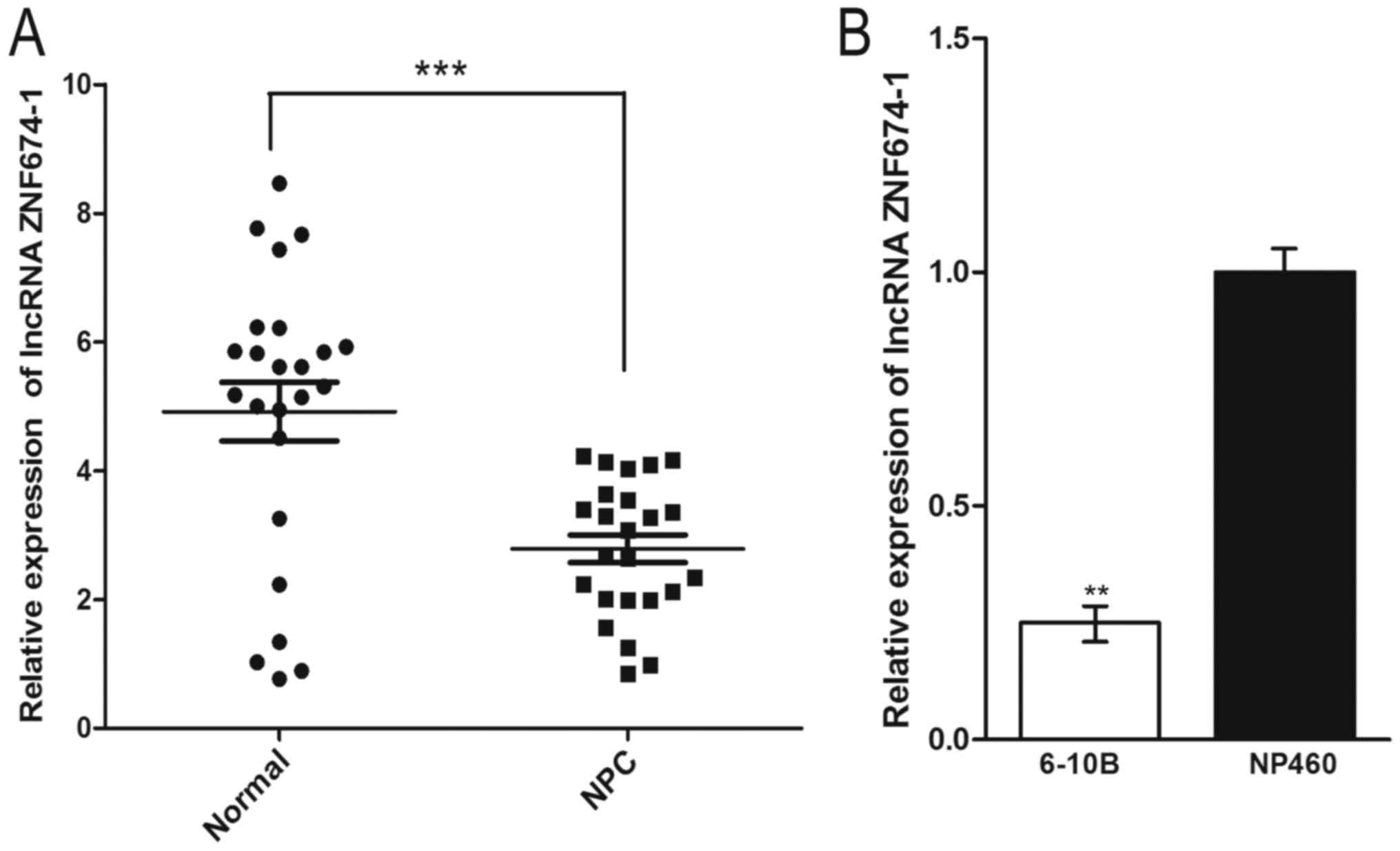
Expression of lncRNA ZNF674-1 was
downregulated in human NPC clinical tissues and NPC cell line
The expression of lncRNA ZNF674-1 was examined in 24
non-cancer nasopharyngeal epithelial specimens and 24 NPC
specimens. The results revealed that lncRNA ZNF674-1 was
significantly downregulated in NPC specimens compared with
non-cancer nasopharyngeal epithelial specimens (P<0.001;
Fig. 1A). As presented in Fig. 1B, the expression of lncRNA ZNF674-1 in
the NPC 6-10B cell line was significantly lower compared with that
in the normal NP460 cell line (P<0.01).
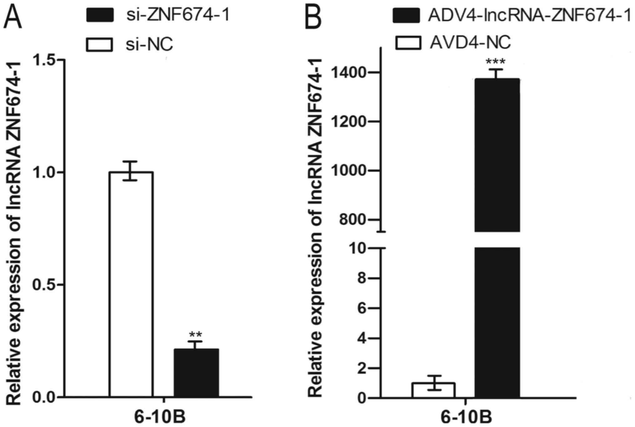
Knockdown and overexpression of lncRNA
ZNF674-1 in 6-10B
To investigate the role lncRNA ZNF674-1 serves in
the progression of NPC, a siRNA transient transfection to knock
down lncRNA ZNF674-1 in 6-10B cells was established. si-ZNF674-1
significantly decreased the expression of lncRNA ZNF674-1 by 78.8%
in 6-10B cells compared with si-NC (P<0.01; Fig. 2A). Next, an adenovirus resulting in
the stable overexpression of lncRNA ZNF674-1 in 6-10B cells was
synthesized. The ADV4-lncRNA-ZNF674-1 adenovirus could overexpress
lncRNA ZNF674-1 at a level 1,370 times higher than that in ADV4-NC
transfected cells in 6-10B cells, a difference which was identified
to be significant (P<0.001; Fig.
2B).
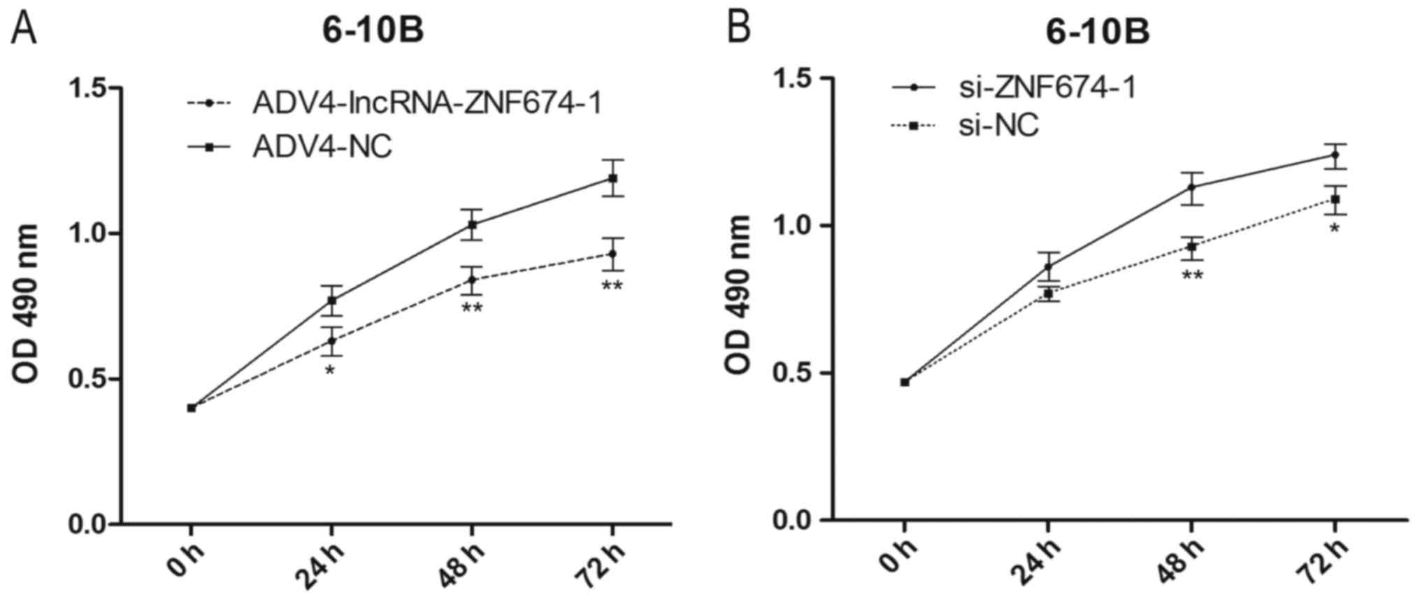
lncRNA ZNF674-1 restrained the
proliferation of NPC cells
A CCK8 assay was used to investigate the effect of
lncRNA ZNF674-1 in NPC cell proliferation. The results revealed
that the cells infected with ADV4-lncRNA-ZNF674-1 exhibited
significantly decreased cell proliferation compared with cells
infected with ADV4-NC (P<0.05; Fig.
3A), while the 6-10B cells transfected with si-ZNF674-1
exhibited significantly increased NPC cell proliferation compared
with cells transfected with si-NC (P<0.05; Fig. 3B).
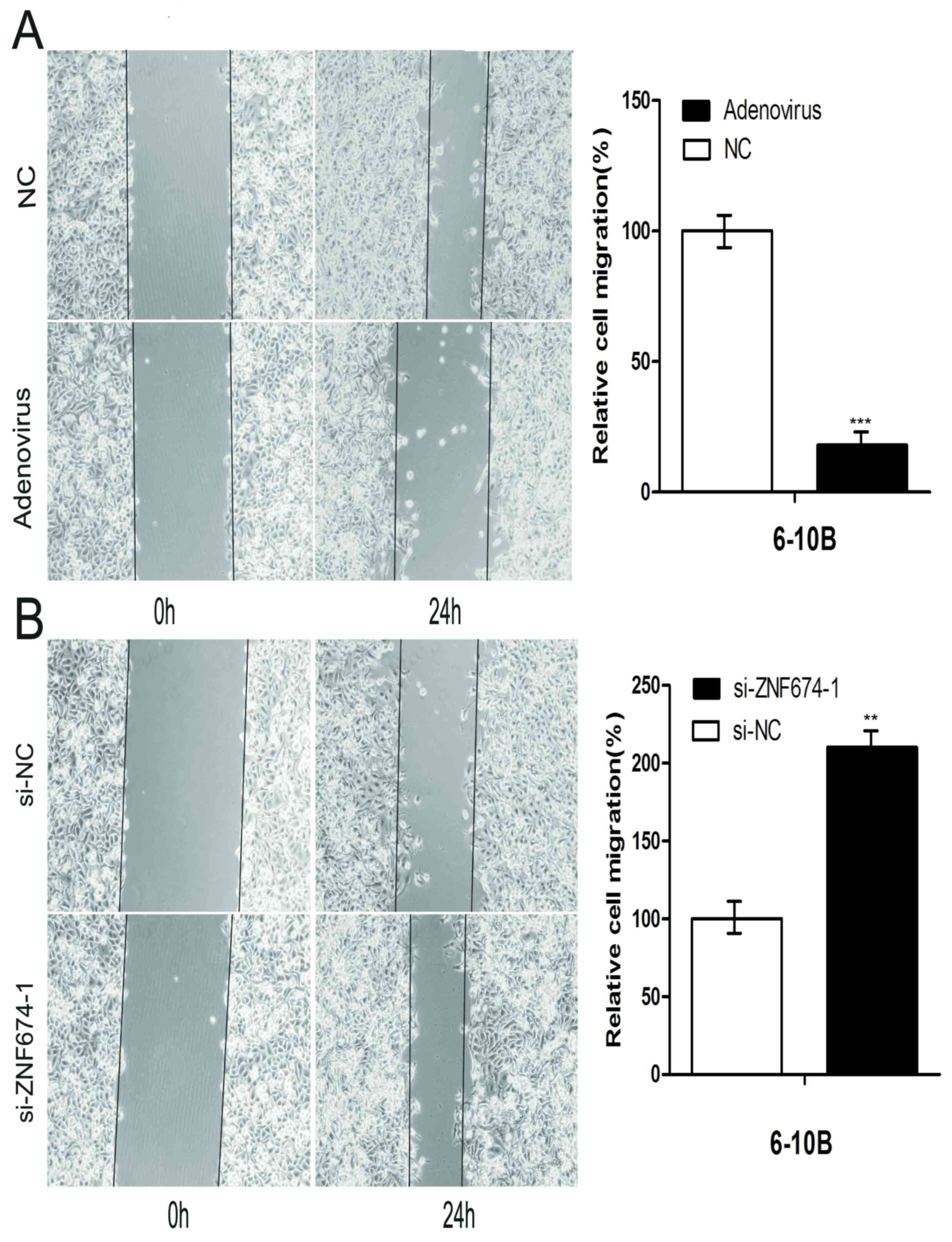
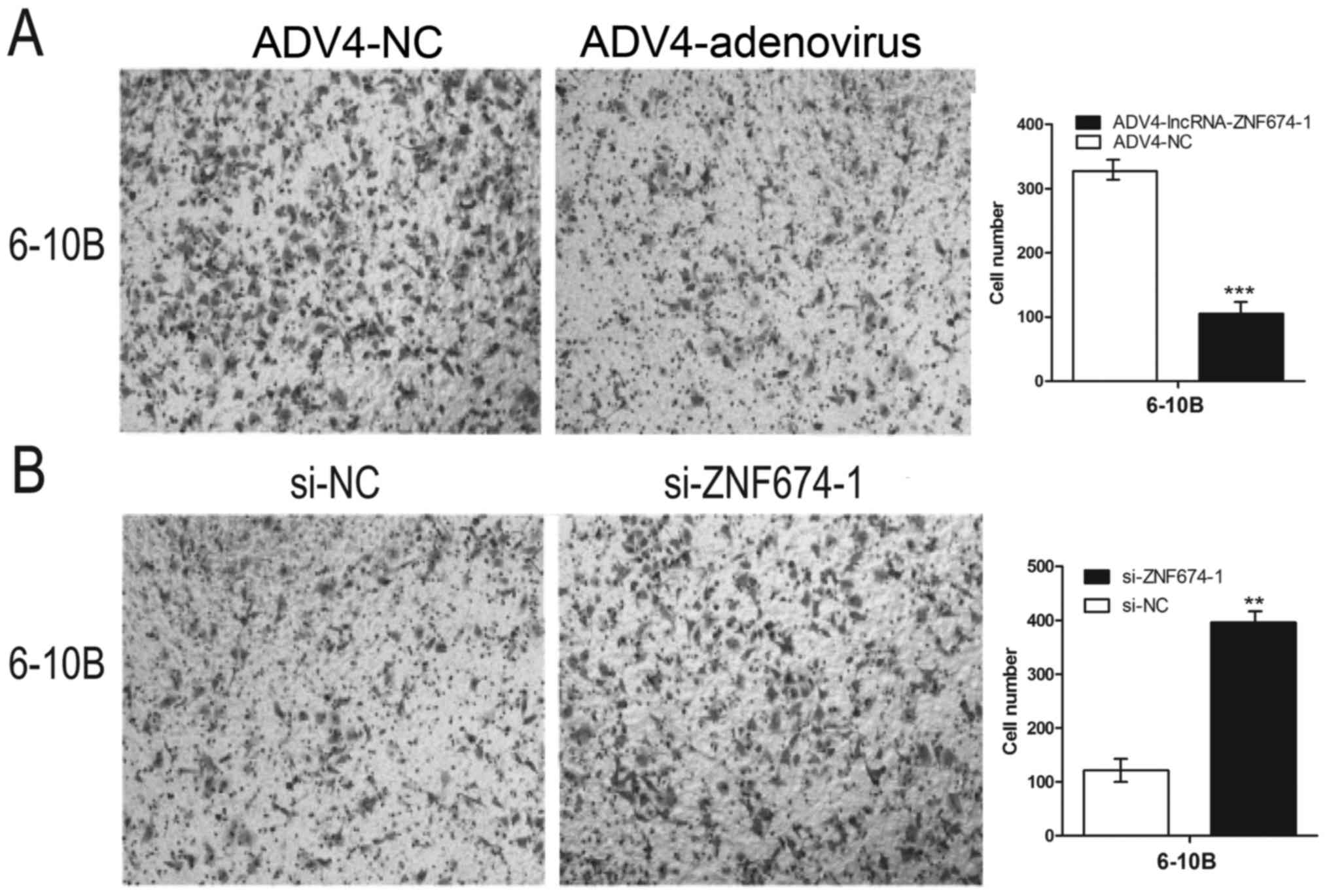
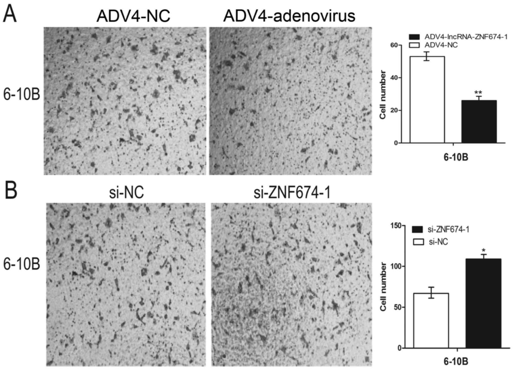
lncRNA ZNF674-1 inhibited the
migration and invasion of NPC cells
The migratory capacity of NPC cells was investigated
through scratch assays and Transwell migration assays. Scratch
assays revealed that the migratory ability of 6-10B cells following
infection with ADV4-lncRNA-ZNF674-1 were significantly reduced
compared with cells infected with the negative control (P<0.001;
Fig. 4A), whereas cell migration was
increased following transfection with si-ZNF674-1 compared with
cells transfected with si-NC (P<0.01; Fig. 4B). A similar result was obtained in
the Transwell migration assays. The migratory ability was
significantly lower in cells infected with ADV4-lncRNA-ZNF674-1
than in those infected with ADV4-NC (P<0.001; Fig. 5A), meanwhile, the migratory ability
was significantly higher in cells transfected with si-ZNF674-1 than
in those transfected with si-NC (P<0.01; Fig. 5B). Transwell invasion assays revealed
that cells infected with ADV4-lncRNA-ZNF674-1 had a significantly
lower invasion ability than did ADV4-NC infected cells (P<0.01;
Fig. 6A). However, cells transfected
with si-ZNF674-1 had a significantly higher invasive capability
than cells transfected with si-NC (P<0.05; Fig. 6B).
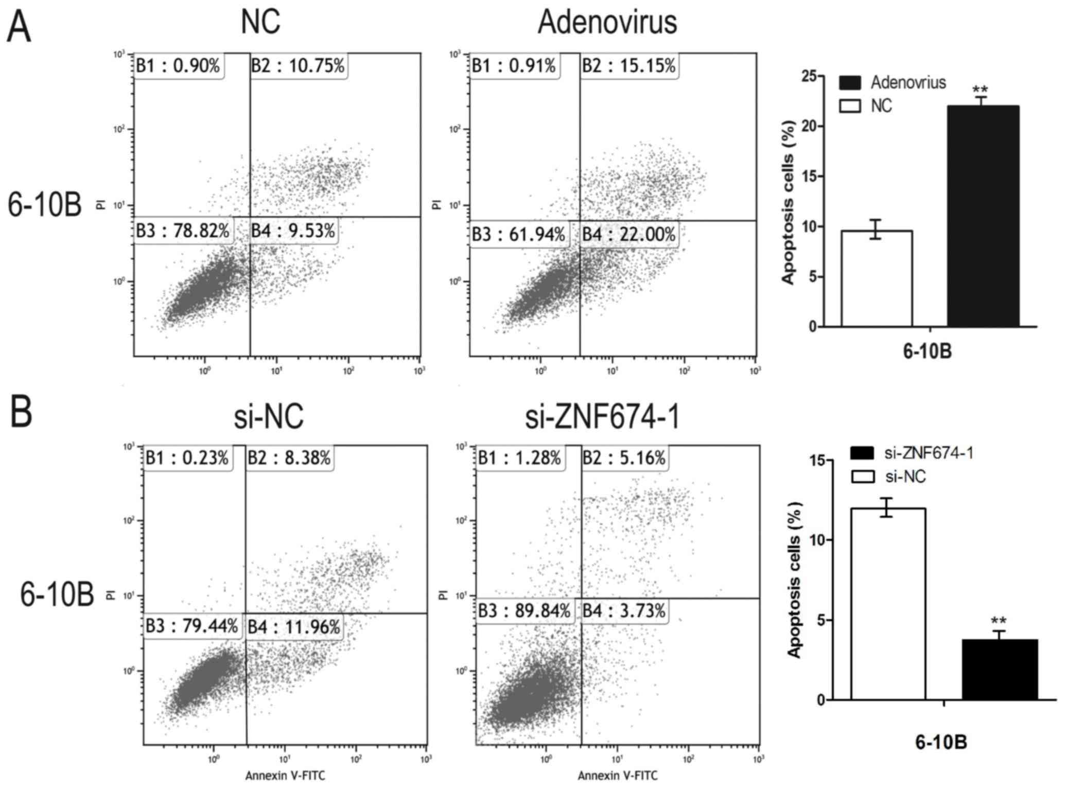
lncRNA ZNF674-1 impaired the apoptosis
of NPC cells
Flow cytometry was used to analyze the role of
lncRNA ZNF674-1 in the apoptosis of NPC cells. As presented in
Fig. 7A, the apoptosis rate of the
cells infected with ADV4-lncRNA-ZNF674-1 was significantly
increased compared with the negative control (P<0.01).
Comparatively, the apoptosis rate of the cells transfected with
si-ZNF674-1 was decreased compared with the cells transfected with
si-NC (Fig. 7B).
Discussion
An epidemiological study has revealed that numerous
factors may affect cell gene expression, which results in the
occurrence of NPC (20). Among all of
these factors, the environment, genes and Epstein-Barr virus
infection status are the most notable (21–24). The
pathological process of NPC remains unclear, so when studying the
pattern of NPC occurrence, the identification of novel biomarkers
could be performed. By combining these biomarkers with the presence
of clinicopathological features, the detection of NPC could occur
earlier, allowing for the earlier treatment of NPC (25,26).
Previously, multiple studies have reported that aberrantly
expressed lncRNAs were closely associated with the tumorigenesis of
malignancies (27–29). A number of these aberrantly expressed
lncRNAs may function as independent biomarkers of the disease,
reflecting the progression of the disease and predicting patient
outcome (30,31). Growth arrest specific 5, the
expression of which is decreased in hepatocellular carcinoma, may
restrain cell proliferation and predict poor patient prognosis
(32,33). MALAT1, originally identified in
non-small-cell lung cancer, has been used as an indicator for the
early detection and judgment of the progress of non-small-cell lung
cancer (15,16). A previous study identified that lncRNA
ZNF674-1 was aberrantly expressed in primary NPC tissues; however,
the function of lncRNA ZNF674-1 in NPC is yet to be investigated
(17). lncRNA ZNF674-1 is situated at
chromosomal location Xp11, and is 626 bp (17) and it has been identified that Xp11.2
translocation is closely associated with renal cell carcinoma
tumorigenesis (34,35). This indicates that lncRNA ZNF674-1 may
be a cancer-associated gene involved the carcinogenesis of NPC. In
the present study, the role of lncRNA ZNF674-1 in NPC was
characterized, to the best of our knowledge, for the first time. In
the present study, the expression of lncRNA ZNF674-1 in 24 primary
NPC specimens, 24 normal nasopharyngeal epithelial specimens, the
normal nasopharyngeal epithelial NP460 cell line and the NPC 6-10B
cell line was examined. It was revealed that lncRNA ZNF674-1 was
significantly downregulated in NPC tissues and NPC cell lines
compared with their respective controls (P<0.01). lncRNA
ZNF674-1 was knocked down or overexpressed to investigate the role
of lncRNA ZNF674-1 in the progression of NPC. lncRNA ZNF674-1
knockdown in 6-10B cells promoted NPC cell proliferation,
migration, invasion and restrained cell apoptosis. On the contrary,
overexpression of lncRNA ZNF674-1 inhibited cell growth, the
invasive and migratory abilities of cells, and promoted cell
apoptosis in vitro.
Taken together, the results of the present study
indicated that lncRNA ZNF674-1 serves a negative function in NPC
carcinogenesis, meaning that it may be a useful diagnostic
biomarker for the early detection and therapy of NPC. However, the
precise molecular mechanisms of lncRNA ZNF674-1 in the
carcinogenesis of NPC requires further study.
Acknowledgements
Not applicable.
Funding
The present study was supported by the Science and
Technology Development Fund Project of Shenzhen (grant no.
JCYJ20150403091443336).
Availability of data and materials
All data generated or analysed for the present study
are included in this published article.
Author's contributions
GHN conceived the experiments. ZL carried out the
molecular genetic studies. HFD and HYH collected clinical
specimens. LL and XFC performed the statistical analysis. WZ
analysed the results and and drafted the manuscript. All authors
read and approved the final manuscript.
Ethics approval and consent to
participate
Ethical approval was obtained from the Research
Ethics Board of Peking University Shenzhen Hospital. Written
informed consent was obtained from all patients prior to the
study.
Consent for publication
Not applicable.
Competing interests
The authors declare that they have no competing
interests.
References
|
1
|
Vokes EE, Liebowitz DN and Weichselbaum
RR: Nasopharyngeal carcinoma. Lancet. 350:1087–1091. 1997.
View Article : Google Scholar : PubMed/NCBI
|
|
2
|
Wei WI and Sham JST: Nasopharyngeal
carcinoma. Lancet. 365:2041–2054. 2005. View Article : Google Scholar : PubMed/NCBI
|
|
3
|
Lo KW, To KF and Huang DP: Focus on
nasopharyngeal carcinoma. Cancer Cell. 5:423–428. 2004. View Article : Google Scholar : PubMed/NCBI
|
|
4
|
Chang ET and Adami HO: The enigmatic
epidemiology of nasopharyngeal carcinoma. Cancer Epidemiol
Biomarkers Prev. 15:1765–1777. 2006. View Article : Google Scholar : PubMed/NCBI
|
|
5
|
Spano JP, Busson P, Atlan D, Bourhis J,
Pignon JP, Esteban C and Armand JP: Nasopharyngeal carcinomas: An
update. Eur J Cancer. 39:2121–2135. 2003. View Article : Google Scholar : PubMed/NCBI
|
|
6
|
Cho WC: Nasopharyngeal carcinoma:
Molecular biomarker discovery and progress. Mol Cancer. 6:12007.
View Article : Google Scholar : PubMed/NCBI
|
|
7
|
Franklin S and Vondriska TM: Genomes,
proteomes, and the central dogma. Circ Cardiovasc Genet. 4:5762011.
View Article : Google Scholar : PubMed/NCBI
|
|
8
|
Balakirev ES and Ayala FJ: Pseudogenes:
Are they ‘junk’ or functional DNA? Annu Rev Genet. 37:123–151.
2003. View Article : Google Scholar : PubMed/NCBI
|
|
9
|
Quan M, Chen J and Zhang D: Exploring the
secrets of long noncoding RNAs. Int J Mol Sci. 16:5467–5496. 2015.
View Article : Google Scholar : PubMed/NCBI
|
|
10
|
Gibb EA, Brown CJ and Lam WL: The
functional role of long non-coding RNA in human carcinomas. Mol
Cancer. 10:382011. View Article : Google Scholar : PubMed/NCBI
|
|
11
|
Fatima R, Akhade VS, Pal D and Rao SM:
Long noncoding RNAs in development and cancer: Potential biomarkers
and therapeutic targets. Mol Cell Ther. 3:52015. View Article : Google Scholar : PubMed/NCBI
|
|
12
|
Nie Y, Liu X, Qu S, Song E, Zou H and Gong
C: Long non-coding RNA HOTAIR is an independent prognostic marker
for nasopharyngeal carcinoma progression and survival. Cancer Sci.
104:458–464. 2013. View Article : Google Scholar : PubMed/NCBI
|
|
13
|
Yao Y, Li J and Wang L: Large intervening
non-coding RNA HOTAIR is an indicator of poor prognosis and a
therapeutic target in human cancers. Int J Mol Sci. 15:18985–18999.
2014. View Article : Google Scholar : PubMed/NCBI
|
|
14
|
Zhang J, Zhang P, Wang L, Piao HL and Ma
L: Long non-coding RNA HOTAIR in carcinogenesis and metastasis.
Acta Biochim Biophys Sin (Shanghai). 46:1–5. 2014. View Article : Google Scholar : PubMed/NCBI
|
|
15
|
Weber DG, Johnen G, Casjens S, Bryk O,
Pesch B, Jöckel KH, Kollmeier J and Brüning T: Evaluation of long
noncoding RNA MALAT1 as a candidate blood-based biomarker for the
diagnosis of non-small cell lung cancer. BMC Res Notes. 6:5182013.
View Article : Google Scholar : PubMed/NCBI
|
|
16
|
Tripathi V, Shen Z, Chakraborty A, Giri S,
Freier SM, Wu X, Zhang Y, Gorospe M, Prasanth SG, Lal A and
Prasanth KV: Long noncoding RNA MALAT1 controls cell cycle
progression by regulating the expression of oncogenic transcription
factor B-MYB. PLoS Genet. 9:e10033682013. View Article : Google Scholar : PubMed/NCBI
|
|
17
|
Gao W, Chan JY and Wong TS: Differential
expression of long noncoding RNA in primary and recurrent
nasopharyngeal carcinoma. Biomed Res Int. 2014:4045672014.
View Article : Google Scholar : PubMed/NCBI
|
|
18
|
Edge SB and Compton CC: The American joint
committee on cancer: The 7th edition of the AJCC cancer staging
manual and the future of TNM. Ann Surg Oncol. 17:1471–1474. 2010.
View Article : Google Scholar : PubMed/NCBI
|
|
19
|
Livak KJ and Schmittgen TD: Analysis of
relative gene expression data using real-time quantitative PCR and
the 2(-Delta Delta C(T)) method. Methods. 25:402–408. 2001.
View Article : Google Scholar : PubMed/NCBI
|
|
20
|
Hila L, Farah F, Ayari H, Ferjaoui M,
Dehria W and Ben Jilani S: Epidemiological study,
immunohistochemistry and in situ hybridization studies of
nasopharyngeal carcinomas: A Tunisian report. Pathol Biol (Paris).
57:427–429. 2009. View Article : Google Scholar : PubMed/NCBI
|
|
21
|
Jia WH and Qin HD: Non-viral environmental
risk factors for nasopharyngeal carcinoma: A systematic review.
Semin Cancer Biol. 22:117–126. 2012. View Article : Google Scholar : PubMed/NCBI
|
|
22
|
Kimura Y, Suzuki D, Tokunaga T,
Takabayashi T, Yamada T, Wakisaka N, Yoshizaki T, Murata H, Miwa K,
Shoujaku H, et al: Epidemiological analysis of nasopharyngeal
carcinoma in the central region of Japan during the period from
1996 to 2005. Auris Nasus Larynx. 38:244–249. 2011. View Article : Google Scholar : PubMed/NCBI
|
|
23
|
Zhou X, Cui J, Macias V, Kajdacsy-Balla
AA, Ye H, Wang J and Rao PN: The progress on genetic analysis of
nasopharyngeal carcinoma. Comp Funct Genomics. 575132007.PubMed/NCBI
|
|
24
|
Young LS and Dawson CW: Epstein-Barr virus
and nasopharyngeal carcinoma. Chin J Cancer. 33:581–590.
2014.PubMed/NCBI
|
|
25
|
Lee AW, Lin JC and Ng WT: Current
management of nasopharyngeal cancer. Semin Radiat Oncol.
22:233–244. 2012. View Article : Google Scholar : PubMed/NCBI
|
|
26
|
Zhang W, Wang L, Zheng F, Zou R, Xie C,
Guo Q, Hu Q, Chen J, Yang X, Yao H, et al: Long noncoding RNA
expression signatures of metastatic nasopharyngeal carcinoma and
their prognostic value. Biomed Res Int. 2015:6189242015. View Article : Google Scholar : PubMed/NCBI
|
|
27
|
Schmitt AM and Chang HY: Long noncoding
RNAs in cancer pathways. Cancer Cell. 29:452–463. 2016. View Article : Google Scholar : PubMed/NCBI
|
|
28
|
Yang G, Lu X and Yuan L: LncRNA: A link
between RNA and cancer. Biochim Biophys Acta. 1839:1097–1109. 2014.
View Article : Google Scholar : PubMed/NCBI
|
|
29
|
Maruyama R and Suzuki H: Long noncoding
RNA involvement in cancer. BMB Rep. 45:604–611. 2012. View Article : Google Scholar : PubMed/NCBI
|
|
30
|
Yarmishyn AA and Kurochkin IV: Long
noncoding RNAs: A potential novel class of cancer biomarkers. Front
Genet. 6:1452015. View Article : Google Scholar : PubMed/NCBI
|
|
31
|
Yan TH, Yang H, Jiang JH, Lu SW, Peng CX,
Que HX, Lu WL and Mao JF: Prognostic significance of long
non-coding RNA PCAT-1 expression in human hepatocellular carcinoma.
Int J Clin Exp Pathol. 8:4126–4131. 2015.PubMed/NCBI
|
|
32
|
Chang L, Li C, Lan T, Wu L, Yuan Y, Liu Q
and Liu Z: Decreased expression of long non-coding RNA GAS5
indicates a poor prognosis and promotes cell proliferation and
invasion in hepatocellular carcinoma by regulating vimentin. Mol
Med Rep. 13:1541–1550. 2016. View Article : Google Scholar : PubMed/NCBI
|
|
33
|
Li L, Li X, The E, Wang LJ, Yuan TY, Wang
SY, Feng J, Wang J, Liu Y, Wu YH, et al: Low expression of
lncRNA-GAS5 is implicated in human primary varicose great saphenous
veins. PLoS One. 10:e01205502015. View Article : Google Scholar : PubMed/NCBI
|
|
34
|
Hodge JC, Pearce KE, Wang X, Wiktor AE,
Oliveira AM and Greipp PT: Molecular cytogenetic analysis for TFE3
rearrangement in Xp11.2 renal cell carcinoma and alveolar soft part
sarcoma: Validation and clinical experience with 75 cases. Mod
Pathol. 27:113–127. 2014. View Article : Google Scholar : PubMed/NCBI
|
|
35
|
Xu L, Yang R, Gan W, Chen X, Qiu X, Fu K,
Huang J, Zhu G and Guo H: Xp11.2 translocation renal cell
carcinomas in young adults. BMC Urol. 15:572015. View Article : Google Scholar : PubMed/NCBI
|