Introduction
As the most malignant type of tumor of the female
reproductive system, ovarian cancer has a marked impact on the
health of females (1) In total, ~90%
of incidences of ovarian cancer are epithelial ovarian cancer, and
its mortality has ranked first among all gynecological cancers due
to its hidden incidence and rapid progression (2). RNA interference (RNAi) may effectively
inhibit cell proliferation, promote cell apoptosis and downregulate
or silence gene expression; as such, RNAi is an attractive prospect
in the genetic treatment of cancer. Scotton et al (3) revealed that the expression of C-X-C
chemokine receptor type 4 (CXCR4) served an important role in the
targeted metastasis of ovarian cancer cells. CXCR4 silencing
with RNAi significantly inhibited breast cancer cell proliferation
and invasion (4). Our previous study
confirmed that CXCR4-short hairpin RNA (shRNA) in human SW626
ovarian cancer cells significantly inhibited CXCR4 mRNA and protein
expression, and additionally suppressed cell proliferation,
metastasis and invasion (5). Since
the mitogen-activated protein kinase (MAPK) signaling pathway
serves a key role in the regulation of various cell functions,
particularly cell proliferation, differentiation and apoptosis
(6), the aim of the present study was
to investigate the effects of shRNA-induced CXCR4 silencing
in the MAPK signal transduction pathway.
Materials and methods
Materials and reagents
The SW626 human epithelial ovarian cancer cell line
was purchased from American Type Culture Collection (Manassas, VA,
USA). Other reagents included the oligonucleotide probe for
CXCR4, pGenesil plasmid and Escherichia coli DH5α
(Wuhan Genesil Biotechnology, Wuhan, China); a one-step plasmid
extraction kit (Shanghai Sangong Pharmaceutical Co., Ltd.,
Shanghai, China); TRIzol RNA kit [Tiangen Biotech (Beijing) Co.,
Ltd, Beijing, China]; Lipofectamine® 2000 transfection
kit (Invitrogen; Thermo Fisher Scientific, Inc., Waltham, MA, USA);
primers for apoptosis signal-regulating kinase 1 (ASK1) and GAPDH
(Shanghai Sangong Pharmaceutical Co., Ltd.); rabbit
anti-extracellular-signal-regulated kinase (ERK)1/2 (cat. no. 9102)
and rabbit anti phosphorylated (p)-c-Jun (Ser73) (cat.
no. 9164s) antibodies (Cell Signaling Technology, Inc., Danvers,
MA, USA) (dilutions, 1:100); PrimeScript® RT reagent kit
(Perfect Real Time) and SYBR® Premix Ex Taq™ II (Perfect
Real Time) (Takara Biotechnology, Co., Ltd., Dalian, China); and a
diaminobenzidine (DAB) kit (PV-9000 kit reagents 1 and 2; Beijing
Zhongshan Golden Bridge Biotechnology Co., Ltd.).
Cell culture
Cells were cultured in high-glucose Dulbecco's
modified Eagle's medium (Hyclone; GE Healthcare Life Sciences,
Logan, MT, USA) with 10% fetal bovine serum at 37°C and 5%
CO2, and were digested by 0.25% trypsin or EDTA for
passaging. Following the adjustment to a proper concentration, the
cells were inoculated into a sterile 6-well plate.
Plasmid transformation, extraction and
purification
In our previous study, we prepared and identified a
DH5α strain containing the CXCR4 (1+2+3) plasmid with three
CXCR4 interference sequences (CXCR41, 5′-AACCCTGTTTCCGTGAAGA-3′;
CXCR42, 5′-ACCATCTACTCCATCATCT-3; CXCR43:
5′-CCTCTATGCTTTCCTTGGA-3′). Cells were preserved at −80°C and
activated prior to use. Following cell culture and transformation,
the plasmids were extracted using the one-step kit and preserved
for subsequent use.
Transient cell transfection
Transient transfection was performed using
Lipofectamine® 2000, according to the manufacturer's
protocol. For transfection, cells were randomly divided into three
groups: Interference group (SW626 cells transfected with the
CXCR4-shRNA vector), empty vector group (SW626 cells transfected
with the blank control vector to exclude the effect of the plasmid
vector on the experimental results) and the blank control group
(untreated SW626 cells). The transfection was performed once the
cells had reached 90% confluence in a 6-well plate, and the
transfection efficiency was observed under a fluorescence
microscope after 48 h of culture.
Reverse transcription-quantitative
polymerase chain reaction (RT-qPCR) determination of ASK1 mRNA
expression
Total RNA extraction, RNA reverse transcription and
cDNA synthesis were performed using the PrimeScript® RT
reagent kit, according to the manufacturer's protocol. The 20 µl
PCR reaction volume contained 2 µl cDNA and 0.8 µl primers and used
SYBR® Premix Ex Taq™ II. The upstream primer sequence
for ASK1 was 5′-CCAGCGTCCTAGCCAATG-3′, and the downstream primer
sequence was 5′-CCCTGACAGAAGAGGCACTAA-3′. The reaction conditions
were 95°C for 30 sec, 95°C for 5 sec and 60°C for 30 sec for a
total of 40 cycles, and the fluorescent signal from the device was
automatically collected. Quantification and normalization was
achieved using the 2−ΔΔCq method (7). At the end of the final cycle, the
melting curve was generated to verify the PCR product
specificity.
Immunocytochemical determination of
ERK1/2 and p-c-Jun protein expression
At 48 h after transfection, cells were collected and
fixed with 3% paraformaldehyde (20 min), incubated with 0.5% Triton
X-100 at room temperature for 20 min, blocked with 5% bovine serum
albumin at 4°C for 20 min, incubated with primary antibody (against
ERK1/2 or p-c-Jun; 1:50) at 4°C overnight, incubated with secondary
antibody at room temperature on the second day for 1 h, washed with
PBS three times, stained with DAB, mounted with 2% Mowiol, observed
and images were captured under a microdissection light microscope
(Leica Microsystems GmbH, Wetzlar, Germany). Image-Pro Plus
software (version 6.0) was used to analyze the mean optical density
(MOD), which was calculated as MOD=(summarized integrated optical
density)/area and the results are presented as the mean ± standard
deviation.
Western blot determination of ERK1/2
and p-c-Jun protein expression
At 48 h after transfection, the cells were collected
and lysed on ice by the phenylmethylsulfonyl fluoride-containing
radioimmunoprecipitation solution, and 50 µg protein samples were
quantified using a bicinchoninic acid kit and separated by SDS-PAGE
(10% gel). The proteins were then transferred onto a nitrocellulose
membrane, blocked with 5% skimmed milk at room temperature for 1 h,
incubated with the primary antibody at 4°C overnight, washed with
0.05% Tris-buffered saline (TBS), incubated with secondary antibody
at 37°C for 1 h, washed by 0.05% TBS again, and stained by DAB. The
experiment was repeated three times, and the gray value ratio
between band ERK1/2 or p-c-Jun and the internal reference (β-actin)
was considered the relative protein expression level. Each
experiment was repeated for three times.
Statistical analysis
SPSS software (version 13.0; SPSS, Inc., Chicago,
IL, USA) was used for the statistical analysis. Data were tested by
one-way analysis of variance, and two-group comparisons were made
using the least significant differences method. P<0.05 was
considered to indicate a statistically significant difference.
Results
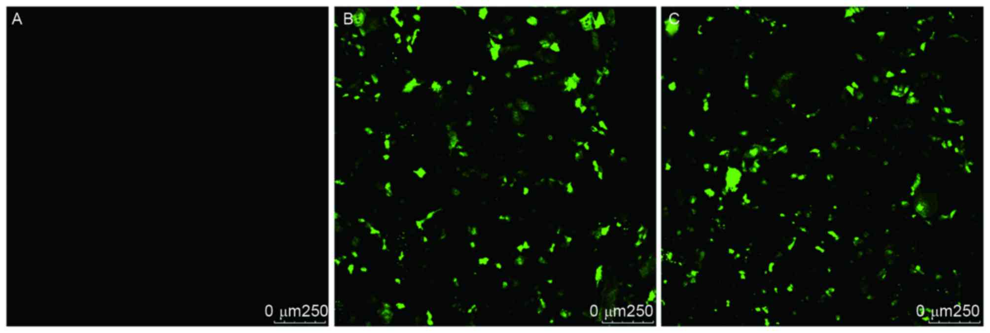
Transient transfection efficiency of
CXCR4-shRNA
The present study successfully obtained
CXCR4-shRNA-containing SW626 cells using cationic liposome-mediated
transfection, and the transfection efficiency was <80% (Fig. 1).
Effect of CXCR4-shRNA on ASK1 mRNA
expression
Following cell transfection, ASK1 mRNA expression
was determined by the ABI PRISM® 7300 amplification
system. As summarized in Table I, the
ASK1 mRNA expression in the interference group was significantly
different compared with that in the blank control and empty vector
groups, and the ΔCq value of the interference group was also
significantly different compared with those of the other two groups
(P<0.001). However, no significant difference was observed
between the blank control and the empty vector group
(P>0.05).
 | Table I.Expression of ASK1 mRNA following
C-X-C chemokine receptor type 4-short hairpin RNA transfection. |
Table I.
Expression of ASK1 mRNA following
C-X-C chemokine receptor type 4-short hairpin RNA transfection.
| Gene | Group | Cq | ΔCq | ΔΔCq | P-value |
|---|
| ASK1 | Blank control | 32.0329±0.8002 | 15.5343 | 0 |
|
|
| Empty vector | 31.5086±1.31723 | 15.1 | −0.4343 | 0.447a |
|
| Interference | 29.4300±0.4452 | 12.1329 | −3.4017 | 0.000b |
| GAPDH | Blank control | 16.4986±0.4831 |
|
|
|
|
| Empty vector | 16.4086±0.4938 |
|
|
|
|
| Interference | 17.2971±0.2880 |
|
|
|
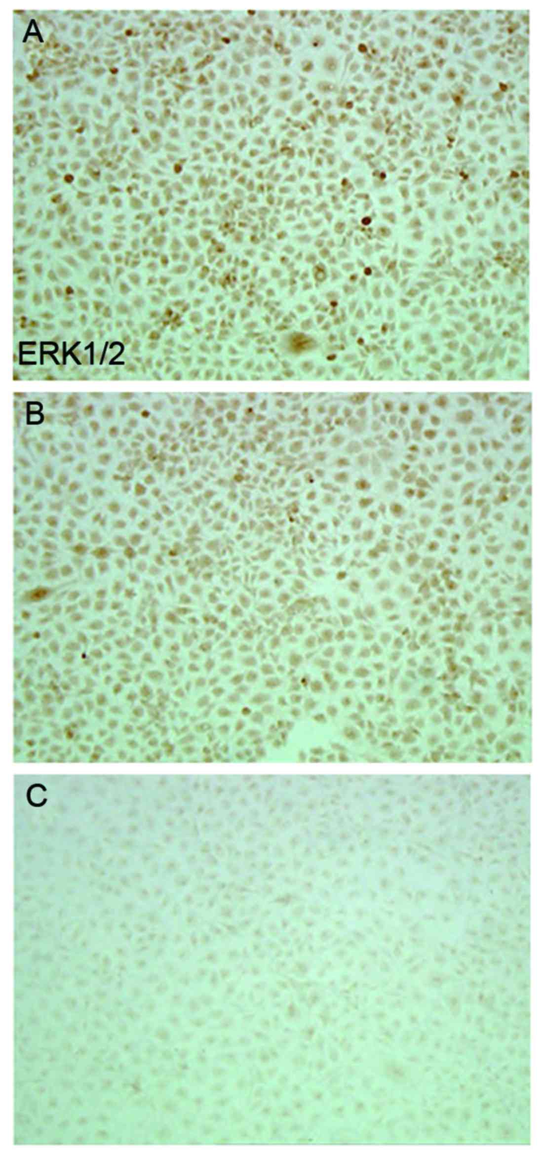
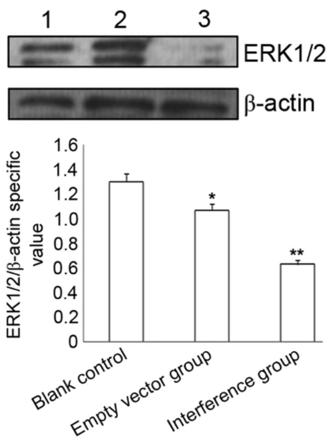
Effect of CXCR4-shRNA transfection on
ERK1/2 protein expression
Immunocytochemistry and western blotting
demonstrated that compared with the blank control and empty vector
groups, SW626 cells transfected with CXCR4-shRNA exhibited
significantly decreased ERK1/2 protein expression levels
(P<0.05), whereas no significant difference was observed between
the blank control and empty vector groups (P>0.05) (Table II; Figs.
2 and 3).
| Table II.Immunocytochemical determination of
extracellular signal-regulated kinase 1/2 protein expression 48 h
post-transfection. |
Table II.
Immunocytochemical determination of
extracellular signal-regulated kinase 1/2 protein expression 48 h
post-transfection.
| Group | Mean optical
density | P-value |
|---|
| Blank control | 71.2117±19.9886 | 0.368a |
| Empty vector | 63.8878±2.3245 | 0.001b |
| Interference | 27.0500±7.3917 |
<0.001c |
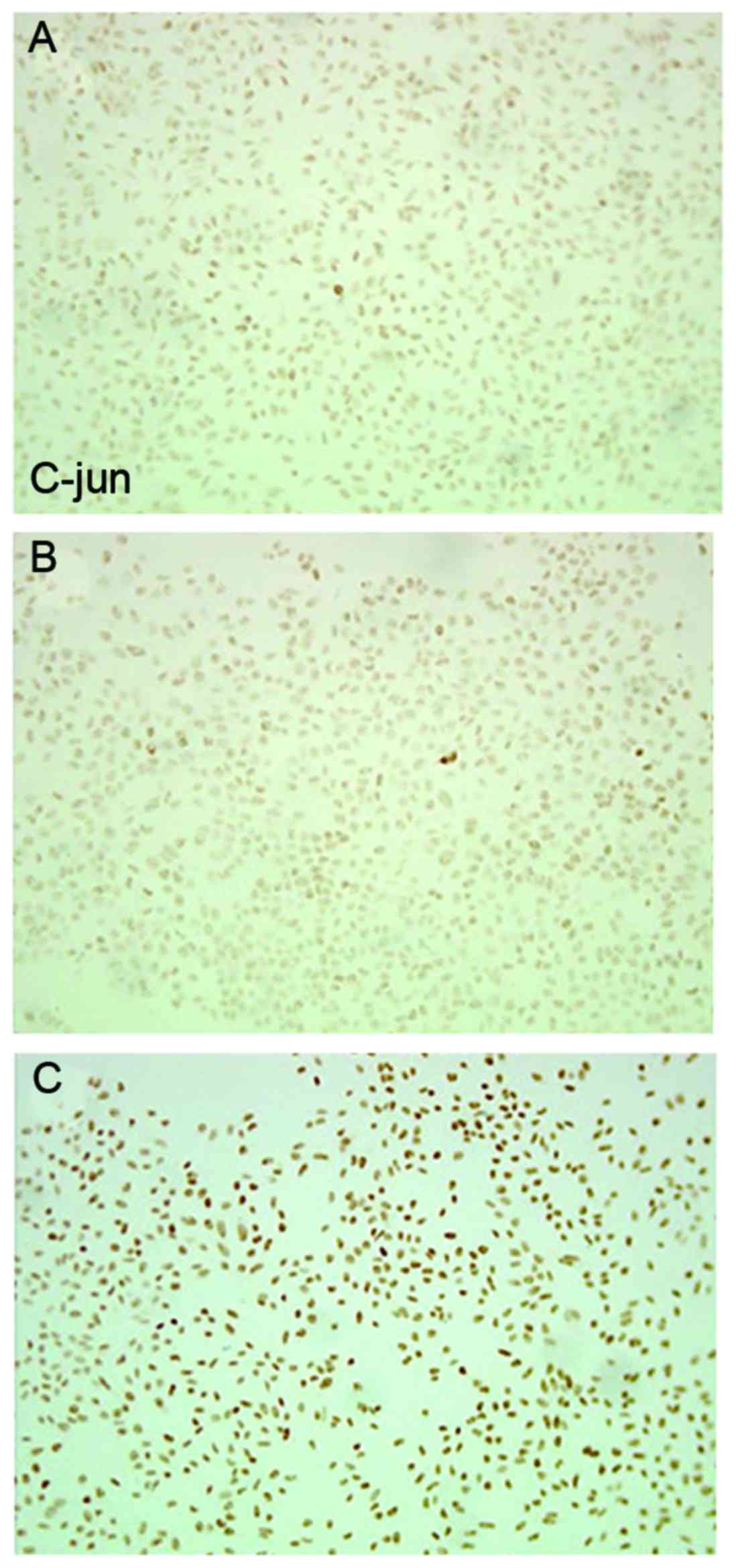
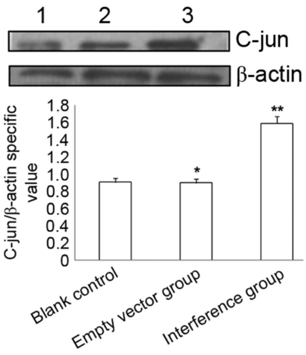
Effect of CXCR4-shRNA transfection on
p-c-Jun protein expression
Immunocytochemistry and western blotting identified
that, compared with the blank control and empty vector group, SW626
cells transfected with CXCR4-shRNA exhibited significantly
increased p-c-Jun protein expression levels (P<0.01), whereas no
significant difference was identified between the blank control and
the empty vector group (P>0.05) (Table III; Figs.
4 and 5).
| Table III.Immunocytochemical determination of
phosphorylated c-Jun protein expression at 48 h after
transfection. |
Table III.
Immunocytochemical determination of
phosphorylated c-Jun protein expression at 48 h after
transfection.
| Group | Mean optical
density | P-value |
|---|
| Blank control | 34.9507±2.3619 |
|
| Empty vector | 37.2107±2.8655 | 0.253a |
| Interference | 57.0649±3.5716 |
<0.001b |
Discussion
MAPK is a serine/threonine protein kinase that
participates in one of the most important types of signal
transduction pathways in eukaryotes, and serves a key role in
regulating gene expression and cytoplasmic function (8). The MAPK signaling pathway primarily
includes ERKs, c-Jun N-terminal kinases (JNKs) and p38 MAPK
(9). The MAPK signaling pathway is
involved in the regulation of various cell functions, particularly
cell proliferation, differentiation and apoptosis (6). A previous study identified that,
compared with normal ovarian tissues and benign ovarian lesions,
MAPK activity in ovarian cancer tissues was significantly increased
(10). Hayakawa et al
(11) demonstrated that the MAPK was
in a continuous activating status in ovarian cancer cells, and
additional phosphorylation antibody detection of upstream kinase
dual-specificity mitogen-activated protein kinase kinase 1 and RAF
proto-oncogene serine/threonine-protein kinase (RAF)1 illustrated
that MAPK activation was associated with upstream kinase
activation. Inhibiting ERK1/2 activity with MAPK blockers promoted
the apoptosis of ovarian cancer cells, which additionally elevated
cellular sensitivity to chemotherapy. The MAPK pathway realizes
signal transduction via the continuous phosphorylation of MAPK
kinase kinase (MAPKKK), MAPK kinase (MAPKK), and MAPK (12). Previous studies identified a number of
MAPK pathways and various extracellular stimulators, including
cytokines, G-protein-coupled receptors, stress signals and
mitogens, may all activate various MAPK signaling transduction
types. As the primary signal proteins involved in the MAPK pathway,
ERK1/2, JNK and p38MAPK exert various biological effects
(13). ERK1/2 activation is primarily
associated with cell proliferation, whereas JNK and p38 primarily
regulate cellular apoptosis.
The ERK1/2 pathway, the earliest identified
Ras-RAF-MAPK signaling pathway (14),
may be activated by a variety of growth factors or cytokines and
regulates cell proliferation and differentiation, so regulating ERK
activation serves a significant role in the occurrence and
progression of cell proliferation (15). A previous study revealed that, upon
extracellular stimulation, ERK is a key factor in determining
whether a cell undergoes end-stage differentiation or apoptosis
(16). ERK activation relies on
signals passing from the cell-surface receptor to the nucleus via
GTPase Ras, RAF1, serine/threonine kinase and MAPK/ERK kinase
dual-specificity kinases (16).
Another study indicated that CXCR3 may promote the metastasis of
SKOV3 ovarian cancer cells via the ERK1/2 pathway, which was
inhibited following ERK1/2 blockage with PD98059 (17). Tarcic and Yarden (18) suggested that the epidermal growth
factor receptor (EGFR) protein may promote cell proliferation via
activating the ERK1/2 signal pathway. In the present study, the
effects of CXCR4-shRNA on ERK1/2 signaling in ovarian cancer cells
were additionally investigated, and it was demonstrated that cells
transfected with CXCR4-shRNA exhibited significantly decreased
expression levels of ERK1/2, whereas no significant difference was
identified between the empty vector group and the blank control.
These results indicate that CXCR4-shRNA may decrease ERK1/2 protein
expression and that such inhibition may be achieved by regulating
EGFR expression to promote cancer cell apoptosis.
ASK1 is a member of the MAPKKK family that performs
significant functions in regulating cell apoptosis and
differentiation, and in the immune response (19,20). In
the MAPK signaling pathway, ASK1 is located upstream of JNK and
p38; as such, once it is activated, it may activate MAPKK and
additionally JNK and p38, thereby causing cell apoptosis via
mitochondria-associated caspase-3 (21). In normal cells, ASK1 activation is
strictly controlled by the phosphorylation/dephosphorylation of
threonine/serine or other protein-protein interactions. In breast
cancer, increased ASK1 expression induced cell apoptosis (22), and subsequent experiments in human
osteosarcoma cells additionally confirmed that the
apoptosis-inducing factors exert their antitumor function through
the proto-oncogene tyrosine-protein kinase reactive oxygen
species/ASK1 pathway (23). The
present study revealed that CXCR4-shRNA-transfected ovarian cancer
cells exhibited significantly increased ASK1 mRNA expression;
therefore, it is hypothesized that the transfection process
activated ASK1 and c-Jun, thereby inducing the apoptosis of cancer
cells.
The oncogene c-Jun encodes the Jun C protein, which
may form homodimers with Jun B or Jun D, or even heterodimers with
proteins from the Fos family. The homodimer or heterodimer formed
by c-Jun or c-Fos proteins is termed activator protein 1. The
expression of c-Jun is regulated by a complex process that
primarily involves JNK as a regulator, and transforming growth
factor-β, bone morphogenetic proteins, hypoxia factors and
oxidative stress as stimulators; c-Jun activation serves
significant roles in cell proliferation, apoptosis, the stress
response and tumor development (24,25). The
present study demonstrated that CXCR4-shRNA transfection increased
p-c-Jun expression in ovarian cancer cells, indicating that
CXCR4-shRNA may induce cancer cell apoptosis by regulating
EGFR-mediated p-c-Jun expression.
The results of the present study suggest that
silencing the CXCR4 gene with shRNA inhibited proliferation
and promoted apoptosis of epithelial ovarian cancer cells, and it
was hypothesized that this was achieved via MAPK signaling, as
decreased MAPK pathway activity was observed. These data provide a
novel theoretical basis for investigating the mechanism by which
CXCR4-shRNA promotes cell apoptosis, and lay a concrete foundation
for use of the CXCR4 gene as a therapy target in the
treatment of ovarian cancer.
References
|
1
|
Weiderpass E and Labreche F: Malignant
tumors of the female reproductive system. Saf Health Work.
3:166–180. 2012. View Article : Google Scholar : PubMed/NCBI
|
|
2
|
Huang CY, Lee CY, Chen MY, Yang WH, Chen
YH, Chang CH, Hsu HC, Fong YC and Tang CH: Stromal cell-derived
factor-1/CXCR4 enhanced motility of human osteosarcoma cells
involves MEK1/2, ERK and NF-kappaB-dependent pathways. J Cell
Physiol. 221:204–212. 2009. View Article : Google Scholar : PubMed/NCBI
|
|
3
|
Scotton CJ, Wilson JL, Milliken D, Stamp G
and Balkwill FR: Epithelial cancer cell migration: A role for
chemokine receptors? Cancer Res. 61:4961–4965. 2001.PubMed/NCBI
|
|
4
|
Chen Y, Stamatoyannopoulos G and Song CZ:
Down-regulation of CXCR4 by inducible small interfering RNA
inhibits breast cancer cell invasion in vitro. Cancer Res.
63:4801–4804. 2003.PubMed/NCBI
|
|
5
|
Chen HY, Wang JM, Wang HY, Zhang YX, Liu
W, Pan L, Wang WH, Chen SF, Jin WG and Wang L: Effect of short
hairpin RNA-induced CXCR4 silence on ovarian cancer cell. Biomed
Pharmacother. 66:549–553. 2012. View Article : Google Scholar : PubMed/NCBI
|
|
6
|
Choi KC, Auersperg N and Leung PC:
Mitogen-activated protein kinases in normal and (pre)neoplastic
ovarian surface epithelium. Reprod Biol Endocrinol. 1:712003.
View Article : Google Scholar : PubMed/NCBI
|
|
7
|
Livak KJ and Schmittgen TD: Analysis of
relative gene expression data using real-time quantitative PCR and
the 2(-Delta Delta C(T)) method. Methods. 25:402–408. 2001.
View Article : Google Scholar : PubMed/NCBI
|
|
8
|
Park J, Song KH and Ha H: Fractalkine
increases mesangial cell proliferation through reactive oxygen
species and mitogen-activated protein kinases. Transplant Proc.
44:1026–1028. 2012. View Article : Google Scholar : PubMed/NCBI
|
|
9
|
Yang SH, Sharrocks AD and Whitmarsh AJ:
MAP kinase signalling cascades and transcriptional regulation.
Gene. 513:1–13. 2013. View Article : Google Scholar : PubMed/NCBI
|
|
10
|
Janku F, Wheler JJ, Westin SN, Moulder SL,
Naing A, Tsimberidou AM, Fu S, Falchook GS, Hong DS, Garrido-Laguna
I, et al: PI3K/AKT/mTOR inhibitors in patients with breast and
gynecologic malignancies harboring PIK3CA mutations. J Clin Oncol.
30:777–782. 2012. View Article : Google Scholar : PubMed/NCBI
|
|
11
|
Hayakawa J, Ohmichi M, Kurachi H, Ikegami
H, Kimura A, Matsuoka T, Jikihara H, Mercola D and Murata Y:
Inhibition of extracellular signal-regulated protein kinase or
c-Jun N-terminal protein kinase cascade, differentially activated
by cisplatin, sensitizes human ovarian cancer cell line. J Biol
Chem. 274:31648–31654. 1999. View Article : Google Scholar : PubMed/NCBI
|
|
12
|
Yoshioka K: Scaffold proteins in mammalian
MAP kinase cascades. J Biochem. 135:657–661. 2004. View Article : Google Scholar : PubMed/NCBI
|
|
13
|
Kim EK and Choi EJ: Pathological roles of
MAPK signaling pathways in human diseases. Biochim Biophys Acta.
1802:396–405. 2010. View Article : Google Scholar : PubMed/NCBI
|
|
14
|
Zhao HB, Tang CL, Hou YL, Xue LR, Li MQ,
Du MR and Li DJ: CXCL12/CXCR4 axis triggers the activation of EGF
receptor and ERK signaling pathway in CsA-induced proliferation of
human trophoblast cells. PLoS One. 7:e383752012. View Article : Google Scholar : PubMed/NCBI
|
|
15
|
Vergara D, Simeone P, Toraldo D, Del
Boccio P, Vergaro V, Leporatti S, Pieragostino D, Tinelli A, De
Domenico S, Alberti S, et al: Resveratrol downregulates Akt/GSK and
ERK signalling pathways in OVCAR-3 ovarian cancer cells. Mol
Biosyst. 8:1078–1087. 2012. View Article : Google Scholar : PubMed/NCBI
|
|
16
|
Porcile C, Bajetto A, Barbero S, Pirani P
and Schettini G: CXCR4 activation induces epidermal growth factor
receptor transactivation in an ovarian cancer cell line. Ann N Y
Acad Sci. 1030:162–169. 2004. View Article : Google Scholar : PubMed/NCBI
|
|
17
|
Shen X, Wang S, Wang H, Liang M, Xiao L
and Wang Z: The role of SDF-1/CXCR4 axis in ovarian cancer
metastasis. J Huazhong Univ Sci Technolog Med Sci. 29:363–367.
2009. View Article : Google Scholar : PubMed/NCBI
|
|
18
|
Tarcic G and Yarden Y: MAP Kinase
activation by receptor tyrosine kinases: In control of cell
migration. Methods Mol Biol. 661:125–135. 2010. View Article : Google Scholar : PubMed/NCBI
|
|
19
|
Komuro Y, Takeda K and Ichijo H:
Regulatiory mechanism of cell dealth through ASK1. Seikagaku.
76:1458–1462. 2004.(In Japanese). PubMed/NCBI
|
|
20
|
Matsuzawa A and Ichijo H: Redox control of
cell fate by MAP kinase: Physiological roles of ASK1-MAP kinase
pathway in stress signaling. Biochim Biophys Acta. 1780:1325–1336.
2008. View Article : Google Scholar : PubMed/NCBI
|
|
21
|
Hatai T, Matsuzawa A, Inoshita S, Mochida
Y, Kuroda T, Sakamaki K, Kuida K, Yonehara S, Ichijo H and Takeda
K: Execution of apoptosis signal-regulating kinase 1 (ASK1)-induced
apoptosis by the mitochondria-dependent caspase activation. J Biol
Chem. 275:26576–26581. 2000. View Article : Google Scholar : PubMed/NCBI
|
|
22
|
Guo Y, Xu X, Liu Z, Zhang T, Zhang X, Wang
L, Wang M, Liu Y, Lu Y, Liu Y, et al: Apoptosis signal-regulating
kinase 1 is associated with the effect of claudin-6 in breast
cancer. Diagn Pathol. 7:1112012. View Article : Google Scholar : PubMed/NCBI
|
|
23
|
Chen JT, Fong YC, Li TM, Liu JF, Hsu CW,
Chang CS and Tang CH: DDTD, an isoflavone derivative, induces cell
apoptosis through the reactive oxygen species/apoptosis
signal-regulating kinase 1 pathway in human osteosarcoma cells. Eur
J Pharmacol. 597:19–26. 2008. View Article : Google Scholar : PubMed/NCBI
|
|
24
|
Qu WS, Tian DS, Guo ZB, Fang J, Zhang Q,
Yu ZY, Xie MJ, Zhang HQ, Lu JG and Wang W: Inhibition of EGFR/MAPK
signaling reduces microglial inflammatory response and the
associated secondary damage in rats after spinal cord injury. J
Neuroinflammation. 9:1782012. View Article : Google Scholar : PubMed/NCBI
|
|
25
|
Zhen YZ, Lin YJ, Gao JL, Zhao YF and Xu
AJ: Rhein lysinate inhibits cell growth by modulating various
mitogen-activated protein kinases in cervical cancer cells. Oncol
Lett. 2:129–133. 2011. View Article : Google Scholar : PubMed/NCBI
|