Introduction
Bladder cancer is one of the most common
malignancies in the urinary system. The biological characteristics
of bladder cancer are recurrence and drug resistance (1). While 50–70% of non-muscle invasive
bladder cancer (NMIBC) patients experience recurrence, 50–80% of
MIBC patients develop progression to distant metastasis (2). A transurethral resection of the bladder
tumor (TURBT) is the primary method of treating NMIBC patients.
TURBT is also an important diagnostic and staging tool in the
management of bladder cancer patients. Radical cystectomy and
pelvic lymph node dissection provide accurate evaluation of MIBC
patients, and this evaluation could avoid errors in clinical
staging (3,4). Meanwhile, the results of animal studies
and clinical trials have suggested that dendritic cell (DC)-based
immunotherapy may have the potential for activating immune
responses and for a therapeutic effect in bladder cancer (5).
DCs are key cells in the initiation of innate and
adaptive immunity, with have the ability to modulate the antitumor
immune response (6). DCs are the most
powerful antigen-presenting cells and only DCs have the ability for
activation of the primary sensitization against specific antigens
in a naïve host (7). However, recent
findings have shown that DCs will be dysfunctional in the tumor
microenvironment (8). Tumor-derived
factors affect DC maturation and differentiation. Immature DCs
(iDCs) fail to supply costimulatory molecules to T cells and
antitumor immune tolerance or anergy may develop. Moreover,
tumor-infiltrating DCs (TIDCs) have been demonstrated to be
associated with low expression of costimulatory signal, ineffective
antigen cross-presentation, and high expression of regulatory
molecules and receptors (9).
Constitutive activation of the signal transducer and
activator of transcription 3 (STAT3) signaling pathway serves a
pivotal role in the growth and invasion of bladder cancer (10). AG490 is a Janus kinase 2 (Jak2)/STAT3
signaling pathway inhibitor, which reverses the inhibitory effect
of hyperphosphorylated STAT3 on DC differentiation (11). Moreover, AG490 also inhibits vascular
endothelial growth factor expression by control over the downstream
target genes of the Jak2/STAT3 pathway. Therefore, AG490 could
provide a basis for targeting anti-angiogenesis and exhibit
therapeutic significance in the treatment of bladder cancer
patients (10).
The purpose of the present study was to investigate
the associations between bladder cancer cells and DCs. Human
bladder cancer pumc-91 cells co-cultured with human peripheral
blood mononuclear cell (PMBC)-derived DCs were used for the DC
surface phenotyping assays, cytokine production assays and
allogeneic mixed leukocyte reaction assay. Furthermore, the study
also investigated how AG490 may reverse the inhibitory effects of
bladder cancer cells on the phenotype of DCs.
Materials and methods
Antibodies
The following antibodies were supplied in PBS and
used without further dilution in the study: Phycoerythrin-labeled,
anti-cluster of differentiation (CD)86 (cat no. 12-0869-42),
anti-CD80 (cat no. 12-0809-42), anti-human leukocyte antigen (HLA)
class I molecules HLA-A, HLA-B and HLA-C antigens (HLA-ABC; cat no.
12-9983-42), anti-HLA-antigen D related (HLA-DR; cat no.
17-9956-42), PE isotype control mAbs (cat no. 12-4724-81), APC
isotype control mAbs (cat no. 17-4714-81), Mouse IgG (cat no.
25-4714-42) and allophycocyanin-labeled anti-CD11c (cat no.
17-0116-42) (all eBioscience; Thermo Fisher Scientific, Inc.,
Waltham, MA, USA).
Cell culture
The human bladder cancer pumc-91 cell line was
provided by Peking Union Medical College Hospital (Beijing, China)
(12). Pumc-91 cells were cultured in
RPMI-1640 medium (Gibco; Thermo Fisher Scientific, Inc.)
supplemented with 10% heat-inactivated fetal bovine serum (FBS;
Ausbian, Sydney, Australia) at 37°C and 5% CO2. Human
PBMCs from healthy volunteers were isolated immediately from 100 ml
buffy coat according to standard protocols using Ficoll-Hypaque
density gradient centrifugation at 700 × g for 30 min at 20°C. To
generate DCs, PBMCs were cultured at a density of 5×10⁶
cells/2 ml in a 6-well plate (Greiner Bio-One, Kremsmünster,
Austria) in AIM-V medium (Gibco; Thermo Fisher Scientific, Inc.)
containing 10% FBS. The plate was incubated at 37°C for 2 h, and
the non-adherent cells were discarded. The adherent cells were
treated for 6 days at 37°C in complete AIM-V medium supplemented
with 10% FBS, 50 ng/ml recombinant human
(rh)-granulocyte-macrophage colony-stimulating factor and 50 ng/ml
rh-interleukin-4 (rhIL-4) (Peprotech, Inc., Rocky Hill, NJ, USA) to
generate iDCs. Half-volume medium replacement with all cytokines
was performed on days 3 and 5. The samples were collected on August
21, 2015, in Beijing Shijitan Hospital, Capital Medicine University
(Beijing, China). The study was approved by the Ethics Board of
Beijing Shijitan Hospital, Capital Medicine University (Beijing,
China).
Bladder cancer cell treatment with
AG490
AG490 was dissolved in dimethyl sulfoxide (DMSO;
both Sigma-Aldrich; Merck KGaA, Darmstadt, Germany) and diluted to
a final concentration of 200 µM. Pumc-91 cells were treated for 24
h with AG490 at 200 µM. Subsequently, AG490-treated pumc-91
(pumc-91/AG490) cells were washed three times and co-cultured with
DCs at 37°C for 24 h. 1.2% DMSO-treated pumc-91 (pumc-91/DMSO)
cells were used as the control group.
DC co-culture with bladder cancer
cells
On day 5 of DC culture, 1×10⁶ pumc-91 or
pumc-91/AG490 cells were added to each well containing the DCs. On
day 6, these cells were washed three times and purified with
microbeads from a QuadroMACS Starting kit (LD columns) and MiniMACS
Starting kit (MS columns) using a Blood Dendritic Isolation kit II
(cat no. 130-091-379, MiltenyiBiotec GmbH, BergischGladbach,
Germany). Non-DC Depletion Cocktail and DC Enrichment Cocktail are
contained within the Blood Dendritic Cell Isolation kit II. The
following antibodies were not pre-diluted and were dissolved in
running buffer at the ratio of 1:3. (cat. no. 130-091-221;
MiltenyiBiotec GmbH, Bergisch Gladbach, Germany). Non-DC Depletion
Cocktail includes monoclonal biotin-conjugated antibodies against
human CD1c, CD14 and CD19. DC Enrichment Cocktail includes
microbeads conjugated to monoclonal antibodies against human CD304,
CD141 and biotin. For iDC activation, 1 µg/ml LPS (Sigma-Aldrich;
Merck KGaA) was added at day 6 and the culture was continued for 24
h to induce mature DCs (mDCs).
DC surface phenotyping
The phenotype of the DCs was analyzed by flow
cytometry. The DCs were harvested and stained with antibodies. DCs
were blocked by mouse IgG κ (cat no. 25-4714-42; eBioscience;
Thermo Fisher Scientific, Inc.) at 4°C for 30 min, then stained
with a PE-labeled, APC-labeled specific mAb or isotype control to
detect the target molecule at 4°C for 30 min. Mouse IgG (5 µl) were
supplied in PBS for each sample and used without further dilution.
DCs were stained with specific monoclonal antibodies, PE isotype
control mAbs or APC isotype control mAbs (5 µl) (as previously
described) at 4°C for 30 min, and then washed twice and resuspended
in phosphate-buffered saline. Stained cells were analyzed by a flow
cytometer (CytoFLEX; Beckman Coulter, Inc., Brea, CA, USA). Data of
mean fluorescence intensity and percentage of positive cells were
acquired and processed using accompanying software (CytExpert 1.0;
Beckman Coulter, Inc.).
Cytokine production assays
Purified mDCs were cultured in a 6-well plate at
1.8×10⁵ cells per well. Human ELISA Ready-SET-Go kits
(eBioscience; Thermo Fisher Scientific, Inc., Waltham, MA, USA)
were used according to the manufacturer's protocols in order to
measure the levels of IL-10 (cat. no. 88-7106) and IL-12p70 (cat.
no. 88-7126).
Allogeneic mixed leukocyte reaction
assay
PBMCs were obtained from healthy human peripheral
blood and isolated by Ficoll-Hypaque density gradient
centrifugation, as aforementioned. Labeled CD3+T cells
were purified from the PBMCs using the Pan T cell Isolation kit
(MiltenyiBiotec GmbH). In a previous experiment, it was found that
DC/T cells at the ratio of 1:20 showed the best result in terms of
T cell proliferation according to the OD value (13). The purified CD3+ T cells
were seeded into a round-bottom 96-well plate at 2.0×10⁵
cells per well. The purified DCs were irradiated (30 Gy for 30
min), incubated with allogeneic CD3+ T cells at DC/T
cell ratios of 1:20 and cultured for 5 days with Cell Counting
kit-8 (Dojindo Molecular Technologies, Inc., Kumamoto, Japan) for 4
h.
Statistical analysis
Data are presented as the mean ± standard deviation
(SD). All the experiments were repeated three times. The
statistical analysis was performed using a one-way analysis of
variance (ANOVA) and least significant difference method with SPSS
17.0 statistical software (SPSS Inc., Chicago, IL, USA). P<0.05
was considered to indicate a statistically significant
difference.
Results
Inhibition of DC phenotype by
co-culture with bladder cancer cells
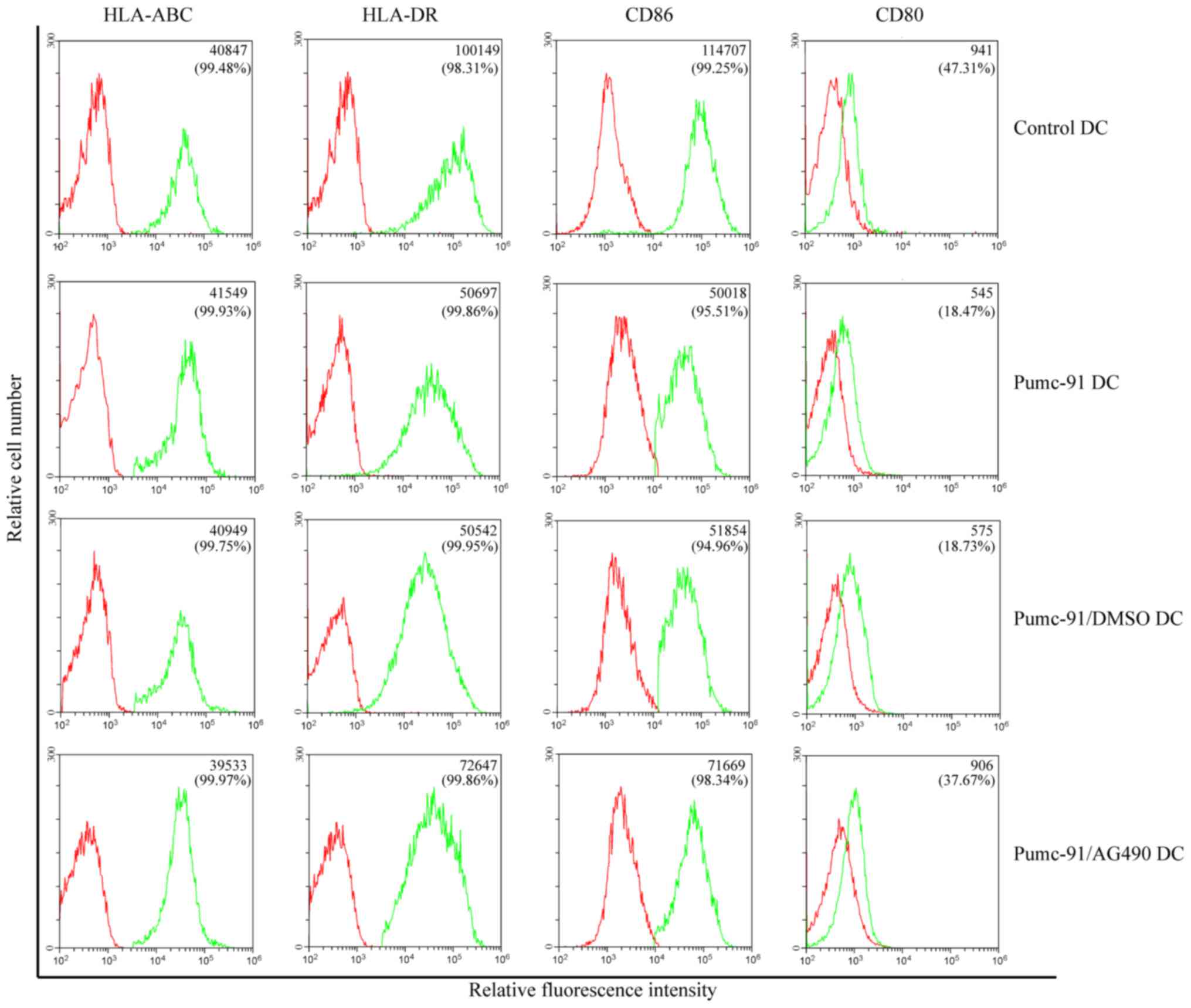
First, the effect of bladder cancer cells on DC
phenotype was assessed. On day 7, the expression of major
histocompatibility complex (MHC) class molecules (HLA-ABC and
HLA-DR) and costimulatory molecules (CD86 and CD80) was determined.
Compared with control DCs, pumc-91 cells co-cultured with DCs
exhibited reduced expression of HLA-DR, CD86 and CD80. However,
there were no differences between pumc-91-exposed DCs and DCs
co-cultured with pumc-91/DMSO. These observations were found based
on mean fluorescence intensity (MFI) results and the percentage of
positive cells (Fig. 1). The DCs in
the three tests were obtained from two donors, but there were no
individual differences. The statistical analysis of MFI was
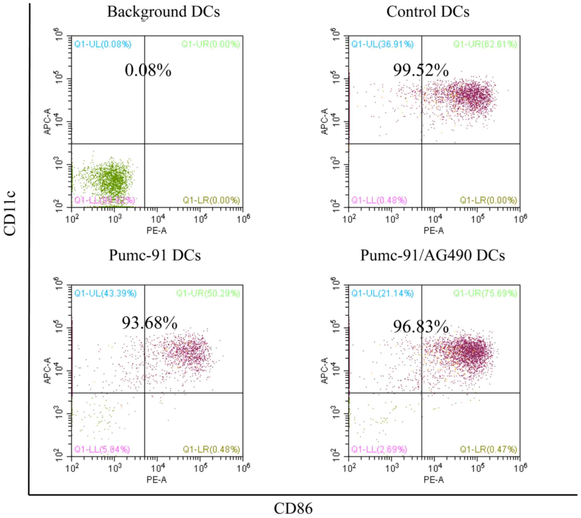
calculated by one-way ANOVA. Furthermore, the purity of the DC
samples was shown to be >93% in all experiments (Fig. 2).
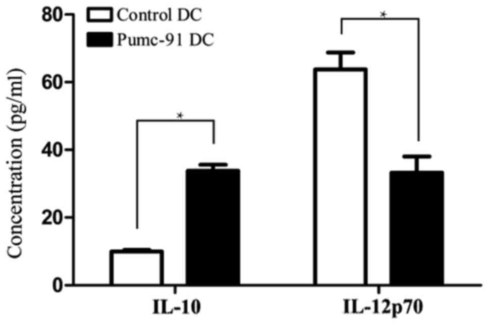
Bladder cancer cells inhibit IL-12p70
production while increasing IL-10 secretion of DCs
In addition to MHC class molecules and costimulatory
molecules, cytokines secreted from DCs serve an important role in
priming the cytotoxic lymphocyte response. IL-10 and IL-12p70 were
detected in supernatants of control DCs and DCs co-cultured with
pumc-91 cells. Upon LPS stimulation, the pumc-91 co-cultured with
DCs produced a marked increase in IL-10 level, while IL-12p70 level
was decreased, compared with that in the control DCs (P<0.05)
(Fig. 3).
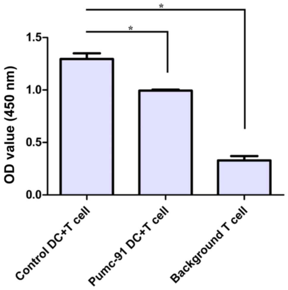
Inefficient T cell stimulation by DCs
co-cultured with pumc-91
Next, the immunostimulatory effect of
pumc-91-exposed DCs was tested. PBMC-derived DCs generated in the
presence or absence of pumc-91 were assessed by measuring their
ability to induce the proliferation of allogeneic CD3+ T
cells. Compared with control DCs, DCs co-cultured with pumc-91
cells showed significantly less potency in stimulating allogeneic
CD3+ T cell proliferation (Fig. 4). Therefore, bladder cancer pumc-91
cells affected the maturation of the DCs along with providing an
inhibitory effect on the function of the DCs.
AG490 reverses the inhibitory effect
of bladder cancer cells on DC phenotype
PBMC-derived DCs were co-cultured with pumc-91 cells
for 24 h with or without pretreatment with AG490. Compared with the
DCs co-cultured with pumc-91 cells, the DCs exposed to
AG490-treated pumc-91 cells exhibited upregulated expression of
HLA-DR, CD86 and CD80 (Fig. 1). This
revealed that blocking pumc-91-induced STAT3 activation by
pretreatment with AG490 reversed the inhibitory effect of the
pumc-91 cells on the DC phenotype.
Discussion
The results of the present study indicate that
bladder cancer cells may evade the host defense response by
inhibiting DC maturation, and by inducing IL-10 secretion and
suppressing IL-12p70 production in DCs. It was also found that
bladder cancer cell co-cultured DCs failed to develop a full
allostimulatory ability for CD3+ T lymphocytes. In
addition, blocking bladder cancer cell-induced STAT3 activation by
AG490 effectively reversed the inhibitory effect on DC
maturation.
DCs are known as the most powerful antigen
presenting cells (14). Previous
studies have suggested that the immunostimulatory ability of DCs
could be conditioned by a number of factors present in the tumor
microenvironment (13,15,16).
Tolerogenic properties of DCs are usually attributed to their
immature status (17). In the present
study, PBMC-derived DCs co-cultured with pumc-91 cells for 24 h
exhibited an immature phenotype. Compared with control DCs, DCs
co-cultured with pumc-91 exhibited low expression of HLA-DR, CD86
and CD80. This may partly explain why TIDCs show a phenotype of low
costimulatory molecule expression, and DCs may be polarized into
immunosuppressive regulatory DCs (18–20).
In addition to the immature phenotype in tumor
lesions, the same finding was obtained in the peripheral blood of
patients with tumors. This result suggests that tumor-associated
soluble factors may prevent DC function (9). One of the cytokines reported to suppress
the function of DCs is IL-10. IL-10 inhibits the capacity of DCs to
stimulate the proliferation of T lymphocytes (21). Overexpression of IL-10 in DCs could
suppress the allogeneic cytotoxic lymphocyte response and IL-12p70
production (22). This is consistent
with the present study findings. It was found that pumc-91 cells
co-cultured with DCs produced a lower level of IL-12p70, but a
higher level of IL-10 compared with controls. The decreased level
of IL-12p70 may prevent the development of the type 1 T helper
response, and result in a lack of IFN-γ supporting the cytotoxic
lymphocyte response (23,24).
It has been well established that the interaction of
CD86 and CD80 with CD28 receptor results in the activation of
costimulatory signals of DCs (25).
These molecules are indispensable to prime the T cytotoxic
lymphocyte response. Therefore, the inhibition of CD86 and CD80 by
pumc-91 cells may suggest that the bladder cancer cells disrupted
the antigen-specific immune-activating abilities of the DCs to
induce a T cell response. Moreover, IL-10 could promote the
apoptosis of DCs and reverse the protection against apoptosis
conferred by tumor necrosis factor-α and CD40 ligand (26). The apoptosis of DCs may reduce the
time window during which they contact with T lymphocytes.
Consistent with these findings, the present observed that pumc-91
cell-exposed DCs could not stimulate proliferation of allogeneic
CD3+ T cells. This also suggests that tumor-associated
soluble factors could impair the antigen-presenting function of
DCs.
Constitutive activation of the STAT3 signal pathway
has been observed in several types of tumors, and it could promote
the proliferation and invasion of tumor cells (27). AG490 is an effective inhibitor of the
Jak2/STAT3 pathway. A previous study showed that prior treatment
with AG490 could reverse human pancreatic cancer cell conditioned
medium-induced inhibition of DC differentiation (11). Moreover, AG490 could induce tumor
apoptosis and promote DC maturation. AG490 did not have any side
effect on activity and function. Therefore, it is suggested that
AG490 could act as an adjuvant used in DC-based immunotherapy
(28). In the present study, compared
with pumc-91 co-cultured DCs, AG490-treated pumc-91-exposed DCs
exhibited upregulation of CD86, CD80 and HLA-DR. This may suggest
that pretreatment with AG490 could inhibit STAT3 activation in
bladder cancer cells and reverse the inhibitory effect of pumc-91
cells on the DC phenotype. Furthermore, Jak2/STAT3 may serve a
vital role in inducing the immunosuppressive effect of bladder
cancer on the function of DCs.
In conclusion, the present study found that bladder
cancer pumc-91 cells could inhibit DC maturation and weaken their
ability to activate T cells. It is possible that this is one of the
reasons for DC-based therapies yielding unsatisfactory therapeutic
effects. Finally, the use of AG490 was demonstrated to partly
reverse DC dysfunction.
Acknowledgements
Not applicable.
Funding
The present study was supported by the Beijing
Natural Science Foundation (grant no. 7172106).
Availability of data and materials
The datasets used and/or analyzed during the current
study are available from the corresponding author on reasonable
request.
Authors' contributions
MZ designed the study. WX, JM and TL performed the
experiments. WX wrote the paper. JM, TL and MZ reviewed and edited
the manuscript. All authors read and approved the manuscript.
Ethics approval and consent to
participate
The study was approved by the Ethics Board of
Beijing Shijitan Hospital, Capital Medicine University (Beijing,
China). All volunteers provided written informed consent.
Patient consent for publication
The volunteers provided consent for publication.
Competing interests
The authors declare that they have no competing
interests.
References
|
1
|
Lei T, Zhao X, Jin S, Meng Q, Zhou H and
Zhang M: Discovery of potential bladder cancer biomarkers by
comparative urine proteomics and analysis. Clin Genitourin Cancer.
11:56–62. 2013. View Article : Google Scholar : PubMed/NCBI
|
|
2
|
Meng Q, Lei T and Zhang M, Zhao J, Zhao XH
and Zhang M: Identification of proteins differentially expressed in
adriamycin-resistant (pumc-91/ADM) and parental (pumc-91) human
bladder cancer cell lines by proteome analysis. J Cancer Res Clin
Oncol. 139:509–519. 2013. View Article : Google Scholar : PubMed/NCBI
|
|
3
|
Stein JP, Lieskovsky G, Cote R, Groshen S,
Feng AC, Boyd S, Skinner E, Bochner B, Thangathurai D, Mikhail M,
et al: Radical cystectomy in the treatment of invasive bladder
cancer: Long-term results in 1,054 patients. J Clin Oncol.
19:666–675. 2001. View Article : Google Scholar : PubMed/NCBI
|
|
4
|
Amling CL, Thrasher JB, Frazier HA, Dodge
RK, Robertson JE and Paulson DF: Radical cystectomy for stages Ta,
Tis and T1 transitional cell carcinoma of the bladder. J Urol.
151:31–35; discussion 35–36. 1994. View Article : Google Scholar : PubMed/NCBI
|
|
5
|
Schuler G, Schuler-Thurner B and Steinman
RM: The use of dendritic cells in cancer immunotherapy. Curr Opin
Immunol. 15:138–147. 2003. View Article : Google Scholar : PubMed/NCBI
|
|
6
|
Si C, Zhang R, Wu T, Lu G, Hu Y, Zhang H,
Xu F, Wei P, Chen K, Tang H, et al: Dendritic cell-derived nitric
oxide inhibits the differentiation of effector dendritic cells.
Oncotarget. 7:74834–74845. 2016. View Article : Google Scholar : PubMed/NCBI
|
|
7
|
Ma J, Usui Y, Takeuchi M, Okunuki Y,
Kezuka T, Zhang L, Mizota A and Goto H: Human uveal melanoma cells
inhibit the immunostimulatory function of dendritic cells. Exp Eye
Res. 91:491–499. 2010. View Article : Google Scholar : PubMed/NCBI
|
|
8
|
Ma Y, Shurin GV, Peiyuan Z and Shurin MR:
Dendritic cells in the cancer microenvironment. J Cancer. 4:36–44.
2013. View
Article : Google Scholar : PubMed/NCBI
|
|
9
|
Pinzon-Charry A, Maxwell T and López JA:
Dendritic cell dysfunction in cancer: A mechanism for
immunosuppression. Immunol Cell Biol. 83:451–461. 2005. View Article : Google Scholar : PubMed/NCBI
|
|
10
|
Joung YH, Na YM, Yoo YB, Darvin P, Sp N,
Kang DY, Kim SY, Kim HS, Choi YH, Lee HK, et al: Combination of
AG490, a Jak2 inhibitor, and methylsulfonylmethane synergistically
suppresses bladder tumor growth via the Jak2/STAT3 pathway. Int J
Oncol. 44:883–895. 2014. View Article : Google Scholar : PubMed/NCBI
|
|
11
|
Bharadwaj U, Li M, Zhang R, Chen C and Yao
Q: Elevated interleukin-6 and G-CSF in human pancreatic cancer cell
conditioned medium suppress dendritic cell differentiation and
activation. Cancer Res. 67:5479–5488. 2007. View Article : Google Scholar : PubMed/NCBI
|
|
12
|
Yu S, Meng Q, Hu H and Zhang M:
Correlation of ANXA1 expression with drug resistance and relapse in
bladder cancer. Int J Clin Exp Pathol. 7:5538–5548. 2014.PubMed/NCBI
|
|
13
|
Xiu W, Ma J, Lei T, Zhang M and Zhou S:
Immunosuppressive effect of bladder cancer on function of dendritic
cells involving of Jak2/STAT3 pathway. Oncotarget. 7:63204–63214.
2016. View Article : Google Scholar : PubMed/NCBI
|
|
14
|
Goyvaerts C and Breckpot K: Pros and cons
of antigen-presenting cell targeted tumor vaccines. J Immunol Res.
2015:7856342015. View Article : Google Scholar : PubMed/NCBI
|
|
15
|
Zhang M, Tang H, Guo Z, An H, Zhu X, Song
W, Guo J, Huang X, Chen T, Wang J and Cao X: Splenic stroma drives
mature dendritic cells to differentiate into regulatory dendritic
cells. Nat Immunol. 5:1124–1133. 2004. View
Article : Google Scholar : PubMed/NCBI
|
|
16
|
Svensson M, Maroof A, Ato M and Kaye PM:
Stromal cells direct local differentiation of regulatory dendritic
cells. Immunity. 21:805–816. 2004. View Article : Google Scholar : PubMed/NCBI
|
|
17
|
Lizée G, Radvanyi LG, Overwijk WW and Hwu
P: Improving antitumor immune responses by circumventing
immunoregulatory cells and mechanisms. Clin Cancer Res.
12:4794–4803. 2006. View Article : Google Scholar : PubMed/NCBI
|
|
18
|
Janco Tran JM, Lamichhane P, Karyampudi L
and Knutson KL: Tumor-infiltrating dendritic cells in cancer
pathogenesis. J Immunol. 194:2985–2991. 2015. View Article : Google Scholar : PubMed/NCBI
|
|
19
|
Harimoto H, Shimizu M, Nakagawa Y,
Nakatsuka K, Wakabayashi A, Sakamoto C and Takahashi H:
Inactivation of tumor-specific CD8+ CTLs by
tumor-infiltrating tolerogenic dendritic cells. Immunol Cell Biol.
91:545–555. 2013. View Article : Google Scholar : PubMed/NCBI
|
|
20
|
Krempski J, Karyampudi L, Behrens MD,
Erskine CL, Hartmann L, Dong H, Goode EL, Kalli KR and Knutson KL:
Tumor-infiltrating programmed death receptor-1+
dendritic cells mediate immune suppression in ovarian cancer. J
Immunol. 186:6905–6913. 2011. View Article : Google Scholar : PubMed/NCBI
|
|
21
|
Steinbrink K, Jonuleit H, Muller G,
Schuler G, Knop J and Enk AH: Interleukin-10-treated human
dendritic cells induce a melanoma-antigen-specific anergy in CD8(+)
T cells resulting in a failure to lyse tumor cells. Blood.
93:1634–1642. 1999.PubMed/NCBI
|
|
22
|
Steinbrink K, Graulich E, Kubsch S, Knop J
and Enk AH: CD4(+) and CD8(+) anergic T cells induced by
interleukin-10-treated human dendritic cells display
antigen-specific suppressor activity. Blood. 99:2468–2476. 2002.
View Article : Google Scholar : PubMed/NCBI
|
|
23
|
Hilkens CM, Kalinski P, de Boer M and
Kapsenberg ML: Human dendritic cells require exogenous
interleukin-12-inducing factors to direct the development of naive
T-helper cells toward the Th1 phenotype. Blood. 90:1920–1926.
1997.PubMed/NCBI
|
|
24
|
Jackson AM, Mulcahy LA, Zhu XW, O'Donnell
D and Patel PM: Tumour-mediated disruption of dendritic cell
function: Inhibiting the MEK1/2-p44/42 axis restores IL-12
production and Th1-generation. Int J Cancer. 123:623–632. 2008.
View Article : Google Scholar : PubMed/NCBI
|
|
25
|
Collins M, Ling V and Carreno BM: The B7
family of immune-regulatory ligands. Genome Biol. 6:2232005.
View Article : Google Scholar : PubMed/NCBI
|
|
26
|
Ludewig B, Graf D, Gelderblom HR, Becker
Y, Kroczek RA and Pauli G: Spontaneous apoptosis of dendritic cells
is efficiently inhibited by TRAP (CD40-ligand) and TNF-alpha, but
strongly enhanced by interleukin-10. Eur J Immunol. 25:1943–1950.
1995. View Article : Google Scholar : PubMed/NCBI
|
|
27
|
Hodge DR, Hurt EM and Farrar WL: The role
of IL-6 and STAT3 in inflammation and cancer. Eur J Cancer.
41:2502–2512. 2005. View Article : Google Scholar : PubMed/NCBI
|
|
28
|
Cirone M, Di Renzo L, Lotti LV, Conte V,
Trivedi P, Santarelli R, Gonnella R, Frati L and Faggioni A:
Primary effusion lymphoma cell death induced by bortezomib and AG
490 activates dendritic cells through CD91. PLoS One. 7:e317322012.
View Article : Google Scholar : PubMed/NCBI
|